Article

Different Expression of Vascularization and Inflammatory Regulators in Cells Derived from Oral Mucosa and Limbus

1
Institute of Biology and Medical Genetics, First Faculty of Medicine, Charles University and General University Hospital in Prague, 128 00 Prague, Czech Republic
2
Department of Pathology of the Third Medical Faculty, University Hospital Kralovske Vinohrady, 100 34 Prague, Czech Republic
3
Ophthalmology Department of the Third Medical Faculty, University Hospital Kralovske Vinohrady, 100 34 Prague, Czech Republic
*
Author to whom correspondence should be addressed.
Bioengineering 2025, 12(7), 688; https://doi.org/10.3390/bioengineering12070688
Submission received: 28 May 2025 / Revised: 20 June 2025 / Accepted: 21 June 2025 / Published: 24 June 2025
(This article belongs to the Special Issue Bioengineering and the Eye—3rd Edition)

Abstract

Bilateral limbal stem cell deficiency (LSCD) can be effectively treated with cultivated oral mucosa epithelial cell transplantation (COMET). However, COMET is associated with greater superficial neovascularization than limbal stem cell (LESC) transplantation, the gold standard for unilateral LSCD. To investigate the intrinsic molecular features of cells intended for grafting, we assessed the in vitro expression of genes involved in vascularization and inflammation using real-time quantitative PCR and multifactorial linear models. Oral mucosal epithelial cells (OMECs) and limbal epithelial cells (LECs) were cultured in either conventional (COM) or xenobiotic-free (XF) media on fibrin substrates. Gene expression profiling revealed distinct transcriptional signatures. The pro-angiogenic genes AGR2, ANGPTL2, CRYAB, EREG, JAM3, and S100A4 were significantly higher in LECs (adjusted p < 0.01), whereas FGF2 was higher in OMECs (adjusted p < 0.001). The anti-angiogenic genes TIMP3 and SERPINF1 were higher in LECs (adjusted p < 0.01), while COL18A1 was higher in OMECs (adjusted p < 0.01). OMECs also showed significantly greater expression of the immunoregulatory genes IL1B, IL6, TNF, CXCL10, and IL1RN (adjusted p < 0.01). Cultivation induced phenotypic changes in OMECs, with COM and XF media exerting comparable effects. These results highlight the contribution of inflammatory mediators to neovascularization following COMET.

1. Introduction

Limbal stem cell deficiency (LSCD) is an ocular disorder characterized by corneal vascularization, opacification, inflammation, and vision loss due to disruption of the limbal barrier [1]. While unilateral LSCD can be treated using ex vivo cultured limbal epithelial cells (LECs) from the contralateral eye [2], bilateral LSCD transplantation presents challenges due to the lack of autologous ocular stem cells. Cultivated oral mucosal epithelial transplantation (COMET) using autologous oral mucosal epithelial cells (OMECs) has emerged as an alternative [3,4].
COMET offers advantages such as cell availability, ease of cell harvesting, and low risk of immune rejection. Its success rate (~70.8%) is comparable to autologous LEC transplantation (~75%) [5]. However, COMET is frequently associated with peripheral corneal neovascularization, which develops to varying degrees in nearly all cases [3,4,6,7,8]. This neovascularization often progresses toward the central cornea within 3–6 months post operation before stabilizing at approximately one year. While this complication can impair visual function, it can be mitigated with immunosuppressive therapy [7].
Corneal avascularity is maintained by anti-angiogenic factors (e.g., pigment epithelium-derived factor, PEDF, encoded by SERPINF1; thrombospondin, THBS1; tissue inhibitor of metalloproteinase 3, TIMP3), while neovascularization is driven by pro-angiogenic mediators (e.g., vascular endothelial growth factor A, VEGFA; fibroblast growth factor 2, FGF2) [9].
Given the vascularized, pro-inflammatory nature of oral mucosa compared to the immune-privileged cornea [10,11,12], a detailed comparison of ex vivo LEC and OMEC sheet grafts is essential. Variations in experimental approaches and methodology affect angiogenic profiles, and the immunomodulatory properties of grafts remain unexplored [10,11,12,13,14]. This study examines the expression of vascularization- and inflammation-related genes in ex vivo LEC and OMEC sheets cultured on fibrin, comparing primary and cultured OMECs and evaluating conventional (COM) vs. xenobiotic-free (XF) culture conditions. Our choice of markers was guided by prior research comparing oral mucosal cells to limbal cells [10,11,12,13,14] as well as widely known indicators of inflammation [9].
We found that ex vivo cultured oral mucosal epithelial cells (OMECs) and LECs exhibit distinct angiogenic-related gene expression patterns, with OMECs showing higher FGF2 and immunomodulatory gene transcription levels. Also, the culturing of OMECs significantly modified their angiogenic and immune profile, increasing the expression of FGF2 and pro-inflammatory cytokines. Although conventional medium promoted earlier outgrowth and higher viability, the choice of culture medium (COM vs. XF) did not significantly impact gene expression patterns.

2. Materials and Methods

2.1. Donor Tissue

This study was conducted in accordance with the principles outlined in the Declaration of Helsinki. The collection of donor tissues complied with all legal requirements in the Czech Republic, including the condition that the donor will not be listed in the national register of individuals opposing the post-mortem removal of tissues and organs. The retrieval of corneoscleral and oral mucosal donor tissues was approved by the local ethical committee.
Eight cadaveric donor corneas (stored under hypothermic conditions) and eight cadaveric oral mucosal samples (6 mm round biopsy from the right and left buccal mucosa) were obtained from the Ocular Tissue Bank, Ophthalmology Department and Department of Pathology, University Hospital Kralovske Vinohrady, Prague, respectively. Specimens were prepared and stored as previously described [15,16]. For donor and tissue details, see Table S1 in Supplementary Material.

2.2. Cell Culture, Growth, and Viability

LECs were expanded as explant cultures [15], while OMECs were prepared from cell suspensions, according to the clinical standard [5,16]. Primary uncultured OMECs were stored for further analysis. Both cell types were cultured on fibrin-coated surfaces (Tisseel Lyo, Baxter, Zurich, Switzerland) in 12-well plates (300 µL fibrin/well), as previously described [15,16], in COM and XF media [15,17]. Cell growth was monitored using an Olympus CKX41 inverted phase-contrast microscope with an EOS 250D camera (Olympus CKX41; Olympus, Tokyo, Japan). Cultures were harvested at 85–95% confluency. The fibrin matrix was digested using 1.0 U/mL dispase II and TrypLE Express (Gibco, Paisley, UK) [18]. After washing and centrifugation (10 min at 250× g), a cell suspension (350,000 cells at least) was prepared and resuspended in 300 μL of RLT lysis buffer (RNeasy Micro Kit; Qiagen, Hilden, Germany) and stored at −80 °C until use. Cell viability and density were assessed using trypan blue staining (0.4%, Bio-Rad, Hercules, CA, USA) and a TC20 Automated Cell Counter (Bio-Rad, Hercules, CA, USA).

2.3. Cell Lysis and Real-Time Quantitative Polymerase Chain Reaction (RT-qPCR)

RNA was extracted using the RNeasy Micro Kit (Qiagen, Hilden, Germany) and diluted with RNase-free water. RNA quality and concentration were assessed via 260/280 and 260/230 nm ratios using an Eppendorf BioPhotometer (Eppendorf, Hamburg, Germany) and verified by agarose gel electrophoresis. Due to variability in RNA yield and quality from post-mortem oral mucosal tissue, only primary OMEC samples with sufficient RNA integrity and quantity were selected for gene expression analysis (n = 5 out of 8). This ensured consistency and reliability in downstream RT-qPCR assays. Samples were diluted to 25 ng/μL. cDNA was synthesized with the iScript cDNA Synthesis Kit (Bio-Rad, Hercules, CA, USA). RT-qPCR was performed in a 96-well plate using the SsoAdvanced Universal SYBR Green Supermix (Bio-Rad, Hercules, CA, USA) on a Bio-Rad CFX Connect system. Three technical replicates were conducted per sample. mRNA levels were normalized to RPL32 and HPRT1 using the ΔΔCt method [19]. Controls without templates and melting curve analysis ensured specificity. A complete list of the genes, their functions, and primer details are provided in Table 1 and Table S2.

2.4. Statistical Analysis

All statistical analyses were performed in R (v.4.4.0) [19]. Comparisons of growth dynamics and viability between LECs and OMECs were performed using t-tests or Wilcoxon rank-sum tests, depending on the normality (Shapiro–Wilk test, p < 0.05). Statistical significance was set at p < 0.05. RT-qPCR log-transformed data were plotted to explore their expression relationships using a hierarchical clustering heatmap and principal component analysis (PCA). Differential expression analysis was conducted using the limma package in R. The duplicate Correlation function was applied using donor identity as a blocking factor for OMECs. A linear model was then fitted to the data using lmFit, incorporating the estimated correlation. Differential expression was assessed through contrast comparisons, and empirical Bayes moderation was applied to improve variance estimation. Multiple testing correction was performed using the false discovery rate method. Adjusted p < 0.05 was considered as a threshold for statistical significance, with F-tests for multiple groups and t-tests for pairwise comparisons. Multifactorial analysis also accounted for age, sex, storage, and sample processing time. Data visualization was performed with the R package ggplot2 v.3.5.2. (https://cran.r-project.org/web/packages/ggplot2/index.html (accessed on 20 June 2025)).

3. Results

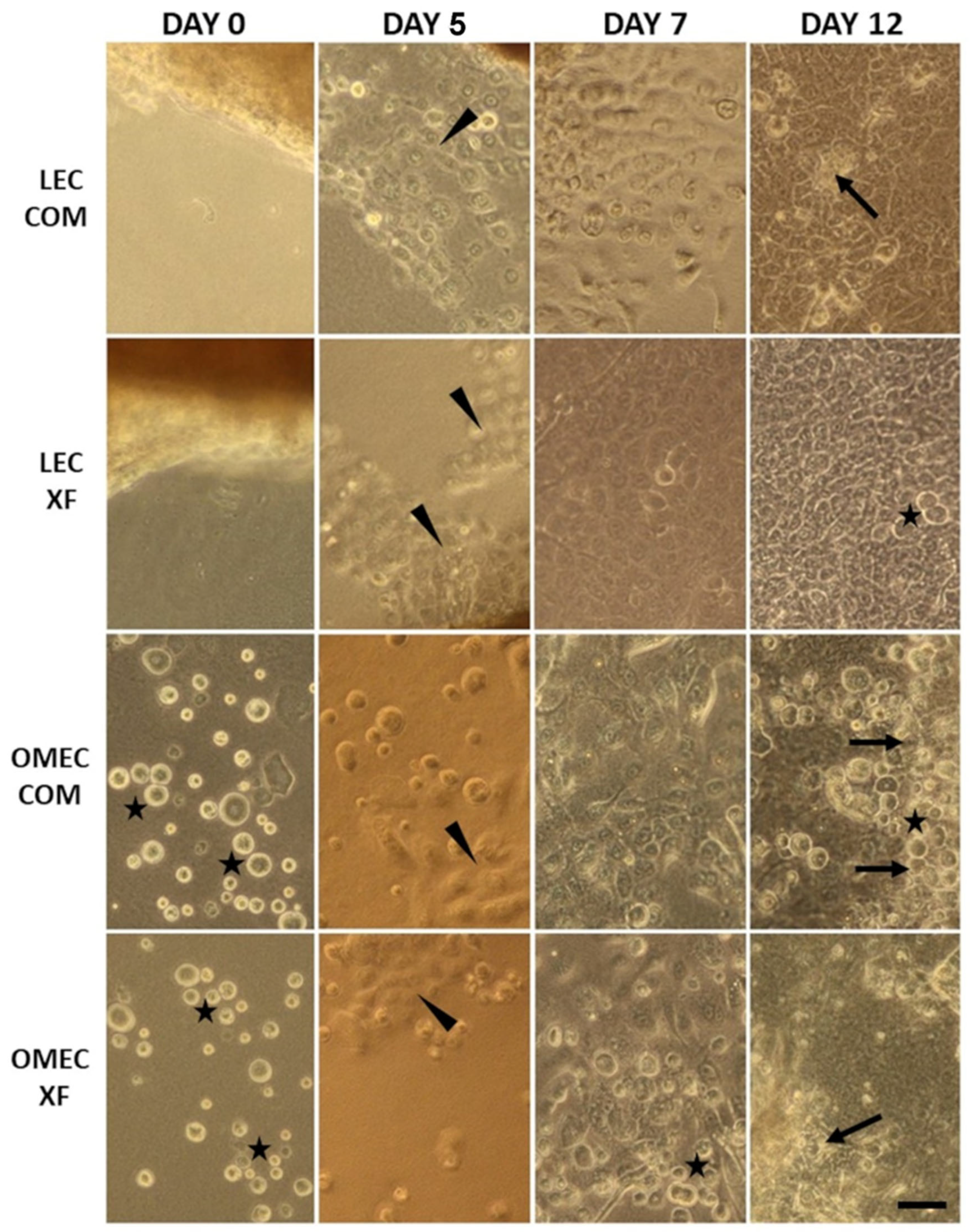
3.1. Cell Growth and Morphology

Limbal explant outgrowth was observed at 3.25 ± 0.89 (average ± SD) days in COM and 4.38 ± 1.06 days in XF conditions, with a significantly earlier onset in COM (paired t-test, p < 0.05) (Table S3). Similarly, in OMECs, cell attachment and proliferation began at 5.13 ± 0.83 days in COM and 5.75 ± 0.89 days in XF (Wilcoxon test, p < 0.05), again favoring COM. These findings indicate that COM supported faster initial growth kinetics than XF in both LECs and OMECs. Additionally, LECs initiated outgrowth earlier than OMECs (unpaired t-test, p < 0.05). The onset of outgrowth was characterized by a significant increase in adherent cell proliferation (Figure 1).
Despite these differences in initial growth, harvesting times were comparable across media and cell types, though viability tended to be higher in LECs than OMECs (Wilcoxon test, p < 0.05 for COM; unpaired t-test, p < 0.01 for XF). LECs reached 85–95% confluence after 10.88 ± 2.64 days in COM and 12.63 ± 2.26 days in XF (p > 0.05), with viability values of 92.5% ± 4.84 and 92.88% ± 5.18, respectively (p > 0.05). Immediately after seeding, OMECs displayed a rounded morphology, and by 7–10 days, they formed stratified sheets with a cobblestone-like morphology, prominent nuclei, and well-defined cell–cell contacts (Figure 1). OMECs reached 85–95% confluence in 11.25 ± 1.67 days in COM and 12.0 ± 1.85 days in XF (p > 0.05). Upon harvesting, OMEC viability was 84.76% ± 8.01 in COM and 79.76% ± 8.11 in XF, with a significant difference favoring COM (paired t-test, p < 0.05) (Table S3).

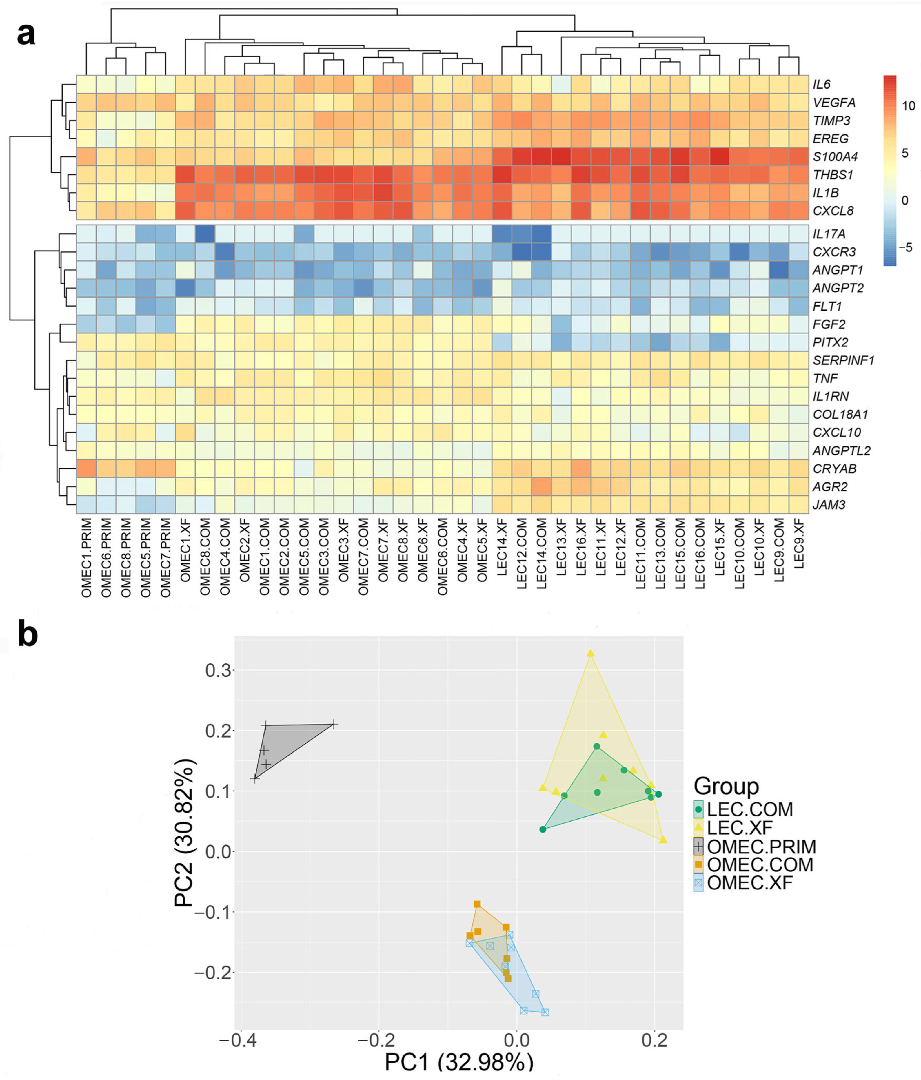
3.2. Angiogenic and Immunomodulatory Gene Expression

The transcriptional profiles of genes regulating vascularization and inflammation were analyzed in LECs and OMECs by RT-qPCR. Bidirectional hierarchical clustering of relative mRNA expressions was used to identify natural groupings across both genes and samples, without imposing predefined categories. This analysis revealed two main sample categories: primary OMECs and cultured cells, with further clustering separating LECs and OMECs (Figure 2a). This outcome was independently confirmed by PCA, which identified three distinct clusters corresponding to primary OMECs, OMECs cultured in COM and XF media, and LECs cultured in COM and XF media. The first two PCA components explained 63.8% of the variance (Figure 2b).
No significant differences in gene expression were observed between COM and XF conditions in either LECs or OMECs (adj. p > 0.05 for all genes). Given the absence of significant media effects, gene expression comparisons were performed between LECs and OMECs, pooling COM and XF samples (Figure 3a). PITX2 was preferentially expressed in OMECs (adj. p < 0.001). LECs had significantly higher expression of anti-angiogenic SERPINF1 (adj. p < 0.01) and TIMP3 (adj. p < 0.001), and also showed higher levels of pro-angiogenic AGR2, ANGPTL2, CRYAB, JAM3, S100A4, and EREG (adj. p < 0.001 for all except EREG, adj. p < 0.01). OMECs exhibited higher expressions of pro-angiogenic FGF2 and anti-angiogenic COL18A1 (adj. p < 0.001 for FGF2, adj. p < 0.05 for COL18A1). Additionally, OMECs demonstrated significantly higher expression of the pro-inflammatory cytokines CXCL10, IL1B, IL6, and TNF (adj. p < 0.01 to <0.001) and the anti-inflammatory IL1RN (adj. p < 0.001). The gene expression of the anti-inflammatory cytokines IL4 and IL10 was undetectable in either cell type. Both LECs and OMECs showed comparable expression of VEGFA, ANGPT1, ANGPT2, FLT1, and THBS1, as well as CXCL3, CXCL8, and IL17A (adj. p > 0.05, Figure 3b).
Comparisons of primary uncultured and cultured OMECs (COM and XF) revealed significant changes in 12 genes (Figure 4). Notable findings included increased expression of the anti-angiogenic THBS1 and TIMP3 (adj. p < 0.001 for both COM and XF). Pro-angiogenic genes such as AGR2, EREG, FGF2, and JAM3 were significantly upregulated in cultured OMECs (adj. p < 0.01 to <0.001). By contrast, there was a downregulation of ANGPTL2 (adj. p < 0.05, COM; adj. p < 0.01, XF) and CRYAB (adj. p < 0.01, COM; adj. p < 0.001, XF). Pro-inflammatory cytokines, including CXCL8, IL1B, IL6, and TNF, were significantly elevated in cultured OMECs (adj. p < 0.001, COM and XF). There was a small positive within-donor correlation (correlation coefficient = 0.24) between primary and cultured OMECs.

4. Discussion

Neovascularization after COMET likely results from an imbalance of angiogenic factors [9]. We compared the angiogenic profile of ex vivo expanded LECs and OMECs [17,18], based on previously suggested markers [10,11,12,13,14], and investigated whether differences in immunoregulation contribute to this phenomenon.
With regard to anti-angiogenic gene expression, there is no consensus in previous studies, possibly due to diverged experimental models and culture conditions including media or cultivation surface. In our study, LECs and OMECs exhibited differential expression in three anti-angiogenic genes. LECs had higher SERPINF1 (PEDF) and TIMP3 mRNA levels, while COL18A (endostatin) was higher in OMECs. Interestingly, we did not find differences in the expression of THBS1, which is considered as the main anti-angiogenic factor maintaining corneal avascularity [14,41], and for which immunostaining was significantly higher in cultured LECs compared to OMECs [14]. We also found similar FLT mRNA levels in human ex vivo expanded LECs and OMECs, in accordance with a study from Sekiyama et al. (2006) comparing sFLT-1 immunoreactivity [14]. However, in the same study, PEDF and endostatin protein levels were all higher in LECs [14]. Using human biopsies, Chen et al. reported higher TIMP3 but not PEDF immunoreactivity in corneal tissue compared to oral mucosa. In addition, healthy corneal tissues showed positive sFLT-1 immunoreactivity, which was absent in oral mucosa and conjunctival biopsies [10]. In vitro, rabbit LECs exhibited higher sFLT protein levels than OMECs, with recombinant sFLT-1 shown to reduce OMEC angiogenic capacity [12]. Therefore, the findings of this study regarding anti-angiogenic factors should be interpreted within the broader context of these existing discrepancies.
Unlike the inconsistencies observed in anti-angiogenic gene expression, all instances of pro-angiogenic factor expression aligned with previous studies. In our study, VEGFA and angiopoietins had similar levels in both cell types, consistent with previous reports [12,14]. Also, AGR2, EREG, and JAM3 transcriptional levels were higher in LECs, as shown previously [13]. Although these genes are associated with angiogenesis and wound healing [13], their higher expression in LECs likely reflects physiological synthesis within the limbal niche [34], although their functions in the cornea may shift during inflammation or injury. A key finding was the significantly higher FGF2 expression in the cultured OMECs than in the LECs, aligning with rabbit studies [12]. Moreover, in support of our findings, when LECs were used as feeder layers for OMECs, FGF2 expression decreased, while SERPINF1 levels were increased compared to OMECs grown on a 3T3 cell surface [54].
To our knowledge, a comprehensive comparison of immunomodulatory mediators between LECs and OMECs has not yet been conducted. We have shown that anti- and pro-vascularization genes are expressed in both cell types. However, OMECs show a significantly higher expression of immunomodulatory genes. It is well known that the angiogenic privilege of the cornea is closely tied to its immune privilege [55], and inflammatory mediators can increase FGF2 and VEGFA mRNA or protein levels [47,56]. Normal corneas maintain low pro-inflammatory cytokine expression, which rises under pathological conditions. For instance, reducing inflammation through cultivated limbal epithelium transplantation for LSCD lowers IL1B mRNA levels in the cornea to levels comparable to healthy tissue [46]. These findings raise the question of whether the higher expression of inflammation-related genes in the oral mucosa may be directly linked to an increased vascularization capacity of OMECs after grafting. Intrinsic differences, related to the immune and angiogenic phenotypes of OMECs, are possibly related to these findings. For example, in normal oral mucosal keratinocytes, similar cytokine expression patterns were found, without prior immune stimulation [57].
We have confirmed that the cultivation of OMECs significantly affected the expression of twelve genes, ten of which were upregulated. The simultaneous upregulation of both pro-angiogenic and anti-angiogenic factors likely reflects a complex adaptation of cultured OMECs to the in vitro environment. This upregulation may result not only from the activation of cell proliferation, as previously confirmed [17], but also from stress responses due to cultivation conditions or a feedback mechanism aimed at balancing angiogenic signaling. CRYAB and ANGPTL2 downregulation may indicate a decline in pro-inflammatory or angiogenic signaling following in vitro expansion and highlight that not all pro-angiogenic genes follow the same pattern in response to cultivation. The observed reduction in ANGLPTL2 expression, linked to inflammation and angiogenesis [22,23], was an unexpected finding, but it is still plausible that this change may be associated with the shift in the inflammatory phenotype observed in OMECs. Moreover, CRYAB gene downregulation may be associated with the regulation of inflammation [58], although the degree of this involvement merits further study.
In our experiments, we compared the growing efficiency of two media, COM and XF, using fibrin as a culture substrate [17]. Despite its potential to induce angiogenesis [59], we preferred this standardized substrate over the more biologically variable amniotic membrane. However, fibrin may still influence the angiogenic potential of both LECs and OMECs. Although harvesting times and final confluence levels were similar across media and cell types, COM significantly promoted earlier outgrowth and higher viability compared to XF, particularly in OMECs. This may indicate intrinsic differences in growth kinetics, possibly influenced by the culture protocol (explants versus primary cell suspensions). Nevertheless, gene expression differences between primary and cultured OMECs or LECs were unaffected by media choice.
The lack of a direct comparison between primary OMECs and LECs can be considered as a limitation of this study. LEC-based grafts are grown from explants [2], unlike OMECs, which are cultivated as suspension cultures [16]. For this reason, they were not included in our study. Another drawback is the use of different donors for OMECs and LECs, potentially introducing variability. Additionally, while these findings provide mechanistic insights, further validation at the protein level is essential.
Future research should aim to characterize OMEC-derived grafts in their final, transplantable form using both transcriptomic and proteomic analyses to determine whether these molecular signatures persist and how they influence clinical outcomes. Moreover, expanding beyond targeted gene panels to include unbiased, pathway-level approaches will be crucial for uncovering additional regulatory mechanisms and optimizing OMEC-based therapies while minimizing adverse events such as neovascularization. Although immunosuppressive strategies could help mitigate post-COMET neovascularization [3], reducing the need for postoperative medication use remains a priority due to the risk of adverse events.

5. Conclusions

In conclusion, ex vivo expanded OMECs and LECs exhibit distinct angiogenic and immunomodulatory gene expression profiles. Our most consistent finding across various models was the higher expression of FGF2 and lower expression of TIMP3 and SERPINF1 in OMECs compared to LECs. This suggests a stronger angiogenic potential in OMECs, highlighting the critical role of these factors in regulating angiogenesis. One of the most notable findings was the significantly higher expression of immunomodulatory genes in cultured OMECs compared to cultured LECs, as well as their upregulation during cultivation. These transcriptional differences provide valuable insights into the functional characteristics of LECs and OMECs and their relevance for regenerative therapy in LSCD.

Supplementary Materials

The following supporting information can be downloaded at https://www.mdpi.com/article/10.3390/bioengineering12070688/s1: Table S1. Details on the donors and samples. Table S2. Genes and primers used for RT-QPCR. Table S3. Growth parameters in limbal epithelial cells (LEC) and oral mucosal epithelial cells (OMEC).

Author Contributions

Conceptualization and methodology: K.J., T.V. and E.V.; investigation: E.V., J.V.C., N.S., V.K., M.N., T.V. and K.J.; software, data curation, and formal analysis: E.V.; visualization: E.V., J.V.C., N.S. and K.J.; writing—original draft, review, and editing: E.V. and K.J.; supervision: T.V. and K.J.; funding acquisition: K.J. All authors have read and agreed to the published version of the manuscript.

Funding

This study was supported by Norway Grants and Technology Agency of the Czech Republic (KAPPA Project No. TO01000099, research project BBMRI_CZ LM2023033). Institutional support (Charles University, Prague) was provided by the Cooperation: Medical Diagnostics and Basic Medical Sciences.

Institutional Review Board Statement

This study was conducted in accordance with the Declaration of Helsinki and approved by the Ethics Committee of General University Hospital in Prague, Czech Republic (IS, 1041/19 S-IV; date of approval: 20 June 2019).

Informed Consent Statement

Patient consent was waived because informed consent is not mandated for the use of donor tissue under Czech law (Law Act No. 372/2011 Coll.) if the data were anonymized before entering the records.

Data Availability Statement

Data are available upon request to the corresponding author. The data are not publicly available due to ongoing analyses and related publications that are currently on preparation.

Acknowledgments

We are grateful to Martina Vacíková and Lukáš Balogh for their valuable contribution throughout the experimental phase of this study.

Conflicts of Interest

The authors declare no conflicts of interest.

Abbreviations

The following abbreviations are used in this manuscript:
COMConventional culture medium
COMETOral mucosa epithelial cell transplantation
FGF2Fibroblast growth factor 2
LECsLimbal epithelial cells
LESCLimbal epithelial stem cell
LSCDBilateral limbal stem cell deficiency
PCAPrincipal component analysis
PEDFPigment epithelium-derived factor
RT-qPCRReal-time quantitative polymerase chain reaction
THBS1Thrombospondin
TIMP3Tissue inhibitor of metalloproteinase 3
VEGFAVascular endothelial growth factor A
XFXenobiotic-free culture medium

References

  1. Bonnet, C.; Roberts, J.S.; Deng, S.X. Limbal Stem Cell Diseases. Exp. Eye Res. 2021, 205, 108437. [Google Scholar] [CrossRef] [PubMed]
  2. Pellegrini, G.; Traverso, C.E.; Franzi, A.T.; Zingirian, M.; Cancedda, R.; De Luca, M. Long-Term Restoration of Damaged Corneal Surfaces with Autologous Cultivated Corneal Epithelium. Lancet 1997, 349, 990–993. [Google Scholar] [CrossRef]
  3. Nakamura, T.; Inatomi, T.; Sotozono, C.; Amemiya, T.; Kanamura, N.; Kinoshita, S. Transplantation of Cultivated Autologous Oral Mucosal Epithelial Cells in Patients with Severe Ocular Surface Disorders. Br. J. Ophthalmol. 2004, 88, 1280–1284. [Google Scholar] [CrossRef] [PubMed]
  4. Kohji, N.; Masayuki, Y.; Yasutaka, H.; Katsuhiko, W.; Kazuaki, Y.; Eijiro, A.; Shigeru, N.; Akihiko, K.; Naoyuki, M.; Hitoshi, W.; et al. Corneal Reconstruction with Tissue-Engineered Cell Sheets Composed of Autologous Oral Mucosal Epithelium. N. Engl. J. Med. 2004, 351, 1187–1196. [Google Scholar] [CrossRef]
  5. Cabral, J.V.; Jackson, C.J.; Utheim, T.P.; Jirsova, K. Ex Vivo Cultivated Oral Mucosal Epithelial Cell Transplantation for Limbal Stem Cell Deficiency: A Review. Stem Cell Res. Ther. 2020, 11, 301. [Google Scholar] [CrossRef]
  6. Inatomi, T.; Nakamura, T.; Kojyo, M.; Koizumi, N.; Sotozono, C.; Kinoshita, S. Ocular Surface Reconstruction with Combination of Cultivated Autologous Oral Mucosal Epithelial Transplantation and Penetrating Keratoplasty. Am. J. Ophthalmol. 2006, 142, 757–764. [Google Scholar] [CrossRef]
  7. Inatomi, T.; Nakamura, T.; Koizumi, N.; Sotozono, C.; Yokoi, N.; Kinoshita, S. Midterm Results on Ocular Surface Reconstruction Using Cultivated Autologous Oral Mucosal Epithelial Transplantation. Am. J. Ophthalmol. 2006, 141, 267–275. [Google Scholar] [CrossRef]
  8. O’Callaghan, A.R.; Daniels, J.T. Concise Review: Limbal Epithelial Stem Cell Therapy: Controversies and Challenges. Stem Cells 2011, 29, 1923–1932. [Google Scholar] [CrossRef]
  9. Lim, P.; Fuchsluger, T.A.; Jurkunas, U.V. Limbal Stem Cell Deficiency and Corneal Neovascularization. Semin. Ophthalmol. 2009, 24, 139–148. [Google Scholar] [CrossRef]
  10. Chen, H.-C.J.; Yeh, L.-K.; Tsai, Y.-J.; Lai, C.-H.; Chen, C.-C.; Lai, J.-Y.; Sun, C.-C.; Chang, G.; Hwang, T.-L.; Chen, J.-K.; et al. Expression of Angiogenesis-Related Factors in Human Corneas after Cultivated Oral Mucosal Epithelial Transplantation. Investig. Ophthalmol. Vis. Sci. 2012, 53, 5615–5623. [Google Scholar] [CrossRef]
  11. Kanayama, S.; Nishida, K.; Yamato, M.; Hayashi, R.; Sugiyama, H.; Soma, T.; Maeda, N.; Okano, T.; Tano, Y. Analysis of Angiogenesis Induced by Cultured Corneal and Oral Mucosal Epithelial Cell Sheets in Vitro. Exp. Eye Res. 2007, 85, 772–781. [Google Scholar] [CrossRef] [PubMed]
  12. Kanayama, S.; Nishida, K.; Yamato, M.; Hayashi, R.; Maeda, N.; Okano, T.; Tano, Y. Analysis of Soluble Vascular Endothelial Growth Factor Receptor-1 Secreted from Cultured Corneal and Oral Mucosal Epithelial Cell Sheets in Vitro. Br. J. Ophthalmol. 2009, 93, 263–267. [Google Scholar] [CrossRef]
  13. Attico, E.; Galaverni, G.; Torello, A.; Bianchi, E.; Bonacorsi, S.; Losi, L.; Manfredini, R.; Lambiase, A.; Rama, P.; Pellegrini, G. Comparison between Cultivated Oral Mucosa and Ocular Surface Epithelia for COMET Patients Follow-Up. Int. J. Mol. Sci. 2023, 24, 11522. [Google Scholar] [CrossRef] [PubMed]
  14. Sekiyama, E.; Nakamura, T.; Kawasaki, S.; Sogabe, H.; Kinoshita, S. Different Expression of Angiogenesis-Related Factors between Human Cultivated Corneal and Oral Epithelial Sheets. Exp. Eye Res. 2006, 83, 741–746. [Google Scholar] [CrossRef]
  15. Brejchova, K.; Trosan, P.; Studeny, P.; Skalicka, P.; Utheim, T.P.; Bednar, J.; Jirsova, K. Characterization and Comparison of Human Limbal Explant Cultures Grown under Defined and Xeno-Free Conditions. Exp. Eye Res. 2018, 176, 20–28. [Google Scholar] [CrossRef] [PubMed]
  16. Zhao, Y.; Ma, L. Systematic Review and Meta-Analysis on Transplantation of Ex Vivo Cultivated Limbal Epithelial Stem Cell on Amniotic Membrane in Limbal Stem Cell Deficiency. Cornea 2015, 34, 592–600. [Google Scholar] [CrossRef]
  17. Cabral, J.V.; Voukali, E.; Smorodinova, N.; Balogh, L.; Kolin, V.; Studeny, P.; Netukova, M.; Jirsova, K. Cultivation and Characterization of Oral Mucosal Epithelial Cells on Fibrin Gel in a Xenobiotic-Free Medium for the Treatment of Limbal Stem Cell Deficiency. Exp. Eye Res. 2025, 253, 110300. [Google Scholar] [CrossRef]
  18. Trousil, J.; Cabral, J.V.; Voukali, E.; Nováčková, J.; Pop-Georgievski, O.; Vacík, T.; Studený, P.; Studenovska, H.; Jirsova, K. Electrospun Poly(l-Lactide-Co-Dl-Lactide) Nanofibrous Scaffold as Substrate for Ex Vivo Limbal Epithelial Cell Cultivation. Heliyon 2024, 10, e30970. [Google Scholar] [CrossRef]
  19. R Core Team. R: A Language and Environment for Statistical Computing; R Foundation for Statistical Computing: Vienna, Austria, 2025; Available online: https://www.R-project.org/ (accessed on 22 June 2025).
  20. Ma, D.H.-K.; Chen, J.-K.; Zhang, F.; Lin, K.-Y.; Yao, J.-Y.; Yu, J.-S. Regulation of Corneal Angiogenesis in Limbal Stem Cell Deficiency. Prog. Retin. Eye Res. 2006, 25, 563–590. [Google Scholar] [CrossRef]
  21. Okada-Ban, M.; Thiery, J.P.; Jouanneau, J. Fibroblast Growth Factor-2. Int. J. Biochem. Cell Biol. 2000, 32, 263–267. [Google Scholar] [CrossRef]
  22. Toyono, T.; Usui, T.; Yokoo, S.; Kimakura, M.; Nakagawa, S.; Yamagami, S.; Miyata, K.; Oike, Y.; Amano, S. Angiopoietin-like Protein 2 Is a Potent Hemangiogenic and Lymphangiogenic Factor in Corneal Inflammation. Investig. Ophthalmol. Vis. Sci. 2013, 54, 4278–4285. [Google Scholar] [CrossRef] [PubMed]
  23. Santulli, G. Angiopoietin-Like Proteins: A Comprehensive Look. Front. Endocrinol. 2014, 5, 4. [Google Scholar] [CrossRef] [PubMed]
  24. Ferrari, G.; Giacomini, C.; Bignami, F.; Moi, D.; Ranghetti, A.; Doglioni, C.; Naldini, L.; Rama, P.; Mazzieri, R. Angiopoietin 2 Expression in the Cornea and Its Control of Corneal Neovascularisation. Br. J. Ophthalmol. 2016, 100, 1005–1010. [Google Scholar] [CrossRef] [PubMed]
  25. Suri, C.; Jones, P.F.; Patan, S.; Bartunkova, S.; Maisonpierre, P.C.; Davis, S.; Sato, T.N.; Yancopoulos, G.D. Requisite Role of Angiopoietin-1, a Ligand for the TIE2 Receptor, during Embryonic Angiogenesis. Cell 1996, 87, 1171–1180. [Google Scholar] [CrossRef]
  26. Guo, H.; Zhu, Q.; Yu, X.; Merugu, S.B.; Mangukiya, H.B.; Smith, N.; Li, Z.; Zhang, B.; Negi, H.; Rong, R.; et al. Tumor-Secreted Anterior Gradient-2 Binds to VEGF and FGF2 and Enhances Their Activities by Promoting Their Homodimerization. Oncogene 2017, 36, 5098–5109. [Google Scholar] [CrossRef]
  27. Delom, F.; Mohtar, M.A.; Hupp, T.; Fessart, D. The Anterior Gradient-2 Interactome. Am. J. Physiol. Cell Physiol. 2020, 318, C40–C47. [Google Scholar] [CrossRef]
  28. Kase, S.; He, S.; Sonoda, S.; Kitamura, M.; Spee, C.; Wawrousek, E.; Ryan, S.J.; Kannan, R.; Hinton, D.R. αB-Crystallin Regulation of Angiogenesis by Modulation of VEGF. Blood 2010, 115, 3398–3406. [Google Scholar] [CrossRef]
  29. Boelens, W.C. Cell Biological Roles of αB-Crystallin. Prog. Biophys. Mol. Biol. 2014, 115, 3–10. [Google Scholar] [CrossRef]
  30. Morita, S.; Shirakata, Y.; Shiraishi, A.; Kadota, Y.; Hashimoto, K.; Higashiyama, S.; Ohashi, Y. Human Corneal Epithelial Cell Proliferation by Epiregulin and Its Cross-Induction by Other EGF Family Members. Mol. Vis. 2007, 13, 2119–2128. [Google Scholar]
  31. Zhang, Y.; Kobayashi, T.; Hayashi, Y.; Yoshioka, R.; Shiraishi, A.; Shirasawa, S.; Higashiyama, S.; Ohashi, Y. Important Role of Epiregulin in Inflammatory Responses during Corneal Epithelial Wound Healing. Investig. Ophthalmol. Vis. Sci. 2012, 53, 2414–2423. [Google Scholar] [CrossRef]
  32. Li, C.; Zhang, F.; Wang, Y. S100A Proteins in the Pathogenesis of Experimental Corneal Neovascularization. Mol. Vis. 2010, 16, 2225–2235. [Google Scholar]
  33. Ambartsumian, N.; Klingelhöfer, J.; Grigorian, M.; Christensen, C.; Kriajevska, M.; Tulchinsky, E.; Georgiev, G.; Berezin, V.; Bock, E.; Rygaard, J.; et al. The Metastasis-Associated Mts1(S100A4) Protein Could Act as an Angiogenic Factor. Oncogene 2001, 20, 4685–4695. [Google Scholar] [CrossRef] [PubMed]
  34. Polisetti, N.; Zenkel, M.; Menzel-Severing, J.; Kruse, F.E.; Schlötzer-Schrehardt, U. Cell Adhesion Molecules and Stem Cell-Niche-Interactions in the Limbal Stem Cell Niche. Stem Cells 2016, 34, 203–219. [Google Scholar] [CrossRef] [PubMed]
  35. Rabquer, B.J.; Amin, M.A.; Teegala, N.; Shaheen, M.K.; Tsou, P.-S.; Ruth, J.H.; Lesch, C.A.; Imhof, B.A.; Koch, A.E. Junctional Adhesion Molecule-C Is a Soluble Mediator of Angiogenesis. J. Immunol. 2010, 185, 1777–1785. [Google Scholar] [CrossRef] [PubMed]
  36. Dawson, D.W.; Volpert, O.V.; Gillis, P.; Crawford, S.E.; Xu, H.; Benedict, W.; Bouck, N.P. Pigment Epithelium-Derived Factor: A Potent Inhibitor of Angiogenesis. Science 1999, 285, 245–248. [Google Scholar] [CrossRef]
  37. Matsui, T.; Nishino, Y.; Maeda, S.; Yamagishi, S. PEDF-Derived Peptide Inhibits Corneal Angiogenesis by Suppressing VEGF Expression. Microvasc. Res. 2012, 84, 105–108. [Google Scholar] [CrossRef]
  38. Ambati, B.K.; Nozaki, M.; Singh, N.; Takeda, A.; Jani, P.D.; Suthar, T.; Albuquerque, R.J.C.; Richter, E.; Sakurai, E.; Newcomb, M.T.; et al. Corneal Avascularity Is Due to Soluble VEGF Receptor-1. Nature 2006, 443, 993–997. [Google Scholar] [CrossRef]
  39. Kenney, M.C.; Chwa, M.; Alba, A.; Saghizadeh, M.; Huang, Z.-S.; Brown, D.J. Localization of TIMP-1, TIMP-2, TIMP-3, Gelatinase A and Gelatinase B in Pathological Human Corneas. Curr. Eye Res. 1998, 17, 238–246. [Google Scholar] [CrossRef]
  40. Ebrahem, Q.; Qi, J.H.; Sugimoto, M.; Ali, M.; Sears, J.E.; Cutler, A.; Khokha, R.; Vasanji, A.; Anand-Apte, B. Increased Neovascularization in Mice Lacking Tissue Inhibitor of Metalloproteinases-3. Investig. Ophthalmol. Vis. Sci. 2011, 52, 6117–6123. [Google Scholar] [CrossRef]
  41. Cursiefen, C.; Masli, S.; Ng, T.F.; Dana, M.R.; Bornstein, P.; Lawler, J.; Streilein, J.W. Roles of Thrombospondin-1 and -2 in Regulating Corneal and Iris Angiogenesis. Investig. Ophthalmol. Vis. Sci. 2004, 45, 1117–1124. [Google Scholar] [CrossRef]
  42. O’Reilly, M.S.; Boehm, T.; Shing, Y.; Fukai, N.; Vasios, G.; Lane, W.S.; Flynn, E.; Birkhead, J.R.; Olsen, B.R.; Folkman, J. Endostatin: An Endogenous Inhibitor of Angiogenesis and Tumor Growth. Cell 1997, 88, 277–285. [Google Scholar] [CrossRef] [PubMed]
  43. Moore, J.E.; McMullen, T.C.B.; Campbell, I.L.; Rohan, R.; Kaji, Y.; Afshari, N.A.; Usui, T.; Archer, D.B.; Adamis, A.P. The Inflammatory Milieu Associated with Conjunctivalized Cornea and Its Alteration with IL-1 RA Gene Therapy. Investig. Ophthalmol. Vis. Sci. 2002, 43, 2905–2915. [Google Scholar]
  44. Gao, N.; Liu, X.; Wu, J.; Li, J.; Dong, C.; Wu, X.; Xiao, X.; Yu, F.-S.X. CXCL10 Suppression of Hem- and Lymph-Angiogenesis in Inflamed Corneas through MMP13. Angiogenesis 2017, 20, 505–518. [Google Scholar] [CrossRef] [PubMed]
  45. Romagnani, P.; Annunziato, F.; Lasagni, L.; Lazzeri, E.; Beltrame, C.; Francalanci, M.; Uguccioni, M.; Galli, G.; Cosmi, L.; Maurenzig, L.; et al. Cell Cycle–Dependent Expression of CXC Chemokine Receptor 3 by Endothelial Cells Mediates Angiostatic Activity. J. Clin. Investig. 2001, 107, 53–63. [Google Scholar] [CrossRef]
  46. Pauklin, M.; Steuhl, K.-P.; Meller, D. Characterization of the Corneal Surface in Limbal Stem Cell Deficiency and after Transplantation of Cultivated Limbal Epithelium. Ophthalmology 2009, 116, 1048–1056. [Google Scholar] [CrossRef] [PubMed]
  47. Biswas, P.S.; Banerjee, K.; Kinchington, P.R.; Rouse, B.T. Involvement of IL-6 in the Paracrine Production of VEGF in Ocular HSV-1 Infection. Exp. Eye Res. 2006, 82, 46–54. [Google Scholar] [CrossRef]
  48. Koch, A.E.; Polverini, P.J.; Kunkel, S.L.; Harlow, L.A.; DiPietro, L.A.; Elner, V.M.; Elner, S.G.; Strieter, R.M. Interleukin-8 as a Macrophage-Derived Mediator of Angiogenesis. Science 1992, 258, 1798–1801. [Google Scholar] [CrossRef]
  49. Strieter, R.M.; Kunkel, S.L.; Elner, V.M.; Martonyi, C.L.; Koch, A.E.; Polverini, P.J.; Elner, S.G. Interleukin-8. A Corneal Factor That Induces Neovascularization. Am. J. Pathol. 1992, 141, 1279–1284. [Google Scholar]
  50. Yoshida, S.; Ono, M.; Shono, T.; Izumi, H.; Ishibashi, T.; Suzuki, H.; Kuwano, M. Involvement of Interleukin-8, Vascular Endothelial Growth Factor, and Basic Fibroblast Growth Factor in Tumor Necrosis Factor Alpha-Dependent Angiogenesis. Mol. Cell. Biol. 1997, 17, 4015–4023. [Google Scholar] [CrossRef]
  51. Chi, H.; Wei, C.; Ma, L.; Yu, Y.; Zhang, T.; Shi, W. The Ocular Immunological Alterations in the Process of High-Risk Corneal Transplantation Rejection. Exp. Eye Res. 2024, 245, 109971. [Google Scholar] [CrossRef]
  52. Volpert, O.V.; Fong, T.; Koch, A.E.; Peterson, J.D.; Waltenbaugh, C.; Tepper, R.I.; Bouck, N.P. Inhibition of Angiogenesis by Interleukin 4. J. Exp. Med. 1998, 188, 1039–1046. [Google Scholar] [CrossRef] [PubMed]
  53. Samolov, B.; Kvanta, A.; van der Ploeg, I. Delayed Neovascularization in Inflammation-Induced Corneal Neovascularization in Interleukin-10-Deficient Mice. Acta Ophthalmol. 2010, 88, 251–256. [Google Scholar] [CrossRef] [PubMed]
  54. Duan, C.-Y.; Xie, H.-T.; Zhao, X.-Y.; Xu, W.-H.; Zhang, M.-C. Limbal Niche Cells Can Reduce the Angiogenic Potential of Cultivated Oral Mucosal Epithelial Cells. Cell. Mol. Biol. Lett. 2019, 24, 3. [Google Scholar] [CrossRef]
  55. Hermankova, B.; Javorkova, E.; Palacka, K.; Holan, V. Perspectives and Limitations of Mesenchymal Stem Cell-Based Therapy for Corneal Injuries and Retinal Diseases. Cell Transplant. 2025, 34, 9636897241312798. [Google Scholar] [CrossRef] [PubMed]
  56. Xie, Y.; Su, N.; Yang, J.; Tan, Q.; Huang, S.; Jin, M.; Ni, Z.; Zhang, B.; Zhang, D.; Luo, F.; et al. FGF/FGFR Signaling in Health and Disease. Signal Transduct. Target. Ther. 2020, 5, 181. [Google Scholar] [CrossRef]
  57. Formanek, M.; Knerer, B.; Temmel, A.; Thurnher, D.; Millesi, W.; Kornfehl, J. Oral Keratinocytes Derived from the Peritonsillar Mucosa Express the Proinflammatory Cytokine IL-6 without Prior Stimulation. J. Oral Pathol. Med. 1998, 27, 202–206. [Google Scholar] [CrossRef]
  58. Fung, G.; Wong, J.; Berhe, F.; Mohamud, Y.; Xue, Y.C.; Luo, H. Phosphorylation and Degradation of αB-Crystallin during Enterovirus Infection Facilitates Viral Replication and Induces Viral Pathogenesis. Oncotarget 2017, 8, 74767–74780. [Google Scholar] [CrossRef]
  59. Dvorak, H.F.; Harvey, V.S.; Estrella, P.; Brown, L.F.; McDonagh, J.; Dvorak, A.M. Fibrin Containing Gels Induce Angiogenesis. Implications for Tumor Stroma Generation and Wound Healing. Lab. Investig. 1987, 57, 673–686. [Google Scholar]
Figure 1. Inverted phase-contrast microscopy of cultured limbal epithelial cells (LECs) and oral mucosal epithelial cells (OMECs) on fibrin substrate in complex (COM) and xenobiotic-free (XF) media. The micrograph shows the progression of proliferation to confluence from the seeding day (0) to 5, 7, and 12 days of cultivation in representative images from different experiments. Note the round morphology of non-attached or proliferating cells (stars), the start of overgrowth from limbal explants or attached OMECs (arrowheads), and the stratification (arrows). Scale bar: 50 μm.
Bioengineering 12 00688 g001
Figure 2. Angiogenesis- and inflammation-related gene expression profiles in limbal epithelial cells (LECs) and oral mucosal epithelial cells (OMECs). (a) Bidirectional hierarchical clustering heatmap of the relative mRNA expression for the tested genes (rows) between all samples (columns). The color scale on the right side of the plot represents higher expression in red and lower expression in blue. (b) Principal component analysis of the relative mRNA expression levels between all samples of LECs and OMECs. COM, complex medium; XF, xenobiotic-free medium; PRIM, primary culture of OMEC; the numbers on sample names refer to matched biological replicates.
Bioengineering 12 00688 g002
Figure 3. Differences and overlaps in selected angiogenesis- and immune-regulating genes between limbal (LECs, n = 16) and oral mucosal epithelial cell (OMECs, n = 16) cultures. (a) Statistically significant differences in the expression of PITX2, and angiogenesis- and immune-regulating genes between LECs and OMECs. Asterisks on the horizontal line above every two boxes indicate statistical significance defined as adj. p < 0.01 (**) and p < 0.001 (***). (b) Genes with higher or comparable expression in cultured LECs and OMECs, grouped according to their function.
Bioengineering 12 00688 g003
Figure 4. Significantly altered gene expression patterns of oral mucosal epithelial cells (OMECs) after culture. Color-coded lines connect data from the same donor. Expression of anti-angiogenic, pro-angiogenic, and immunomodulatory genes in primary cells before culture (PRIM, n = 5) and after culture in complex (COM, n = 8) or xenobiotic-free (XF, n = 8) medium. Asterisks indicate statistical significance defined as adj. p < 0.05 (*), p < 0.01 (**), and p < 0.001 (***).
Bioengineering 12 00688 g004
Table 1. Gene functions and relevance to vascularization, inflammation, or oral mucosa identity.
GeneFull NameFunction
Pro-angiogenic
FGF2Fibroblast growth factor 2Promotes angiogenesis, wound healing, and cell proliferation [9,20,21]
VEGFAVascular endothelial growth factor AKey driver of blood vessel formation under hypoxic or inflammatory conditions [9,20]
ANGPTL2Angiopoietin like 2Induces inflammation and neovascularization in tissue remodeling and cancer [22,23]
ANGPT2Angiopoietin 2Destabilizes blood vessels, facilitating VEGF-driven angiogenesis [24]
ANGPT1Angiopoietin 1Stabilizes blood vessels and supports endothelial cell survival [25]
AGR2Anterior gradient 2, protein disulphide isomerase family memberPromotes epithelial cell growth and has roles in wound healing and tumor angiogenesis [26,27]
CRYABCrystallin alpha BActs as a molecular chaperone; involved in protection against stress and may support angiogenesis [28,29]
EREGEpiregulinEGFR ligand that promotes epithelial repair, proliferation, and angiogenesis [30,31]
S100A4S100 calcium binding protein A4Associated with motility, invasion, and angiogenesis in cancer and inflammation [32,33]
JAM3Junctional adhesion molecule 3Mediates cell–cell adhesion and contributes to angiogenesis [34,35]
Anti-angiogenic
SERPINF1Serpin family F member 1 (PEDF)Potent anti-angiogenic factor that inhibits VEGF signaling [36,37]
FLT1Fms related receptor tyrosine kinase 1 (VEGFR1)Acts as a decoy receptor for VEGF, limiting angiogenesis [38]
TIMP3TIMP metallopeptidase inhibitor 3Inhibits matrix metalloproteinases and angiogenesis [39,40]
THBS1Thrombospondin 1Suppresses angiogenesis through interaction with CD36 and other receptors [14,41]
COL18A1Collagen type XVIII alpha 1 chainEncodes endostatin, a known inhibitor of angiogenesis [42]
Immuno-regulatory
IL1RNInterleukin 1 receptor antagonistAnti-inflammatory cytokine that blocks IL-1 signaling [43]
CXCL10C-X-C motif chemokine ligand 10Chemokine that recruits immune cells and can inhibit angiogenesis [44]
CXCR3C-X-C motif chemokine receptor 3Receptor for CXCL9/10/11 involved in T-cell trafficking and inflammation [45]
IL1BInterleukin 1 betaKey pro-inflammatory cytokine promoting leukocyte recruitment [9,46]
IL6Interleukin 6Pro-inflammatory cytokine with roles in acute phase response and angiogenesis [9,47]
CXCL8C-X-C motif chemokine ligand 8 (IL-8)Promotes neutrophil chemotaxis and angiogenesis [9,48,49,50]
IL17AInterleukin 17AStimulates pro-inflammatory responses and angiogenesis [51]
TNFTumor necrosis factorMaster regulator of inflammation, promotes angiogenesis under pathological conditions [9,50]
IL4Interleukin 4Anti-inflammatory cytokine, promotes Th2 immune response [52]
IL10Interleukin 10Anti-inflammatory cytokine that limits immune responses [53]
Oral mucosa identity
PITX2Paired like homeodomain 2Transcription factor involved in oral epithelial identity and development [13]
Disclaimer/Publisher’s Note: The statements, opinions and data contained in all publications are solely those of the individual author(s) and contributor(s) and not of MDPI and/or the editor(s). MDPI and/or the editor(s) disclaim responsibility for any injury to people or property resulting from any ideas, methods, instructions or products referred to in the content.

