Article

Predictive Model of Gemtuzumab Ozogamicin Response in Childhood Acute Myeloid Leukemia on Event-Free Survival: Data Analysis Based on Trial AAML0531

1
Department of Hematology/Oncology, Children’s Medical Center, Sun Yat-sen Memorial Hospital, Sun Yat-sen University, Guangzhou 510120, China
2
Guangdong Provincial Key Laboratory of Malignant Tumor Epigenetics and Gene Regulation, Sun Yat-sen Memorial Hospital, Sun Yat-sen University, Guangzhou 510120, China
*
Authors to whom correspondence should be addressed.
These authors contributed equally to this work.
Bioengineering 2025, 12(3), 297; https://doi.org/10.3390/bioengineering12030297
Submission received: 26 January 2025 / Revised: 6 March 2025 / Accepted: 10 March 2025 / Published: 14 March 2025
(This article belongs to the Section Biosignal Processing)

Abstract

Purpose: We aimed to develop a simple nomogram and online calculator that can identify the optimal subpopulation of pediatric acute myeloid leukemia (AML) patients who would benefit most from gemtuzumab ozogamicin (GO) therapy. Methods: Within the framework of the phase Ⅲ AAML0531 randomized trial for GO, the event-free survival (EFS) probability was calculated using a predictor-based nomogram to evaluate GO treatment impact on EFS in relation to baseline characteristics. Nomogram performance was assessed by the area under the receiver operating characteristic curve (AUC) and the calibration curve with 500 bootstrap resample validations. Decision curve analysis (DCA) was performed to evaluate the clinical utility of the nomogram. Results: A total of 705 patients were randomly assigned to two arms: the No-GO arm (n = 358) and the GO arm (n = 347). We performed a nomogram model for EFS among childhood AML. The AUC (C statistic) of the nomogram was 0.731 (95%CI: 0.614–0.762) in the development group and 0.700 (95% CI: 0.506–0.889) in the validation group. DCA showed that the model in the development and validation groups had a net benefit when the risk thresholds were 0–0.75 and 0–0.75, respectively. Notably, an intriguing observation emerged wherein pediatric patients with AML exhibited a favorable outcome in the GO arm when the predicted 5-year EFS probability fell below 60%, demonstrating a superior EFS compared to the No-GO Arm. Conclusions: We have developed a nomogram and online calculator that can be used to predict EFS among childhood AML based on trial AAML0531, and this might help deciding which patients can benefit from GO.

1. Introduction

Acute myeloid leukemia (AML) is a heterogeneous disorder characterized by unfavorable prognosis, accounting for 15–20% of childhood leukemias. While treatment outcomes have improved over the past decades, approximately 30–40% of pediatric AML patients still experience relapse, highlighting the need for novel therapeutic approaches [1,2,3]. Childhood AML exhibits distinct biological characteristics compared to adult AML, including higher frequencies of core-binding factor translocations (e.g., t(8;21) and inv(16)) and lower incidence of adverse genetic markers like FLT3-ITD, which necessitate age-specific treatment strategies [4,5,6].
The cell-surface antigen CD33 is expressed in over 90% of pediatric AML cases, with expression patterns showing developmental stage-dependent variations that differ from adult populations [7,8,9]. Gemtuzumab ozogamicin (GO), a humanized anti-CD33 antibody conjugated to calicheamicin, has shown particular promise in pediatric AML. Initial phase II studies (AAML00P2 and AAML03P1) demonstrated the safety and efficacy of GO combined with chemotherapy in children, achieving 3-year event-free survival (EFS) rates of 53–63% [10,11,12]. The Children’s Oncology Group (COG) AAML0531 phase III trial, the largest randomized study specifically designed for pediatric AML, established that GO addition improved 3-year EFS from 46.9% to 53.1% (p = 0.04), though this benefit was not uniformly distributed across all risk groups [13].
Despite these advances, childhood AML presents unique challenges in treatment optimization. The developing hematopoietic system’s increased sensitivity to chemotherapy toxicity, combined with the long-term sequelae of intensive therapy in growing children, underscores the need for precision medicine approaches [14,15,16]. Current risk stratification systems, primarily based on cytogenetics and treatment response, fail to adequately predict which patients will benefit most from targeted therapies like GO. This limitation is particularly relevant given the recent FDA reapproval of GO for CD33-positive AML in 2017, which included pediatric indications but lacked age-specific guidance [17,18,19].
A critical knowledge gap persists in identifying the optimal pediatric subpopulation for GO therapy. While meta-analyses of adult trials suggest particular benefit in favorable-risk AML, pediatric studies have shown conflicting results regarding the interaction between molecular subtypes and GO response [20,21,22]. Furthermore, the long-term follow-up data from AAML0531 (median 4.9 years) revealed diminishing GO benefits over time, emphasizing the need for robust predictive tools specific to childhood AML [23,24,25,26,27,28].
This study addresses a critical gap in pediatric AML management by developing the first GO response prediction model specifically for childhood AML, integrating molecular markers (WT1/CEBPA) with clinical parameters. Our web-based nomogram represents a novel precision medicine tool that outperforms existing risk stratification systems in identifying GO-responsive subgroups.

2. Patients and Methods

2.1. Patients

Pediatric patients newly diagnosed with AML who participated in the COG AAML0531 phase III trial, spanning from August 2006 to June 2010, were eligible for the present analysis. The findings presented herein are derived, in whole or in part, from the data generated by the Therapeutically Applicable Research to Generate Effective Treatments (TARGET) initiative (https://ocg.cancer.gov/programs/target (accessible on 1 January 2025)) under the identifier phs000218. The data utilized for this analysis can be accessed at https://portal.gdc.cancer.gov/projects (accessible on 1 January 2025). Notably, the primary distinction between this study and prior investigations lies in its exclusive enrollment of pediatric patients with AML, specifically those under the age of 18 years.
The demographic characteristics were compared between the No-GO and GO arms of the study. Inclusion criteria included newly diagnosed de novo AML, centralized cytogenetic/molecular profiling, and full treatment adherence per protocol. Key exclusions included acute promyelocytic leukemia (APL) or prior chemotherapy. A total of 705 patients with de novo acute myeloid leukemia (AML), aged 0 to 18 years, were randomly allocated to either the no-GO arm (n = 358) or the GO arm (n = 347). Of the entire study population, 51.5% (363 patients) were male and 48.5% (342 patients) were female, with a median age of 10 years. The randomization process resulted in well-balanced study arms, with no significant differences observed between the two arms in terms of gender, age, white blood cell count (WBC), the French–American–British (FAB) category, peripheral blood (PB) blast count, bone marrow (BM) blast count, karyotype, risk group, or gene mutation status.
After each induction course, remission was evaluated thoroughly. Upon completion of IND1, there were comparable rates of CR, not-in-CR status, and death. At the conclusion of IND2, no statistically significant difference in CR rates was observed between the two arms. In summary, the overall induction remission rates were similar across both arms, indicating that the two arms were comparable in this regard. The demographic information of 705 eligible pediatric patients with AML, as classified by the GO, is presented in Table 1.

2.2. Study Design

In total, 705 patients diagnosed with de novo AML, within the age range of 0 to 18 years, were randomly assigned to two distinct treatment arms: the standard five-course chemotherapy arm (designated as the No-GO arm; comprising 358 patients) and the same chemotherapy regimen augmented with two doses of GO at 3 mg/m2 per dose (designated as the GO arm; consisting of 347 patients). All patients were treated under the Children’s Oncology Group (COG) AAML0531 phase III trial (NCT00372593), which mandated a fixed chemotherapy backbone for all participants. No modifications to chemotherapeutic agents, dosing, or scheduling occurred during the trial period (2006–2014). Protocol amendments were restricted to supportive care (e.g., antimicrobial prophylaxis), with no changes to the chemotherapy backbone.
This allocation was carried out during the first induction course (IND1) and the second intensification phase (as detailed in Supplemental Table S1). Prior to the administration of GO, chemotherapy-induced cytoreduction was performed to optimize CD33 target saturation, rather than escalating the dosage of GO. Concurrent administration of anthracyclines was avoided to mitigate the potential for additive hepatotoxicity. Risk stratification within both treatment arms guided the decision to proceed with SCT, based on diagnostic molecular/cytogenetic risk factors and the disease response observed after IND1, as specified below.
Low risk (LR) was defined based on the presence of t(8;21)(q22;q22), inv(16)(p13.1q22), or t(16;16)(p13.1;q22). Patients with LR were not considered for SCT. High risk (HR) was determined by the presence of monosomy 7, monosomy 5/5q deletion, or persistent disease (PD) with bone marrow blasts exceeding 15% at the end of IND1. Following enrollment of 374 eligible patients, the presence of FLT-3 internal tandem duplication with a high allelic ratio (>0.4; FLT3-ITD HAR) was added as a criterion for HR classification. Cytogenetics was the primary determinant in risk classification, with FLT3-ITD HAR overriding favorable cytogenetics. All HR patients underwent best allogeneic SCT (nonsyngeneic matched family donor or unrelated donor) following INT1 or INT3. HR patients without suitable donors continued with their assigned chemotherapy regimen. Standard risk (SR) was defined by the absence of low- or high-risk factors, and these patients only received SCT if a matched family donor was available. Patients allocated to SCT underwent the procedure after INT1. Consequently, patients randomly assigned to the GO arm received only one dose during IND1. Response classification was determined by morphologic examination of bone marrow blasts, with complete remission (CR) defined as less than 5% blasts, partial remission (PR) as 5% to 15% blasts, and PD as more than 15% blasts. Patients with refractory disease (RD) were excluded from the protocol therapy. RD was defined as the presence of central nervous system (CNS) disease after IND1, bone marrow blasts ≥ 5%, or any extramedullary disease at the end of IND2. The clinical protocol was approved by the institutional review boards of all participating institutions, and the COG Myeloid Disease Biology Committee approved the research study. The trial was conducted in accordance with the Declaration of Helsinki and was registered at www.clinicaltrials.gov under NCT00372593.

2.3. Cytogenetic and Molecular Profiling

Centralized analysis was performed at COG-certified laboratories using conventional karyotyping and FISH for core abnormalities (t(8;21), inv(16), 11q23/MLL rearrangements), standard techniques in pediatric AML during the 2000s. FLT3-ITD status was determined via fragment analysis (sensitivity: 5% mutant alleles), consistent with era-specific guidelines.

2.4. Treatment Response and Follow-Up

Patients were deemed to be in CR if they exhibited less than 5% blasts and the absence of extramedullary disease following a single course of induction chemotherapy. Minimal residual disease (MRD) was determined using flow cytometry and was considered positive if 0.1% or more of the disease was detected at the conclusion of induction I. Clinical outcome data for patients enrolled in the COG AAML051 trial were analyzed as of 30 September 2014. The median follow-up period for eligible patients who were alive at the time of last contact and included in our analysis was 4.9 years (ranging from 0 to 9.6 years). EFS was defined as the duration from study entry until the occurrence of death, induction failure, or any type of relapse. Patients who were lost to follow-up were censored at the date of their last known contact.

2.5. Statistical Analyses

Univariable analyses were performed to identify prognostic factors associated with event-free survival (EFS). Categorical variables (e.g., sex, FAB subtype) were compared using Chi-square test or Fisher’s exact test as appropriate. Continuous variables (e.g., WBC, age) were analyzed with Mann–Whitney U test or Kruskal–Wallis test based on data distribution, using forward stepwise selection with Akaike information criterion. Variables reaching p < 0.05 were included in the multivariable Cox proportional hazards model. The final prediction model was developed using three core components: 1. Discrimination: ROC curve analysis (AUC) evaluating model’s ability to distinguish high/low-risk patients. 2. Calibration: bootstrap-corrected curves comparing predicted vs. observed 5-year EFS probabilities. 3. Clinical utility: decision curve analysis quantifying net benefit across clinical decision thresholds. Internal validation was performed using 1000 bootstrap resamples to prevent overfitting. All analyses were conducted with R 4.2.2 (survival and rms packages).

3. Results

3.1. Univariable and Multivariable Analyses

In the analysis, risk factors that were deemed significant during univariable analysis were incorporated into multivariable models, with the aim of elucidating the independent effects of these factors on EFS among pediatric patients with AML, as outlined in Table 2. Through multivariate analysis, it was determined that WBC; (p < 0.001) and WT1 mutation (p < 0.001) emerged as independent factors that have a detrimental impact on EFS. Conversely, favorable effects on EFS for the entire cohort were observed in association with FAB M7 subtype (p = 0.017), inv(16) cytogenetic abnormality (p = 0.014), t(8;21) translocation (p = 0.008), and CEBPA mutation (p = 0.011). Notably, no significant association was detected between GO treatment and EFS (p = 0.818). Additionally, a comparable 5-year EFS rate was observed between patients who did not receive GO treatment and those who did (44.8% versus 45.5%, respectively; p = 0.7; refer to Supplementary Figure S1 for further details).

3.2. Development and Assessment of the Predictive Nomogram

3.2.1. Model Development

Given the absence of a discernible difference in 5-year EFS between the No-GO and GO arms, we have undertaken further research to develop a predictive model aimed at identifying the optimal patient population for the GO arms. To achieve this, subjects were randomly assigned to either the development group (n = 529; 75%) or the validation group. Initially, utilizing the finalized multivariable model, we created a nomogram. This was accomplished by assigning a weighted score to each factor associated with 5-year EFS in pediatric AML. Each predictor was assigned a specific score on a rating scale, and the total points for each variable were summed. A vertical line was then drawn downward from the total points to indicate the corresponding probability of EFS. Notably, a higher score corresponded to a lower probability of EFS, as depicted in Figure 1.
The area under the curve (AUC) of the receiver operating characteristic (ROC) curve for this comprehensive nomogram was determined to be 0.731, with a 95%CI ranging from 0.614 to 0.762 (Supplementary Figure S2A). Subsequent to 1000 bootstrap iterations, the calibration curves demonstrated a satisfactory alignment between the observed and predicted probabilities within the nomogram, thereby attesting to the stability and precision of the predictive models (Supplementary Figure S3A). Furthermore, the curves revealed that the model yielded a net benefit when the risk threshold was situated within the range of 0 to 0.75 (Supplementary Figure S4A).

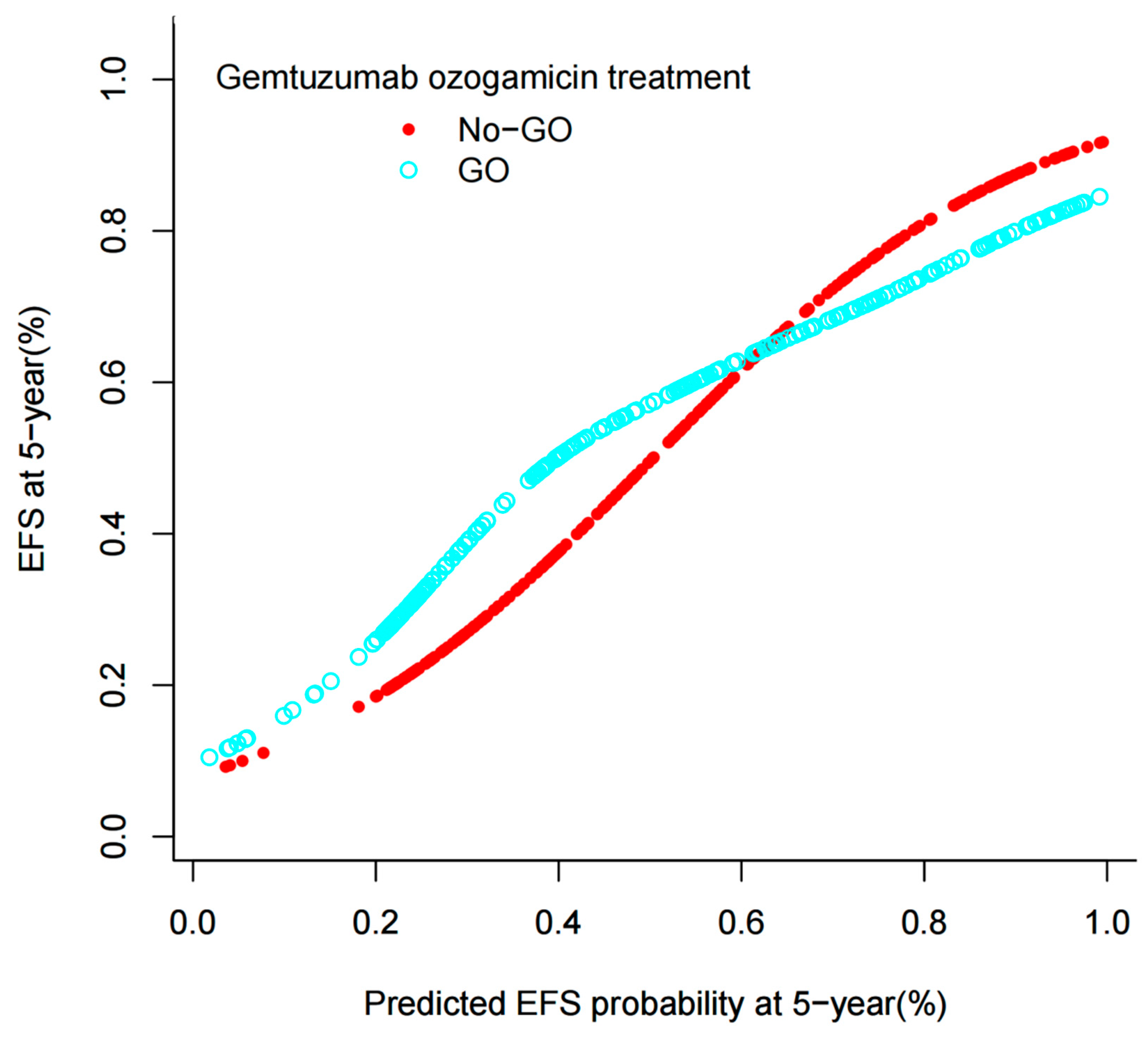
3.2.2. Model Validation

To evaluate the internal validity of the model, it was implemented on the remaining 25% of the randomly selected internal validation dataset. The assessment of internal validity in childhood AML patients was conducted utilizing the C-statistic, calibration, and decision curve analysis. The results of the 25% random internal validation indicated a commendable predictive performance and stability of the nomogram models. Specifically, the AUC (C-statistic) of the model within the validation cohort was 0.700 (95% CI: 0.506–0.889), as depicted in Supplementary Figure S2B. Additionally, the nomogram calibration curve of the validation cohort demonstrated satisfactory calibration, as shown in Supplementary Figure S3B. The decision curves further revealed that the model yielded a net benefit when the risk threshold ranged from 0 to 0.75, as illustrated in Supplementary Figure S4B.

3.3. Survival Analysis for the Entire Cohort

As depicted in the aforementioned nomogram models, the identified independent predictors were subsequently employed to estimate the 5-year EFS probability for patients in both the No-GO and GO arms. Notably, an intriguing observation emerged wherein pediatric patients with AML exhibited a favorable outcome in the GO arm when the predicted 5-year EFS probability fell below 60%, demonstrating a superior EFS compared to the No-GO Arm. Conversely, this advantage dissipated in the GO arm patients when the predicted EFS probability surpassed 60%, accompanied by a marked decline in EFS (Figure 2). The statistical significance of the interaction between the predicted EFS and the GO arm treatment arm was established (p < 0.05), underscoring the variability in the GO arm’s therapeutic benefit contingent upon the predicted EFS. Subsequent to bootstrap resampling, the significance of the interaction term persisted (p < 0.05). Importantly, when the predicted EFS probability exceeded 60%, no discernible difference was observed between the two arms (p > 0.05). Furthermore, we have developed an online calculator accessible at http://www.empowerstats.net/pmodel/?m=11688_PredictiveModelofGemtuzumabOzogamicinResponse, accessed on 25 January 2025 (Figure 3), which facilitates the prediction of 5-year EFS among pediatric AML patients, thereby aiding pediatricians in making informed decisions regarding the initiation of GO treatment.

4. Discussion

Based on several randomized trials, the recent US Food and Drug Administration approval of GO, a CD33-directed agent, represents significant advancements in AML treatment. Nevertheless, the efficacy of GO in pediatric AML remains a matter of debate. The TARGET program, a collaboration between the COG and the National Cancer Institute (NCI), offers a robust platform for studying pediatric cancer cohorts [29]. Consequently, we accessed the TARGET dataset to retrieve trial AAML0531 data. The AAML0531 trial, the largest randomized pediatric de novo AML trial to date and the sole pediatric randomized controlled trial, to incorporate GO into induction and intensification, demonstrated that GO significantly enhanced EFS at the 3-year mark (53.1% versus 46.9%; p = 0.04) [26]. However, our analysis within the context of the COG AAML0531 trial revealed no statistically significant improvement in 5-year EFS with GO (p = 0.7). These discrepancies with prior research can be attributed to our extended follow-up period and our study’s exclusive focus on patients under the age of 18. Given the lack of difference in 5-year EFS between the No-GO and GO arms, we have developed a predictive model aimed at identifying the optimal population for GO administration.
Previous research has conclusively demonstrated that EFS in pediatric AML is not merely a single risk factor, but rather a complex interplay of numerous factors [30,31]. The nomogram’s incorporation of WBC and the WT1 mutation aligns with prior pediatric AML studies, where hyperleukocytosis and WT1 aberrations correlate with chemoresistance and relapse. High WBC and WT1 mutations identified subgroups deriving significant GO benefit. Mechanistically, elevated WBC may reflect CD33-high blast populations susceptible to GO, while WT1 mutations are linked to chemoresistance pathways counteracted by GO’s DNA damage mechanism. Conversely, CEBPA mutations and inv(16) predicted favorable outcomes independent of GO, suggesting limited added value in these subgroups.
Importantly, the lack of GO benefit in high-EFS subgroups suggests that CD33-independent pathways may dominate in biologically favorable AML, warranting further investigation. Notably, our current study reveals that elevated WBC levels and the presence of the WT1 mutation are predictive of poorer EFS in childhood AML, whereas CEBPA mutation, as well as the presence of inv(16) and t(8;21) karyotypic abnormalities, are indicative of a more favorable prognosis. These findings are in line with previous investigations, emphasizing the strong correlation between these factors and EFS in pediatric AML [31,32,33].
In the current study, we have devised and rigorously tested a nomogram to predict EFS among pediatric patients with AML. This predictive model is based on several key factors: WBC, FAB category, karyotype, WT1 mutation status, and CEBPA mutation status. The AUC values obtained in the training and validation cohorts were 0.731 and 0.700, respectively, underscoring the excellent accuracy and robustness of the nomogram. The usability of this model is enhanced by the limited number of predictors required and the straightforwardness of the computational procedures, making it a comprehensive and sensitive tool for identifying pediatric AML patients with a poor prognosis.
Our nomogram model is capable of deriving the cumulative score of risk factors and the probability of EFS, thereby facilitating pediatricians in providing more pertinent recommendations to patients diagnosed with AML. Notably, prior research endeavors have also developed predictive models aimed at forecasting EFS among pediatric AML cases [34,35]. Nevertheless, these prior models were constructed primarily based on data sourced from single-center studies or limited sample sizes. Consequently, they may be constrained by the absence of a comprehensive dataset derived from a multi-center, randomized phase Ⅲ trial akin to the COG AAML0531 study. In contrast, our model exhibits enhanced precision and credibility in comparison to these previous studies.
In our comprehensive assessment, we have deemed it necessary to meticulously balance the potential advantages of GO treatment against its individual-level drawbacks. Consequently, our current report offers invaluable insights to the existing literature, carrying significant clinical ramifications. Specifically, we have leveraged an online calculator to determine an individual’s predicted EFS, thereby facilitating an accurate decision on the suitability of GO treatment. For instance, if the nomogram’s total score corresponds to a predicted EFS of 70%, the EFS for the No-GO arm surpasses that of the GO arm, but the difference is not significant. Based on risk-benefit assessment, we advise the patient to opt for the No-GO arm. Conversely, if the nomogram predicts an EFS of 50% and the EFS for the GO arm is higher than that of the No-GO arm, the patient should undergo GO treatment. This approach empowers pediatricians and patients to make well-informed clinical decisions when evaluating the role of GO treatment in AML.
We acknowledge the fact that our study has some limitations. First of all, our study did not incorporate CD33 expression levels, a critical determinant of GO efficacy, due to data unavailability in the TARGET cohort. Future validation in pediatric cohorts with CD33 quantification (e.g., flow cytometry, transcriptomics) is essential to refine predictive accuracy. Secondly, the prediction model of this study was only internally validated and not externally validated. Thirdly, retrospective analysis of biomarker data introduces potential selection bias, though all samples were prospectively collected. Future work should do the following: (1) validate the model in multicenter cohorts; (2) incorporate emerging biomarkers (e.g., FLT3-ITD allelic ratio); (3) assess cost-effectiveness in resource-limited settings.
In conclusion, our nomogram and online calculator are simple to use and able to predict the EFS among childhood AML based on trial AAML0531. The novel nomogram total score corresponds to predicted 5-year EFS, and this might help deciding which patients can benefit from GO treatment, so as to achieve the purpose of precise treatment of pediatric AML.

Supplementary Materials

The following supporting information can be downloaded at: https://www.mdpi.com/article/10.3390/bioengineering12030297/s1, Supplementary Figure S1. Patients’ event-free survival according to the GO treatment. Supplementary Figure S2. Receiver operator characteristic (ROC) curves of the nomogram. (A) ROC curves of the nomogram in the developing group. (B) ROC curve of the nomogram in the validation group. Supplementary Figure S3. Calibration curves of the nomogram. (A) Calibration curves of the nomogram in the developing group. (B) Calibration curve of the nomogram in the validation group. Supplementary Figure S4. Decision curve of the nomogram. (A) Decision curves of the nomogram in the developing group. (B) Decision curves of the nomogram in the validation group. Supplementary Table S1. AAML0531 Therapeutic Regimen.

Author Contributions

X.-Y.L. and K.-Y.Q. wrote the manuscript and performed the study. J.-P.F. and D.-H.Z. reviewed the manuscript. All authors have read and agreed to the published version of the manuscript.

Funding

This work was supported by Guangdong Basic and Applied Basic Research Foundation (NO. 2024A1515012445), Special topic of science and technology for agriculture and social development of Guangzhou key R&D plan (2024B03J1247), Bethune Medical Scientific Research Fund Project (No. SCE111DS), Guangdong Medical Scientific Research Foundation (A2024057), Basic Research Project (Dengfeng Hospital) jointly funded by Universities (Institutes) in Guangzhou (No. 202201020310), Guangzhou Basic and Applied Basic Research Foundation (2024A04J4686), Yat-sen Excellent Young Scientists Fund (2024A03J1185), Fundamental Research Funds for the Central Universities, Sun Yat-sen University (24qnpy314), and Sun Yat-sen Pilot Scientific Research Fund (YXQH202205).

Institutional Review Board Statement

The clinical protocol was approved by the institutional review boards of all participating institutions, and the COG Myeloid Disease Biology Committee approved the research study.

Informed Consent Statement

Not applicable.

Data Availability Statement

The data in the current study are available from the corresponding author on reasonable request.

Acknowledgments

The study was supported by grant from Guangdong Science and Technology Department (2020B1212060018).

Conflicts of Interest

The authors declare no conflicts of interest.

References

  1. Döhner, H.; Wei, A.H.; Löwenberg, B. Towards precision medicine for AML. Nat. Rev. Clin. Oncol. 2021, 18, 577–590. [Google Scholar] [CrossRef]
  2. Daver, N.; Schlenk, R.F.; Russell, N.H.; Levis, M.J. Targeting FLT3 mutations in AML: Review of current knowledge and evidence. Leukemia 2019, 33, 299–312. [Google Scholar] [CrossRef]
  3. Kayser, S.; Levis, M.J. Updates on targeted therapies for acute myeloid leukaemia. Br. J. Haematol. 2022, 196, 316–328. [Google Scholar] [CrossRef]
  4. Röllig, C. Gemtuzumab ozogamicin in AML: The next chapter. Blood 2023, 142, 1673–1674. [Google Scholar] [CrossRef]
  5. Lambert, J.; Pautas, C.; Terré, C.; Raffoux, E.; Turlure, P.; Caillot, D.; Legrand, O.; Thomas, X.; Gardin, C.; Gogat-Marchant, K.; et al. Gemtuzumab ozogamicin for de novo acute myeloid leukemia: Final efficacy and safety updates from the open-label, phase III ALFA-0701 trial. Haematologica 2019, 104, 113–119. [Google Scholar] [CrossRef]
  6. Freeman, S.D.; Thomas, A.; Thomas, I.; Hills, R.K.; Vyas, P.; Gilkes, A.F.; Metzner, M.; Jakobsen, N.A.; Kennedy, A.; Moore, R.; et al. Fractionated vs single-dose gemtuzumab ozogamicin with determinants of benefit in older patients with AML: The UK NCRI AML18 trial. Blood 2023, 142, 1697–1707. [Google Scholar] [CrossRef]
  7. Dhillon, S. Inotuzumab Ozogamicin. First Pediatric Approval. Paediatr. Drugs 2024, 26, 59–467. [Google Scholar] [CrossRef]
  8. Fenwarth, L.; Fournier, E.; Cheok, M.; Boyer, T.; Gonzales, F.; Castaigne, S.; Boissel, N.; Lambert, J.; Dombret, H.; Preudhomme, C.; et al. Biomarkers of Gemtuzumab Ozogamicin Response for Acute Myeloid Leukemia Treatment. Int. J. Mol. Sci. 2020, 21, 5626. [Google Scholar] [CrossRef]
  9. Wang, H.; Yang, S.; Chen, L.; Li, Y.; He, P.; Wang, G.; Dong, H.; Ma, P.; Ding, G. Tumor diagnosis using carbon-based quantum dots: Detection based on the hallmarks of cancer. Bioact. Mater. 2023, 33, 174–222. [Google Scholar] [CrossRef]
  10. Wijnen, N.E.; Koedijk, J.B.; Klein, K.; Luesink, M.; Goemans, B.F.; Zwaan, C.M.; Kaspers, G.J. Treating CD33-Positive de novo Acute Myeloid Leukemia in Pediatric Patients: Focus on the Clinical Value of Gemtuzumab Ozogamicin. Onco Targets Ther. 2023, 16, 297–308. [Google Scholar] [CrossRef]
  11. Aplenc, R.; Alonzo, T.A.; Gerbing, R.B.; Lange, B.J.; Hurwitz, C.A.; Wells, R.J.; Bernstein, I.; Buckley, P.; Krimmel, K.; Smith, F.O.; et al. Safety and efficacy of gemtuzumab ozogamicin in combination with chemotherapy for pediatric acute myeloid leukemia: A report from the Children’s Oncology Group. J. Clin. Oncol. 2008, 26, 2390–3295. [Google Scholar] [CrossRef] [PubMed]
  12. Cooper, T.M.; Franklin, J.; Gerbing, R.B.; Alonzo, T.A.; Hurwitz, C.; Raimondi, S.C.; Hirsch, B.; Smith, F.O.; Mathew, P.; Arceci, R.J.; et al. AAML03P1, a pilot study of the safety of gemtuzumab ozogamicin in combination with chemotherapy for newly diagnosed childhood acute myeloid leukemia: A report from the Children’s Oncology Group. Cancer 2012, 118, 761–769. [Google Scholar] [CrossRef] [PubMed]
  13. Kantarjian, H.M.; DiNardo, C.D.; Kadia, T.M.; Daver, N.G.; Altman, J.K.; Stein, E.M.; Jabbour, E.; Schiffer, C.A.; Lang, A.; Ravandi, F. Acute myeloid leukemia management and research in 2025. CA Cancer J. Clin. 2025, 75, 46–67. [Google Scholar] [CrossRef] [PubMed]
  14. Singh, A.; Thota, S.; Bradley, T.; Griffiths, E.A.; Faber, M.G.; Sadek, S.; Przespolewski, A.; Thompson, J.E.; Baron, J.; Cronin, T.; et al. Outcomes of Adult Acute Myeloid Leukemia Treated with Gemtuzumab-Ozogamicin: Cue to Optimized Chemotherapy Backbone. Clin. Lymphoma Myeloma Leuk. 2021, 21, 613–620. [Google Scholar] [CrossRef]
  15. Petersdorf, S.H.; Kopecky, K.J.; Slovak, M.; Willman, C.; Nevill, T.; Brandwein, J.; Larson, R.A.; Erba, H.P.; Stiff, P.J.; Stuart, R.K.; et al. A phase 3 study of gemtuzumab ozogamicin during induction and postconsolidation therapy in younger patients with acute myeloid leukemia. Blood 2013, 121, 4854–4860. [Google Scholar] [CrossRef]
  16. Burnett, A.; Cavenagh, J.; Russell, N.; Hills, R.; Kell, J.; Jones, G.; Nielsen, O.J.; Khwaja, A.; Thomas, I.; Clark, R. Defining the dose of gemtuzumab ozogamicin in combination with induction chemotherapy in acute myeloid leukemia: A comparison of 3 mg/m2 with 6 mg/m2 in the NCRI AML17 Trial. Haematologica 2016, 101, 724–731. [Google Scholar] [CrossRef]
  17. Bornhäuser, M.; Schliemann, C.; Schetelig, J.; Röllig, C.; Kramer, M.; Glass, B.; Platzbecker, U.; Burchert, A.; Hänel, M.; Müller, L.P.; et al. Allogeneic Hematopoietic Cell Transplantation vs Standard Consolidation Chemotherapy in Patients With Intermediate-Risk Acute Myeloid Leukemia: A Randomized Clinical Trial. JAMA Oncol. 2023, 9, 519–526. [Google Scholar] [CrossRef]
  18. Burnett, A.K.; Hills, R.K.; Grimwade, D.; Jovanovic, J.V.; Craig, J.; McMullin, M.F.; Kell, J.; Wheatley, K.; Yin, J.A.L.; Hunter, A.; et al. Inclusion of chemotherapy in addition to anthracycline in the treatment of acute promyelocytic leukaemia does not improve outcomes: Results of the MRC AML15 trial. Leukemia 2013, 27, 843–851. [Google Scholar] [CrossRef]
  19. Burnett, A.K.; Hills, R.K.; E Hunter, A.; Milligan, D.; Kell, W.J.; Wheatley, K.; Yin, J.; McMullin, M.F.; Dignum, H.; Bowen, D. The addition of gemtuzumab ozogamicin to low-dose Ara-C improves remission rate but does not significantly prolong survival in older patients with acute myeloid leukaemia: Results from the LRF AML14 and NCRI AML16 pick-a-winner comparison. Leukemia 2013, 27, 75–81. [Google Scholar] [CrossRef]
  20. Burnett, A.K.; Hills, R.K.; Milligan, D.; Kjeldsen, L.; Kell, J.; Russell, N.H.; Yin, J.A.; Hunter, A.; Goldstone, A.H.; Wheatley, K. Identification of patients with acute myeloblastic leukemia who benefit from the addition of gemtuzumab ozogamicin: Results of the MRC AML15 trial. J. Clin. Oncol. 2011, 29, 369–377. [Google Scholar] [CrossRef]
  21. Burnett, A.K.; Russell, N.H.; Hills, R.K.; Kell, J.; Freeman, S.; Kjeldsen, L.; Hunter, A.E.; Yin, J.; Craddock, C.F.; Dufva, I.H.; et al. Addition of gemtuzumab ozogamicin to induction chemotherapy improves survival in older patients with acute myeloid leukemia. J. Clin. Oncol. 2012, 30, 3924–3931. [Google Scholar] [CrossRef] [PubMed]
  22. Castaigne, S.; Pautas, C.; Terré, C.; Raffoux, E.; Bordessoule, D.; Bastie, J.N.; Legrand, O.; Thomas, X.; Turlure, P.; Reman, O.; et al. Effect of gemtuzumab ozogamicin on survival of adult patients with de-novo acute myeloid leukaemia (ALFA-0701): A randomised, open-label, phase 3 study. Lancet 2012, 379, 1508–1516. [Google Scholar] [CrossRef] [PubMed]
  23. Delaunay, J.; Recher, C.; Pigneux, A.; Witz, F.; Vey, N.; Blanchet, O.; Lefebvre, P.; Luquet, I.; Guillerme, I.; Volteau, C.; et al. Addition of gemtuzumab ozogamycin to chemotherapy improves event-free survival but not overall survival of AML patients with intermediate cytogenetics not eligible for allogeneic transplantation: Results of the GOELAMS AML. Blood 2011, 118, 79. [Google Scholar] [CrossRef]
  24. Hills, R.K.; Castaigne, S.; Appelbaum, F.R.; Delaunay, J.; Petersdorf, S.; Othus, M.; Estey, E.H.; Dombret, H.; Chevret, S.; Ifrah, N.; et al. Addition of gemtuzumab ozogamicin to induction chemotherapy in adult patients with acute myeloid leukaemia: A meta-analysis of individual patient data from randomised controlled trials. Lancet Oncol. 2014, 15, 986–996. [Google Scholar] [CrossRef] [PubMed]
  25. Mort, J.F.; Brighton, D.; DiBenedetto, S.; Wells, L.; Clark, S.M.; Reid, J.; Patel, I.; Jackson, C.; Yelvington, B.; Miller, R.; et al. Evaluation of Gemtuzumab Ozogamicin and Anthracycline Dosing for Favorable Risk Acute Myeloid Leukemia. Eur. J. Haematol. 2025, 114, 481–494. [Google Scholar] [CrossRef]
  26. Gamis, A.S.; Alonzo, T.A.; Meshinchi, S.; Sung, L.; Gerbing, R.B.; Raimondi, S.C.; Hirsch, B.A.; Kahwash, S.B.; Heerema-McKenney, A.; Winter, L.; et al. Gemtuzumab ozogamicin in children and adolescents with de novo acute myeloid leukemia improves event-free survival by reducing relapse risk: Results from the randomized phase III Children’s Oncology Group trial AAML0531. J. Clin. Oncol. 2014, 32, 3021–3032. [Google Scholar] [CrossRef] [PubMed]
  27. Jen, E.Y.; Ko, C.-W.; Lee, J.E.; Del Valle, P.L.; Aydanian, A.; Jewell, C.; Norsworthy, K.J.; Przepiorka, D.; Nie, L.; Liu, J. FDA Approval: Gemtuzumab Ozogamicin for the Treatment of Adults with Newly Diagnosed CD33-Positive Acute Myeloid Leukemia. Clin. Cancer Res. 2018, 24, 3242–3246. [Google Scholar] [CrossRef]
  28. Norsworthy, K.J.; Ko, C.-W.; Lee, J.E.; Liu, J.; John, C.S.; Przepiorka, D.; Farrell, A.T.; Pazdur, R. FDA Approval Summary: Mylotarg for Treatment of Patients with Relapsed or Refractory CD33-Positive Acute Myeloid Leukemia. Oncologist 2018, 23, 1103–1108. [Google Scholar] [CrossRef]
  29. Bolouri, H.; Farrar, J.E.; Triche, T., Jr.; Ries, R.E.; Lim, E.L.; Alonzo, T.A.; Ma, Y.; Moore, R.; Mungall, A.J.; Marra, M.A.; et al. The molecular landscape of pediatric acute myeloid leukemia reveals recurrent structural alterations and age-specific mutational interactions. Nat. Med. 2018, 24, 103–112. [Google Scholar] [CrossRef]
  30. Elgarten, C.W.; Aplenc, R. Pediatric acute myeloid leukemia: Updates on biology, risk stratification, and therapy. Curr. Opin. Pediatr. 2020, 32, 57–66. [Google Scholar] [CrossRef]
  31. Liao, X.Y.; Fang, J.P.; Zhou, D.H.; Qiu, K.Y. CEBPA are independent good prognostic factors in pediatric acute myeloid leukemia. Hematol. Oncol. 2022, 40, 258–268. [Google Scholar] [CrossRef]
  32. Qiu, K.-Y.; Liao, X.-Y.; Li, Y.; Huang, K.; Xu, H.-G.; Fang, J.-P.; Zhou, D.-H. Outcome and prognostic factors of CBF pediatric AML patients with t(8;21) differ from patients with inv(16). BMC Cancer 2023, 23, 476. [Google Scholar] [CrossRef] [PubMed]
  33. Qiu, K.Y.; Liao, X.Y.; Liu, Y.; Huang, K.; Li, Y.; Fang, J.P.; Zhou, D.H. Poor outcome of pediatric patients with acute myeloid leukemia harboring high FLT3/ITD allelic ratios. Nat. Commun. 2022, 13, 3679. [Google Scholar] [CrossRef] [PubMed]
  34. Mądry, K.; Lis, K.; Biecek, P.; Młynarczyk, M.; Rytel, J.; Górka, M.; Kacprzyk, P.; Dutka, M.; Rodzaj, M.; Bołkun, Ł.; et al. Predictive Model for Infection Risk in Myelodysplastic Syndromes, Acute Myeloid Leukemia, and Chronic Myelomonocytic Leukemia Patients Treated with Azacitidine; Azacitidine Infection Risk Model: The Polish Adult Leukemia Group Study. Clin. Lymphoma Myeloma Leuk. 2019, 19, 264–274.e4. [Google Scholar] [CrossRef]
  35. Chen, M.; Zeng, Z.; Qin, W.; Cai, X.; Lu, X.; Chen, S. A novel prognostic model of methylation-associated genes in acute myeloid leukemia. Clin. Transl. Oncol. 2023, 25, 1719–1728. [Google Scholar] [CrossRef]
Figure 1. Nomogram to predict the probability of 5-year EFS in childhood AML patients.
Bioengineering 12 00297 g001
Figure 2. EFS rate plotted against predicted probability of EFS at 5-year after diagnosis. The lines for No-GO and GO cross at 60%.
Bioengineering 12 00297 g002
Figure 3. The online calculation for predicting probability of EFS at 5-year among childhood AML.
Bioengineering 12 00297 g003
Table 1. Baseline characteristics of study participants by GO status classification.
CharacteristicsTotalGO Statusp Value
No-GO (n = 358)GO (n = 347)
Gender, n (%) 0.248
Male363 (51.5%) 192 (53.6%)171 (49.3%)
Female342 (48.5%) 166 (46.4%)176 (50.7%)
Age (y), median (range)10.0 (0.0–18.0) 10.2 (0.0–18.0)9.7 (0.0–17.9)0.442
FAB category 0.798
M024 (4.1%) 12 (4.2%)12 (4.0%)
M178 (13.3%) 42 (14.6%)36 (12.0%)
M2156 (26.6%) 77 (26.8%)79 (26.3%)
M4161 (27.4%) 78 (27.2%)83 (27.7%)
M5128 (21.8%) 56 (19.5%)72 (24.0%)
M613 (2.2%) 8 (2.8%)5 (1.7%)
M727 (4.6%) 14 (4.9%)13 (4.3%)
Initial WBC (×109/L), median (range)33.5 (0.2–519.0)35.3 (0.2–473.1)32.7 (0.8–519.0)0.612
PB blast (%)48.0 (0.0–98.0)47.0 (0.0–98.0)49.0 (0.0–98.0)0.645
BM blast (%)71.0 (0.0–100.0)71.0 (0.0–99.0)71.0 (3.0–100.0)0.331
Karyotype 0.526
Normal167 (24.3%) 81 (23.1%)86 (25.6%)
inv(16)91 (13.3%) 46 (13.1%)45 (13.4%)
MLL125 (18.2%) 62 (17.7%)63 (18.8%)
t(8;21)109 (15.9%) 64 (18.3%)45 (13.4%)
Other194 (28.3%) 97 (27.7%)97 (28.9%)
Risk group, n (%) 0.568
Low risk262 (38.2%) 140 (40.1%)122 (36.2%)
Standard risk305 (44.5%) 151 (43.3%)154 (45.7%)
High risk119 (17.3%) 58 (16.6%)61 (18.1%)
CNSL, n (%) 0.541
No654 (92.8%) 330 (92.2%)324 (93.4%)
Yes51 (7.2%) 28 (7.8%)23 (6.6%)
FLT3-ITD status, n (%) 0.879
FLT3-ITD wild-type555 (78.7%) 281 (78.5%)274 (79.0%)
FLT3-ITD mutation150 (21.3%) 77 (21.5%)73 (21.0%)
CEBPA status, n (%) 0.898
CEBPA wild-type1743 (94.5%)342 (96.6%)323 (94.4%)
CEBPA mutation101 (5.5%)12 (3.4%)19 (5.6%)
NPM1 status, n (%) 0.480
NPM1 wild-type639 (91.4%) 321 (90.7%)318 (92.2%)
NPM1 mutation60 (8.6%) 33 (9.3%)27 (7.8%)
WT1 status, n (%) 0.923
WT1 wild-type647 (92.6%) 328 (92.7%)319 (92.5%)
WT1 mutation52 (7.4%) 26 (7.3%)26 (7.5%)
End of IND1 response 0.103
CR513 (73.6%) 257 (72.2%)256 (75.1%)
Not in CR176 (25.3%) 92 (25.8%)84 (24.6%)
Death8 (1.1%) 7 (2.0%)1 (0.3%)
End of IND2 response 0.099
CR600 (86.8%) 300 (84.5%)300 (89.3%)
Not in CR79 (11.4%) 46 (13.0%)33 (9.8%)
Death12 (1.7%) 9 (2.5%)3 (0.9%)
SCT in first CR 0.633
No518 (81.8%) 265 (82.6%)253 (81.1%)
Yes115 (18.2%) 56 (17.4%)59 (18.9%)
Abbreviations: WBC, white blood cell counts; PB blast, peripheral blood blast; BM, bone marrow blast; CNSL, central nervous system leukemia; CEBPA, CCAAT/enhancer-binding protein alpha; FLT3-ITD, fms-related tyrosine kinase 3; NPM1, nucleophosmin 1; WT1, Wilms tumor 1; CR, complete remission; IND, induction course; SCT, stem cell transplantation.
Table 2. Univariable and multivariable analyses on EFS.
VariablesUnivariable AnalysisMultivariable Analysis
HR (95%CI)p ValueHR (95%CI)p Value
Gender
MaleRef. Ref.
Female0.9 (0.7, 1.1) 0.1430.8 (0.6, 1.0) 0.107
Age1.0 (1.0, 1.0) 0.9951.0 (1.0, 1.0)0.084
Risk group
Low riskRef. Ref.
Standard risk3.1 (2.4, 4.0) <0.0011.7 (0.7, 3.9) 0.225
High risk3.3 (2.4, 4.4) <0.0011.1 (0.5, 2.6) 0.800
WBC1.0 (1.0, 1.0)<0.0011.0 (1.0, 1.0) <0.001
BM blast (%)1.0 (1.0, 1.0) <0.0011.0 (1.0, 1.0) 0.109
Peripheral blasts (%)1.0 (1.0, 1.0) 0.0921.0 (1.0, 1.0) 0.989
CNSL
NoRef. Ref.
Yes0.9 (0.6, 1.4) 0.6491.2 (0.8, 1.9)0.437
FAB category
M0Ref. Ref.
M10.5 (0.3, 0.9) 0.0120.8 (0.4, 1.4) 0.418
M20.3 (0.2, 0.6) <0.0010.9 (0.5, 1.8) 0.854
M40.5 (0.3, 0.8) 0.0050.9 (0.5, 1.7) 0.804
M50.6 (0.4, 0.9)0.0240.6 (0.3, 1.0) 0.065
M60.8 (0.3, 1.7) 0.4970.7 (0.3, 1.7) 0.416
M70.1 (0.1, 0.4)<0.0010.3 (0.1, 0.8) 0.017
Karyotype
NormalRef. Ref.
inv(16)0.5 (0.3, 0.8) <0.0010.3 (0.1, 0.8) 0.014
MLL1.2 (0.9, 1.7) 0.1380.8 (0.5, 1.3) 0.316
t(8;21)0.4 (0.3, 0.6) <0.0010.3 (0.1, 0.7) 0.008
Other1.2 (0.9, 1.6)0.1220.8 (0.5, 1.1) 0.190
FLT3-ITD status
FLT3-ITD wild-typeRef. Ref.
FLT3-ITD mutation1.6 (1.2, 2.0) <0.0011.0 (0.6, 1.5) 0.875
NPM1 status
NPM1 wild-typeRef. Ref.
NPM1 mutation0.5 (0.3, 0.8) 0.0030.5 (0.2, 1.0) 0.055
CEBPA status
CEBPA wild-typeRef. Ref.
CEBPA mutation0.4 (0.2, 0.8) 0.0080.2 (0.1, 0.7) 0.011
WT1 status
WT1 wild-typeRef. Ref.
WT1 mutation2.3 (1.7, 3.2) <0.0012.1 (1.4, 3.2) <0.001
Gemtuzumab ozogamicin treatment
No-GORef. Ref.
GO0.9 (0.8, 1.1) 0.5381.0 (0.8, 1.3)0.818
Abbreviations: WBC, white blood cell counts; BM, bone marrow blast; CNSL, central nervous system leukemia; CEBPA, CCAAT/enhancer-binding protein alpha; FLT3-ITD, fms-related tyrosine kinase 3; NPM1, nucleophosmin 1; WT1, Wilms tumor 1.
Disclaimer/Publisher’s Note: The statements, opinions and data contained in all publications are solely those of the individual author(s) and contributor(s) and not of MDPI and/or the editor(s). MDPI and/or the editor(s) disclaim responsibility for any injury to people or property resulting from any ideas, methods, instructions or products referred to in the content.

