A Sensor Localization and Orientation Method for OPM-MEG Based on Rigid Coil Structures and Magnetic Dipole Fitting Models
Abstract
1. Introduction
2. Principles and Methods
2.1. Magnetic Field Modeling in Space
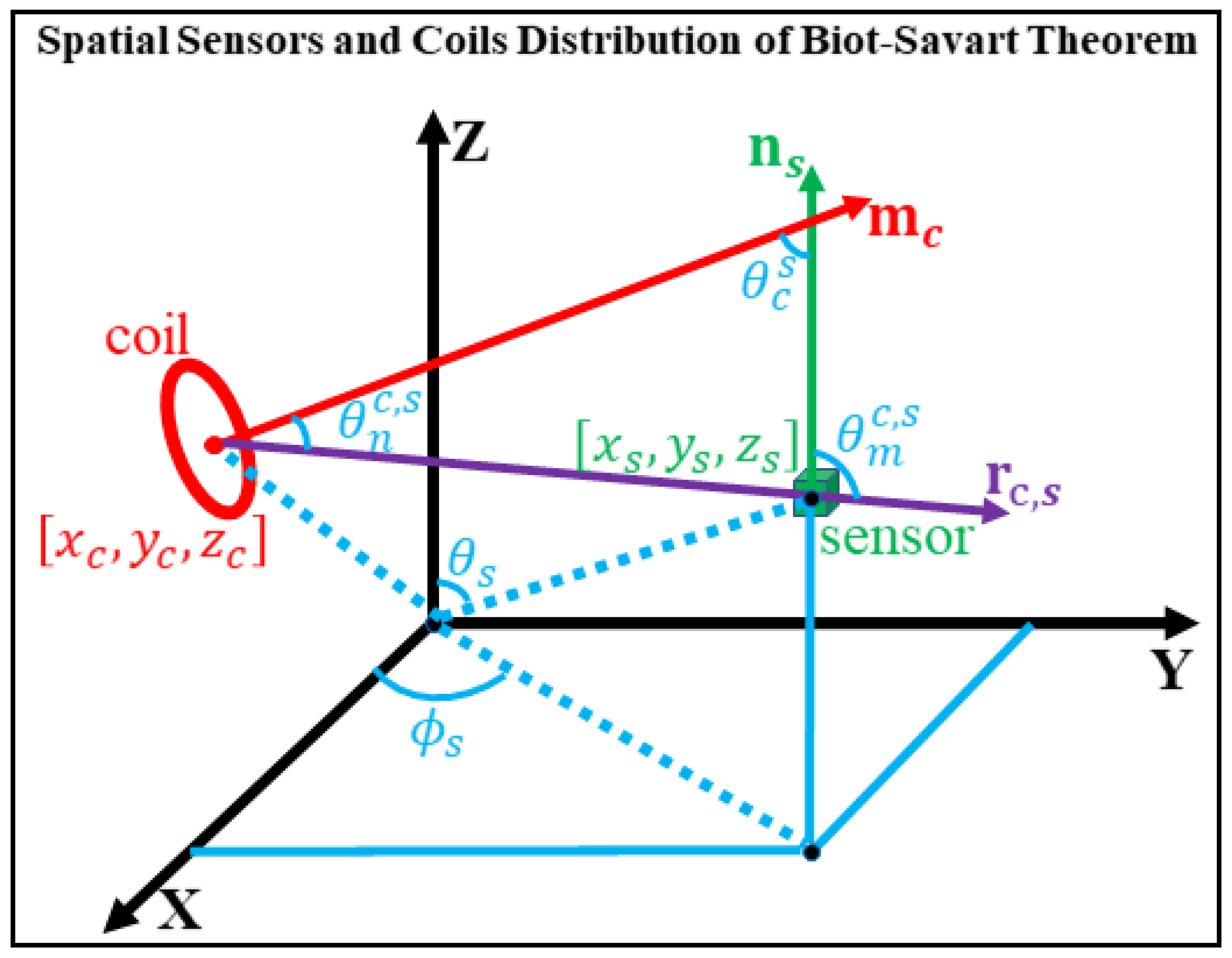
- Coil–sensor distance : The dipole field decays as while the error term is dominated by quadrupole term. As the sensor approaches the coil, the dipole approximation degrades rapidly.
- Angle : Governs the radial component of the dipole field. When , the magnetic moment is perpendicular to the observation direction. In this case, the radial component of the dipole field (the first term in Equation (2)) becomes zero, while the tangential component (the second term) remains non-zero. Therefore, the total dipole field does not vanish but changes direction.
- Angle : Determines the projection of the dipole field onto the sensor’s sensitive axis. When , the sensor’s sensitive axis is perpendicular to the observation direction, and the projection of the dipole field onto the sensor’s sensitive axis becomes minimal, although the total field itself is not zero.
- Angle : Describes the coupling between the dipole moment and the sensor axis. When or , the coupling is maximized, while intermediate values can produce destructive interference, amplifying local modeling errors.
2.2. Selection of Objective Functions
2.3. Ideal Single-Turn Coil Model
3. Simulation Analysis and System Design
3.1. Simulation of Magnetic Dipole Fitting in RCS Channels
3.2. Simulation Analysis of Sensor Localization and Orientation Using RCS
3.3. Simulation with Assembly Errors
4. Discussion
Limitations and Future Work
5. Conclusions
Author Contributions
Funding
Data Availability Statement
Conflicts of Interest
Appendix A
| Symbol | Description | Unit |
|---|---|---|
| permeability of vacuum | 4π × 10−7 H/m | |
| position vector of the sensor | mm | |
| position vector of coil | mm | |
| unit vector of the sensor’s sensitive axis | _ | |
| Magnetic moment vector of the coil | A·m2 | |
| position vector of the sensor | mm | |
| angle between the coil’s magnetic moment direction and the observation direction | rad | |
| angle between the sensor’s sensitive axis and the observation direction | rad | |
| angle between the magnetic moment and the sensor’s sensitive axis | rad | |
| polar angle of the sensor’s spherical coordinates | rad | |
| azimuth angle of the sensor’s spherical coordinates | rad | |
| X-axis coordinate of the sensor in the Cartesian coordinate system | mm | |
| Y-axis coordinate of the sensor in the Cartesian coordinate system | mm | |
| Z-axis coordinate of the sensor in the Cartesian coordinate system | mm | |
| instantaneous current of the coil | A | |
| scaling factor proportional to the coil driving current | _ | |
| measured magnetic field | T | |
| predicted or reference magnetic field | T | |
| quantify the fit between the dipole-model field and the ideal magnetic field | _ |
Appendix B
References
- Baillet, S. Magnetoencephalography for brain electrophysiology and imaging. Nat. Neurosci. 2017, 20, 327–339. [Google Scholar] [CrossRef]
- Hämäläinen, M.; Hari, R.; Ilmoniemi, R.J.; Knuutila, J.; Lounasmaa, O.V. Magnetoencephalography-theory, instrumentation, and applications to noninvasive studies of the working human brain. Rev. Mod. Phys. 1993, 65, 413–497. [Google Scholar] [CrossRef]
- McNabb, C.B.; Driver, I.D.; Hyde, V.; Hughes, G.; Chandler, H.L.; Thomas, H.; Allen, C.; Messaritaki, E.; Hodgetts, C.J.; Hedge, C.; et al. WAND: A multi-modal dataset integrating advanced MRI, MEG, and TMS for multi-scale brain analysis. Sci. Data 2025, 12, 220. [Google Scholar] [CrossRef] [PubMed]
- Yang, Y.; Luo, S.; Wang, W.; Gao, X.; Yao, X.; Wu, T. From bench to bedside: Overview of magnetoencephalography in basic principle, signal processing, source localization and clinical applications. NeuroImage-Clinical 2024, 42, 103608. [Google Scholar] [CrossRef] [PubMed]
- Dehgan, A.; Abdelhedi, H.; Hadid, V.; Rish, I.; Jerbi, K. Artificial neural networks for magnetoencephalography: A review of an emerging field. J. Neural. Eng. 2025, 22, 1. [Google Scholar] [CrossRef] [PubMed]
- Boto, E.; Holmes, N.; Leggett, J.; Roberts, G.; Shah, V.; Meyer, S.S.; Muñoz, L.D.; Mullinger, K.J.; Tierney, T.M.; Bestmann, S.; et al. Moving magnetoencephalography towards real-world applications with a wearable system. Nature 2018, 555, 657–661. [Google Scholar] [CrossRef]
- Iivanainen, J.; Stenroos, M.; Parkkonen, L. Measuring MEG closer to the brain: Performance of on-scalp sensor arrays. NeuroImage 2017, 147, 542–553. [Google Scholar] [CrossRef]
- Hill, R.M.; Boto, E.; Rea, M.; Holmes, N.; Leggett, J.; Coles, L.A.; Papastavrou, M.; Everton, S.K.; Hunt, B.A.E.; Sims, D.; et al. Multi-channel whole-head OPM-MEG: Helmet design and a comparison with a conventional system. NeuroImage 2020, 219, 116995. [Google Scholar] [CrossRef]
- Rivero, G.R.; Tanner, Z.; Rier, L.; Hill, R.M.; Shah, V.; Rea, M.; Doyle, C.; Osborne, J.; Bobela, D.; Morris, P.G.; et al. OPM-MEG reveals dynamics of beta bursts underlying attentional processes in sensory cortex. Sci. Rep. 2025, 15, 30471. [Google Scholar] [CrossRef]
- Boto, E.; Seedat, Z.A.; Holmes, N.; Leggett, J.; Hill, R.M.; Roberts, G.; Shah, V.; Fromhold, T.M.; Mullinger, K.J.; Tierney, T.M.; et al. Wearable neuroimaging: Combining and contrasting magnetoencephalography and electroencephalography. NeuroImage 2021, 241, 118400. [Google Scholar] [CrossRef]
- Kraus, R.H.; Flynn, E.R.; Espy, M.A.; Matlashov, A.; Overton, W.; Peters, M.; Ruminer, P. First results for a novel superconducting imaging-surface sensor array. IEEE Trans. Appl. Supercond. 1999, 9, 2927–2930. [Google Scholar] [CrossRef]
- Hari, R.; Salmelin, R. Magnetoencephalography: From SQUIDs to neuroscience: Neuroimage 20th anniversary special edition. NeuroImage 2012, 61, 386–396. [Google Scholar] [CrossRef]
- Tierney, T.M.; Holmes, N.; Meyer, S.S.; Boto, E.; Roberts, G.; Leggett, J.; Buck, S.; Duque-Muñoz, L.; Litvak, V.; Bestmann, S.; et al. Cognitive neuroscience using wearable magnetometer arrays: Non-invasive assessment of language function. NeuroImage 2018, 181, 513–520. [Google Scholar] [CrossRef]
- Vivekananda, U.; Mellor, S.; Tierney, T.M.; Holmes, N.; Boto, E.; Leggett, J.; Roberts, G.; Hill, R.M.; Litvak, V.; Brookes, M.J.; et al. Optically pumped magnetoencephalography in epilepsy. Ann. Clin. Transl. Neur. 2020, 7, 397–401. [Google Scholar] [CrossRef]
- Pfeiffer, C.; Ruffieux, S.; Jönsson, L.; Chukharkin, M.L.; Kalaboukhov, A.; Xie, M.; Winkler, D.; Schneiderman, J.F. A 7-channel high-Tc SQUID-based on-scalp MEG system. bioRxiv 2019. [Google Scholar] [CrossRef]
- Schneiderman, J.F. Information content with low-vs. high-Tc SQUID arrays in MEG recordings: The case for high-Tc SQUID-based MEG. J. Neurosci. Meth. 2014, 222, 42–46. [Google Scholar] [CrossRef] [PubMed]
- Cao, F.; An, N.; Xu, W.; Wang, W.; Li, W.; Wang, C.; Xiang, M.; Gao, Y.; Ning, X. Optical co-registration method of triaxial OPM-MEG and MRI. IEEE Trans. Med. Imaging 2023, 42, 2706–2713. [Google Scholar] [CrossRef]
- Iivanainen, J.; Zetter, R.; Parkkonen, L. Potential of on-scalp MEG: Robust detection of human visual gamma-band responses. NeuroImage 2020, 207, 116430. [Google Scholar] [CrossRef] [PubMed]
- Seymour, R.A.; Alexander, N.; Mellor, S.; O’Neill, G.C.; Tierney, T.M.; Barnes, G.R.; Maguire, E.A. Using OPMs to measure neural activity in standing, mobile participants. NeuroImage 2022, 253, 119060. [Google Scholar] [CrossRef]
- Liu, Y.; Huang, B.; Li, J.; Li, R.; Zhai, Y. Flow rate estimation based on magnetic particle detection using a miniaturized high-sensitivity OPM. IEEE Trans. Instrum. Meas. 2025, 74, 9518010. [Google Scholar] [CrossRef]
- Gu, W.; Ru, X.; Li, D.; He, K.; Cui, Y.; Sheng, J.; Gao, J.-H. Automatic coregistration of MRI and on-scalp MEG. J. Neurosci. Meth. 2021, 358, 109181. [Google Scholar] [CrossRef]
- Cao, F.; An, N.; Xu, W.; Wang, W.; Yang, Y.; Xiang, M.; Gao, Y.; Ning, X. Co-registration comparison of on-scalp magnetoencephalography and magnetic resonance imaging. Front. Neurosci. 2021, 15, 706785. [Google Scholar] [CrossRef]
- Hill, R.M.; Boto, E.; Holmes, N.; Hartley, C.; Seedat, Z.A.; Leggett, J.; Roberts, G.; Shah, V.; Tierney, T.M.; Woolrich, M.W.; et al. A tool for functional brain imaging with lifespan compliance. Nat. Commun. 2019, 10, 4785. [Google Scholar] [CrossRef]
- Zetter, R.; Iivanainen, J.; Parkkonen, L. Optical co-registration of MRI and on-scalp MEG. Sci. Rep. 2019, 9, 5490. [Google Scholar] [CrossRef]
- Pfeiffer, C.; Andersen, L.M.; Lundqvist, D.; Hämäläinen, M.; Schneiderman, J.F.; Oostenveld, R. Localizing on-scalp MEG sensors using an array of magnetic dipole coils. PLoS ONE 2018, 13, 0191111. [Google Scholar] [CrossRef]
- Pfeiffer, C.; Ruffieux, S.; Andersen, L.M.; Kalabukhov, A.; Winkler, D.; Oostenveld, R.; Lundqvist, D.; Schneiderman, J.F. On-scalp MEG sensor localization using magnetic dipole-like coils: A method for highly accurate co-registration. NeuroImage 2020, 212, 116686. [Google Scholar] [CrossRef]
- Nolte, G. The magnetic lead field theorem in the quasi-static approximation and its use for magnetoencephalography forward calculation in realistic volume conductors. Phys. Med. Biol. 2003, 48, 3637–3652. [Google Scholar] [CrossRef] [PubMed]
- Jackson, J.D. Classical Electrodynamics, 3rd ed.; Wiley: New York, NY, USA, 1998. [Google Scholar]
- Sarvas, J. Basic mathematical and electromagnetic concepts of the biomagnetic inverse problem. Phys. Med. Biol. 1987, 32, 11–22. [Google Scholar] [CrossRef] [PubMed]
- Wang, Z.; Bovik, A.C.; Sheikh, H.R.; Simoncelli, E.P. Image quality assessment: From error visibility to structural similarity. IEEE Trans. Image Process. 2004, 13, 600–612. [Google Scholar] [CrossRef] [PubMed]
- Iivanainen, J.; Borna, A.; Zetter, R.; Carter, T.R.; Stephen, J.M.; McKay, J.; Parkkonen, L.; Taulu, S.; Schwindt, P.D.D. Calibration and Localization of Optically Pumped Magnetometers Using Electromagnetic Coils. Sensors 2022, 22, 3059. [Google Scholar] [CrossRef]
- Hill, R.M.; Reina Rivero, G.; Tyler, A.J.; Schofield, H.; Doyle, C.; Osborne, J.; Bobela, D.; Rier, L.; Gibson, J.; Tanner, Z.; et al. Determining Sensor Geometry and Gain in a Wearable MEG System. Imaging Neurosci. 2025, 3, 00535. [Google Scholar] [CrossRef] [PubMed]


| |E| | Number of Point | Proportion |
|---|---|---|
| <0.01 | 2387 | 80.24% |
| <0.02 <0.03 <0.04 <0.05 | 2606 2690 2756 2799 | 87.60% 90.42% 92.64% 94.08% |
| |E| | Number of Point | Proportion |
|---|---|---|
| <0.01 | 1554 | 52.24% |
| <0.02 <0.03 <0.04 <0.05 | 1629 1648 1655 1657 | 54.76% 55.39% 55.63% 55.70% |
| Objective Function | Average Positioning Error (mm) | Average Angle Error (°) |
|---|---|---|
| Frobenius norm | Group 1: 0.18 Group 2: 0.19 Group 3: 0.18 | Group 1: 0.55 Group 2: 0.49 Group 3: 1.02 |
| Weighted | Group 1: 0.13 Group 2: 0.16 Group 3: 0.16 | Group 1: 0.27 Group 2: 0.15 Group 3: 0.58 |
| SSIM | Group 1: 0.16 Group 2: 0.18 Group 3: 0.17 | Group 1: 0.36 Group 2: 0.34 Group 3: 0.49 |
| Objective Function | Average Positioning Error (mm) | Average Angle Error (°) |
|---|---|---|
| Frobenius norm | Group 1: 0.60 Group 2: 0.52 Group 3: 0.93 | Group 1: 0.55 Group 2: 0.49 Group 3: 1.02 |
| Weighted | Group 1: 0.26 Group 2: 0.24 Group 3: 0.56 | Group 1: 0.27 Group 2: 0.15 Group 3: 0.58 |
| SSIM | Group 1: 0.35 Group 2: 0.27 Group 3: 0.47 | Group 1: 0.36 Group 2: 0.34 Group 3: 0.49 |
Disclaimer/Publisher’s Note: The statements, opinions and data contained in all publications are solely those of the individual author(s) and contributor(s) and not of MDPI and/or the editor(s). MDPI and/or the editor(s) disclaim responsibility for any injury to people or property resulting from any ideas, methods, instructions or products referred to in the content. |
© 2025 by the authors. Licensee MDPI, Basel, Switzerland. This article is an open access article distributed under the terms and conditions of the Creative Commons Attribution (CC BY) license (https://creativecommons.org/licenses/by/4.0/).
Share and Cite
Xu, W.; Wang, W.; Cao, F.; An, N.; Li, W.; Xiang, M.; Ning, X.; Liu, Y.; Wang, B. A Sensor Localization and Orientation Method for OPM-MEG Based on Rigid Coil Structures and Magnetic Dipole Fitting Models. Bioengineering 2025, 12, 1198. https://doi.org/10.3390/bioengineering12111198
Xu W, Wang W, Cao F, An N, Li W, Xiang M, Ning X, Liu Y, Wang B. A Sensor Localization and Orientation Method for OPM-MEG Based on Rigid Coil Structures and Magnetic Dipole Fitting Models. Bioengineering. 2025; 12(11):1198. https://doi.org/10.3390/bioengineering12111198
Chicago/Turabian StyleXu, Weinan, Wenli Wang, Fuzhi Cao, Nan An, Wen Li, Min Xiang, Xiaolin Ning, Ying Liu, and Baosheng Wang. 2025. "A Sensor Localization and Orientation Method for OPM-MEG Based on Rigid Coil Structures and Magnetic Dipole Fitting Models" Bioengineering 12, no. 11: 1198. https://doi.org/10.3390/bioengineering12111198
APA StyleXu, W., Wang, W., Cao, F., An, N., Li, W., Xiang, M., Ning, X., Liu, Y., & Wang, B. (2025). A Sensor Localization and Orientation Method for OPM-MEG Based on Rigid Coil Structures and Magnetic Dipole Fitting Models. Bioengineering, 12(11), 1198. https://doi.org/10.3390/bioengineering12111198


