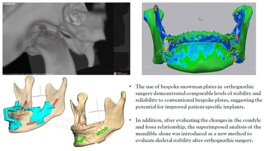
Clinical Stability of Bespoke Snowman Plates for Fixation following Sagittal Split Ramus Osteotomy of the Mandible
Abstract
1. Introduction
2. Materials and Methods
2.1. Patients
2.2. Virtual Surgery (VS), Designing and Creating Patient-Specific Materials, and Actual Surgery
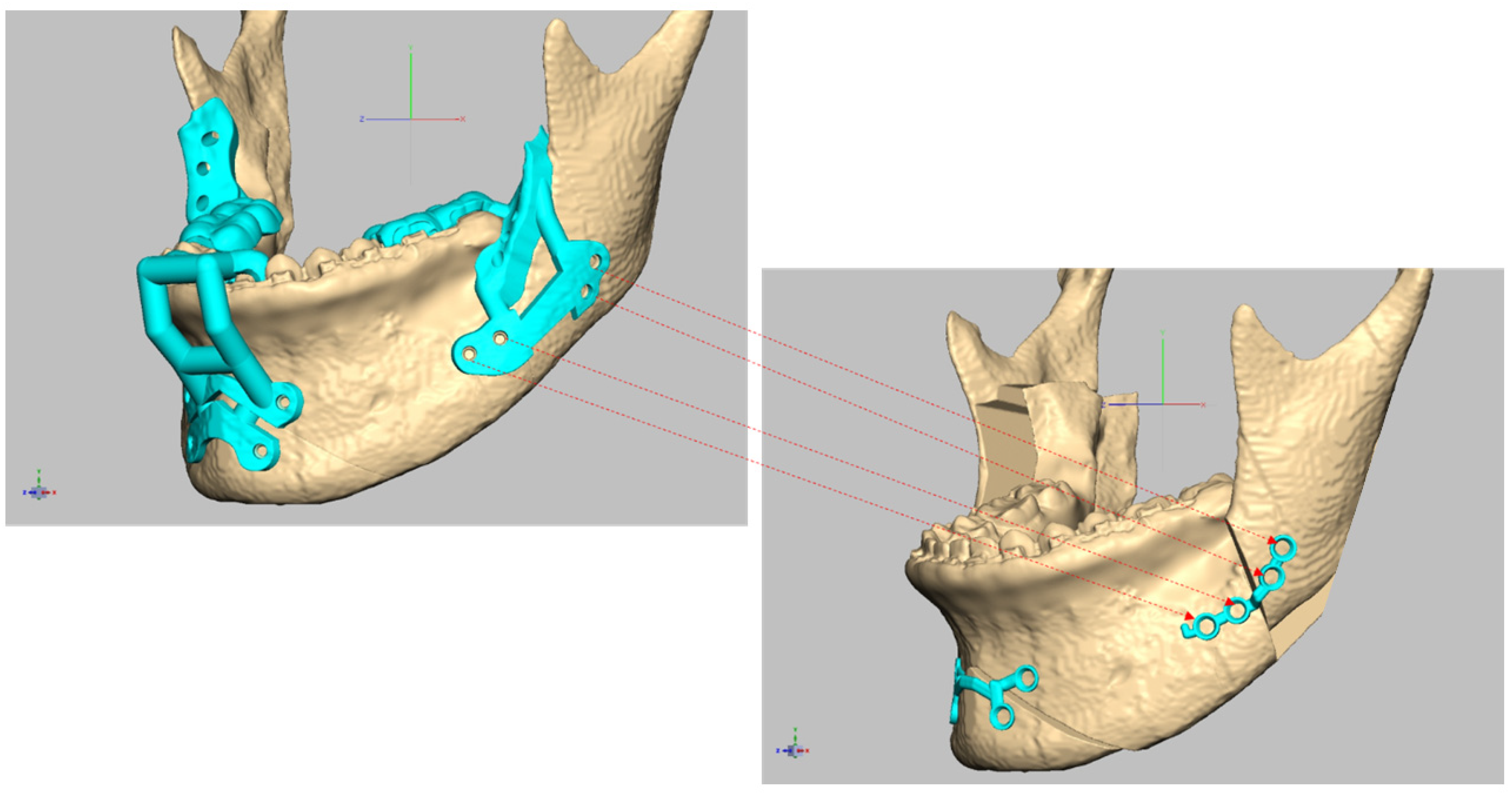
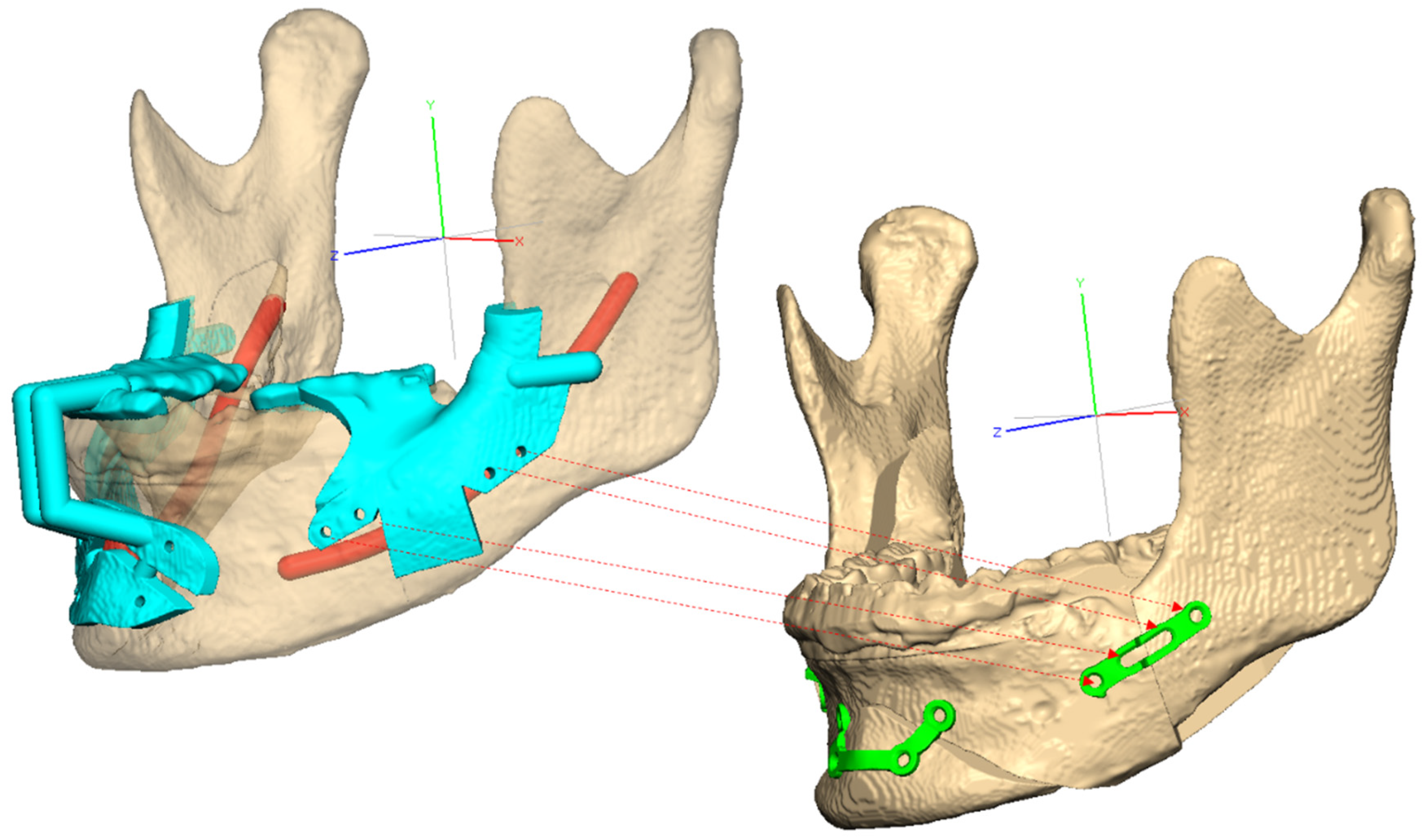
2.2.1. Virtual Surgical Planning (VSP), including Preoperative Preparation and Creating Patient-Specific Surgical Guides and PSPs
2.2.2. Surgery
2.3. Methods
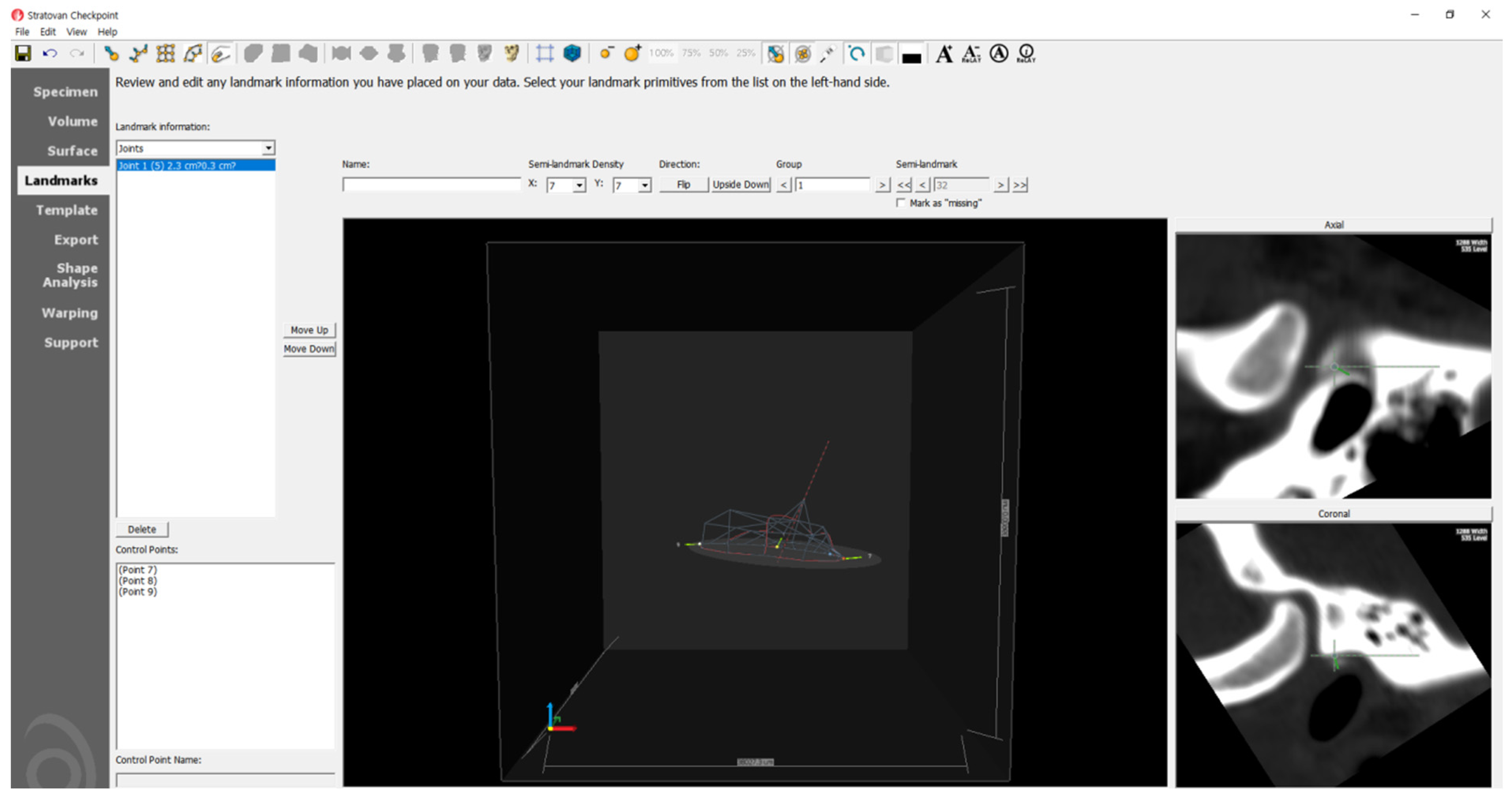
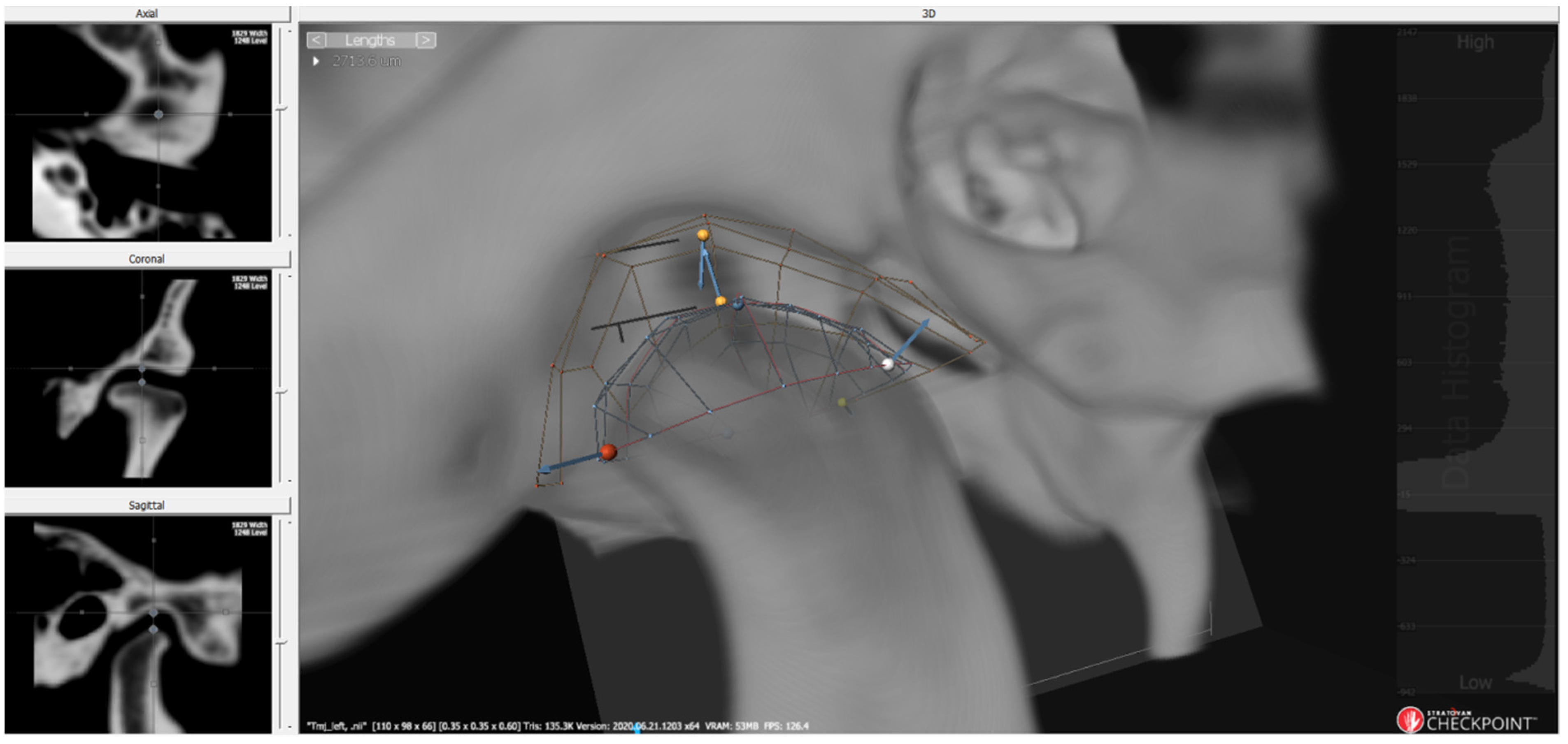
2.3.1. 3D Condyle–Fossa Relationship Analysis
2.3.2. Post-Superimposition Analysis of the Mandible
2.4. Statistical Analysis
3. Results
3.1. Patient Composition
3.2. 3D Condyle–Fossa Relationship Analysis Results
3.3. Post-Superimposition Analysis Results of the Mandible
4. Discussion
5. Conclusions
Supplementary Materials
Author Contributions
Funding
Institutional Review Board Statement
Informed Consent Statement
Data Availability Statement
Acknowledgments
Conflicts of Interest
References
- Cottrell, D.A.; Farrell, B.; Ferrer-Nuin, L.; Ratner, S. Surgical correction of maxillofacial skeletal deformities. J. Oral Maxillofac. Surg. 2017, 75, e94–e125. [Google Scholar] [CrossRef] [PubMed]
- Trauner, R. Zur Operationstechnik bei der Progenie und anderen unterkieferanormalien. Dtsch. Zahn Mund Kieferheilk 1955, 23, 1–26. [Google Scholar]
- Trauner, R.; Obwegeser, H. The surgical correction of mandibular prognathism and retrognathia with consideration of genioplasty: Part I. Surgical procedures to correct mandibular prognathism and reshaping of the chin. Oral Surg. Oral Med. Oral Pathol. 1957, 10, 677–689. [Google Scholar] [CrossRef] [PubMed]
- Obwegeser, H. The one time forward movement of the maxilla and backward movement of the mandible for the correction of extreme prognathism. Schweiz. Monatsschrift Zahnheilkd. 1970, 80, 547. [Google Scholar]
- Obwegeser, H.L. Orthognathic surgery and a tale of how three procedures came to be: A letter to the next generations of surgeons. Clin. Plast. Surg. 2007, 34, 331–355. [Google Scholar] [CrossRef]
- Perez, D.; Ellis, E. Sequencing bimaxillary surgery: Mandible first. J. Oral Maxillofac. Surg. 2011, 69, 2217–2224. [Google Scholar]
- Kuik, K.; De Ruiter, M.; De Lange, J.; Hoekema, A. Fixation methods in sagittal split ramus osteotomy: A systematic review on in vitro biomechanical assessments. Int. J. Oral Maxillofac. Surg. 2019, 48, 56–70. [Google Scholar] [CrossRef]
- Chen, C.-M.; Hwang, D.-S.; Hsiao, S.-Y.; Chen, H.-S.; Hsu, K.-J. Skeletal stability after mandibular setback via sagittal split ramus osteotomy verse intraoral vertical ramus osteotomy: A systematic review. J. Clin. Med. 2021, 10, 4950. [Google Scholar] [CrossRef]
- Lee, C.-H.; Cho, S.-W.; Kim, J.-W.; Ahn, H.-J.; Kim, Y.-H.; Yang, B.-E. Three-dimensional assessment of condylar position following orthognathic surgery using the centric relation bite and the ramal reference line: A retrospective clinical study. Medicine 2019, 98, e14931. [Google Scholar] [CrossRef]
- Dai, Z.; Connelly, S.T.; Silva, R.; Gupta, R.J. Condylar Position is Maintained in Maxillomandibular Advancement Surgery Utilizing Custom Cutting Guides and Plates. J. Oral Maxillofac. Surg. 2023, 81, 156–164. [Google Scholar] [CrossRef]
- Lim, Y.N.; Park, I.-Y.; Kim, J.-C.; Byun, S.-H.; Yang, B.-E. Comparison of changes in the condylar volume and morphology in Skeletal Class III deformities undergoing orthognathic surgery using a customized versus conventional miniplate: A retrospective analysis. J. Clin. Med. 2020, 9, 2794. [Google Scholar] [CrossRef]
- Jang, W.-S.; Byun, S.-H.; Cho, S.-W.; Park, I.-Y.; Yi, S.-M.; Kim, J.-C.; Yang, B.-E. Correction of Condylar Displacement of the Mandible Using Early Screw Removal following Patient-Customized Orthognathic Surgery. J. Clin. Med. 2021, 10, 1597. [Google Scholar] [CrossRef] [PubMed]
- Michelet, F.; Benoit, J.; Festal, F.; Despujols, P.; Bruchet, P.; Arvor, A. Fixation without blocking of sagittal osteotomies of the rami by means of endo-buccal screwed plates in the treatment of antero-posterior abnormalities. Rev. Stomatol. Chir. Maxillo-Faciale 1971, 72, 531. [Google Scholar]
- Böckmann, R.; Meyns, J.; Dik, E.; Kessler, P. The modifications of the sagittal ramus split osteotomy: A literature review. Plast. Reconstr. Surg. Glob. Open 2014, 2, e271. [Google Scholar] [CrossRef] [PubMed]
- Michelet, F.X.; Deymes, J.; Dessus, B. Osteosynthesis with miniaturized screwed plates in maxillo-facial surgery. J. Maxillofac. Surg. 1973, 1, 79–84. [Google Scholar] [CrossRef]
- Champy, M. Die Behandlunk von Mandibularfrakturen mittels Osteosyutheseohne intermaxillare Ruhigstellung nach der Technik von FX Michelet. Zahn Mund Kiefer-Heilk 1975, 63, 339. [Google Scholar]
- Thiele, O.C.; Kreppel, M.; Bittermann, G.; Bonitz, L.; Desmedt, M.; Dittes, C.; Dörre, A.; Dunsche, A.; Eckert, A.W.; Ehrenfeld, M. Moving the mandible in orthognathic surgery–A multicenter analysis. J. Cranio-Maxillofac. Surg. 2016, 44, 579–583. [Google Scholar] [CrossRef]
- Chen, J.M. Bespoke surgery: We’re virtually there. J. Thorac. Cardiovasc. Surg. 2018, 155, 1743–1744. [Google Scholar] [CrossRef]
- Baron, M.; Karwowska, N.; Buchbinder, Z.; Buchbinder, D. Patient-Specific Orthognathic Solutions (PSOS) Survey: Global Trends in Orthognathic Surgery. J. Oral Maxillofac. Surg. 2022, 80, S26–S28. [Google Scholar] [CrossRef]
- Kim, J.-W.; Kim, J.-C.; Jeong, C.-G.; Cheon, K.-J.; Cho, S.-W.; Park, I.-Y.; Yang, B.-E. The accuracy and stability of the maxillary position after orthognathic surgery using a novel computer-aided surgical simulation system. BMC Oral Health 2019, 19, 18. [Google Scholar] [CrossRef]
- Steenen, S.; Becking, A. Bad splits in bilateral sagittal split osteotomy: Systematic review of fracture patterns. Int. J. Oral Maxillofac. Surg. 2016, 45, 887–897. [Google Scholar] [CrossRef] [PubMed]
- Larson, B.E.; Lee, N.K.; Jang, M.J.; Jo, D.W.; Yun, P.Y.; Kim, Y.K. Comparative evaluation of the sliding plate technique for fixation of a sagittal split ramus osteotomy: Finite element analysis. Oral Surg. Oral Med. Oral Pathol. Oral Radiol. 2017, 123, e148–e152. [Google Scholar] [CrossRef] [PubMed]
- Ghang, M.-H.; Kim, H.-M.; You, J.-Y.; Kim, B.-H.; Choi, J.-P.; Kim, S.-H.; Choung, P.-H. Three-dimensional mandibular change after sagittal split ramus osteotomy with a semirigid sliding plate system for fixation of a mandibular setback surgery. Oral Surg. Oral Med. Oral Pathol. Oral Radiol. 2013, 115, 157–166. [Google Scholar] [CrossRef] [PubMed]
- Kim, J.-W.; Park, K.-N.; Lee, C.-H.; Kim, Y.-S.; Kim, Y.-H.; Yang, B.-E. Finite element stress analysis of Keyhole Plate System in sagittal split ramus osteotomy. Sci. Rep. 2018, 8, 8971. [Google Scholar] [CrossRef]
- Ku, J.-K.; Choi, S.-K.; Lee, J.-G.; Yu, H.-C.; Kim, S.-Y.; Kim, Y.-K.; Shin, Y.; Lee, N.-K. Analysis of sagittal position changes of the condyle after mandibular setback surgery across the four different types of plating systems. J. Craniofacial Surg. 2021, 32, 2441. [Google Scholar] [CrossRef] [PubMed]
- Lee, J.-Y.; Kim, Y.-K.; Yun, P.-Y.; Lee, N.-K.; Kim, J.-W.; Choi, J.-H. Evaluation of stability after orthognathic surgery with minimal orthodontic preparation: Comparison according to 3 types of fixation. J. Craniofacial Surg. 2014, 25, 911–915. [Google Scholar] [CrossRef]
- Lee, H.; Agpoon, K.; Besana, A.; Lim, H.; Jang, H.; Lee, E. Mandibular stability using sliding or conventional four-hole plates for fixation after bilateral sagittal split ramus osteotomy for mandibular setback. Br. J. Oral Maxillofac. Surg. 2017, 55, 378–382. [Google Scholar] [CrossRef]
- Sevitt, S. Bone Repair and Fracture Healing in Man; Churchill Livingstone: London, UK, 1981. [Google Scholar]
- Hunsuck, E. A modified intraoral sagittal splitting technic for correction of mandibular prognathism. J. Oral Surg. 1968, 26, 49–52. [Google Scholar]
- Zeynalzadeh, F.; Shooshtari, Z.; Eshghpour, M.; Zarch, S.H.H.; Tohidi, E.; Samieirad, S. Dal Pont vs Hunsuck: Which Technique Can Lead to a Lower Incidence of Bad Split during Bilateral Sagittal Split Osteotomy? A Triple-blind Randomized Clinical Trial. World J. Plast. Surg. 2021, 10, 25. [Google Scholar] [CrossRef]
- Kim, J.-W.; Kim, J.-C.; Cheon, K.-J.; Cho, S.-W.; Kim, Y.-H.; Yang, B.-E. Computer-aided surgical simulation for yaw control of the mandibular condyle and its actual application to orthognathic surgery: A one-year follow-up study. Int. J. Environ. Res. Public Health 2018, 15, 2380. [Google Scholar] [CrossRef]
- Christensen, A.M.; Weimer, K.; Beaudreau, C.; Rensberger, M.; Johnson, B. The digital thread for personalized craniomaxillofacial surgery. In Digital Technologies in Craniomaxillofacial Surgery; Springer: New York, NY, USA, 2018; pp. 23–45. [Google Scholar]
- Ikeda, R.; Oberoi, S.; Wiley, D.F.; Woodhouse, C.; Tallman, M.; Tun, W.W.; McNeill, C.; Miller, A.J.; Hatcher, D. Novel 3-dimensional analysis to evaluate temporomandibular joint space and shape. Am. J. Orthod. Dentofac. Orthop. 2016, 149, 416–428. [Google Scholar] [CrossRef] [PubMed]
- Muto, T.; Takahashi, M.; Akizuki, K. Evaluation of the mandibular ramus fracture line after sagittal split ramus osteotomy using 3-dimensional computed tomography. J. Oral Maxillofac. Surg. 2012, 70, e648–e652. [Google Scholar] [CrossRef]
- Cunha, G.; Oliveira, M.; Salmen, F.; Gabrielli, M.; Gabrielli, M. How does bone thickness affect the split pattern of sagittal ramus osteotomy? Int. J. Oral Maxillofac. Surg. 2020, 49, 218–223. [Google Scholar] [CrossRef] [PubMed]
- Fahradyan, A.; Wolfswinkel, E.M.; Clarke, N.; Park, S.; Tsuha, M.; Urata, M.M.; Hammoudeh, J.A.; Yamashita, D.-D.R. Impact of the distance of maxillary advancement on horizontal relapse after orthognathic surgery. Cleft Palate-Craniofacial J. 2018, 55, 546–553. [Google Scholar] [CrossRef] [PubMed]
- Haan, I.d.; Ciesielski, R.; Nitsche, T.; Koos, B. Evaluation of relapse after orthodontic therapy combined with orthognathic surgery in the treatment of skeletal class III. J. Orofac. Orthop. Fortschritte Kieferorthopadie 2013, 74, 362–369. [Google Scholar] [CrossRef]
- Romero, L.G.; Mulier, D.; Orhan, K.; Shujaat, S.; Shaheen, E.; Willems, G.; Politis, C.; Jacobs, R. Evaluation of long-term hard tissue remodelling after skeletal class III orthognathic surgery: A systematic review. Int. J. Oral Maxillofac. Surg. 2020, 49, 51–61. [Google Scholar] [CrossRef]
- Panchal, N.; Ellis, C.; Tiwana, P. Stability and relapse in orthognathic surgery. Sel. Read. Oral Maxillofac. Surg. 2017, 24, 1–16. [Google Scholar]
- Joss, C.U.; Vassalli, I.M. Stability after bilateral sagittal split osteotomy advancement surgery with rigid internal fixation: A systematic review. J. Oral Maxillofac. Surg. 2009, 67, 301–313. [Google Scholar] [CrossRef]
- Roh, Y.-C.; Shin, S.-H.; Kim, S.-S.; Sandor, G.K.; Kim, Y.-D. Skeletal stability and condylar position related to fixation method following mandibular setback with bilateral sagittal split ramus osteotomy. J. Cranio-Maxillofac. Surg. 2014, 42, 1958–1963. [Google Scholar] [CrossRef]
- Rubens, B.C.; Stoelinga, P.J.; Blijdorp, P.A.; Schoenaers, J.H.; Politis, C. Skeletal stability following sagittal split osteotomy using monocortical miniplate internal fixation. Int. J. Oral Maxillofac. Surg. 1988, 17, 371–376. [Google Scholar] [CrossRef]
- Karanxha, L.; Rossi, D.; Hamanaka, R.; Giannì, A.B.; Baj, A.; Moon, W.; Del Fabbro, M.; Romano, M. Accuracy of splint vs splintless technique for virtually planned orthognathic surgery: A voxel-based three-dimensional analysis. J. Cranio-Maxillofac. Surg. 2021, 49, 1–8. [Google Scholar] [CrossRef] [PubMed]
- Pascal, E.; Majoufre, C.; Bondaz, M.; Courtemanche, A.; Berger, M.; Bouletreau, P. Current status of surgical planning and transfer methods in orthognathic surgery. J. Stomatol. Oral Maxillofac. Surg. 2018, 119, 245–248. [Google Scholar] [CrossRef] [PubMed]
- Heufelder, M.; Wilde, F.; Pietzka, S.; Mascha, F.; Winter, K.; Schramm, A.; Rana, M. Clinical accuracy of waferless maxillary positioning using customized surgical guides and patient specific osteosynthesis in bimaxillary orthognathic surgery. J. Cranio-Maxillofac. Surg. 2017, 45, 1578–1585. [Google Scholar] [CrossRef] [PubMed]
- Hsu, S.S.-P.; Gateno, J.; Bell, R.B.; Hirsch, D.L.; Markiewicz, M.R.; Teichgraeber, J.F.; Zhou, X.; Xia, J. Accuracy of a computer-aided surgical simulation protocol for orthognathic surgery: A prospective multicenter study. J. Oral Maxillofac. Surg. 2013, 71, 128–142. [Google Scholar] [CrossRef] [PubMed]
- Arcas, A.; Vendrell, G.; Cuesta, F.; Bermejo, L. Advantages of performing mentoplasties with customized guides and plates generated with 3D planning and printing. Results from a series of 23 cases. J. Cranio-Maxillofac. Surg. 2018, 46, 2088–2095. [Google Scholar] [CrossRef]
- Hanafy, M.; Akoush, Y.; Abou-ElFetouh, A.; Mounir, R. Precision of orthognathic digital plan transfer using patient-specific cutting guides and osteosynthesis versus mixed analogue–digitally planned surgery: A randomized controlled clinical trial. Int. J. Oral Maxillofac. Surg. 2020, 49, 62–68. [Google Scholar] [CrossRef]
- Van den Bempt, M.; Liebregts, J.; Maal, T.; Bergé, S.; Xi, T. Toward a higher accuracy in orthognathic surgery by using intraoperative computer navigation, 3D surgical guides, and/or customized osteosynthesis plates: A systematic review. J. Cranio-Maxillofac. Surg. 2018, 46, 2108–2119. [Google Scholar] [CrossRef]
- Shakoori, P.; Yang, R.; Nah, H.-D.; Scott, M.; Swanson, J.W.; Taylor, J.A.; Bartlett, S.P. Computer-aided Surgical Planning and Osteosynthesis Plates for Bimaxillary Orthognathic Surgery: A Study of 14 Consecutive Patients. Plast. Reconstr. Surg. Glob. Open 2022, 10, e4609. [Google Scholar] [CrossRef]
- Valls-Ontañón, A.; Ascencio-Padilla, R.; Vela-Lasagabaster, A.; Sada-Malumbres, A.; Haas-Junior, O.; Masià-Gridilla, J.; Hernández-Alfaro, F. Relevance of 3D virtual planning in predicting bony interferences between distal and proximal fragments after sagittal split osteotomy. Int. J. Oralmaxillofacial Surg. 2020, 49, 1020–1028. [Google Scholar] [CrossRef]
- Hosoki, M.; Nishigawa, K.; Miyamoto, Y.; Ohe, G.; Matsuka, Y. Allergic contact dermatitis caused by titanium screws and dental implants. J. Prosthodont. Res. 2016, 60, 213–219. [Google Scholar] [CrossRef]
- Fage, S.W.; Muris, J.; Jakobsen, S.S.; Thyssen, J.P. Titanium: A review on exposure, release, penetration, allergy, epidemiology, and clinical reactivity. Contact Dermat. 2016, 74, 323–345. [Google Scholar]
- Abeloos, J.; De Clercq, C.; Neyt, L. Skeletal stability following miniplate fixation after bilateral sagittal split osteotomy for mandibular advancement. J. Oral Maxillofac. Surg. 1993, 51, 366–369. [Google Scholar] [CrossRef] [PubMed]
- Baker, D.L.; Stoelinga, P.J.; Blijdorp, P.A.; Brouns, J.J. Long-term stability after inferior maxillary repositioning by miniplate fixation. Int. J. Oral Maxillofac. Surg. 1992, 21, 320–326. [Google Scholar] [CrossRef]
- Sun, Y.; Tian, L.; Luebbers, H.-T.; Politis, C. Relapse tendency after BSSO surgery differs between 2D and 3D measurements: A validation study. J. Cranio-Maxillofac. Surg. 2018, 46, 1893–1898. [Google Scholar] [CrossRef]
- Shaw, K.; McIntyre, G.; Mossey, P.; Menhinick, A.; Thomson, D. Validation of conventional 2D lateral cephalometry using 3D cone beam CT. J. Orthod. 2013, 40, 22–28. [Google Scholar] [CrossRef] [PubMed]
- Van Hemelen, G.; Van Genechten, M.; Renier, L.; Desmedt, M.; Verbruggen, E.; Nadjmi, N. Three-dimensional virtual planning in orthognathic surgery enhances the accuracy of soft tissue prediction. J. Cranio-Maxillofac. Surg. 2015, 43, 918–925. [Google Scholar] [CrossRef]
- Brunso, J.; Franco, M.; Constantinescu, T.; Barbier, L.; Santamaría, J.A.; Alvarez, J. Custom-machined miniplates and bone-supported guides for orthognathic surgery: A new surgical procedure. J. Oral Maxillofac. Surg. 2016, 74, 1061.e1–1061.e12. [Google Scholar] [CrossRef]
- Figueiredo, C.; Paranhos, L.; da Silva, R.; Herval, Á.; Blumenberg, C.; Zanetta-Barbosa, D. Accuracy of orthognathic surgery with customized titanium plates–Systematic review. J. Stomatol. Oral Maxillofac. Surg. 2020, 122, 88–97. [Google Scholar] [CrossRef]
- Suojanen, J.; Leikola, J.; Stoor, P. The use of patient-specific implants in orthognathic surgery: A series of 32 maxillary osteotomy patients. J. Cranio-Maxillofac. Surg. 2016, 44, 1913–1916. [Google Scholar] [CrossRef]
- Yang, B.-E. The Latest Update of Digital “Bespoke” Orthognathic Surgery (BOGS) in Korea. Available online: https://youtu.be/BfB4v2NYb1c (accessed on 1 July 2023).
- Yang, B.-E. Orthognathic surgery with new computerized orthognathic simulation and patient-specific osteosynthesis plates. In Contemporary Digital Orthodontics; Quintessence Korea Publishing Co., Ltd.: Seoul, Republic of Korea, 2021; pp. 140–166. [Google Scholar]
- Mazzoni, S.; Bianchi, A.; Schiariti, G.; Badiali, G.; Marchetti, C. Computer-aided design and computer-aided manufacturing cutting guides and customized titanium plates are useful in upper maxilla waferless repositioning. J. Oral Maxillofac. Surg. 2015, 73, 701–707. [Google Scholar] [CrossRef]
- Au, S.W.; Li, D.T.S.; Su, Y.-X.; Leung, Y.Y. Accuracy of self-designed 3D-printed patient-specific surgical guides and fixation plates for advancement genioplasty. Int. J. Comput. Dent. 2022, 25, 369–376. [Google Scholar] [PubMed]
- Goodson, A.; Parmar, S.; Ganesh, S.; Zakai, D.; Shafi, A.; Wicks, C.; O’Connor, R.; Yeung, E.; Khalid, F.; Tahim, A. Printed titanium implants in UK craniomaxillofacial surgery. Part II: Perceived performance (outcomes, logistics, and costs). Br. J. Oral Maxillofac. Surg. 2021, 59, 320–328. [Google Scholar] [CrossRef] [PubMed]
- Oh, J.-h. Recent advances in the reconstruction of cranio-maxillofacial defects using computer-aided design/computer-aided manufacturing. Maxillofac. Plast. Reconstr. Surg. 2018, 40, 2. [Google Scholar] [CrossRef]
- Veyssiere, A.; Leprovost, N.; Ambroise, B.; Prévost, R.; Chatellier, A.; Bénateau, H. Study of the mechanical reliability of an S-shaped adjustable osteosynthesis plate for bilateral sagittal split osteotomies. Study on 15 consecutive cases. J. Stomatol. Oral Maxillofac. Surg. 2018, 119, 19–24. [Google Scholar] [CrossRef] [PubMed]



| Group | Patient No. | Age | Gender | Operation | Diagnosis |
|---|---|---|---|---|---|
| Pt 1 | 29 | M | 2jaw | III | |
| Pt 2 | 23 | M | 2jaw/gen | III | |
| Pt 3 | 29 | F | 1jaw/gen | III, FA | |
| Pt 4 | 20 | F | 2jaw | III, FA | |
| Control | Pt 5 | 21 | M | 2jaw | III, FA |
| Pt 6 | 18 | F | 2jaw | III | |
| Pt 7 | 26 | M | 2jaw/gen | III, FA | |
| Pt 8 | 22 | F | 2jaw | FA | |
| Pt 9 | 22 | F | 2jaw/gen | III, FA | |
| Pt 10 | 27 | F | 2jaw | III, FA | |
| Pt 11 | 19 | F | 2jaw/gen | III, FA | |
| Pt 1 | 26 | M | 2jaw | III, FA | |
| Pt 2 | 20 | F | 1jaw | III, FA | |
| Pt 3 | 22 | M | 2jaw | FA | |
| Pt 4 | 20 | F | 1jaw | III, FA | |
| Study | Pt 5 | 22 | M | 1jaw | III, FA |
| Pt 6 | 18 | F | 2jaw | III, FA | |
| Pt 7 | 32 | M | 1jaw | III, FA | |
| Pt 8 | 19 | F | 1jaw/gen | III, FA | |
| Pt 9 | 21 | M | 2jaw | FA | |
| Pt 10 | 21 | F | 2jaw | III, FA | |
| Pt 11 | 21 | M | 1jaw/gen | III, FA |
| Group | Total | χ2 | p-Value | |||
|---|---|---|---|---|---|---|
| Control | Study | |||||
| Age | 23.27 ± 1.18 | 22 ± 1.18 | 6.333 | 0.706 | ||
| Gender | Male | 4 (36.4) | 6 (54.5) | 10 (45.5) | 0.733 | 0.392 |
| Female | 7 (63.6) | 5 (45.5) | 12 (54.5) | |||
| 1jaw/2jaw | 1jaw | 1 (9.1) | 6 (54.5) | 7 (31.8) | 5.238 | 0.022 |
| 2jaw | 10 (90.9) | 5 (45.5) | 15 (68.2) | |||
| FA | FA (−) | 3 (27.3) | 0 (0) | 3 (13.6) | 3.474 | 0.062 |
| FA ( + ) | 8 (72.7) | 11 (100) | 19 (86.4) | |||
| Total | 11 (100) | 11 (100) | 22 (100) | |||
| Source | Sum of Squares (SS) | df | Mean Squares (MS) | F | p-Value |
|---|---|---|---|---|---|
| Time | 0.442 | 1 | 0.442 | 4.004 | 0.048 |
| Time × group | 0.015 | 1 | 0.015 | 0.137 | 0.712 |
| Time × position of joint space | 0.186 | 2 | 0.093 | 0.841 | 0.434 |
| Error | 13.905 | 126 | 0.110 |
| Group | Position of Joint Space | Time | N | Average (mm) | Standard Error | t | p-Value |
|---|---|---|---|---|---|---|---|
| Control | AJS | T0 | 22 | 1.95 | 0.70 | −1.063 | 0.294 |
| T2 | 22 | 2.16 | 0.63 | ||||
| SJS | T0 | 22 | 2.40 | 1.01 | −0.305 | 0.762 | |
| T2 | 22 | 2.49 | 0.96 | ||||
| PJS | T0 | 22 | 2.15 | 0.88 | 0.058 | 0.954 | |
| T2 | 22 | 2.13 | 0.67 | ||||
| Study | AJS | T0 | 22 | 1.80 | 0.71 | −0.504 | 0.617 |
| T2 | 22 | 1.90 | 0.60 | ||||
| SJS | T0 | 22 | 2.31 | 1.18 | 0 | 1 | |
| T2 | 22 | 2.31 | 0.90 | ||||
| PJS | T0 | 22 | 2.02 | 0.92 | −0.412 | 0.683 | |
| T2 | 22 | 2.12 | 0.68 |
| Source | Sum of Squares | df | MEAN Squares | F | p-Value | |
|---|---|---|---|---|---|---|
| Group | X diff. | 0.009 | 1 | 0.009 | 0.165 | 0.685 |
| Y diff. | 0.029 | 1 | 0.029 | 1.374 | 0.243 | |
| Z diff. | 0.003 | 1 | 0.003 | 0.087 | 0.768 | |
| Surface diff. | 0.039 | 1 | 0.039 | 0.449 | 0.503 | |
| Point | X diff. | 0.38 | 4 | 0.095 | 1.813 | 0.128 |
| Y diff. | 0.226 | 4 | 0.056 | 2.678 | 0.033 | |
| Z diff. | 0.295 | 4 | 0.074 | 1.851 | 0.121 | |
| Surface diff. | 0.795 | 4 | 0.199 | 2.262 | 0.064 | |
| Time | X diff. | 0.001 | 1 | 0.001 | 0.019 | 0.891 |
| Y diff. | 0.046 | 1 | 0.046 | 2.192 | 0.14 | |
| Z diff. | 0.937 | 1 | 0.937 | 23.525 ** | 0 | |
| Surface diff. | 0.559 | 1 | 0.559 | 6.365 | 0.012 | |
| Group × point | X diff. | 0.109 | 4 | 0.027 | 0.518 | 0.722 |
| Y diff. | 0.011 | 4 | 0.003 | 0.126 | 0.973 | |
| Z diff. | 0.063 | 4 | 0.016 | 0.394 | 0.813 | |
| Surface | 0.096 | 4 | 0.024 | 0.273 | 0.895 | |
| Group × time | X diff. | 0.018 | 1 | 0.018 | 0.34 | 0.561 |
| Y diff. | 0.033 | 1 | 0.033 | 1.542 | 0.216 | |
| Z diff. | 0.046 | 1 | 0.046 | 1.143 | 0.286 | |
| Surface diff. | 0.071 | 1 | 0.071 | 0.81 | 0.369 | |
| Point × time | X diff. | 1.591 | 4 | 0.398 | 7.588 ** | 0 |
| Y diff. | 0.174 | 4 | 0.043 | 2.061 | 0.087 | |
| Z diff. | 8.279 | 4 | 2.07 | 51.965 ** | 0 | |
| Surface diff. | 11.646 | 4 | 2.911 | 33.149 ** | 0 | |
| Group × point × time | X diff. | 0.104 | 4 | 0.026 | 0.496 | 0.739 |
| Y diff. | 0.03 | 4 | 0.008 | 0.357 | 0.839 | |
| Z diff. | 0.104 | 4 | 0.026 | 0.655 | 0.624 | |
| Surface diff. | 0.253 | 4 | 0.063 | 0.72 | 0.58 | |
| Error | X diff. | 10.484 | 200 | 0.052 | ||
| Y diff. | 4.217 | 200 | 0.021 | |||
| Z diff. | 7.966 | 200 | 0.04 | |||
| Surface diff. | 17.566 | 200 | 0.088 |
| Group | Time diff. | Average (mm) | Standard Error | N | |
|---|---|---|---|---|---|
| X diff. | Control | ΔT2 | 0.017 | 0.222 | 55 |
| ΔT1 | 0.004 | 0.226 | 55 | ||
| Study | ΔT2 | 0.012 | 0.271 | 55 | |
| ΔT1 | 0.034 | 0.247 | 55 | ||
| Y diff. | Control | ΔT2 | 0.035 | 0.116 | 55 |
| ΔT1 | 0.030 | 0.140 | 55 | ||
| Study | ΔT2 | 0.082 | 0.179 | 55 | |
| ΔT1 | 0.028 | 0.146 | 55 | ||
| Z diff. | Control | ΔT2 | −0.070 | 0.233 | 55 |
| ΔT1 | 0.089 | 0.328 | 55 | ||
| Study | ΔT2 | −0.059 | 0.242 | 55 | |
| ΔT1 | 0.071 | 0.309 | 55 | ||
| surface diff. | Control | ΔT2 | 0.049 | 0.308 | 55 |
| ΔT1 | −0.088 | 0.443 | 55 | ||
| Study | ΔT2 | 0.039 | 0.335 | 55 | |
| ΔT1 | −0.025 | 0.399 | 55 |
| Group | Point | Time diff. | Average (mm) | Standard Error (SE) | N | |
|---|---|---|---|---|---|---|
| X diff. | Control | Lt. mental fo | ΔT2 | 0.08 | 0.17 | 11 |
| ΔT1 | −0.15 | 0.15 | 11 | |||
| Lt. molar root | ΔT2 | −0.01 | 0.22 | 11 | ||
| ΔT1 | 0.09 | 0.32 | 11 | |||
| Rt. mental fo | ΔT2 | −0.06 | 0.20 | 11 | ||
| ΔT1 | 0.13 | 0.20 | 11 | |||
| Rt. molar root | ΔT2 | 0.03 | 0.33 | 11 | ||
| ΔT1 | −0.13 | 0.13 | 11 | |||
| Incisor root | ΔT2 | 0.06 | 0.16 | 11 | ||
| ΔT1 | 0.07 | 0.14 | 11 | |||
| Study | Lt. mental fo | ΔT2 | 0.13 | 0.17 | 11 | |
| ΔT1 | −0.16 | 0.13 | 11 | |||
| Lt. molar root | ΔT2 | −0.14 | 0.31 | 11 | ||
| ΔT1 | 0.08 | 0.32 | 11 | |||
| Rt. mental fo | ΔT2 | −0.06 | 0.20 | 11 | ||
| ΔT1 | 0.15 | 0.19 | 11 | |||
| Rt. molar root | ΔT2 | 0.01 | 0.34 | 11 | ||
| ΔT1 | 0.00 | 0.30 | 11 | |||
| Incisor root | ΔT2 | 0.12 | 0.23 | 11 | ||
| ΔT1 | 0.09 | 0.17 | 11 | |||
| Y diff. | Control | Lt. mental fo | ΔT2 | 0.00 | 0.02 | 11 |
| ΔT1 | −0.02 | 0.06 | 11 | |||
| Lt. molar root | ΔT2 | 0.02 | 0.10 | 11 | ||
| ΔT1 | −0.02 | 0.20 | 11 | |||
| Rt. mental fo | ΔT2 | 0.04 | 0.06 | 11 | ||
| ΔT1 | 0.06 | 0.10 | 11 | |||
| Rt. molar root | ΔT2 | 0.01 | 0.19 | 11 | ||
| ΔT1 | 0.08 | 0.10 | 11 | |||
| Incisor root | ΔT2 | 0.10 | 0.13 | 11 | ||
| ΔT1 | 0.04 | 0.19 | 11 | |||
| Study | Lt. mental fo | ΔT2 | 0.00 | 0.08 | 11 | |
| ΔT1 | 0.00 | 0.05 | 11 | |||
| Lt. molar root | ΔT2 | 0.06 | 0.22 | 11 | ||
| ΔT1 | −0.01 | 0.20 | 11 | |||
| Rt. mental fo | ΔT2 | 0.12 | 0.14 | 11 | ||
| ΔT1 | 0.08 | 0.17 | 11 | |||
| Rt. molar root | ΔT2 | 0.06 | 0.21 | 11 | ||
| ΔT1 | 0.08 | 0.08 | 11 | |||
| Incisor root | ΔT2 | 0.17 | 0.19 | 11 | ||
| ΔT1 | −0.01 | 0.17 | 11 | |||
| Z diff. | Control | Lt. mental fo | ΔT2 | 0.09 | 0.16 | 11 |
| ΔT1 | −0.17 | 0.15 | 11 | |||
| Lt. molar root | ΔT2 | −0.03 | 0.23 | 11 | ||
| ΔT1 | 0.09 | 0.30 | 11 | |||
| Rt. mental fo | ΔT2 | 0.04 | 0.16 | 11 | ||
| ΔT1 | −0.11 | 0.16 | 11 | |||
| Rt. molar root | ΔT2 | −0.09 | 0.14 | 11 | ||
| ΔT1 | 0.08 | 0.13 | 11 | |||
| Incisor root | ΔT2 | −0.46 | 0.39 | 11 | ||
| ΔT1 | 0.72 | 0.48 | 11 | |||
| Study | Lt. mental fo | ΔT2 | 0.16 | 0.17 | 11 | |
| ΔT1 | −0.16 | 0.13 | 11 | |||
| Lt. molar root | ΔT2 | −0.11 | 0.23 | 11 | ||
| ΔT1 | 0.08 | 0.30 | 11 | |||
| Rt. mental fo | ΔT2 | 0.04 | 0.16 | 11 | ||
| ΔT1 | −0.12 | 0.15 | 11 | |||
| Rt. molar root | ΔT2 | −0.01 | 0.24 | 11 | ||
| ΔT1 | 0.05 | 0.16 | 11 | |||
| Incisor root | ΔT2 | −0.41 | 0.40 | 11 | ||
| ΔT1 | 0.47 | 0.38 | 11 | |||
| surface diff. | Control | Lt. mental fo | ΔT2 | −0.12 | 0.23 | 11 |
| ΔT1 | 0.23 | 0.22 | 11 | |||
| Lt. molar root | ΔT2 | 0.01 | 0.29 | 11 | ||
| ΔT1 | 0.00 | 0.38 | 11 | |||
| Rt. mental fo | ΔT2 | −0.08 | 0.26 | 11 | ||
| ΔT1 | 0.17 | 0.25 | 11 | |||
| Rt. molar root | ΔT2 | 0.06 | 0.34 | 11 | ||
| ΔT1 | −0.17 | 0.20 | 11 | |||
| Incisor root | ΔT2 | 0.37 | 0.16 | 11 | ||
| ΔT1 | −0.72 | 0.48 | 11 | |||
| Study | Lt. mental fo | ΔT2 | −0.21 | 0.24 | 11 | |
| ΔT1 | 0.23 | 0.19 | 11 | |||
| Lt. molar root | ΔT2 | 0.13 | 0.30 | 11 | ||
| ΔT1 | −0.05 | 0.44 | 11 | |||
| Rt. mental fo | ΔT2 | −0.08 | 0.26 | 11 | ||
| ΔT1 | 0.20 | 0.24 | 11 | |||
| Rt. molar root | ΔT2 | 0.05 | 0.46 | 11 | ||
| ΔT1 | −0.03 | 0.36 | 11 | |||
| Incisor root | ΔT2 | 0.33 | 0.17 | 11 | ||
| ΔT1 | −0.50 | 0.38 | 11 |
Disclaimer/Publisher’s Note: The statements, opinions and data contained in all publications are solely those of the individual author(s) and contributor(s) and not of MDPI and/or the editor(s). MDPI and/or the editor(s) disclaim responsibility for any injury to people or property resulting from any ideas, methods, instructions or products referred to in the content. |
© 2023 by the authors. Licensee MDPI, Basel, Switzerland. This article is an open access article distributed under the terms and conditions of the Creative Commons Attribution (CC BY) license (https://creativecommons.org/licenses/by/4.0/).
Share and Cite
Byun, S.-H.; Park, S.-Y.; Yi, S.-M.; Park, I.-Y.; On, S.-W.; Jeong, C.-K.; Kim, J.-C.; Yang, B.-E. Clinical Stability of Bespoke Snowman Plates for Fixation following Sagittal Split Ramus Osteotomy of the Mandible. Bioengineering 2023, 10, 914. https://doi.org/10.3390/bioengineering10080914
Byun S-H, Park S-Y, Yi S-M, Park I-Y, On S-W, Jeong C-K, Kim J-C, Yang B-E. Clinical Stability of Bespoke Snowman Plates for Fixation following Sagittal Split Ramus Osteotomy of the Mandible. Bioengineering. 2023; 10(8):914. https://doi.org/10.3390/bioengineering10080914
Chicago/Turabian StyleByun, Soo-Hwan, Sang-Yoon Park, Sang-Min Yi, In-Young Park, Sung-Woon On, Chun-Ki Jeong, Jong-Cheol Kim, and Byoung-Eun Yang. 2023. "Clinical Stability of Bespoke Snowman Plates for Fixation following Sagittal Split Ramus Osteotomy of the Mandible" Bioengineering 10, no. 8: 914. https://doi.org/10.3390/bioengineering10080914
APA StyleByun, S.-H., Park, S.-Y., Yi, S.-M., Park, I.-Y., On, S.-W., Jeong, C.-K., Kim, J.-C., & Yang, B.-E. (2023). Clinical Stability of Bespoke Snowman Plates for Fixation following Sagittal Split Ramus Osteotomy of the Mandible. Bioengineering, 10(8), 914. https://doi.org/10.3390/bioengineering10080914


