


Fast Optical Signals for Real-Time Retinotopy and Brain Computer Interface

 ,
,  , , , and
Abstract
1. Introduction
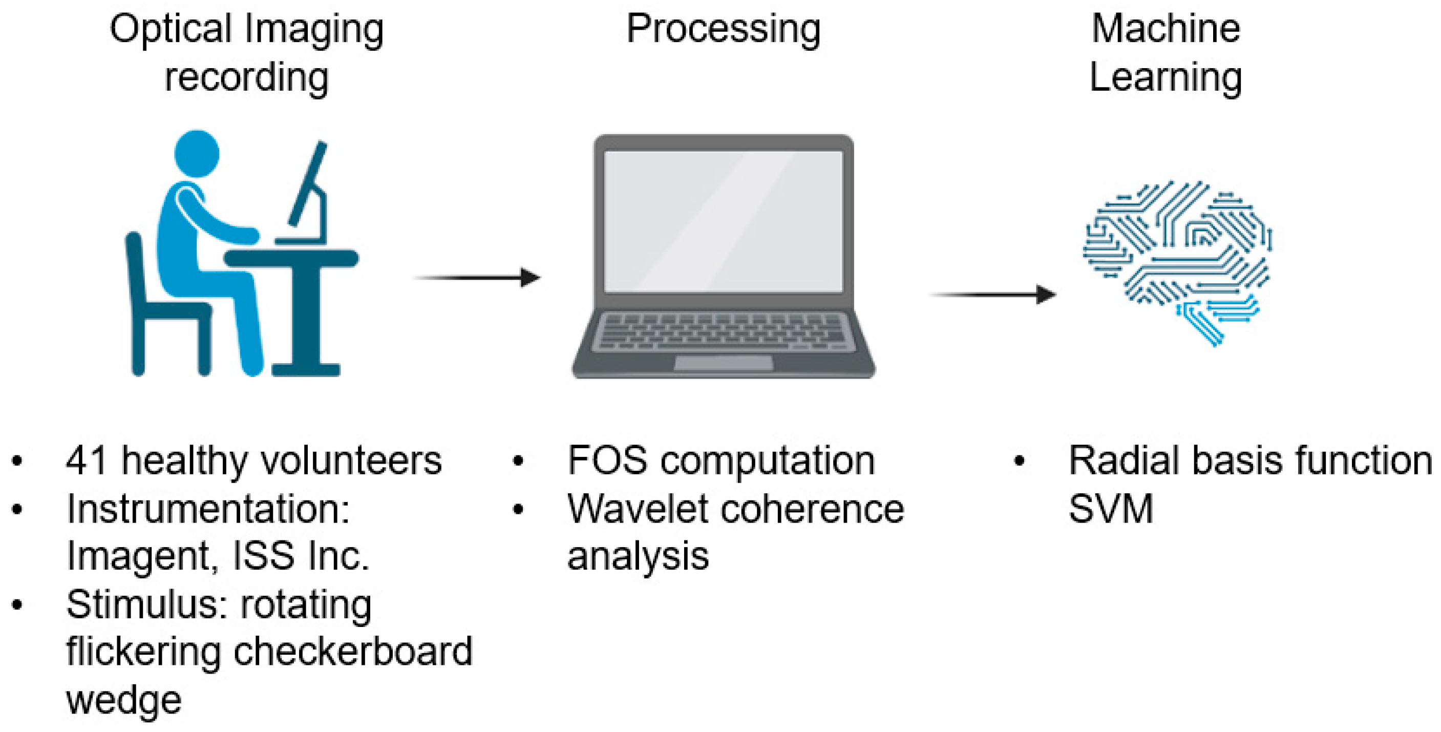
2. Materials and Methods
2.1. Participants
2.2. Visual Stimulation
2.3. Optical Imaging Recording
2.4. Fast Optical Signal Preprocessing
2.5. Machine Learning Approach
3. Results
4. Discussion
5. Conclusions
Author Contributions
Funding
Institutional Review Board Statement
Informed Consent Statement
Data Availability Statement
Conflicts of Interest
Nomenclature
| AC | Alternating Current light intensity |
| AUC | Area Under the Curve |
| BCI | Brain–computer interface |
| DC | Direct Current light intensity |
| EEG | Electroencephalography |
| fNIRS | Functional near-infrared spectroscopy |
| FOS | Fast optical signals |
| ITR | Information transfer rate |
| ML | Machine learning |
| PH | Phase delay |
| RBF | Radial Basis Function |
| ROC | Receiver Operating Characteristic |
| SNR | Signal-to-noise ratio |
| SVM | Support vector machine |
References
- Wolpaw, J.R.; Birbaumer, N.; Heetderks, W.J.; McFarland, D.J.; Peckham, P.H.; Schalk, G.; Donchin, E.; Quatrano, L.A.; Robinson, C.J.; Vaughan, T.M. Brain-Computer Interface Technology: A Review of the First International Meeting. IEEE Trans. Rehabil. Eng. 2000, 8, 164–173. [Google Scholar] [CrossRef] [PubMed]
- Hekmatmanesh, A.; Nardelli, P.H.; Handroos, H. Review of the State-of-the-Art of Brain-Controlled Vehicles. IEEE Access 2021, 9, 110173–110193. [Google Scholar] [CrossRef]
- Nicolas-Alonso, L.F.; Gomez-Gil, J. Brain Computer Interfaces, a Review. Sensors 2012, 12, 1211–1279. [Google Scholar] [CrossRef] [PubMed]
- Rasheed, S. A Review of the Role of Machine Learning Techniques towards Brain–Computer Interface Applications. Mach. Learn. Knowl. Extr. 2021, 3, 835–862. [Google Scholar] [CrossRef]
- Hallez, H.; Vanrumste, B.; Grech, R.; Muscat, J.; De Clercq, W.; Vergult, A.; D’Asseler, Y.; Camilleri, K.P.; Fabri, S.G.; Van Huffel, S.; et al. Review on Solving the Forward Problem in EEG Source Analysis. J. NeuroEng. Rehabil. 2007, 4, 46. [Google Scholar] [CrossRef]
- Lotte, F.; Bougrain, L.; Cichocki, A.; Clerc, M.; Congedo, M.; Rakotomamonjy, A.; Yger, F. A Review of Classification Algorithms for EEG-Based Brain–Computer Interfaces: A 10 Year Update. J. Neural Eng. 2018, 15, 031005. [Google Scholar] [CrossRef]
- Ferracuti, F.; Freddi, A.; Iarlori, S.; Longhi, S.; Peretti, P. Auditory Paradigm for a P300 BCI System Using Spatial Hearing. In Proceedings of the 2013 IEEE/RSJ International Conference on Intelligent Robots and Systems, Tokyo, Japan, 3–7 November 2013; pp. 871–876. [Google Scholar]
- Pinti, P.; Tachtsidis, I.; Hamilton, A.; Hirsch, J.; Aichelburg, C.; Gilbert, S.; Burgess, P.W. The Present and Future Use of Functional Near-infrared Spectroscopy (FNIRS) for Cognitive Neuroscience. Ann. N. Y. Acad. Sci. 2020, 1464, 5–29. [Google Scholar] [CrossRef]
- Forcione, M.; Chiarelli, A.M.; Davies, D.J.; Perpetuini, D.; Sawosz, P.; Merla, A.; Belli, A. Cerebral Perfusion and Blood-Brain Barrier Assessment in Brain Trauma Using Contrast-Enhanced near-Infrared Spectroscopy with Indocyanine Green: A Review. J. Cereb. Blood Flow Metab. 2020, 40, 1586–1598. [Google Scholar] [CrossRef]
- Stepnoski, R.A.; LaPorta, A.; Raccuia-Behling, F.; Blonder, G.E.; Slusher, R.E.; Kleinfeld, D. Noninvasive Detection of Changes in Membrane Potential in Cultured Neurons by Light Scattering. Proc. Natl. Acad. Sci. USA 1991, 88, 9382–9386. [Google Scholar] [CrossRef]
- Rector, D.M.; Poe, G.R.; Kristensen, M.P.; Harper, R.M. Light Scattering Changes Follow Evoked Potentials From Hippocampal Schaeffer Collateral Stimulation. J. Neurophysiol. 1997, 78, 1707–1713. [Google Scholar] [CrossRef]
- Lee, J.; Kim, S.J. Spectrum Measurement of Fast Optical Signal of Neural Activity in Brain Tissue and Its Theoretical Origin. NeuroImage 2010, 51, 713–722. [Google Scholar] [CrossRef]
- Gratton, G.; Corballis, P.M.; Cho, E.; Fabiani, M.; Hood, D.C. Shades of Gray Matter: Noninvasive Optical Images of Human Brain Reponses during Visual Stimulation. Psychophysiology 1995, 32, 505–509. [Google Scholar] [CrossRef]
- Radhakrishnan, H.; Vanduffel, W.; Deng, H.P.; Ekstrom, L.; Boas, D.A.; Franceschini, M.A. Fast Optical Signal Not Detected in Awake Behaving Monkeys. NeuroImage 2009, 45, 410–419. [Google Scholar] [CrossRef]
- Medvedev, A.V.; Kainerstorfer, J.; Borisov, S.V.; Barbour, R.L.; VanMeter, J. Event-Related Fast Optical Signal in a Rapid Object Recognition Task: Improving Detection by the Independent Component Analysis. Brain Res. 2008, 1236, 145–158. [Google Scholar] [CrossRef]
- Medvedev, A.V.; Kainerstorfer, J.M.; Borisov, S.V.; Gandjbakhche, A.H.; VanMeter, J.W. Seeing Electroencephalogram through the Skull: Imaging Prefrontal Cortex with Fast Optical Signal. JBO 2010, 15, 061702. [Google Scholar] [CrossRef]
- Chiarelli, A.M.; Di Vacri, A.; Romani, G.L.; Merla, A. Fast Optical Signal in Visual Cortex: Improving Detection by General Linear Convolution Model. Neuroimage 2013, 66, 194–202. [Google Scholar] [CrossRef]
- Gratton, G.; Fabiani, M. Fast Optical Imaging of Human Brain Function. Front. Hum. Neurosci. 2010, 4, 52. [Google Scholar] [CrossRef]
- Gratton, G.; Fabiani, M.; Goodman-Wood, M.R.; Desoto, M.C. Memory-Driven Processing in Human Medial Occipital Cortex: An Event-Related Optical Signal (EROS) Study. Psychophysiology 1998, 35, 348–351. [Google Scholar] [CrossRef]
- Gratton, E.; Fantini, S.; Franceschini, M.A.; Gratton, G.; Fabiani, M. Measurements of Scattering and Absorption Changes in Muscle and Brain. Philos. Trans. R. Soc. Lond. Ser. B Biol. Sci. 1997, 352, 727–735. [Google Scholar] [CrossRef]
- Yi, H. Efficient Machine Learning Algorithm for Electroencephalogram Modeling in Brain–Computer Interfaces. Neural Comput. Appl. 2022, 34, 9233–9243. [Google Scholar] [CrossRef]
- Hashmi, M.F.; Kene, J.D.; Kotambkar, D.M.; Matte, P.; Keskar, A.G. An Efficient P300 Detection Algorithm Based on Kernel Principal Component Analysis-Support Vector Machine. Comput. Electr. Eng. 2022, 97, 107608. [Google Scholar] [CrossRef]
- Sweeti. Attentional Load Classification in Multiple Object Tracking Task Using Optimized Support Vector Machine Classifier: A Step towards Cognitive Brain–Computer Interface. J. Med. Eng. Technol. 2022, 46, 69–77. [Google Scholar] [CrossRef] [PubMed]
- Awad, M.; Khanna, R. Support Vector Regression. In Efficient Learning Machines: Theories, Concepts, and Applications for Engineers and System Designers; Awad, M., Khanna, R., Eds.; Apress: Berkeley, CA, USA, 2015; pp. 67–80. ISBN 978-1-4302-5990-9. [Google Scholar]
- Perpetuini, D.; Filippini, C.; Zito, M.; Cardone, D.; Merla, A. Altered Microcirculation in Alzheimer’s Disease Assessed by Machine Learning Applied to Functional Thermal Imaging Data. Bioengineering 2022, 9, 492. [Google Scholar] [CrossRef]
- Hekmatmanesh, A. Investigation of EEG Signal Processing for Rehabilitation Robot Control. Ph.D. Thesis, LUT University, Lappeenranta, Finland, 2019. [Google Scholar]
- Hekmatmanesh, A.; Wu, H.; Motie-Nasrabadi, A.; Li, M.; Handroos, H. Combination of Discrete Wavelet Packet Transform with Detrended Fluctuation Analysis Using Customized Mother Wavelet with the Aim of an Imagery-Motor Control Interface for an Exoskeleton. Multimed. Tools Appl. 2019, 78, 30503–30522. [Google Scholar] [CrossRef]
- Proulx, N.; Samadani, A.-A.; Chau, T. Online Classification of the Near-Infrared Spectroscopy Fast Optical Signal for Brain-Computer Interfaces. Biomed. Phys. Eng. Express 2018, 4, 065010. [Google Scholar] [CrossRef]
- Gratton, G.; Fabiani, M. The Event-Related Optical Signal (EROS) in Visual Cortex: Replicability, Consistency, Localization, and Resolution. Psychophysiology 2003, 40, 561–571. [Google Scholar] [CrossRef]
- Chen, X.; Gupta, R.S.; Gupta, L. Exploiting the Cone of Influence for Improving the Performance of Wavelet Transform-Based Models for ERP/EEG Classification. Brain Sci. 2023, 13, 21. [Google Scholar] [CrossRef]
- Chiarelli, A.M.; Romani, G.L.; Merla, A. Fast Optical Signals in the Sensorimotor Cortex: General Linear Convolution Model Applied to Multiple Source–Detector Distance-Based Data. NeuroImage 2014, 85, 245–254. [Google Scholar] [CrossRef]
- Maclin, E.; Low, K.; Fabiani, M.; Gratton, G. Improving the Signal-to-Noise Ratio of Event-Related Optical Signals. IEEE Eng. Med. Biol. Mag. Q. Mag. Eng. Med. Biol. Soc. 2007, 26, 47–51. [Google Scholar] [CrossRef]
- Morren, G.; Wolf, M.; Lemmerling, P.; Wolf, U.; Choi, J.H.; Gratton, E.; De Lathauwer, L.; Van Huffel, S. Detection of Fast Neuronal Signals in the Motor Cortex from Functional near Infrared Spectroscopy Measurements Using Independent Component Analysis. Med. Biol. Eng. Comput. 2004, 42, 92–99. [Google Scholar] [CrossRef]
- Wolf, M.; Wolf, U.; Choi, J.H.; Toronov, V.; Adelina Paunescu, L.; Michalos, A.; Gratton, E. Fast Cerebral Functional Signal in the 100-Ms Range Detected in the Visual Cortex by Frequency-Domain near-Infrared Spectrophotometry. Psychophysiology 2003, 40, 521–528. [Google Scholar] [CrossRef]
- Franceschini, M.A.; Boas, D.A. Noninvasive Measurement of Neuronal Activity with Near-Infrared Optical Imaging. NeuroImage 2004, 21, 372–386. [Google Scholar] [CrossRef]
- Gratton, G.; Fabiani, M.; Friedman, D.; Franceschini, M.A.; Fantini, S.; Corballis, P.; Gratton, E. Rapid Changes of Optical Parameters in the Human Brain During a Tapping Task. J. Cogn. Neurosci. 1995, 7, 446–456. [Google Scholar] [CrossRef]
- Gratton, G.; Brumback, C.R.; Gordon, B.A.; Pearson, M.A.; Low, K.A.; Fabiani, M. Effects of Measurement Method, Wavelength, and Source-Detector Distance on the Fast Optical Signal. NeuroImage 2006, 32, 1576–1590. [Google Scholar] [CrossRef]
- Chiarelli, A.M.; Maclin, E.L.; Low, K.A.; Mathewson, K.E.; Fabiani, M.; Gratton, G. Combining Energy and Laplacian Regularization to Accurately Retrieve the Depth of Brain Activity of Diffuse Optical Tomographic Data. J. Biomed. Opt. 2016, 21, 36008. [Google Scholar] [CrossRef]
- Kubler, A.; Mushahwar, V.K.; Hochberg, L.R.; Donoghue, J.P. BCI Meeting 2005-Workshop on Clinical Issues and Applications. IEEE Trans. Neural Syst. Rehabil. Eng. 2006, 14, 131–134. [Google Scholar] [CrossRef]
- Strangman, G.; Franceschini, M.A.; Boas, D.A. Factors Affecting the Accuracy of Near-Infrared Spectroscopy Concentration Calculations for Focal Changes in Oxygenation Parameters. NeuroImage 2003, 18, 865–879. [Google Scholar] [CrossRef]
- Chiarelli, A.M.; Perpetuini, D.; Croce, P.; Greco, G.; Mistretta, L.; Rizzo, R.; Vinciguerra, V.; Romeo, M.F.; Zappasodi, F.; Merla, A. Fiberless, Multi-Channel FNIRS-EEG System Based on Silicon Photomultipliers: Towards Sensitive and Ecological Mapping of Brain Activity and Neurovascular Coupling. Sensors 2020, 20, 2831. [Google Scholar] [CrossRef]
- Chiarelli, A.M.; Zappasodi, F.; Pompeo, F.D.; Merla, A. Simultaneous Functional Near-Infrared Spectroscopy and Electroencephalography for Monitoring of Human Brain Activity and Oxygenation: A Review. Neurophotonics 2017, 4, 041411. [Google Scholar] [CrossRef]
- Coyle, S.; Ward, T.; Markham, C. Physiological Noise in Near-Infrared Spectroscopy: Implications for Optical Brain Computer Interfacing. In Proceedings of the 26th Annual International Conference of the IEEE Engineering in Medicine and Biology Society, San Francisco, CA, USA, 1–5 September 2004; Volume 2, pp. 4540–4543. [Google Scholar]
- Obrig, H.; Kohl-Bareis, M.; Steinbrink, J.; Malak, J.; Uludağ, K.; Villringer, A. Noninvasive Monitoring of Cerebral Blood Flow by a Dye Bolus Method: Separation of Brain from Skin and Skull Signals. JBO 2002, 7, 464–470. [Google Scholar] [CrossRef]
- Cecotti, H.; Marathe, A.R.; Ries, A.J. Optimization of Single-Trial Detection of Event-Related Potentials Through Artificial Trials. IEEE Trans. Biomed. Eng. 2015, 62, 2170–2176. [Google Scholar] [CrossRef] [PubMed]


| Contrast | Optical Metric | Accuracy (%) | p-Value | ITR (bpm) |
|---|---|---|---|---|
| Top vs. bottom | PH 830 | 59.20 | 0.005 | 2.82 |
| Top vs. bottom | DC 830 | 62.95 | 0.001 | 5.92 |
| Top vs. bottom | PH 690 | 55.75 | 0.071 | 1.25 |
| Top vs. bottom | DC 690 | 58.00 | 0.021 | 2.23 |
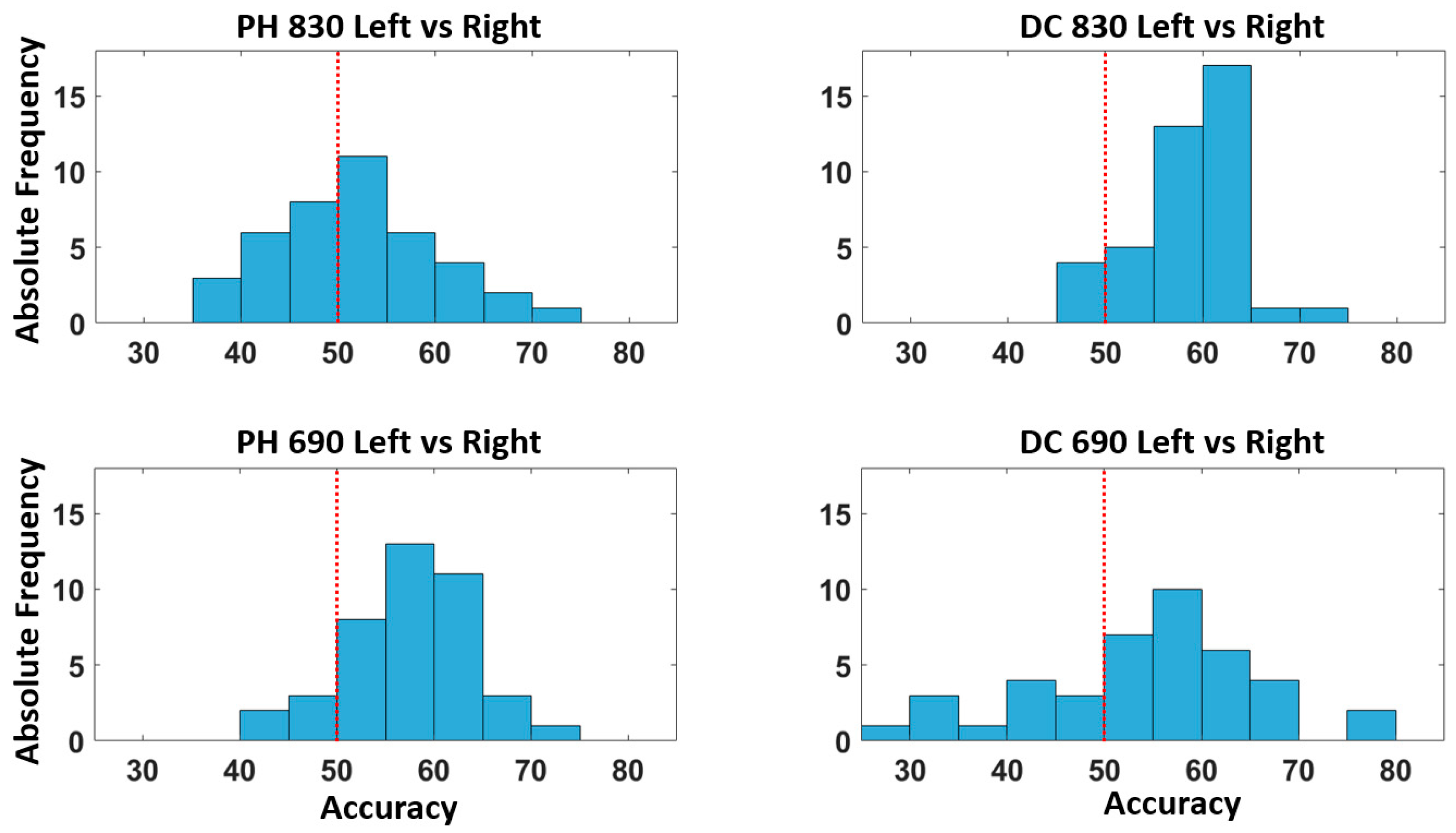
| Left vs. right | PH 830 | 55.00 | 0.082 | 0.87 |
| Left vs. right | DC 830 | 58.00 | 0.016 | 2.23 |
| Left vs. right | PH 690 | 56.25 | 0.037 | 1.70 |
| Left vs. right | DC 690 | 56.20 | 0.039 | 1.70 |
Disclaimer/Publisher’s Note: The statements, opinions and data contained in all publications are solely those of the individual author(s) and contributor(s) and not of MDPI and/or the editor(s). MDPI and/or the editor(s) disclaim responsibility for any injury to people or property resulting from any ideas, methods, instructions or products referred to in the content. |
© 2023 by the authors. Licensee MDPI, Basel, Switzerland. This article is an open access article distributed under the terms and conditions of the Creative Commons Attribution (CC BY) license (https://creativecommons.org/licenses/by/4.0/).
Share and Cite
Perpetuini, D.; Günal, M.; Chiou, N.; Koyejo, S.; Mathewson, K.; Low, K.A.; Fabiani, M.; Gratton, G.; Chiarelli, A.M. Fast Optical Signals for Real-Time Retinotopy and Brain Computer Interface. Bioengineering 2023, 10, 553. https://doi.org/10.3390/bioengineering10050553
Perpetuini D, Günal M, Chiou N, Koyejo S, Mathewson K, Low KA, Fabiani M, Gratton G, Chiarelli AM. Fast Optical Signals for Real-Time Retinotopy and Brain Computer Interface. Bioengineering. 2023; 10(5):553. https://doi.org/10.3390/bioengineering10050553
Chicago/Turabian StylePerpetuini, David, Mehmet Günal, Nicole Chiou, Sanmi Koyejo, Kyle Mathewson, Kathy A. Low, Monica Fabiani, Gabriele Gratton, and Antonio Maria Chiarelli. 2023. "Fast Optical Signals for Real-Time Retinotopy and Brain Computer Interface" Bioengineering 10, no. 5: 553. https://doi.org/10.3390/bioengineering10050553
APA StylePerpetuini, D., Günal, M., Chiou, N., Koyejo, S., Mathewson, K., Low, K. A., Fabiani, M., Gratton, G., & Chiarelli, A. M. (2023). Fast Optical Signals for Real-Time Retinotopy and Brain Computer Interface. Bioengineering, 10(5), 553. https://doi.org/10.3390/bioengineering10050553