Article

Fast Trace Detection of Chlorpyrifos Vapors Using a Handheld Ion Mobility Spectrometer Operated near Ambient Temperature

by
Victor Bocoș-Bințințan
1,2,*,†,
Ancuța-Maria Dodea
1,†,
Tomáš Rozsypal
3,
Adrian Pătruț
4,5,
Gheorghe Roșian
1,
Aurel-Vasile Martiniuc
2,
Alin-Gabriel Moraru
1,
Simina Vasc
1 and
Maria-Paula Bocoș-Bințințan
6,*
1
Faculty of Environmental Science and Engineering, Babeș-Bolyai University, Str. Fântânele 30, 400294 Cluj-Napoca, Romania
2
SC Transcend SRL, Str. Arinilor 13, 400568 Cluj-Napoca, Romania
3
Nuclear, Biological and Chemical Defence Institute, University of Defence, Vita Nejedleho 1, 68203 Vyskov, Czech Republic
4
Faculty of Chemistry & Chemical Engineering, Babeș-Bolyai University, Str. Arany János 11, 400028 Cluj-Napoca, Romania
5
Raluca Ripan Institute for Research in Chemistry, Babeș-Bolyai University, Str. Fântânele 30, 400294 Cluj-Napoca, Romania
6
Faculty of Veterinary Medicine, University of Agricultural Sciences and Veterinary Medicine, Calea Mănăştur 3-5, 400372 Cluj-Napoca, Romania
*
Authors to whom correspondence should be addressed.
These authors contributed equally to this work.
Toxics 2025, 13(10), 843; https://doi.org/10.3390/toxics13100843
Submission received: 15 September 2025 / Revised: 29 September 2025 / Accepted: 1 October 2025 / Published: 2 October 2025
(This article belongs to the Section Agrochemicals and Food Toxicology)

Highlights

What are the main findings?
  • Detection of Chlorpyrifos vapors at levels below 1 ppmv was accomplished using a ToF IMS operated under near-ambient temperature.
  • Both excellent sensitivity and speed in the detection of Chlorpyrifos were achieved using the handheld ToF IMS.
  • Ammonia doping of the IMS ensures good selectivity.
  • Formation of two product ions represents a strong point for Chlorpyrifos identification.
What is the implication of the main finding?
  • Rapid sensing of Chlorpyrifos at trace levels with highly portable IMS devices, demonstrating real-time response (seconds), was shown.
  • Potential applications in real-world screening have been highlighted.

Abstract

Chlorpyrifos CPF (O,O-diethyl O-(3,5,6-trichloro-2-pyridyl) phosphorothioate), known also as Chlorpyrifos-ethyl, is one of the most utilized organophosphorus pesticides worldwide. Additionally, CPF could be used as a chemical warfare agent surrogate. Although its acute toxicity is not high, it is responsible for both a large number of intoxications and chronic, delayed neurological effects. In this work, it is reported for the first time the qualitative and quantitative response produced by CPF vapors, using a pocket-held Time-of-Flight Ion Mobility Spectrometer (ToF IMS) with a non-radioactive ionization source and ammonia doping, model LCD-3.2E (Smiths Detection Ltd.), operated near ambient temperature (below 30 °C). Spectra of CPF in positive ion mode included two distinct product ion peaks; thus, identification of CPF vapors by IMS relies on these peaks—the monomer M·NH4+ with reduced ion mobility K0 = ca. 1.76 cm2 V−1 s−1 and the dimer M2·NH4+ with K0 = ca. 1.47 cm2 V−1 s−1 (where M may be assignable to CPF molecule)—and positive reactant ions (Pos RIP) have K0 = ca. 2.25 cm2 V−1 s−1. Excellent sensitivity, with a limit of detection LOD of 0.72 ppbv (10.5 μg m−3) and a limit of quantification LOQ of 2.41 ppbv (35.1 μg m−3), has been noticed; linear response was up to 100 ppbv, while saturation occurs over ca. 1000 ppbv (14.6 mg m−3). Our results demonstrate that this method provides a robust tool for both off-site and on-site detecting and quantifying CPF vapors at trace levels, which has strong implications for either industrial hygiene or forensic investigations concerning the pesticide Chlorpyrifos, as well as for monitoring of environmental contamination by organophosphorus pesticides.

Graphical Abstract

1. Introduction

Chlorpyrifos (CPF), known also as Chlorpyrifos-ethyl, is an organophosphorus (OP) insecticide with large spectrum, widely used against a variety of pests (mainly insects, but also worms, larvae, fleas, locusts—in both outdoor and indoor applications) and is considered as one of the most used insecticides in agriculture both in the U.S. and in Europe [1,2]. CPF was introduced in 1965 by Dow Chemical Company (Dow AgroSciences), then manufactured and marketed in huge amounts (thousands of tons per year), especially under the tradenames Dursban™ and Lorsban™.
Organophosphorus (OP) compounds induce acute neurotoxic effects by inhibiting the enzyme acetylcholinesterase in neurons’ synapses; this way, cholinergic hyperstimulation is caused. Main symptoms of acute intoxications with OP compounds (either pesticides or nerve chemical warfare agents) are miosis, salivation, lacrimation, urination and defecation, then muscle twitching, convulsions and, as ultimate effect, death [3]. Infants and children are particularly vulnerable to chronic adverse effects generated by OP pesticides; thus, their exposure to OP pesticides (in utero and/or post-birth) has been associated with issues related to neuro-development, decreasing the normal gestation period and alteration of fetal growth [4]. Poisoning with CPF may also affect, besides the central nervous system, the cardiovascular and respiratory systems. While CPF is far less toxic than organophosphorus nerve chemical warfare agents, it shares similar toxicological effects, making it, therefore, a useful tool for research and training related to nerve chemical weapons exposure.
There are also many adverse eco-toxicological effects related to Chlorpyrifos, for instance, toxicity towards pollinating insects, birds, fish and other aquatic organisms, organisms living in soils and domestic animals. Like most OPs, CPF has, fortunately, a relatively short half-life inside biological tissues; for instance, a single exposure to CPF results in ca. 24 h residence time in blood and ca. 60 h in fat. Also, CPF does not have a high potential for bioaccumulation in mammals. The half-life of CPF in indoor environments has been estimated to be approximately one month [5]. However, investigations concerning the treatment with CPF demonstrated that it was still present up to 8 years post application; CPF has persisted into/onto various objects and surfaces (such as toys, pavements or furniture) for up to two weeks after its indoor application [5].
The most relevant physico–chemical and toxicological properties of Chlorpyrifos are summarized in Table 1.
It is important to emphasize that Chlorpyrifos is sensitive to light, moisture, various alkaline compounds (e.g., bleach) and also to microbial degradation. The half-life of Chlorpyrifos in water is pretty short—from several days to a couple of weeks. CPF may be adsorbed to sediments and organic matter; thus, its half-life in soil may usually be 2 to 4 months, but it may extend to over one year. The granular commercial formulations of CPF are found to be even more persistent (up to 6 months). The major biological metabolite and environmental breakdown product of CPF hydrolysis is 3,5,6-trichloro-2-pyridinol (TCP: C5H2Cl3NO), accompanied by either diethyl phosphorothioate (DETP: C4H11O3PS) or diethyl phosphate (DEP: C4H11O3PO) [5,6].
Detection of various OP compounds present as vapors in air samples can be realized by using a diversity of analytical techniques, from simple colorimetric techniques and photoionization detectors (PIDs) to more complex instruments such as gas chromatographs coupled often with mass spectrometers (GC-MSs), flame photometry detectors (FPDs), or surface acoustic wave (SAW) sensors [7]. For most toxic OP compounds—like the nerve chemical warfare agents—the preferred analytical technology used on-field nowadays is ion mobility spectrometry (IMS) [8].
Ion mobility spectrometry (IMS) has been continuously building an increasing recognition during the last decades and has become a mature analytical technology. IMS possesses extreme versatility, being perfectly suited for trace detection and identification of many classes of chemicals—either present in air samples as vapors, or vaporized from liquid and solid samples—after their soft ionization at atmospheric pressure [8]. IMS possesses a superb sensitivity, often presenting detection limits of low ppbv for a great variety of compounds, with no pre-concentration. The high speed of ion separation, which takes place at atmospheric pressure and in the gaseous phase, can also be regarded as the main operational advantage of IMS; since a single ion mobility spectrum is obtained in 20 milliseconds, the whole analysis run usually takes just several seconds. IMS instrumentation is pretty simple, which has translated towards highly compact, hand- and even pocket-held rugged IMS devices. The most important fields of IMS applicability are detection and identification of all types of chemical weapons [9], fast sensing of explosives (including acetone-based homemade ones) [10,11], illegal drugs plus their associated precursors [12,13] and a wide range of toxic industrial chemicals [14,15]. IMS technology was also successfully used in other domains—forensic approaches, various industries for quality control and quality assurance, toxicology, bio-medicine, environmental protection and monitoring or even space research [8].
IMS relies on two main stages. The first and crucial one is the ionization of neutral compounds, occurring at atmospheric pressure and in the gas phase. The second stage is the ions’ separation in a neutral drift gas, under the influence of a direct-current electric field with relatively low intensity (E < 500 V/cm). Nowadays, this IMS analytical technique is named the Drift Tube (DT) IMS, or Time-of-Flight (ToF) IMS. Therefore, in ToF IMS, the created ions are separated in time by the longitudinally applied electrical field, while the analytical output is a practical, very fast chromatogram. In ToF IMS, separation of different ions occurs because they possess different speeds and mobilities when traveling against a flow of neutral drift gas (recirculated purified air in portable IMS instruments). The “motor” of ions’ displacement is the longitudinal d.c. electric field applied onto the IMS cell. Formation of ions in IMS is a fast but complex process, involving two successive steps—firstly, the generation of primary (reactant) ions starting from molecules present in the drift gas (usually purified air), and secondly, the formation of secondary (product) ions, which will include the entire molecule of the target analyte. Therefore, when an ionization source is used in air, a series of fast and complex ion–molecule collisional processes will produce the initial batch of reactant ions. For instance, in positive ion operation mode, with a radioactive ionization source and by using water vapor chemistry, the positive reactant ions formed will be predominantly hydrated protons (H2O)xH+, accompanied by (H2O)yNH4+ and (H2O)zNO+, while in negative ion mode, the dominant negative reactant ions will be the (H2O)nO2 clusters. After reactant ions are generated, if the target analyte has a proton affinity (or electron affinity in the negative ion mode) higher than that of the reactant ions, the latter can transfer their electrical charge to the molecules of the analyte, and this leads to the formation of product ions that include the whole analyte molecule. The concentration level of water vapor present in the IMS cell is paramount and must therefore be strictly controlled and kept at a low level, since water vapor has a crucial role in the whole ion–molecule chemistry that occurs at atmospheric pressure [14]; otherwise, the IMS analytical performance will degrade significantly.
The constant that links the drift speed vd of an ion to the electric field intensity E that propels that ion through the IMS cell is called “ion mobility” K, according to the following equation: vd = K·E = ld/td (where ld = drift length of the IMS cell and td is the drift time of an ion). Of course, one may further infer that K = vd/E = ld/(E·td). By normalizing ion mobility for both pressure and temperature inside the IMS measurement cell one shall obtain the so-called “reduced ion mobility K0”, a parameter that is used in a consistent manner for accomplishing qualitative characterization of the compound that generates that/those product ion(s): K0 = K·(Tambient/Tcell)·(Pcell/Patmospheric) [8].
The main aim of the present paper is to demonstrate the fast detection and quantification of vapors of Chlorpyrifos present at ultra-trace levels (<1 ppmv) in air samples by employing a handheld ToF IMS instrument with corona discharge ionization and ammonia doping, operated near ambient temperature. Both the risk of acute/chronic intoxications to humans and other potential ecotoxicological effects of Chlorpyrifos fully justify the need for fast trace detection of its vapors using the rapid analytical technique of IMS. Even if the commercial use of CPF has been restricted since 2000, this necessity remains stringent. The literature on the detection of CPF by IMS is relatively scarce, especially as it approaches solid and liquid matrices, as discussed later.
Our intention was to demonstrate, through this study, the qualitative and quantitative capabilities of portable IMS devices with corona discharge non-radioactive ionization operated near ambient temperature for the fast sensing of CPF vapors. Evaluating the performance of such handheld portable IMS devices constitutes an important novelty and a strong point of the paper, especially since previous studies have employed only bulky stationary desktop IMS instruments or even more complex desktop hyphenated GC–IMS systems. The application of portable IMS technology for rapid on-site detection of CPF vapors was highlighted. Our research is based on preparing a series of standard atmospheres with known concentrations of CPF vapors, in the ultra-trace (under 1 ppmv) range.
A large number of studies have approached the analysis of CPF in complex environmental matrices, and we comprehensively and critically reviewed them; all these studies provide valuable insight into CPF analysis in various environmental (water, fruits and vegetables) and particulate samples. However, to the best of our knowledge, none of the previous reports have investigated CPF vapors using a handheld IMS instrument operated near ambient temperature, as all prior IMS studies employed stationary instruments operating at high temperatures (150–240 °C). Our current work is therefore intended to significantly complement the existing literature by using a portable IMS device suitable for real-time CPF vapor sensing at relatively low (ambient) temperature.

2. Materials and Methods

2.1. The IMS Instrument

For detection and quantification of CPF vapors, a highly portable, pocket-sized commercial ToF IMS instrument (size ca. 18.0 × 11.5 × 4.5 cm and weight of ca. 0.6 kg), model LCD-3.2E (Lightweight Chemical Detector), manufactured by the company Smiths Detection Ltd., Watford, UK, was used. The instrument is built around a twin-design miniature IMS cell, which means that two IMS cells are used simultaneously. Both cells use a classical design consisting of alternating conducting elements placed onto an insulating cylinder, and each has a drift length of ca. 3 cm; the intensity of d.c. electric field E is ca. 270 V cm−1. On the basis of this “twin cell” configuration, this IMS platform generates both positive and negative ion mobility spectra at the same time, which is a consistent advantage. The LCD-3.2E is operated near ambient temperature, at ca. 301 K (28 °C), while the pressure inside the measurement cells is atmospheric pressure, of ca. 1000 mbar. A non-radioactive ionization source, based on a point-to-plane corona discharge, was the one employed in the LCD-3.2E system. For a series of reasons, a source based on corona discharge is a more viable ionization alternative to the almost universally used radioactive sources based on beta isotopes (mainly Ni-63, or more rarely tritium H-3); one major advantage to quote here is the lack of any legal and bureaucratic burdens associated with manipulating, using or transporting any radioactive material. Besides that, the ionization sources with corona discharge generate a higher amount of reactant ions (hence a higher signal) compared to radioactive sources, while their ionization chemistry is quite similar to that of radioactive sources. The LCD-3.2E uses dry air as drift gas, which is continuously recirculated through a filter based on molecular sieve. Also worth mentioning is that the same purifying filter delivers, in a continuous manner, low amounts of gaseous ammonia, which is the dopant that seriously improves selectivity in positive ion mode. To the best of our knowledge, this model of IMS instrumentation is maybe the most compact and miniaturized commercial ToF IMS system that exists currently on the market and is perfectly capable of sensing and identifying rapidly, in a matter of seconds, both gases and vapors belonging to the TIC and CWA categories. The LCD-3.2E instrument has already been described in reference [15], including its schematic diagram.
This IMS system was connected, through the proprietary software TrimScan2, ver. 0.4.0 (Smiths Detection Ltd., UK), to a PC. This way, all resulting experimental data were saved as ion mobility spectra (positive and negative) on the hard drive of the PC; then, the spectra were exported to MS Excel files.
If ammonia is being used as a dopant in IMS, then the major positive reactant ions shift from hydrated proton clusters (H2O)nH+ towards ammonia-hydrated ones, having the general formula (H2O)mNH4+. When a small volume of air sample with CPF vapors is sent into the IMS cell, the non-radioactive ionization source using the corona discharge ionizes the molecules of CPF in a soft manner and eventually generates product ions that include the molecule of the target analyte. All formed ions—both reactant and product—are injected inside the drift (separation) region by the opening of the shutter grid, then they “fly” through the distance from the shutter grid to the detector (the drift length) and eventually attain a drift speed vd that has a constant value of several meters per second. When a specific ion (either reactant or product ions) arrives at the detector, a very low ion current, usually in the range of tens of pA, is produced, then amplified and measured. As a consequence, every compound will have its own characteristic drift speed when traveling through the neutral drift gas (air at atmospheric pressure), and for this reason, its drift time can be utilized for its identification, similar to the retention time in chromatography.

2.2. Reagents, Sampling and Work Flow Procedure

A liquid standard reference material (SRM) containing CPF in hexane, with a concentration of 2 mg L−1 (Dr. Ehrenstorfer GmbH, Augsburg, Germany), was used in order to create test atmospheres that have very low concentrations of CPF vapors in air. These standard atmospheres were produced by injecting known volumes of standard solution inside 0.72 L glass jars with screw caps; a series of microliter GC syringes with volumes of 5, 10, 20, 50, 100 and 500 μL were used for this purpose.
Glass jars were stored in a heating oven for 30 min, at a temperature of ca. 50 °C, in order to allow vaporization of CPF. Between the jar’s opening and metallic screw cap, a triple sheet of 10 micron-thick aluminum foil was inserted; after the equilibration period, the cap was removed, then a 1 cm hole was made in the aluminum layers, and the analytical sampling nozzle of the IMS spectrometer was inserted in order to sample.
Therefore, in order to perform the necessary IMS measurements, the standard atmospheres with known, low concentrations of CPF vapors (<1 ppmv) were produced by using a static method. Using the described static method, firstly, a series of six different test atmospheres containing very low concentrations (≤50 ppbv) of CPF vapors were generated: 2.5 ppbv, 5 ppbv, 10 ppbv, 20 ppbv, 40 ppbv and 100 ppbv, respectively.
Higher concentrations of CPF vapors, up to ca. 1000 ppbv, were obtained using a commercial pesticide formulation called Nurelle-D (manufactured by Agriphar S.A., Ougree, Belgium), which is an emulsified liquid concentrate containing 500 g CPF L−1. The same experimental procedure was used as for low concentrations, after repeatedly diluting the Nurelle-D with hexane in order to decrease the CPF concentration in the standard solution to 0.5 mg cm−3.
Table 2 presents succinctly the experimental conditions used for generating all the above-mentioned standard/test atmospheres.

3. Results

The experimental data obtained from the IMS model LCD-3.2E (ion mobility spectra, in positive ion mode) were recorded step by step, from the lowest concentration to the highest one, for every trace concentration of vapors of CPF obtained in the afferent static test atmosphere. IMS measurements for any concentration level were performed in triplicate; standard deviations between 5% and 7% were observed.
The results of our research are shown synthetically in Table 3, where CCPF is the concentration of CPF vapors in the standard atmosphere (see Table 2).
For low CPF vapor concentrations (≤50 ppbv), ion mobility spectra showed only one product ion peak (PIP) in the positive ion mode, observed at a drift time td = ca. 6.1 ms and assignable to the monomer product ion, with probable structure CPF·NH4+. At higher CPF concentrations (≥100 ppbv), a dimer product ion (assignable to CPF2·NH4+) appeared at a drift time td = ca. 7.3 ms. The peak of positive reactant ions (Pos RIP) was noticed to appear at a drift time td = ca. 4.7 ms. All positive ion mode IMS spectra for low vapor concentrations of CPF (from 1.5 to 800 ppbv) are presented in Figure 1; spectra clearly illustrate electrical charge conservation principle—the intensity of the product ion peaks produced by CPF increases when vapor concentration of CPF increases, and, consequently, at the same time, the intensity of the positive reactant ion peak declines.
The appearance of proton-bound dimer product ions is observed for several classes of compounds, such as ketones or organophosphorus compounds, and typically occurs when the analyte concentration exceeds a certain threshold [8]. The phenomenon results, therefore, from the enrichment of vapor-phase analyte molecules at higher concentrations, correlated with the life of the dimer product ion long enough for this ionic species to reach the detector and hence to generate an ion current.
CPF did not produce an IMS response (spectrum) in the negative operation mode.
Because n-hexane used as a solvent for CPF possesses a proton affinity, PA, far lower than that of ammonia (the PA of hexane is ca. 676 kJ mol−1, while PA of ammonia dopant is ca. 854 kJ mol−1), molecules of n-hexane will not become ionized by collisional charge transfer with ammonia-based positive reactant ions and hence they will not generate any peak in the positive ion mode IMS spectrum.
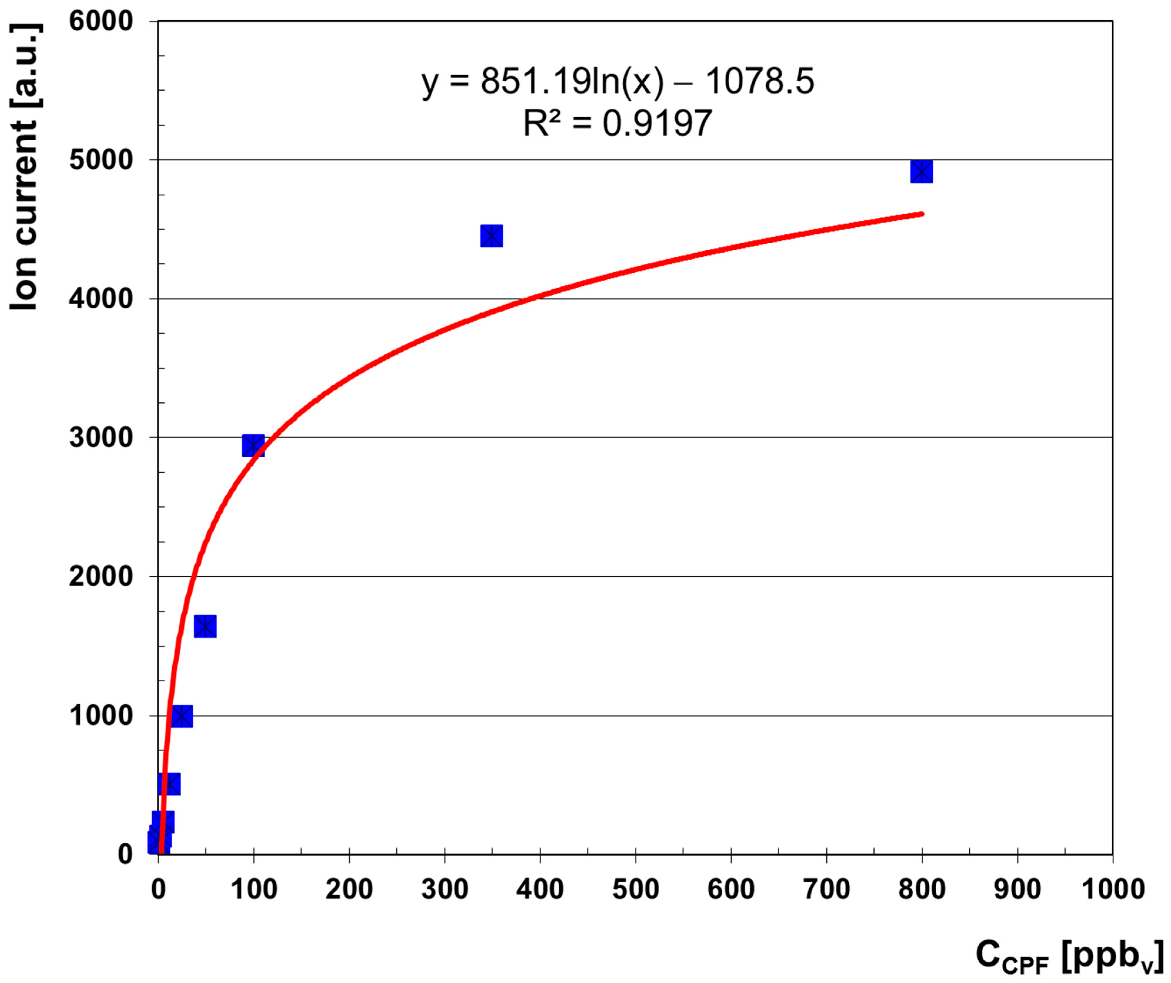
Quantitative information (peak intensities) has been utilized to build the afferent calibration curve and then to evaluate the quantitative response offered by the LCD-3.2E IMS instrument to vapors of CPF. All nine concentrations of CPF vapors were employed—the six very low concentrations + the three larger concentrations. This calibration curve is presented in Figure 2; the ion current (analytical signal) is considered as the sum of the ion current of the monomer product ion and that of the dimer product ion. A logarithmic-type trend line equation has also been included.
IMS spectra contain the qualitative information (that includes the drift time td of a specific ion and its reduced mobility K0) and also the quantitative information that resides in peak height. Drift time is relatively proportional to the ion’s mass and its size and inversely proportional to the ion’s electrical charge. Summarizing and simplifying, any ion mobility peak in the spectrum can be characterized by using three numbers: (1) drift time td (in ms); (2) reduced ion mobility K0 (in cm2 V−1 s−1); and (3) peak height hmax (in a.u.).
In Table 4, the relevant qualitative information is presented, which includes both drift times and the associated reduced ion mobilities for a positive reactant ion peak and, of course, for product ion peaks of CPF (both monomer and dimer). A common feature of OP compounds is the formation, when vapor concentration increases, of a pair of two product ion peaks [8,9]—a monomer and a dimer—and CPF does not make an exception.
Reduced ion mobility K0 has been checked and re-calculated using the accepted mobility standard compound 2,4-lutidine (2,4-dimethylpyridine), from which the “IMS cell constant” has been calculated. This method has the consistent advantage of considering all possible inhomogeneity of the electric field E that propels the ions inside the IMS measurement cell. Moreover, by using this cell constant in calculating K0, the need for highly accurate measurements for relevant instrumental parameters (electric field intensity E and drift length ld) and environmental ones (pressure and temperature inside the IMS cell) is eliminated. The compound 2,4-lutidine has already been largely accepted as a chemical standard for mobility in the positive ion mode of operation, with the following known reduced ion mobilities: K0 of standard (Lutidine monomer) = 1.95 cm2 V−1 s−1 and K0 of standard (Lutidine dimer) = 1.43 cm2 V−1 s−1, respectively [16]. The constant of the ion mobility cell (noted here with A) is, in fact, nothing but the product between the reduced mobility K0 of the chemical standard and the drift time td of the product ion generated by that chemical standard. Consequently, the following simple equation is used in order to find the reduced ion mobility of peaks associated with CPF·vapors: A = K0 of standard (Lutidine dimer)·td of standard (Lutidine dimer) = K0 of analyte (CPF)·td of analyte (CPF). Since the measured td of standard (Lutidine dimer) = 7.46 ms, then the cell constant of the IMS instrument will be calculated as follows: A = 1.43 cm2 V−1 s−1·7.46·10−3 s = 10.668·10−3 cm2 V−1.
The normalization of drift time of both product ions (monomer and dimer) against the drift time of positive reactant ion peak (Pos RIP) is also of interest; thus, the ratio between the drift times of PIPs and Pos RIP is as follows: td PIP 1/td Pos RIP = K0 Pos RIP/K0 PIP1 = 1.278 for the monomer product ion CPF·NH4+, and td PIP 2/td Pos RIP = K0 Pos RIP/K0 PIP2 = 1.536 for the dimer product ion CPF2·NH4+, respectively.
Resolving power of the IMS instrument model LCD-3.2E, RIMS, is the ratio between the drift time of an ion and its width at half height (RIMS = td/Δtd) and has been determined for all peaks existing in the ion mobility spectrum; the found results are given in Table 5. These resolving power-calculated values, which ranged between 13.5 and 17.3, are perfectly normal for handheld commercial instruments with short IMS measurement cells, like the case of the LCD-3.2E device.

Validation

Validation has been performed by using a simple procedure to quickly assess the suitability of the IMS-based analytical method employed. Thus, the evaluated parameters of the proposed method using IMS were as follows: limit of detection, limit of quantitation, sensitivity, linear response range, and accuracy.
Limit of detection (LOD) is the lowest concentration that produces a signal-to-noise (S/N) ratio equal to 3, and limit of quantification (LOQ) is defined as the lowest concentration that generates an S/N ratio of 10. Sensitivity S is the change in signal Y (peak height) that appears when the analyte’s concentration is incrementally changed (S = ΔY/ΔC). The background signal—described as the average of the background noise—has been calculated by using the latest 500 data points (those output ion currents obtained for drift times ranging from 10.00 to 20.00 ms, with increments of 0.02 milliseconds) for the blank IMS spectra; this average value of the signal (ion current) in positive ion operation mode has been found to be of ca. 8 a.u. The relevant figures of merit related to quantification of CPF vapors (LOD, LOQ, linear range and sensitivity S), which were determined based on the IMS response over the linear range (≤50 ppbv CPF), are as follows:
  • Limit of detection LOD = 0.72 ppbv
  • Limit of quantification LOQ = 2.41 ppbv
  • Linear range: 2.41—50 ppbv
  • Equation of linear regression: Y = 33.155·X + 49.622 (R2 = 0.9893)
  • Sensitivity S = 32.1 a.u./ppbv.
Precision has been evaluated using analyses in triplicate (see Table 3). Accuracy was assessed by using relative standard deviation, RSD (also known as coefficient of variation, CV), which was determined to be from 5% to 7% for the product ion peaks, PIPs. The repeatability of results was good, since RSD was less than 10%.

4. Discussion

The logarithmic-type allure of the built calibration curve (Figure 2) is characteristic for any IMS-type response [8]. There is also a good resemblance between the quantitative IMS response provided by the portable ToF IMS device equipped with a non-radioactive (corona discharge) ionization source that has been used by us (the LCD-3.2E) and that generated by other ToF IMS systems that rely on radioactive ionization sources.
IMS spectra obtained for the highest vapor concentration of CPF of 800 ppbv indicate that the saturation threshold was almost reached, since at this concentration level, the reactant ions peak was still present but had a much-diminished intensity of just about 10% of its initial height. Total saturation means that the whole amount of reactant ions has been depleted; its consequence is the total disappearance of the peak of reactant ions from the IMS spectrum. It must be strongly emphasized that saturation must be avoided, since it will lead to the persistent contamination of both the IMS cell and all the internal surfaces that came in contact with the vapors of the analyte; in other words, this type of contamination generates totally unwanted false alarms and memory effects.
Potential interferences from other chemicals are always a potential issue, and this may also happen in the case of IMS instruments. On the other hand, because CPF produces, at trace levels of just hundreds of ppbv, two peaks in the positive ion mode simultaneously, the qualitative identification of CPF using two time windows (monomer + dimer product ions) is clearly much more reliable, as compared to that using a single product ion peak. Common solvents and the majority of other vapors are very unlikely to form positive product ions having exactly the same drift times simultaneously (and reduced ion mobilities) as the two product ions generated by CPF, as most of these compounds have a proton affinity lower than that of ammonia-based reactant ions (854 kJ mol−1). Therefore, significant interferences from usual solvents under our experimental conditions are not anticipated at all. This enhanced chemical selectivity in positive ion mode is a crucial advantage that occurs from modifying chemical ionization processes at atmospheric pressure when using dopants such as ammonia.
Reduced ion mobilities for the monomer and dimer product ion peaks generated by Chlorpyrifos (CPF) vapors in the positive mode have been determined as K0 = ca. 1.76 cm2 V−1 s−1 for the monomer (assumed to be CPF·NH4+) and K0 = ca. 1.47 cm2 V−1 s−1 for the dimer (assumed to be CPF2·NH4+), using 2,4-lutidine as mobility standard.
By examining the quantitative response (Figure 2) and all ion mobility spectra obtained for the whole range of CPF vapor concentrations (Figure 1), one may conclude the following:
  • Minimum measured concentration of CPF vapors was 1.5 ppbv (0.022 mg m−3).
  • Linear dynamic range is from 2.4 ppbv (0.035 mg m−3) to ca. 100 ppbv (1.46 mg m−3) CPF.
  • Saturation is estimated to appear at >1000 ppbv (14.57 mg m−3) CPF.
The IMS responses for CPF were all obtained in the positive ion mode, which strongly suggests that CPF has a proton affinity (PA) higher than that of ammonium positive reactant ions of 854 kJ mol−1. The pocket-held IMS instrument model LCD-3.2E responded quickly, in real time (several seconds only), to vapors of target analyte CPF.
A comprehensive literature survey about the detection and quantification of CPF using various IMS instruments is summarized in Table 6. One may rapidly observe that almost all references have provided information regarding CPF extraction from various environmental samples (water, fruits and vegetables) with subsequent detection by IMS.
Information concerning detection and quantification of CPF using DT-IMS, summarized in Table 6, reveals a series of conclusions: there is a large packet of 21 papers (ref. [17,18,19,20,21,22,23,24,25,26,27,28,29,30,31,32,33,34,35,36,37]) by several groups of Iranian researchers, which, in fact, heavily focus on their efforts towards analyzing CPF present in various liquid and solid samples (water, fruits and vegetables), using a variety of elaborated and time-consuming methods of extraction and pre-concentration, most of them relying on micro-extraction. However, an objective but critical examination of all these papers clearly indicates that the information related to IMS detection is relatively poor and/or incomplete, since the authors only included the IMS spectrum, but they did not provide even the exact drift time of the CPF peak; it has to be mentioned that drift times included in Table 6 for ref. [17,18,19,20,21,22,23,24,25,26,27,28,29,30,31,32,33,34,35,36,37] were estimated visually by us from the spectra included in those papers. The reduced ion mobility K0 of the CPF product ion was also not indicated, with the exception of two papers that have reported the K0 of CPF using nicotinamide as mobility standard—ref. [30], with K0 = 1.37 cm2 V−1 s−1 and ref. [32], with K0 = 1.27 cm2 V−1 s−1. All these papers ([17,18,19,20,21,22,23,24,25,26,27,28,29,30,31,32,33,34,35,36,37]) claim to have detected CPF in the positive ion mode and at high temperatures of the IMS cell, between 150 and 235 °C, but none of them accomplished the net identification of CPF using a mass spectrum (e.g., by employing the tandem GC-MS), which is another potential drawback. Another common finding is the presence of just one product ion peak generated by CPF, although in the IMS spectrum from ref. [35,36], a second small peak emerges. Even if the authors of previous studies have not provided the reduced ion mobilities for the product ion assigned to CPF, based on their included information regarding the IMS instruments used (drift length ld; temperature of the IMS cell TIMS, pressure inside the IMS cell PIMS—assumed it as 760 Torr everywhere—and intensity of the d.c. electric field E, respectively), the K0 was calculated by us in order to compare these values with our findings. In this regard, the calculated reduced ion mobilities are as follows: K0 = 1.180 cm2 V−1 s−1 for [17]; K0 = 1.133 cm2 V−1 s−1 for [18]; K0 = 1.086 cm2 V−1 s−1 for [19]; K0 = 1.232 cm2 V−1 s−1 for [20]; K0 = 1.395 cm2 V−1 s−1 for [21]; K0 = 1.414 cm2 V−1 s−1 for [22]; K0 = 1.332 cm2 V−1 s−1 for [23]; K0 = 0.913 cm2 V−1 s−1 for [24]; K0 = 1.521 cm2 V−1 s−1 for [25]; K0 = 1.380 cm2 V−1 s−1 for [26]; K0 = 1.076 cm2 V−1 s−1 for [27]; K0 = 0.847 cm2 V−1 s−1 for [28]; K0 = 1.675 cm2 V−1 s−1 for [31]; K0 = 2.098 cm2 V−1 s−1 for [33]; K0 = 0.907 cm2 V−1 s−1 for [34]; K0 = 1.251 cm2 V−1 s−1 for [35]; K0 = 1.257 cm2 V−1 s−1 for [36] and K0 = 1.204 cm2 V−1 s−1 for [37]. One easily observes that calculated reduced ion mobilities K0 range from 0.847 to 2.098 cm2 V−1 s−1, so there is an interval of about 1.25 cm2 V−1 s−1 for K0 values, which is not reasonable. The only two papers that reported K0 based on using nicotinamide as mobility standard (of 1.37 [30] and 1.27 cm2 V−1 s−1 [32], respectively) seem, however, to be the most reliable in terms of K0 determination; these reduced mobilities are the closest to our found value for the dimer product ion of CPF (ca. 1.47 cm2 V−1 s−1). In other words, it is very possible that the unique peak described by the Iranian researchers is in fact the dimer product ion peak of CPF.
Apart of this avalanche of papers written by Iranian scientists, only one distinct article dealing with CPF detection using DT (ToF) IMS was found; this particular work has also been performed using a commercial desktop IMS instrument with radioactive ionization source and operated at high temperature; oddly enough, the authors report that CPF produced an IMS response in the negative ion mode, with just one product ion peak having a reduced ion mobility K0 = 1.56 cm2 V−1 s−1 [38].
Therefore, based on our rather exhaustive literature survey (Table 6), it seems that the current approach of sensing CPF vapors using a handheld DT IMS instrument with a corona discharge ionization source and operating near ambient temperature is the first one described in the literature. As mentioned already, all papers dealing so far with IMS detection of Chlorpyrifos [17,18,19,20,21,22,23,24,25,26,27,28,29,30,31,32,33,34,35,36,37,38] accomplish the IMS analysis at high temperatures (from 150 °C to ca. 240 °C), while the analyzed samples with CPF were either liquids (in most cases) or solid particles.
Assigning the identity of ions (both reactant and product) generated inside the IMS cell is possible with a high degree of reliability only by coupling the IMS with a mass spectrometer, which performs the identification of the ions formed in the IMS instrument. The hyphenated IMS-MS systems were utilized, especially with the goal of revealing the real identity of ions [14]. Assigning the identity of ions produced by CPF vapors inside the IMS cell with LCD-3.2E was therefore not intended and also not feasible in this study. On the other hand, previous studies found that in IMS systems using NH3 as dopant and at temperatures lower than 50 °C, as in the case of the LCD-3.2E instrument used by us in this work, the monomer and dimer product ions are not protonated, but ammoniated species; this was demonstrated using mass spectrometry for product ions from DMMP (dimethyl methyl phosphonate), a known surrogate of nerve agents [39]; in conclusion, it may confidently be assumed that both product ions generated by CPF were, most probably, the ammoniated species CPF·NH4+ and CPF2·NH4+, respectively.
Concerning the figures of merit, LOD and LOQ obtained for Chlorpyrifos using the handheld ToF IMS instrument model LCD-3.2E (with ammonia doping, corona discharge ionization source and operated near ambient temperature) are very close to those related to the organophosphorus compound DIMP (diisopropyl methylphosphonate), which is simultaneously a surrogate compound for nerve chemical warfare agents Sarin and Soman and a precursor of Sarin; if in the case of DIMP the LOD and LOQ were found to be 0.24 ppbv and 0.80 ppbv (1.8 and 6 μg m−3) [40], for Chlorpyrifos (this work) LOD and LOQ were calculated as 0.72 ppbv and 2.41 ppbv (10.5 and 35.1 μg m−3), respectively.

5. Conclusions

This paper comes to report, for the first time, the successful IMS detection and quantification of CPF vapors in positive ion mode and in real time (seconds), using a handheld ToF IMS instrument operated near ambient temperature. Characteristic ion mobility spectra that included two product ion peaks were obtained. Reduced ion mobilities were determined using 2,4-lutidine as an ion mobility standard and were found to be as follows: K0 = ca. 1.76 cm2 V−1 s−1 (which may be assigned to the ammoniated monomer product ion) and K0 = ca. 1.47 cm2 V−1 s−1 (the dimer product ion). The identification of CPF by IMS is therefore reliably feasible using simultaneously both product ion peaks observed in the ion mobility spectrum.
The detection limit for CPF vapors was determined as LOD = 0.72 ppbv (10.5 μg m−3), and the limit of quantification was LOQ = 2.41 ppbv (35.1 μg m−3) CPF. Saturation of IMS response is thought to develop above 1000 ppbv (14.6 mg m−3) CPF.
Through this study, it was also successfully proven that rapid sensing and quantifying vapors of CPF at ultra-trace levels is perfectly feasible using a pocket-held ToF IMS instrument with a non-radioactive (corona discharge) ionization source and operated near ambient temperature. This provides a series of strategic advantages—like excellent sensitivity, possibility for on-field analysis, low cost per analysis and real-time response—compared with many other analytical techniques.
Potential shortcoming of this work may be, most probably, related to potential interferences coming from other OP pesticides present in the air sample interrogated by this handheld ToF IMS instrument. However, a different OP molecule will generate product ions having different shapes (and hence reduced ion mobilities) than ions produced by CPF.
Finally, the main conclusion is that fast detection of vapors of CPF organophosphorus pesticide in the field, at ultra-trace levels (<1 ppmv), can be achieved quickly using handheld, highly integrated IMS instruments operated near ambient temperature, as is the case with the Lightweight Chemical Detector model LCD-3.2E. Furthermore, the applicability of this IMS-based method can successfully cover, for instance, rapid environmental research (for example, studies on CPF persistence), but also forensic or industrial hygiene investigations. Besides the mentioned applications, rapid trace detection of CPF vapors could probably be useful in studying and assessing the behavior of utterly toxic nerve chemical warfare agents, making it therefore a useful tool for research and training related to chemical weapons exposure.

Author Contributions

Conceptualization, V.B.-B., A.-M.D. and A.P.; Data curation, V.B.-B., A.-M.D., A.P., G.R., A.-G.M. and M.-P.B.-B.; Formal analysis, V.B.-B., A.-M.D., T.R., A.P., A.-V.M. and M.-P.B.-B.; Funding acquisition, V.B.-B.; Investigation, V.B.-B., A.-M.D., S.V. and M.-P.B.-B.; Methodology, V.B.-B., A.-M.D., T.R. and M.-P.B.-B.; Project administration, V.B.-B.; Resources, V.B.-B.; Software, V.B.-B., G.R., A.-V.M. and A.-G.M.; Supervision, V.B.-B.; Validation, V.B.-B., T.R., A.P., G.R. and S.V.; Visualization, V.B.-B., A.-V.M. and A.-G.M.; Writing—original draft, V.B.-B., A.-M.D., T.R., G.R., A.-V.M. and S.V.; Writing—review and editing, V.B.-B., T.R. and M.-P.B.-B. All authors have read and agreed to the published version of the manuscript.

Funding

This research received no funding.

Institutional Review Board Statement

Not applicable.

Informed Consent Statement

Not applicable.

Data Availability Statement

The original contributions presented in the study are included in the article; further inquiries can be directed to the corresponding authors.

Conflicts of Interest

Authors Victor Bocoș-Bințințan and Aurel-Vasile Martiniuc was employed by the company SC Transcend SRL. The remaining authors declare that the research was conducted in the absence of any commercial or financial relationships that could be construed as a potential conflict of interest.

References

  1. Saunders, M.; Magnanti, B.L.; Carreira, C.S.; Yang, A.; Alamo-Hernández, U.; Riojas-Rodriguez, H.; Calamandrei, G.; Koppe, J.G.; Krayer von Krauss, M.; Keune, H.; et al. Chlorpyrifos and neurodevelopmental effects: A literature review and expert elicitation on research and policy. Environ. Health 2012, 11 (Suppl. S1), S5. [Google Scholar] [CrossRef]
  2. Venerosi, A.; Tait, S.; Stecca, L.; Chiarotti, F.; De Felice, A.; Cometa, M.F.; Volpe, M.T.; Calamandrei, G.; Ricceri, L. Effects of maternal Chlorpyrifos diet on social investigation and brain neuroendocrine markers in the offspring—A mouse study. Environ. Health 2015, 14, 32. [Google Scholar] [CrossRef]
  3. Lee, I.; Eriksson, P.; Fredriksson, A.; Buratovic, S.; Viberg, H. Developmental neurotoxic effects of two pesticides: Behavior and biomolecular studies on Chlorpyrifos and Carbaryl. Toxicol. Appl. Pharmacol. 2015, 288, 429–438. [Google Scholar] [CrossRef]
  4. Quirós-Alcalá, L.; Bradman, A.; Nishioka, M.; Harnly, M.E.; Hubbard, A.; McKone, T.E.; Ferber, J.; Eskenazi, B. Pesticides in house dust from urban and farmworker households in California: An observational measurement study. Environ. Health 2011, 10, 19. [Google Scholar] [CrossRef]
  5. PubChem—National Library of Medicine, National Center of Biotechnology Information. Compound Summary: Chlorpyrifos. Available online: https://pubchem.ncbi.nlm.nih.gov/compound/2730 (accessed on 23 July 2025).
  6. Solomon, K.R.; Williams, W.M.; Mackay, D.; Purdy, J.; Giddings, J.M.; Giesy, J.P. Properties and uses of chlorpyrifos in the United States. Res. Environ. Contam. Toxicol. 2014, 231, 13–34. [Google Scholar] [CrossRef]
  7. Pacsial-Ong, E.J.; Aguilar, Z.P. Chemical warfare agent detection: A review of current trends and future perspective. Front. Biosci. 2013, 5, 516–543. [Google Scholar] [CrossRef] [PubMed]
  8. Eiceman, G.; Karpas, Z. Ion Mobility Spectrometry, 2nd ed.; CRC Press: Boca Raton, FL, USA; Taylor & Francis: Abingdon, UK, 2005. [Google Scholar] [CrossRef]
  9. Mäkinen, M.A.; Anttalainen, O.A.; Sillanpää, M.E.T. Ion Mobility Spectrometry and Its Applications in Detection of Chemical Warfare Agents. Anal. Chem. 2010, 82, 9594–9600. [Google Scholar] [CrossRef]
  10. Du, Z.; Sun, T.; Zhao, J.; Wang, D.; Zhang, Z.; Yu, W. Development of a plug-type IMS-MS instrument and its applications in resolving problems existing in in-situ detection of illicit drugs and explosives by IMS. Talanta 2018, 184, 65–72. [Google Scholar] [CrossRef]
  11. Giannoukos, S.; Brkić, B.; Taylor, S.; Marshall, A.; Verbeek, G.F. Chemical Sniffing Instrumentation for Security Applications. Chem. Rev. 2016, 116, 8146–8172. [Google Scholar] [CrossRef] [PubMed]
  12. Reiss, R.; Hauser, F.; Ehlert, S.; Pütz, M.; Zimmermann, R. Comparison of Different Analytical Methods for the On-Site Analysis of Traces at Clandestine Drug Laboratories. Appl. Sci. 2021, 11, 3754. [Google Scholar] [CrossRef]
  13. Sisco, E.; Verkouteren, J.; Staymates, J.; Lawrence, J. Rapid detection of fentanyl, fentanyl analogues, and opioids for on-site or laboratory based drug seizure screening using thermal desorption DART-MS and ion mobility spectrometry. Forensic Chem. 2017, 4, 108–115. [Google Scholar] [CrossRef] [PubMed]
  14. Bocos-Bintintan, V.; Brittain, A.; Thomas, C.L.P. The response of a membrane inlet ion mobility spectrometer to chlorine and the effect of water contamination of the drying media on ion mobility spectrometric responses to chlorine. Analyst 2001, 126, 1539–1544. [Google Scholar] [CrossRef]
  15. Bocos-Bintintan, V.; Ratiu, I.A. Fast Sensing of Hydrogen Cyanide (HCN) Vapors Using a Hand-Held Ion Mobility Spectrometer with Nonradioactive Ionization Source. Sensors 2021, 21, 5045. [Google Scholar] [CrossRef]
  16. Eiceman, G.A.; Nazarov, E.G.; Stone, J.A. Chemical standards in ion mobility spectrometry. Anal. Chim. Acta 2003, 493, 185–194. [Google Scholar] [CrossRef]
  17. Ghotbadini-Bahraman, N.; Sheibani, A.; Shishehbore, M.R. Off-line coupling of QuEChERS sample preparation to ion mobility spectrometry for the determination of chlorpyrifos residue in pistachio oil. Int. J. Ion Mobil. Spectrom. 2017, 20, 41–45. [Google Scholar] [CrossRef]
  18. Tabibi, A.; Jafari, M.T. High efficient solid-phase microextraction based on a covalent organic framework for determination of trifluralin and chlorpyrifos in water and food samples by GC-CD-IMS. Food Chem. 2022, 373, 131527. [Google Scholar] [CrossRef] [PubMed]
  19. Khorshidi, L.; Aibaghi, B. Synthesis and characterization of a copper-based metal-organic framework and its application in microextraction and determination of chlorpyrifos by ion mobility spectrometry. Microchem. J. 2024, 207, 111794. [Google Scholar] [CrossRef]
  20. Amouei, J.; Bazmandegan-Shamili, A.; Ranjbar-Karimi, R.; Moghadam, M.R. Ultrasound-assisted dispersive liquid-liquid microextraction combined with ion mobility spectrometry for the simultaneous preconcentration and determination of dimethoate and chlorpyrifos in fruit, vegetable, and water samples. Anal. Lett. 2024, 57, 58–70. [Google Scholar] [CrossRef]
  21. Amini, S.; Ebrahimzadeh, H.; Seidi, S.; Jalilian, N. Preparation of Polyacrylonitrile/Ni-MOF electrospun nanofiber as an efficient fiber coating material for headspace solid-phase microextraction of diazinon and chlorpyrifos followed by CD-IMS analysis. Food Chem. 2021, 350, 129242. [Google Scholar] [CrossRef]
  22. Rezayat, M.R.; Jafari, M.T.; Rahmanian, F. Thin film nanofibers containing ZnTiO3 nanoparticles for rapid evaporation of extraction solvent: Application to the preconcentration of chlorpyrifos prior to its quantification by ion mobility spectrometry. Microchim. Acta 2019, 186, 35. [Google Scholar] [CrossRef]
  23. Karami, K.; Mardaniboldaji, A.; Rezayat, M.R.; Bayat, P.; Jafari, M.T. Novel UiO-66-NH2/Gly/GO Nanocomposite Adsorbent for Ultra-trace Analyzing of Chlorpyrifos Pesticide by Ion Mobility Spectrometry. ChemistrySelect 2021, 6, 3370–3377. [Google Scholar] [CrossRef]
  24. Kalhor, H.; Hashemipour, S.; Yaftian, M.R. Ultrasound-Assisted Emulsification-Microextraction/Ion Mobility Spectrometry Combination: Application for Analysis of Organophosphorus Pesticide Residues in Rice Samples. Food Anal. Methods 2016, 9, 3006–3014. [Google Scholar] [CrossRef]
  25. Heydari, M.; Jafari, M.T.; Saraji, M.; Soltani, R.; Dinari, M. Covalent triazine-based framework-grafted functionalized fibrous silica sphere as a solid-phase microextraction coating for simultaneous determination of fenthion and chlorpyrifos by ion mobility spectrometry. Microchim. Acta 2021, 188, 4. [Google Scholar] [CrossRef]
  26. Mehrani, Z.; Ebrahimzadeh, H.; Aliakbar, A.R.; Asgharinezhad, A.A. A poly(4-nitroaniline)/poly(vinyl alcohol) electrospun nanofiber as an efficient nanosorbent for solid phase microextraction of diazinon and chlorpyrifos from water and juice samples. Microchim. Acta 2018, 185, 384. [Google Scholar] [CrossRef]
  27. Kermani, M.; Jafari, M.T.; Saraji, M. Porous magnetized carbon sheet nanocomposites for dispersive solid-phase microextraction of organophosphorus pesticides prior to analysis by gas chromatography-ion mobility spectrometry. Microchim. Acta 2019, 186, 88. [Google Scholar] [CrossRef]
  28. Saraji, M.; Jafari, M.T.; Mossaddegh, M. Carbon nanotubes@silicon dioxide nanohybrids coating for solid-phase microextraction of organophosphorus pesticides followed by gas chromatography–corona discharge ion mobility spectrometric detection. J. Chrom. A 2016, 1429, 30–39. [Google Scholar] [CrossRef]
  29. Jafari, M.T.; Saraji, M.; Kermani, M. Sol-gel electrospinning preparation of hybrid carbon silica nanofibers for extracting organophosphorus pesticides prior to analyzing them by gas chromatography-ion mobility spectrometry. J. Chrom. A 2018, 1558, 1–13. [Google Scholar] [CrossRef]
  30. Mohammadi, V.; Jafari, M.T.; Saraji, M. Solvent holder-assisted liquid-phase microextraction using nano-structure biomass-derived carbonaceous aerogel combined with ion mobility spectrometry for simultaneous determination of ethion and chlorpyrifos. Microchim. Acta 2020, 187, 232. [Google Scholar] [CrossRef]
  31. Bahrami, H.; Rezaei, B.; Jafari, M.T. Coupling of a novel electrospun polyacrylonitrile/amino-Zr-MOF nanofiber as a thin film for microextraction-corona discharge-ion mobility spectrometry for the analysis of chlorpyrifos in water samples. Anal. Meth. 2019, 11, 1073–1079. [Google Scholar] [CrossRef]
  32. Saraji, M.; Jafari, M.T.; Sherafatmand, H. Sol–gel/nanoclay composite as a solid-phase microextraction fiber coating for the determination of organophosphorus pesticides in water samples. Anal. Bioanal. Chem. 2015, 407, 1241–1252. [Google Scholar] [CrossRef] [PubMed]
  33. Aladaghlo, Z.; Fakhari, A.R. Development of a new solvent-assisted dispersive solid-phase extraction followed by ion mobility spectrometry for trace determination of organophosphorus pesticides in environmental water samples. Sep. Sci. Plus 2019, 2, 291–299. [Google Scholar] [CrossRef]
  34. Amini, S.; Amiri, M.; Ebrahimzadeh, H.; Seidi, S.; Hejabri Kandeh, S. Synthesis of magnetic Cu/CuFe2O4@MIL-88A(Fe) nanocomposite and application to dispersive solid-phase extraction of chlorpyrifos and phosalone in water and food samples. J. Food Comp. Anal. 2021, 104, 104128. [Google Scholar] [CrossRef]
  35. Rezayat, M.R.; Jafari, M.T.; Mohammadipour, L. A configuration for cooling assisted organic solvent coated thin film microextraction after dispersive liquid-liquid microextraction method: A microextraction method for ultra-trace analyzing of volatile sample. Heliyon 2024, 10, e33230. [Google Scholar] [CrossRef]
  36. Kermani, M.; Jafari, M.T.; Saraji, M. Self-rotating stir mesh screen sorptive extraction for analyzing chlorpyrifos by ion mobility spectrometry. Anal. Meth. 2021, 13, 2631–2644. [Google Scholar] [CrossRef]
  37. Heidarbeigi, M.; Saraji, M.; Jafari, M.T. Silica aerogel modified electrospun polyacrylonitrile as a sorbent for thin-film microextraction of chlorpyrifos from real samples coupled with corona discharge ion mobility spectrometry detection. Anal. Meth. 2022, 14, 4106–4112. [Google Scholar] [CrossRef]
  38. Gallart-Mateu, D.; Armenta, S.; de la Guardia, M. Indoor and outdoor determination of pesticides in air by ion mobility spectrometry. Talanta 2016, 161, 632–639. [Google Scholar] [CrossRef] [PubMed]
  39. Hauck, B.C.; Davis, E.J.; Clark, A.E.; Siems, W.F.; Harden, C.S.; McHugh, V.M.; Hill, H.H. Determining the water content of a drift gas using reduced ion mobility measurements. Int. J. Mass Spectrom. 2014, 368, 37–44. [Google Scholar] [CrossRef]
  40. Bocoș-Bințințan, V.; Bocoș-Bințințan, P.-F.; Rozsypal, T.; Beldean-Galea, M.S. Trace Detection of Di-Isopropyl Methyl Phosphonate DIMP, a By-Product, Precursor, and Simulant of Sarin, Using Either Ion Mobility Spectrometry or GC-MS. Toxics 2025, 13, 102. [Google Scholar] [CrossRef] [PubMed]
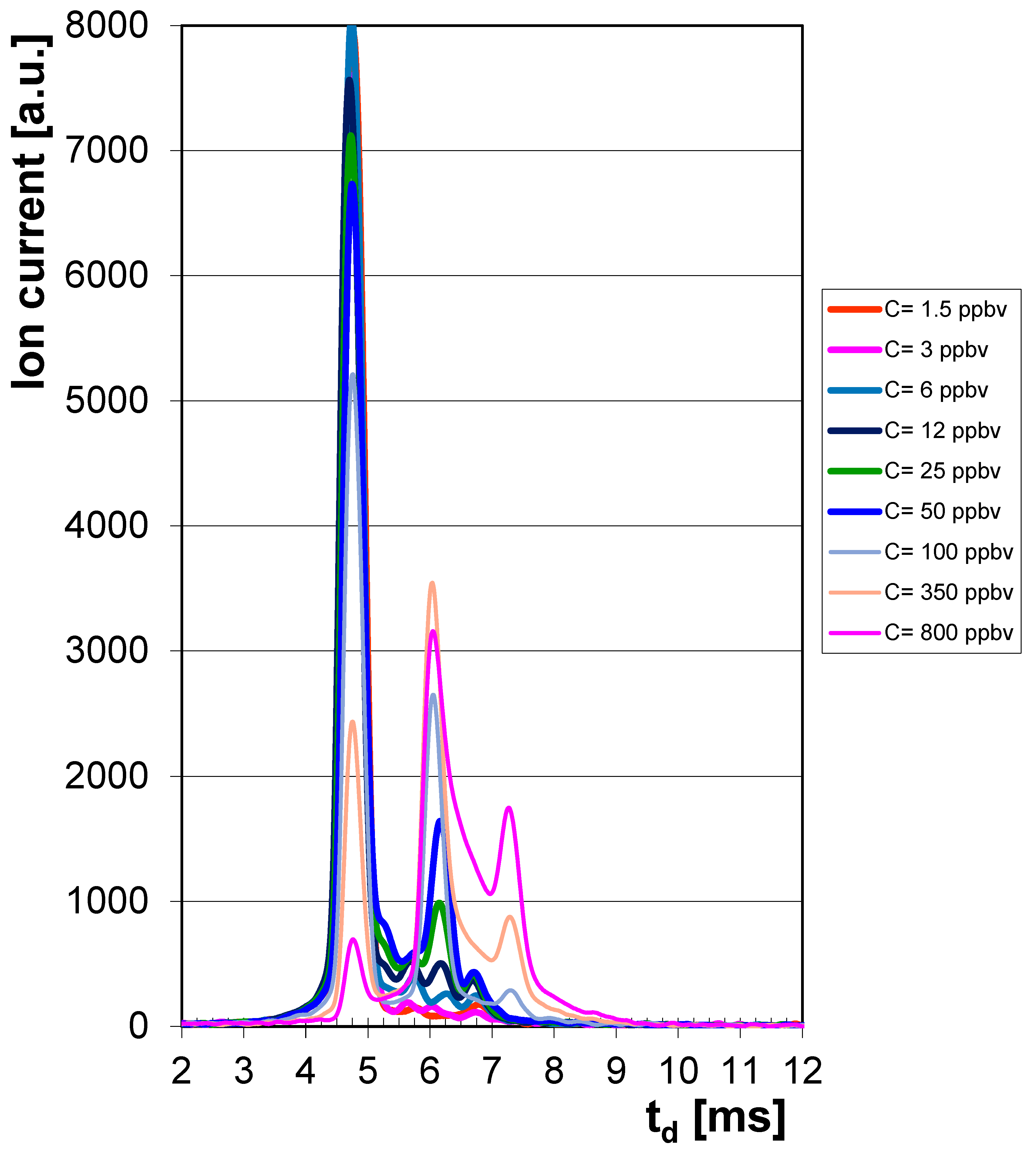
Figure 1. Ion mobility spectra from CPF, obtained in the positive ion mode, for vapor concentrations between 1.5 and 800 ppbv. Positive RIP (reactant ion peak) is the first peak in the spectrum, at td = ca. 4.7 ms, then the monomer product ion peak is the second feature (at td = ca. 6.1 ms) and finally the dimer product ion peak from CPF is the third feature in the spectrum, at td = ca. 7.3 ms. The scale of drift time has been narrowed, from 2 to 12 milliseconds, for reasons of clarity.
Toxics 13 00843 g001
Figure 2. Calibration for CPF, in the positive ion mode.
Toxics 13 00843 g002
Table 1. Main properties of OP pesticide Chlorpyrifos (O,O-diethyl O-(3,5,6-trichloro-2-pyridyl) phosphorothioate) (adapted information from ref. [5]).
Substance Name and FormulaProperties

Chlorpyrifos

O,O-diethyl
O-(3,5,6-trichloro-2-pyridyl)
phosphorothioate

C9H11Cl3NO3PS

CAS#: 2921-88-2
EC#: 220-864-4
Molecular mass: 350.57 g mol−1
Melting point: ca. 41…42 °C
Boiling point: decomposes at approx. 160 °C
Physical state: solid, white/colorless crystals
Density: 1.4 g cm−3 (@ 20 °C)

Relative density of vapors: 12.1 (air = 1)
Vapor pressure: ca. 2·10−5 Torr @ 25 °C
Volatility: 0.38 mg m−3 @ 25 °C

Octanol–water partition coefficient (log Kow): 4.9…5.2
Solubility in water: low—ca. 1.4 mg L−1 (@ 25 °C)
Soluble in organic solvents (acetone, benzene, carbon disulfide, etc.)

Acute toxicity: medium to high, with LD50 of ca. 150 mg kg−1 (rat, oral), ca. 500…1000 mg kg−1 (goats, oral), ca. 1200 mg kg−1 (rabbit, dermal) and LC50 of ca. 550 mg m−3 (rat, inhalation, 4 h)
Minimum risk level: 0.003 mg kg−1 day−1 (acute, oral) and 0.001 mg kg−1 day−1 (chronic, oral)
Reported fatal dose: 300 mg kg−1 (human, adult)

Occupational air level (OSHA): 0.04 mg m−3 (8 h)
TWA: 0.2 mg m−3 (UK); STEL: 0.6 mg m−3 (UK)
NIOSH recommended exposure limit (REL): 0.2 mg m−3 (10 h, skin); 0.6 mg m−3 (15 min, skin)
TLV (threshold limit value)–TWA: 0.1 mg m−3 (8 h; inhalable fraction and vapor), skin
EPA limit in air: 1 μg m−3 (adults) and 0.5 μg m−3 (children)
Estimated AVDI (average daily food intake) in the U.S.: 0.04·10−3 mg day−1 in 1980; 0.8·10−3 mg day−1 in 1990.

Conversion: 1 ppmv = 14.57 mg m−3 (20 °C)
Table 2. Concentrations of CPF vapors in the standard atmospheres, correlated to volumes of injected standard reference material (with 2 ng μL−1 CPF) (for vapor concentrations < 100 ppbv), respectively, to volumes of injected standard solution (with 0.5 μg μL−1 CPF) obtained from Nurelle-D commercial product: CCPF vapors = (Vstandard sol. × Cstandard sol.)/Vjar (1 ppbv = 14.57 μg m−3).
Vstandard sol.−SRM
[μL]
CCPF [μg m−3]CCPF [ppbv]
8221.5
16443
32896
6317512
13236625
26573650
Vstandard sol.−Nurelle D
[μL]
CCPF [μg m−3]CCPF [ppbv]
2.11458100
7.45140350
16.811,667800
Table 3. Summary of experimental results obtained with CPF vapors using the LCD-3.2E handheld ToF IMS instrument in positive ion mode (in order to calculate standard deviation, three replicates were used for peak height). Notes: Peak of positive reactant ion (RIP) has td = 4.74 ms and its height is hmax = 8000 a.u. TIMS = 301.5 K and PIMS = 971.5 mbar = 729 mm Hg.
CCPF [ppbv]Drift Time Monomer td [ms]Peak Height Monomer hmax [a.u.]Drift Time Dimer
td [ms]
Peak Height Dimer hmax [a.u.]
1.56.1485 ± 6--
36.14130 ± 9--
66.14230 ± 14--
126.16500 ± 26--
256.14990 ± 58--
506.161640 ± 84--
1006.062650 ± 1527.28290 ± 16
3506.063540 ± 1947.28890 ± 48
8006.063160 ± 1687.261750 ± 92
Table 4. Reduced ion mobilities K0 calculated for ions produced by CPF vapors.
Operation ModeIon Drift Time,
td [ms]
Reduced Ion Mobility 1,
K0 [cm2 V−1 s−1]
Reduced Ion Mobility 2,
K0 [cm2 V−1 s−1]
Pos RIP: 4.74 2.278 2.251
PositivePIP 1 (monomer):6.06 1.783 1.760
PIP 2 (dimer):7.28 1.484 1.465
where 1—Calculated by the IMS software TrimScan. 2—Calculated using the IMS cell constant (A): K0 = (A/td). PIP—product ion peak; RIP—reactant ion peak.
Table 5. Resolving power of the LCD-3.2E IMS instrument for CPF (spectrum for 100 ppbv).
Ion Drift Time,
td [ms]
Peak Width at Half Maximum, Δtd [ms]Resolving Power, RIMS
Pos RIP:4.74 0.35 13.5
PIP 1 (monomer):6.06 0.36 16.8
PIP 2 (dimer):7.28 0.42 17.3
Table 6. Detection and quantification of CPF using various IMS and hyphenated GC-IMS systems. RS—IMS instrument equipped with a radioactive ionization source; NRS—IMS instrument equipped with a non-radioactive ionization source.
Application/Instrument UsedK0 [cm2 V−1 s−1]Quant.Ref.
Determination of CPF residue in pistachio oil after liquid extraction by IMS:
Home-built IMS (Isfahan Univ. of Technology) with corona discharge ionization (NRS). Drift length ld = 11 cm. E = 500 V cm−1. TIMS cell = 190 °C. Drift gas: N2.
Extraction of CPF from pistachio oil was performed with hexane. Liquid extract was injected into the IMS at 220 °C.
Linear dynamic range LDR: 0.4 to 20 μg g−1.
Only one product ion peak was noticed, at td = ca. 11 ms (POS mode).
K0 not reported!
LOD 0.1 μg g−1
LOQ 0.3 μg g−1
[17]
Determination of CPF in water and food samples by tandem GC-IMS:
CPF was extracted by SPME from various matrices (fruits, vegetables, water), and then, analysis by GC—IMS was performed.
NRS IMS (corona discharge) was used. TIMS cell = 200 °C. Drift length ld = 11 cm (?). E = 500 V cm−1. Drift gas: N2.
Linear dynamic range LDR: 0.50 to 25 μg L−1.
Only one product ion peak was observed (td = ca. 11.2 ms), overlapping with that produced by Trifluralin, at td ca. 11.4 ms (POS mode).
K0 not reported!
LOD 0.15 μg L−1
LOQ 0.50 μg L−1
[18]
Determination of CPF after microextraction using Cu COF by IMS:
Samples: water, soil, pear fruits.
IMS model IMS 300 (TOF Tech., Iran) with NRS (corona discharge). TIMS cell = 200 °C. Drift length ld = 11 cm (?). E = 500 V cm−1 (?).
Linear dynamic range LDR: 1.0 to 400.0 ng mL−1.
Only one product ion peak was noticed, at td = ca. 11.7 ms (POS mode).
K0 not reported!
LOD 0.65 ng mL−1[19]
Determination of CPF after ultrasound-assisted microextraction using IMS:
Samples: water, food (vegetables, fruits).
IMS model IMS 400 (TOF Tech. Pars Co., Iran) with NRS (corona discharge). Drift length ld = 11 cm; drift gas: dry air. TIMS cell = 200 °C. E = 500 V cm−1.
Linear dynamic range LDR: 5.0 to 200.0 μg L−1.
Only one product ion peak was noticed (POS mode), at td = ca. 10.3 ms.
K0 not reported!
LOD 1.3 μg L−1[20]
Determination of CPF after headspace SPME by IMS:
Various aqueous samples (river, farm, groundwater) and fruit juices.
IMS model CD-1400 (TOF Tech. Pars Co., Isfahan, Iran) with NRS (corona discharge). Drift length ld = 11 cm; drift gas: nitrogen. TIMS cell = 190 °C. E = 500 V cm−1 (?).
Linear dynamic range LDR: 0.5 to 300.0 ng mL−1.
Only one product ion peak was noticed, at td = ca. 9.3 ms (POS mode).
K0 not reported!
LOD 0.2 ng mL−1[21]
Determination of CPF after DLLME by IMS:
Samples of water, fruits (apples), vegetables (tomatoes).
IMS (Teif Azmon Espadana Co., Isfahan, Iran) with NRS (corona discharge). Drift length ld = 11 cm; drift gas: nitrogen. TIMS cell = 160 °C. E = 450 V cm−1.
Linear dynamic range LDR: 0.1 to 3.0 μg L−1.
Only one product ion peak was reported, at td = ca. 10.9 ms (and at 12.2 ms in spiked samples!) (POS mode).
K0 not reported!
LOD 0.04 μg L−1[22]
Determination of CPF after SPME by IMS:
Sample: standard solution of CPF.
IMS (Teif Azmon Espadana Co., Isfahan, Iran) with NRS (corona discharge). Drift length ld = 11 cm; drift gas: nitrogen. TIMS cell = 160 °C. E= 420 V cm−1.
Linear dynamic range LDR: 0.5 to 20.0 μg L−1.
Only one product ion peak was noticed, at td = ca. 12.4 ms (POS mode).
K0 not reported!
LOD 0.15 μg L−1
LOQ 0.5 μg L−1
[23]
Determination of CPF after ultrasound-assisted emulsification microextraction using IMS:
Samples: rice paddy water, rice.
Home-built IMS with RS (63Ni). Drift length ld = 10 cm; drift gas: nitrogen. TIMS cell = 200 °C. E = 550 V cm−1.
Linear dynamic range LDR: 8.9 to 750.0 μg L−1.
Only one product ion peak was reported, at td = ca. 11.5 ms (POS mode).
K0 not reported!
LOD 3.2 μg L−1[24]
Determination of CPF after SPME by IMS:
Samples: water, fruits (grape, tangerine).
IMS (Teif Azmon Espadana Co., Isfahan Univ. of Technology, Iran) with NRS (corona discharge). Drift length ld = 11 cm; drift gas: nitrogen. TIMS cell = 160 °C. E = 400 V cm−1.
Linear dynamic range LDR: 0.1 to 10.0 μg L−1.
Only one product ion peak was noticed, at td = ca. 11.4 ms (POS mode).
K0 not reported!
LOD 0.05 μg L−1
LOQ 0.10 μg L−1
[25]
Determination of CPF after SPME by IMS:
Samples: water, fruit juices.
IMS model CD-1400 (Theif Azmoon Espadana Co., Isfahan, Iran) with NRS (corona discharge). Drift length ld = 11 cm; drift gas: nitrogen. TIMS cell = 200 °C. E = 500 V cm−1 (?).
Linear dynamic range LDR: 2 to 250.0 ng mL−1.
Only one product ion peak was noticed, at td = ca. 9.2 ms (POS mode).
K0 not reported!
LOD 0.6 ng mL−1[26]
Determination of CPF by tandem GC-IMS after SPME:
CPF was extracted by dispersive SPME from various matrices (water, fruits, vegetables), and then, analysis by GC—IMS was performed.
IMS manufactured by Teif Azmon Espadana Co., Isfahan, Iran, with NRS (corona discharge) was used. Drift length ld = 11 cm. TIMS cell = 200 °C. Drift gas: N2. E = 500 V cm−1.
Linear dynamic range LDR: 2 to 1000 μg L−1.
Only one product ion peak was observed (td = ca. 11.8 ms), overlapped with peak produced by Malathion (ca. 12.0 ms).—POS mode.
K0 not reported!
LOD 0.85 μg L−1
LOQ 2 μg L−1
[27]
Determination of CPF after SPME by tandem GC-IMS:
CPF was extracted by SPME from various matrices (water, fruits, vegetables), and then, analysis by GC—IMS was performed.
Home-built IMS (Isfahan Univ., Iran), with NRS (corona discharge) used. Drift length ld = 11 cm; drift gas: nitrogen. TIMS cell = 230 °C. E = 500 V cm−1.
Linear dynamic range LDR: 0.025 to 2.0 μg L−1 (river water, wastewater); 0.75 to 20.0 μg kg−1 (pears, grapes); 0.50 to 15.0 μg kg−1 (eggplants).
Only one product ion peak was observed, with td = ca. 14.1 ms (POS mode).
K0 not reported!
LOD 0.010 μg L−1
LOQ 0.025 μg L−1 (water)

LOD 0.30 μg L−1
LOQ 0.75 μg L−1 (pears, grapes)
[28]
Determination of CPF after SPME by tandem GC-IMS:
CPF was extracted by SPME from various matrices (water, milk, serum, fruits), and then, analysis by GC—IMS was performed.
Home-built IMS (Isfahan Univ. of Technology, Iran), with NRS (corona discharge). Drift length ld = 11 cm; drift gas: N2. TIMS cell = 200 °C. E = 500 V cm−1.
Linear dynamic range LDR: 0.05 to 20.0 μg L−1 (water).
No IMS spectrum was provided.
K0 not reported!
LOD 0.019 μg L−1 (water)
LOQ 0.050 μg L−1 (water)
[29]
Determination of CPF after liquid phase micro-extraction by IMS:
Samples: water, vegetables.
IMS (Teif Azmon Espadana Co., Isfahan, Iran) with NRS (secondary electrospray ionization SESI). Drift length ld = 11 cm; drift gas: N2. TIMS cell = 150 °C. E = 567 V cm−1.
Linear dynamic range LDR: 1 to 70.0 μg L−1.
Only one product ion peak was noticed, at td = 12.28 ms (POS mode).
K0 = 1.37 cm2 V−1 s−1 (with nicotinamide as mobility standard)
LOD 0.21 μg L−1
LOQ 0.70 μg L−1
[30]
Determination of CPF after micro-extraction by IMS:
Sample: water.
Home-built IMS (Isfahan Univ., Iran), with NRS (corona discharge) was used. Drift length ld = 11 cm; drift gas: N2. TIMS cell = 150 °C. E = 400 V cm−1.
Linear dynamic range LDR: 2 to 200 μg L−1.
Only one product ion peak was noticed, at td = ca. 10.6 ms (POS mode).
K0 not reported!
LOD 0.6 μg L−1
LOQ 2.0 μg L−1
[31]
Determination of CPF after SPME by tandem GC-IMS:
Sample: water.
Home-built GC-IMS (Isfahan Univ., Iran), with NRS (corona discharge) was used. Drift gas: N2. TIMS cell = 235 °C. E = 500 V cm−1.
Linear dynamic range LDR: 0.02 to 5 μg L−1.
Only one product ion peak was noticed, at td = ca. 12.1 ms (POS mode).
K0 = 1.27 cm2 V−1 s−1 (with nicotinamide as mobility standard).
LOD 0.012 μg L−1[32]
Determination of CPF after dispersive solid-phase extraction by IMS:
Samples: water, vegetables.
Home-built IMS model 1000 (Isfahan Univ. of Technology, Iran), with NRS (corona discharge) was used. Drift length ld = 16 cm; drift gas: N2. TIMS cell = 200 °C. E = 500 V cm−1. Reactant ion: NH4+.
Linear dynamic range LDR: 1 to 500 ng mL−1.
Only one product ion peak was noticed, at td = ca. 8.8 ms.
K0 not reported!
LOD 0.3 ng mL−1
LOQ 1 ng mL−1
[33]
Determination of CPF after dispersive solid-phase extraction by IMS:
Samples: water, fruit juice, vegetables.
IMS model CD-1400 (TOF Tech, Isfahan, Iran), with NRS (corona discharge), was used. Drift length ld = 11 cm; drift gas: N2. TIMS cell = 200 °C. E = 636 V cm−1.
Linear dynamic range LDR: 0.6 to 300.0 ng mL−1.
Only one product ion peak was noticed, at td = ca. 11 ms (POS mode).
K0 not reported!
LOD 0.2 ng mL−1[34]
Determination of CPF after dispersive liquid–liquid microextraction by IMS:
Samples: water, vegetables (potato).
IMS model CD-1400 (Teif Azmon Espadana Co., Isfahan, Iran), with NRS (corona discharge) was used. Drift length ld = 11 cm; electric field E = 420 V cm−1; drift gas: N2. TIMS cell = 160 °C.
Linear dynamic range LDR: 0.1 to 7.0 μg L−1.
Only one product ion peak was noticed, at td = ca. 13.2 ms (POS mode).
K0 not reported!
LOD 0.03 μg L−1
LOQ 0.1 μg L−1
[35]
Determination of CPF after stir mesh screen sorptive extraction (SMSE) by IMS:
Samples: water (river, well, agricultural wastewater); fruits (apple).
IMS with thermal desorption unit (Teif Azmon Espadana Co., Isfahan Univ. of Technology, Iran), with NRS (corona discharge) was used. Drift length ld = 11 cm; electric field E = 500 V cm−1; drift gas: nitrogen. TIMS cell = 150 °C.
Linear dynamic range LDR: 0.1 to 20.0 μg L−1.
Only one product ion peak was noticed, at td = ca. 11.3 ms (POS mode).
K0 not reported!
LOD 0.035 μg L−1
LOQ 0.100 μg L−1
[36]
Determination of CPF after thin film micro-extraction (SMSE) by IMS:
Samples: water (well, river, agricultural wastewater); fruits (tangerine).
IMS with thermal desorption unit (Isfahan Univ. of Technology, Iran), with NRS (corona discharge) was used. Drift length ld = 11 cm; electric field E = 450 V cm−1; drift gas: N2. TIMS cell = 160 °C.
Linear dynamic range LDR: 1 to 100 μg L−1.
Only one product ion peak was noticed, at td = ca. 12.8 ms (POS mode).
K0 not reported!
LOD 0.3 μg L−1[37]
Indoor and outdoor determination of pesticides in air after active sampling on Teflon membranes by IMS:
Samples: air.
Commercial IMS with RS (63Ni) with thermal desorption, model Ionscan-LS (Smiths Detection, Morristown, NJ, USA). Drift Tube length: 7 cm; electric field E = 252 V cm−1; drift gas: dry air. TIMS cell = 237 °C.
Linear dynamic range LDR—over 2 to 10 ng.
One product ion peak in the Negative ion mode, at td = 11.93 ms.
K0 = 1.561 cm2 V−1 s−1 (using 4-nitrobenzonitrile as mobility standard, with K0 = 1.655 cm2 V−1 s−1).
LOD 600 pg

LOQ 1960 pg
[38]
Determination of CPF vapors by IMS:
ToF IMS, model LCD-3.2E (NRS—corona discharge), operated near ambient T (ca. 28 °C); ammonia doped.
Calibration: from 1.5 to 800 ppbv
Linear range: from 2.5 to 100 ppbv
Saturation: >1000 ppbv
1.727 (monomer)

1.461 (dimer)
LOD 0.72 ppbv

LOQ 2.41 ppbv
this work
Note: LOD is the limit of detection and LOQ the limit of quantification, respectively.
Disclaimer/Publisher’s Note: The statements, opinions and data contained in all publications are solely those of the individual author(s) and contributor(s) and not of MDPI and/or the editor(s). MDPI and/or the editor(s) disclaim responsibility for any injury to people or property resulting from any ideas, methods, instructions or products referred to in the content.