
Effects of DEHP, DEHT and DINP Alone or in a Mixture on Cell Viability and Mitochondrial Metabolism of Endothelial Cells In Vitro

 ,
,
Abstract
:
1. Introduction
2. Materials and Methods
2.1. Human Microvascular Endothelial Cell Line (HMEC-1) Culture
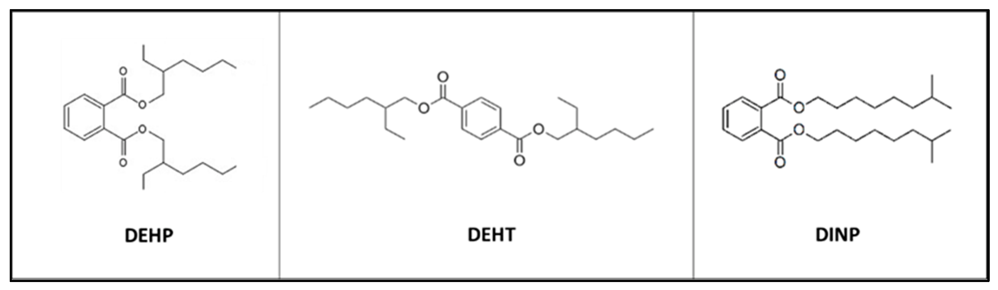
2.2. Studied Plasticizers
2.3. Solution Preparations
2.4. Reagents and Standards
2.5. Plasticizer Treatments and Exposure Procedures on HMEC-1 Cells
2.6. Cell Viability
2.7. Cell Counting
2.8. Total Intracellular Glutathione Content
2.9. Intracellular Adenosine Amounts
2.10. Cell Metabolism Assays
2.11. Statistical Analyses
3. Results
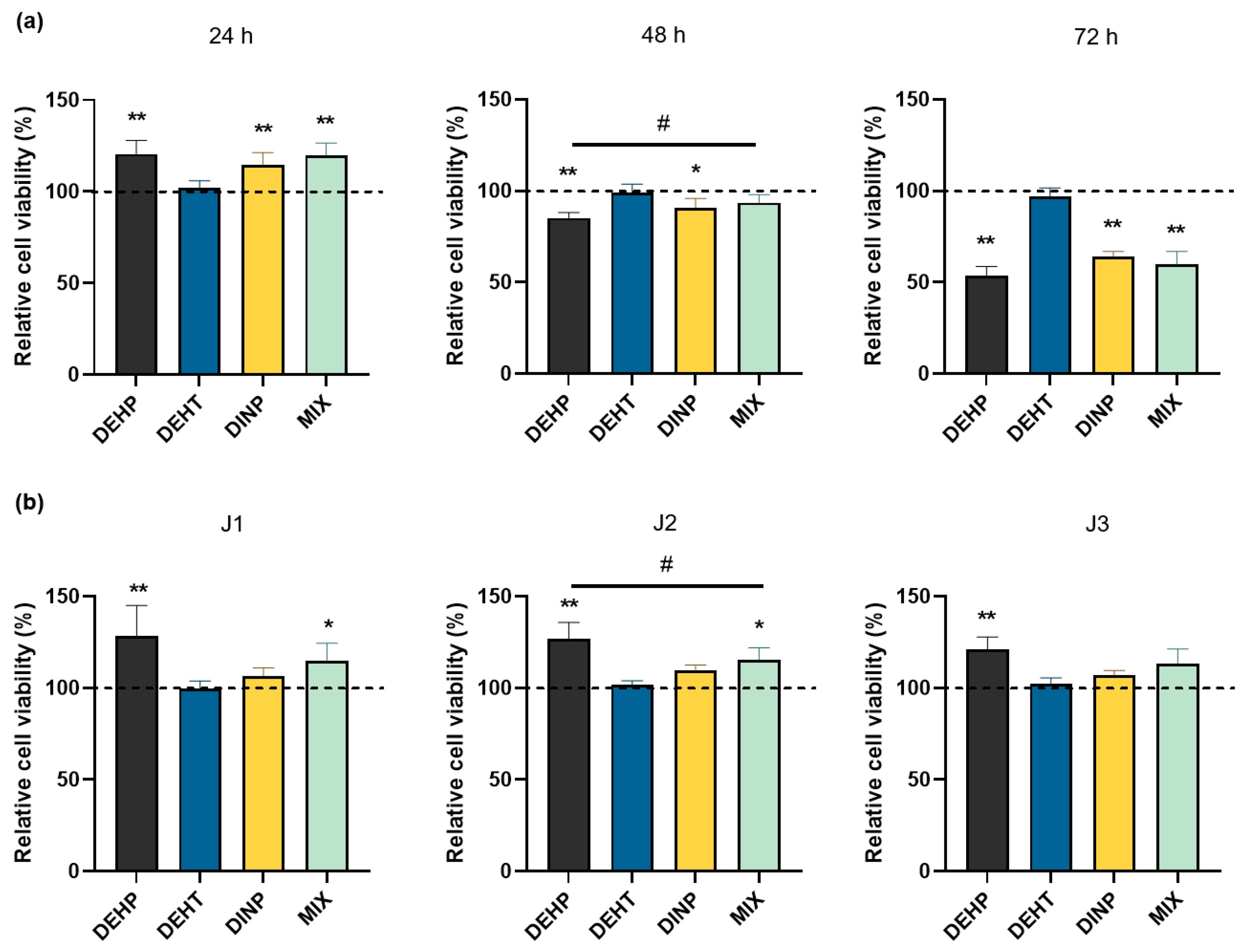
3.1. Modification of the HMEC-1 Cell Viability Following Individual and Combined Plasticizer Exposures
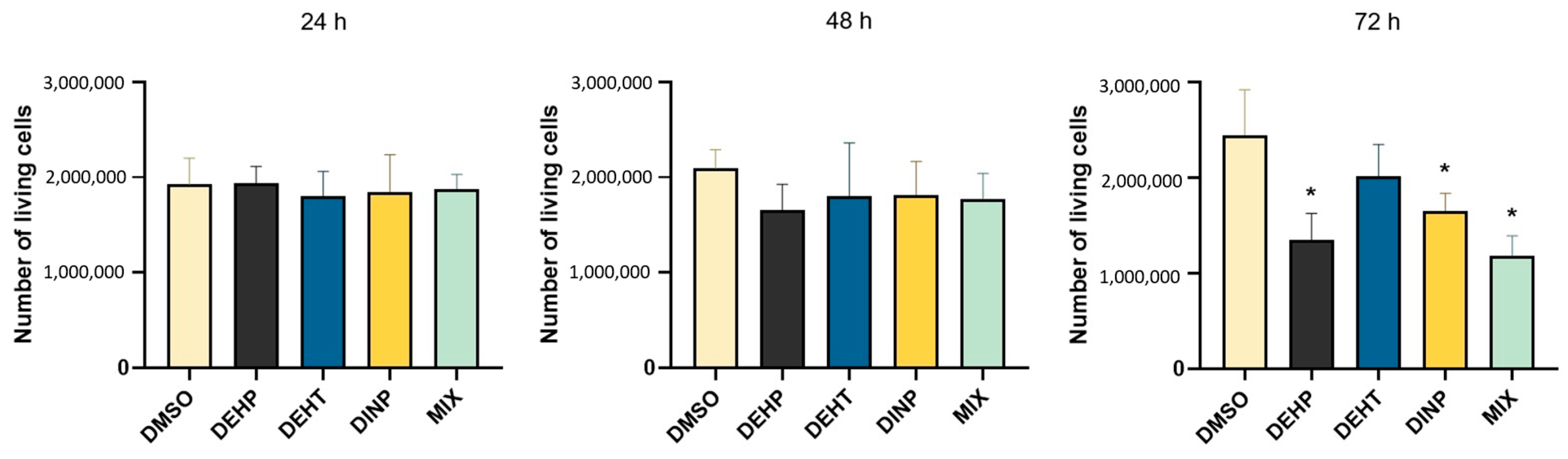
3.2. Effects of Individual and Combined Plasticizer Exposures on HMEC-1 Cell Counting
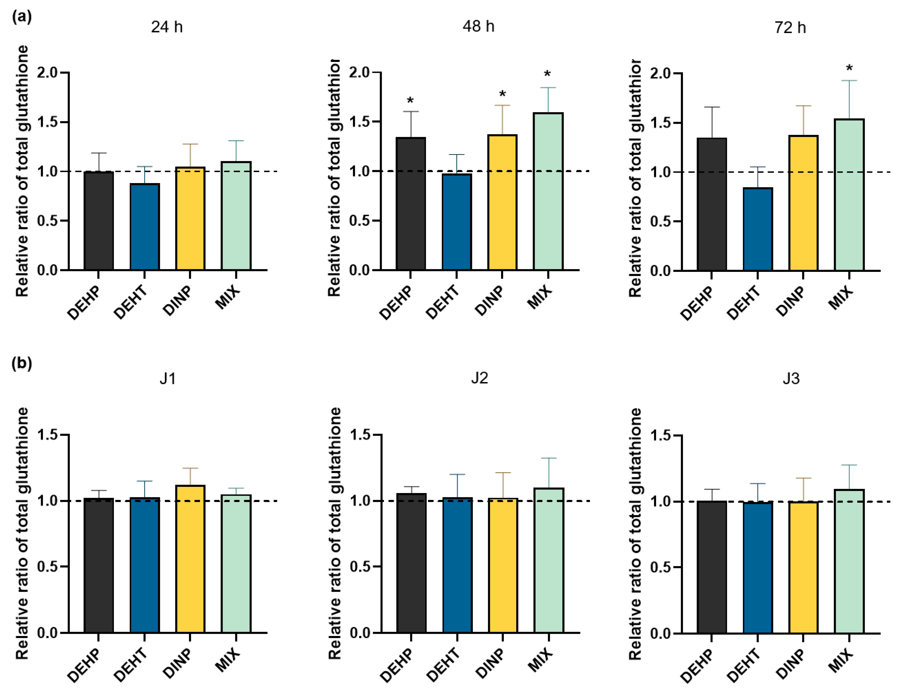
3.3. Effects of Individual and Combined Plasticizer Exposure on the Total Intracellular Glutathione in HMEC-1 Cells
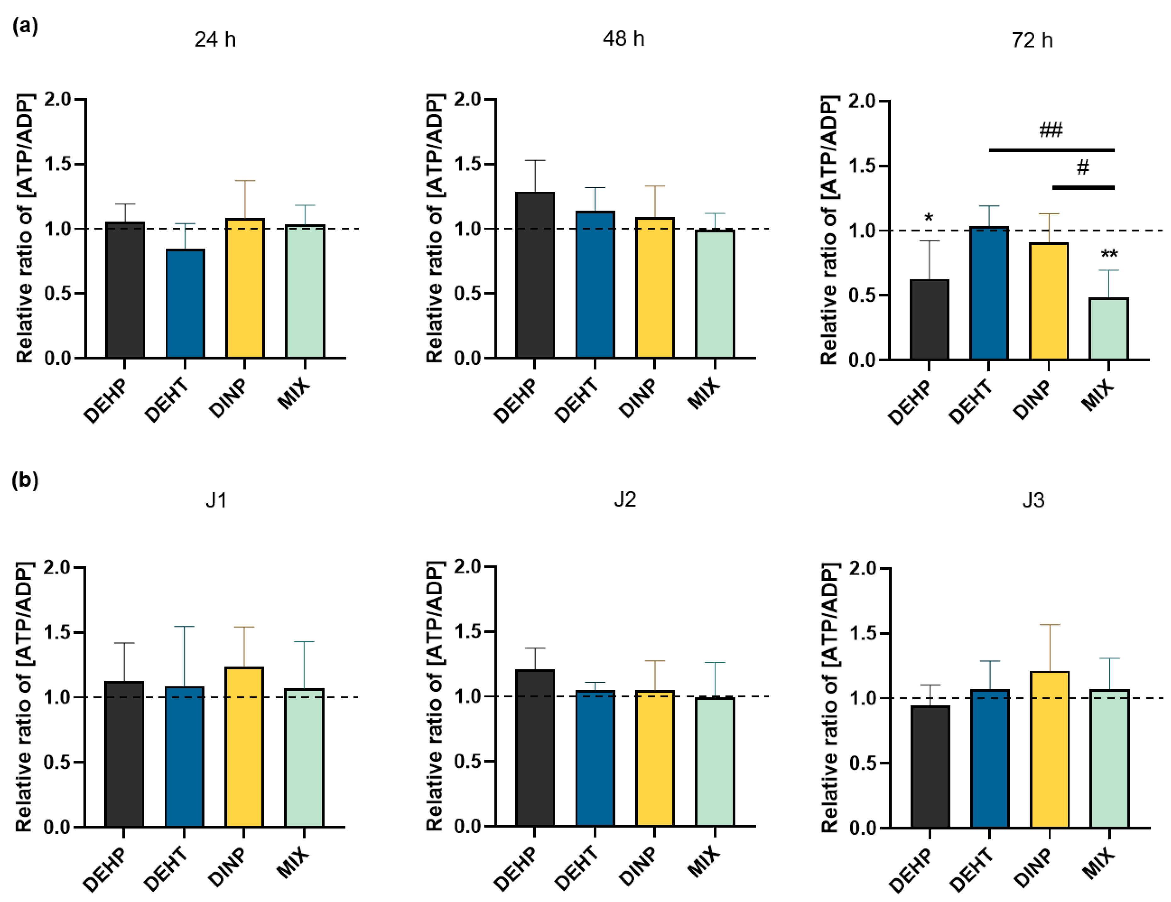
3.4. Effects of Individual and Combined Plasticizer Exposure on the Amount of Intracellular Adenosines in HMEC-1 Cells
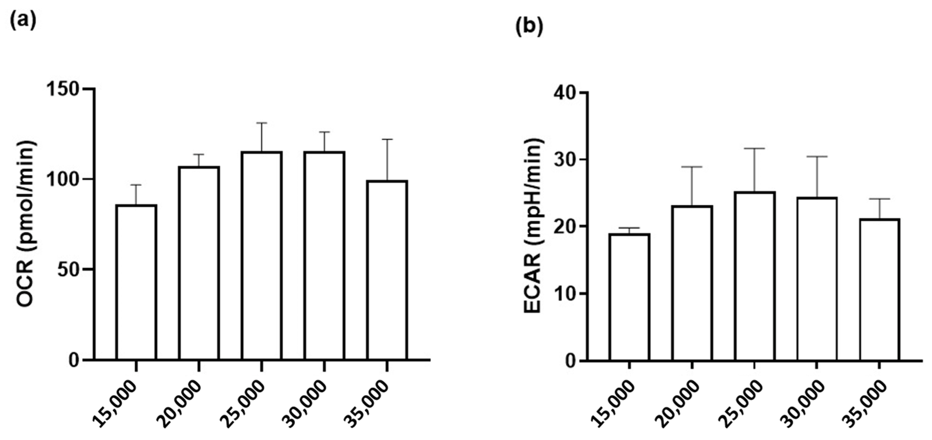
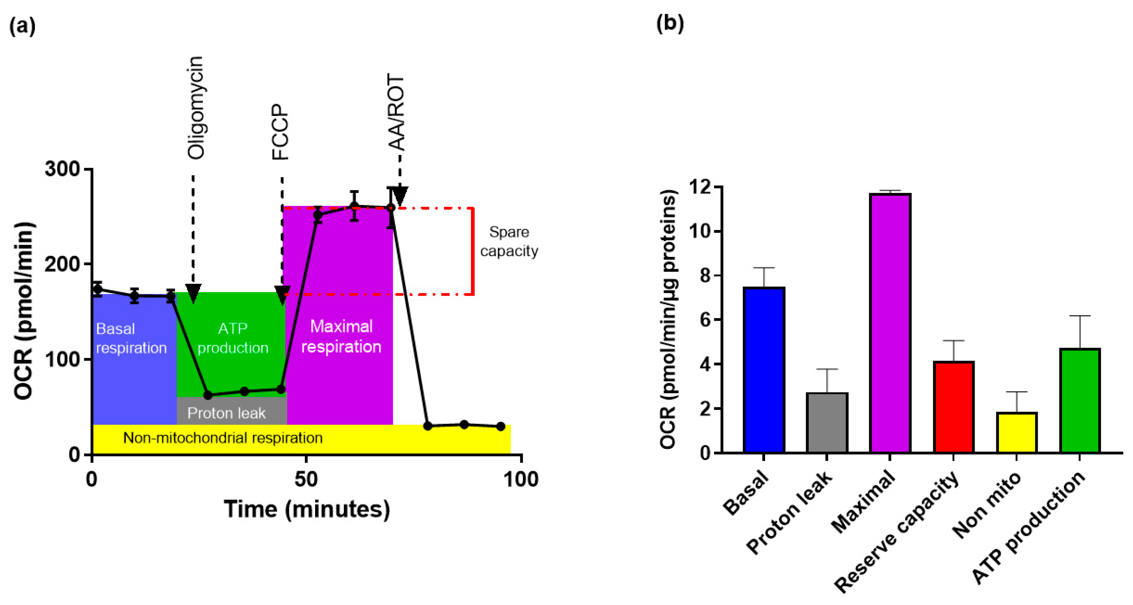
3.5. Measurements of Mitochondrial Function in HMEC-1 Cells
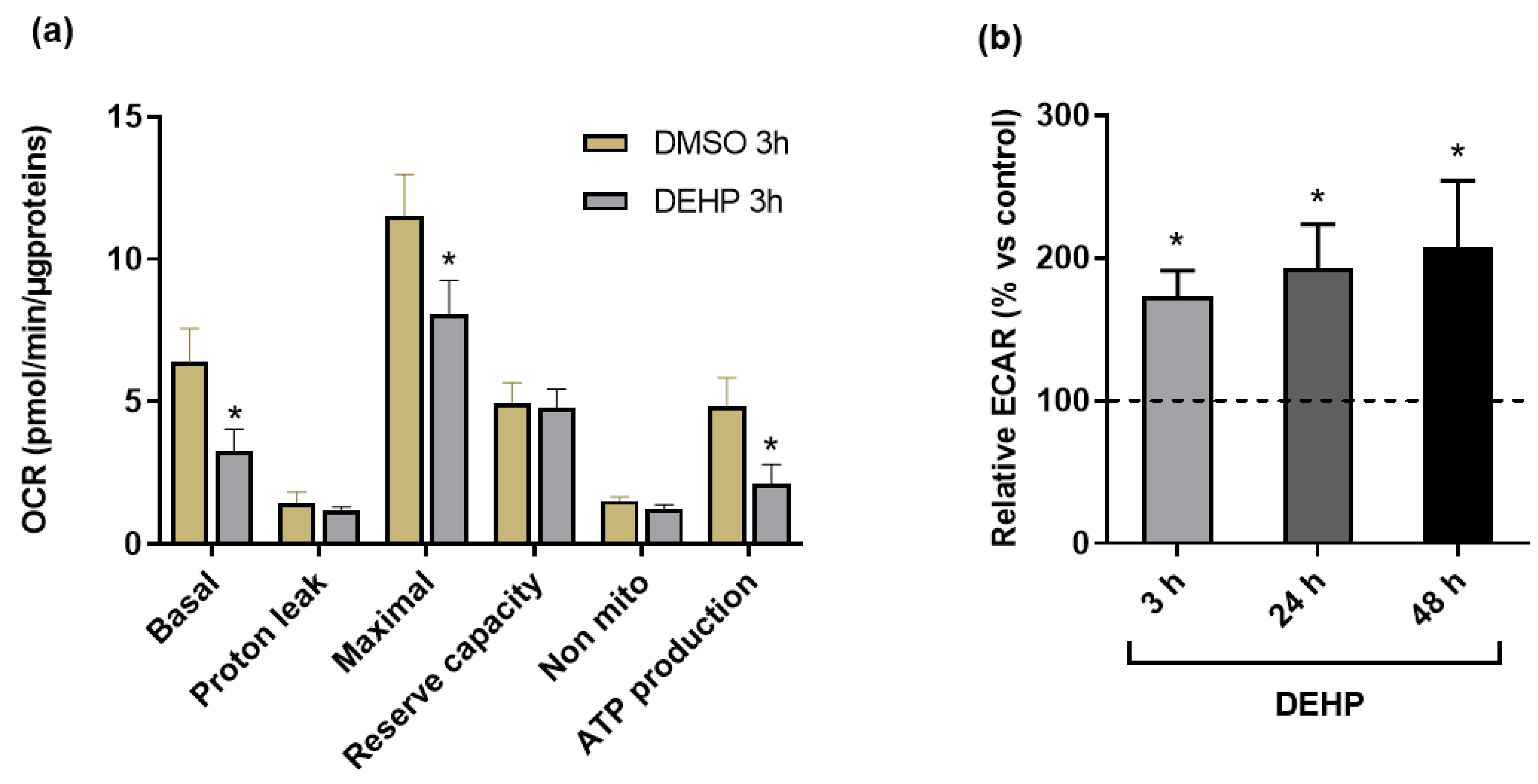
3.6. Effects of Plasticizers on Mitochondrial Function
4. Discussion
5. Conclusions
Author Contributions
Funding
Institutional Review Board Statement
Informed Consent Statement
Data Availability Statement
Conflicts of Interest
References
- Plasticisers-Information Center. Available online: https://www.plasticisers.org/plasticisers/ (accessed on 20 August 2021).
- Krauskopf, L.; Godwin, A. Chapter 5: Plasticizers. In PVC Handbook; Hanser Publisher: Munich, Germany, 2005; pp. 173–193. [Google Scholar]
- Petersen, J.H.; Jensen, L.K. Phthalates in soft PVC products used in food production equipment and in other food contact materials on the Danish and the Nordic Market 2013–2014. Int. J. Food Contam. 2016, 3, 3. [Google Scholar] [CrossRef] [Green Version]
- Xie, M.; Wu, Y.; Little, J.C.; Marr, L.C. Phthalates and alternative plasticizers and potential for contact exposure from children’s backpacks and toys. J. Expo. Sci. Environ. Epidemiol. 2016, 26, 119–124. [Google Scholar] [CrossRef] [PubMed]
- Malarvannan, G.; Onghena, M.; Verstraete, S.; van Puffelen, E.; Jacobs, A.; Vanhorebeek, I.; Verbruggen, S.C.A.T.; Joosten, K.F.M.; Van den Berghe, G.; Jorens, P.G.; et al. Phthalate and alternative plasticizers in indwelling medical devices in pediatric intensive care units. J. Hazard. Mater. 2019, 363, 64–72. [Google Scholar] [CrossRef] [PubMed]
- Poitou, K.; Rogez-Florent, T.; Lecoeur, M.; Danel, C.; Regnault, R.; Vérité, P.; Monteil, C.; Foulon, C. Analysis of Phthalates and Alternative Plasticizers in Gloves by Gas Chromatography–Mass Spectrometry and Liquid Chromatography–UV Detection: A Comparative Study. Toxics 2021, 9, 200. [Google Scholar] [CrossRef]
- Kim, D.Y.; Chun, S.-H.; Jung, Y.; Mohamed, D.F.M.S.; Kim, H.-S.; Kang, D.-Y.; An, J.-W.; Park, S.-Y.; Kwon, H.-W.; Kwon, J.-H. Phthalate Plasticizers in Children’s Products and Estimation of Exposure: Importance of Migration Rate. Int. J. Environ. Res. Public Health 2020, 17, 8582. [Google Scholar] [CrossRef]
- Fromme, H.; Schütze, A.; Lahrz, T.; Kraft, M.; Fembacher, L.; Siewering, S.; Burkardt, R.; Dietrich, S.; Koch, H.M.; Völkel, W. Non-phthalate plasticizers in German daycare centers and human biomonitoring of DINCH metabolites in children attending the centers (LUPE 3). Int. J. Hyg. Environ. Health 2016, 219, 33–39. [Google Scholar] [CrossRef]
- Eckert, E.; Müller, J.; Höllerer, C.; Purbojo, A.; Cesnjevar, R.; Göen, T.; Münch, F. Plasticizer exposure of infants during cardiac surgery. Toxicol. Lett. 2020, 330, 7–13. [Google Scholar] [CrossRef]
- Chao, K.-P.; Huang, C.-S.; Wei, C.-Y. Health risk assessments of DEHP released from chemical protective gloves. J. Hazard. Mater. 2015, 283, 53–59. [Google Scholar] [CrossRef]
- Mariana, M.; Feiteiro, J.; Verde, I.; Cairrao, E. The effects of phthalates in the cardiovascular and reproductive systems: A review. Environ. Int. 2016, 94, 758–776. [Google Scholar] [CrossRef]
- Axelsson, J.; Rylander, L.; Rignell-Hydbom, A.; Lindh, C.H.; Jönsson, B.A.G.; Giwercman, A. Prenatal phthalate exposure and reproductive function in young men. Environ. Res. 2015, 138, 264–270. [Google Scholar] [CrossRef]
- Ferguson, K.K.; Rosen, E.M.; Rosario, Z.; Feric, Z.; Calafat, A.M.; McElrath, T.F.; Vélez Vega, C.; Cordero, J.F.; Alshawabkeh, A.; Meeker, J.D. Environmental phthalate exposure and preterm birth in the PROTECT birth cohort. Environ. Int. 2019, 132, 105099. [Google Scholar] [CrossRef] [PubMed]
- Yuan, X.-Q.; Du, Y.-Y.; Liu, C.; Guo, N.; Teng, X.-M.; Hua, X.; Yao, Y.-C.; Deng, Y.-L.; Zeng, Q.; Deng, T.-R.; et al. Phthalate metabolites and biomarkers of oxidative stress in the follicular fluid of women undergoing in vitro fertilization. Sci. Total Environ. 2020, 738, 139834. [Google Scholar] [CrossRef] [PubMed]
- Capela, D.; Mhaouty-Kodja, S. Effects of pubertal exposure to low doses of di-(2-ethylexyl)phthalate on reproductive behaviors in male mice. Chemosphere 2021, 263, 128191. [Google Scholar] [CrossRef] [PubMed]
- Chiang, C.; Flaws, J.A. Subchronic Exposure to Di(2-ethylhexyl) Phthalate and Diisononyl Phthalate During Adulthood Has Immediate and Long-Term Reproductive Consequences in Female Mice. Toxicol. Sci. 2019, 168, 620–631. [Google Scholar] [CrossRef] [PubMed]
- Khasin, L.G.; Della Rosa, J.; Petersen, N.; Moeller, J.; Kriegsfeld, L.J.; Lishko, P.V. The Impact of Di-2-Ethylhexyl Phthalate on Sperm Fertility. Front. Cell Dev. Biol. 2020, 8, 426. [Google Scholar] [CrossRef]
- Ma, T.; Zhou, Y.; Xia, Y.; Jin, H.; Wang, B.; Wu, J.; Ding, J.; Wang, J.; Yang, F.; Han, X.; et al. Environmentally relevant perinatal exposure to DBP disturbs testicular development and puberty onset in male mice. Toxicology 2021, 459, 152860. [Google Scholar] [CrossRef]
- Saillenfait, A.-M.; Sabaté, J.-P.; Robert, A.; Cossec, B.; Roudot, A.-C.; Denis, F.; Burgart, M. Adverse effects of diisooctyl phthalate on the male rat reproductive development following prenatal exposure. Reprod. Toxicol. 2013, 42, 192–202. [Google Scholar] [CrossRef]
- Adir, M.; Combelles, C.M.H.; Mansur, A.; Ophir, L.; Hourvitz, A.; Orvieto, R.; Dor, J.; Machtinger, R. Dibutyl phthalate impairs steroidogenesis and a subset of LH-dependent genes in cultured human mural granulosa cell in vitro. Reprod. Toxicol. 2017, 69, 13–18. [Google Scholar] [CrossRef]
- Mariana, M.; Cairrao, E. Phthalates Implications in the Cardiovascular System. J. Cardiovasc. Dev. Dis. 2020, 7, 26. [Google Scholar] [CrossRef]
- Trasande, L.; Liu, B.; Bao, W. Phthalates and attributable mortality: A population-based longitudinal cohort study and cost analysis. Environ. Pollut. 2022, 292, 118021. [Google Scholar] [CrossRef]
- Bourdeaux, D.; Yessaad, M.; Chennell, P.; Larbre, V.; Eljezi, T.; Bernard, L.; Sautou, V. Analysis of PVC plasticizers in medical devices and infused solutions by GC–MS. J. Pharm. Biomed. Anal. 2016, 118, 206–213. [Google Scholar] [CrossRef] [PubMed]
- Fernandez-Canal, C.; Pinta, P.-G.; Eljezi, T.; Larbre, V.; Kauffmann, S.; Camilleri, L.; Cosserant, B.; Bernard, L.; Pereira, B.; Constantin, J.-M.; et al. Patients’ exposure to PVC plasticizers from ECMO circuits. Expert Rev. Med. Devices 2018, 15, 377–383. [Google Scholar] [CrossRef] [PubMed]
- Deng, T.; Xie, X.; Duan, J.; Chen, M. Di-(2-ethylhexyl) phthalate induced an increase in blood pressure via activation of ACE and inhibition of the bradykinin-NO pathway. Environ. Pollut. 2019, 247, 927–934. [Google Scholar] [CrossRef]
- Zhao, J.-F.; Hsiao, S.-H.; Hsu, M.-H.; Pao, K.-C.; Kou, Y.R.; Shyue, S.-K.; Lee, T.-S. Di-(2-ethylhexyl) phthalate accelerates atherosclerosis in apolipoprotein E-deficient mice. Arch. Toxicol. 2016, 90, 181–190. [Google Scholar] [CrossRef] [PubMed]
- Wang, J.; Liao, Y.; Fan, J.; Ye, T.; Sun, X.; Dong, S. Apigenin inhibits the expression of IL-6, IL-8, and ICAM-1 in DEHP-stimulated human umbilical vein endothelial cells and in vivo. Inflammation 2012, 35, 1466–1476. [Google Scholar] [CrossRef] [PubMed]
- Liu, N.; Jiang, L.; Sun, X.; Yao, X.; Zhai, X.; Liu, X.; Wu, X.; Bai, Y.; Wang, S.; Yang, G. Mono-(2-ethylhexyl) phthalate induced ROS-dependent autophagic cell death in human vascular endothelial cells. Toxicol. Vitro 2017, 44, 49–56. [Google Scholar] [CrossRef] [PubMed]
- Xu, J.; Wang, L.; Zhang, L.; Zheng, F.; Wang, F.; Leng, J.; Wang, K.; Héroux, P.; Shen, H.-M.; Wu, Y.; et al. Mono-2-ethylhexyl phthalate drives progression of PINK1-parkin-mediated mitophagy via increasing mitochondrial ROS to exacerbate cytotoxicity. Redox Biol 2021, 38, 101776. [Google Scholar] [CrossRef] [PubMed]
- Ban, J.-B.; Fan, X.-W.; Huang, Q.; Li, B.-F.; Chen, C.; Zhang, H.-C.; Xu, S.-Q. Mono-(2-ethylhexyl) phthalate induces injury in human umbilical vein endothelial cells. PLoS ONE 2014, 9, e97607. [Google Scholar] [CrossRef] [Green Version]
- Incalza, M.A.; D’Oria, R.; Natalicchio, A.; Perrini, S.; Laviola, L.; Giorgino, F. Oxidative stress and reactive oxygen species in endothelial dysfunction associated with cardiovascular and metabolic diseases. Vasc. Pharmacol. 2018, 100, 1–19. [Google Scholar] [CrossRef]
- Wu, X.; Jiang, L.; Sun, X.; Yao, X.; Bai, Y.; Liu, X.; Liu, N.; Zhai, X.; Wang, S.; Yang, G. Mono(2-ethylhexyl) phthalate induces autophagy-dependent apoptosis through lysosomal-mitochondrial axis in human endothelial cells. Food Chem. Toxicol. 2017, 106, 273–282. [Google Scholar] [CrossRef]
- Wormuth, M.; Scheringer, M.; Vollenweider, M.; Hungerbühler, K. What are the sources of exposure to eight frequently used phthalic acid esters in Europeans? Risk Anal. 2006, 26, 803–824. [Google Scholar] [CrossRef] [PubMed]
- Bui, T.T.; Giovanoulis, G.; Cousins, A.P.; Magnér, J.; Cousins, I.T.; de Wit, C.A. Human exposure, hazard and risk of alternative plasticizers to phthalate esters. Sci. Total Environ. 2016, 541, 451–467. [Google Scholar] [CrossRef] [PubMed]
- Howdeshell, K.L.; Wilson, V.S.; Furr, J.; Lambright, C.R.; Rider, C.V.; Blystone, C.R.; Hotchkiss, A.K.; Gray Jr, L.E. A Mixture of Five Phthalate Esters Inhibits Fetal Testicular Testosterone Production in the Sprague-Dawley Rat in a Cumulative, Dose-Additive Manner. Toxicol. Sci. 2008, 105, 153–165. [Google Scholar] [CrossRef] [PubMed] [Green Version]
- Zhou, C.; Flaws, J.A. Effects of an Environmentally Relevant Phthalate Mixture on Cultured Mouse Antral Follicles. Toxicol. Sci. 2017, 156, 217–229. [Google Scholar] [CrossRef] [PubMed]
- Repouskou, A.; Stamatakis, A.; Kitraki, E. In utero exposure to phthalates and reproductive toxicity in rodents. Best Pract. Res. Clin. Endocrinol. Metab. 2021, 35, 101512. [Google Scholar] [CrossRef] [PubMed]
- Hannas, B.R.; Howdeshell, K.L.; Furr, J.; Earl Gray, L. In utero phthalate effects in the female rat: A model for MRKH syndrome. Toxicol. Lett. 2013, 223, 315–321. [Google Scholar] [CrossRef] [PubMed] [Green Version]
- Bocqué, M.; Voirin, C.; Lapinte, V.; Caillol, S.; Robin, J.-J. Petro-based and bio-based plasticizers: Chemical structures to plasticizing properties. J. Polym. Sci. A Polym. Chem. 2016, 54, 11–33. [Google Scholar] [CrossRef]
- Deng, T.; Xie, X.; Duan, J.; Chen, M. Exposure to diisononyl phthalate induced an increase in blood pressure through activation of the ACE/ AT1R axis and inhibition of NO production. Toxicol. Lett. 2019, 309, 42–50. [Google Scholar] [CrossRef]
- Ahmadpour, D.; Mhaouty-Kodja, S.; Grange-Messent, V. Disruption of the blood-brain barrier and its close environment following adult exposure to low doses of di(2-ethylhexyl)phthalate alone or in an environmental phthalate mixture in male mice. Chemosphere 2021, 282, 131013. [Google Scholar] [CrossRef]
- ECHA (European Chemicals Agency). ECHA Substance Information on Bis(2-ethylhexyl) Phthalate. Available online: https://echa.europa.eu/fr/substance-information/-/substanceinfo/100.003.829 (accessed on 18 March 2022).
- ECHA (European Chemicals Agency). ECHA Substance Information on Bis(2-ethylhexyl) Terephthalate. Available online: https://echa.europa.eu/fr/substance-information/-/substanceinfo/100.026.524 (accessed on 18 March 2022).
- ECHA (European Chemicals Agency). ECHA Substance Information on Di-“isononyl” Phthalate. Available online: https://echa.europa.eu/fr/substance-information/-/substanceinfo/100.044.602 (accessed on 18 March 2022).
- Muñoz-Vega, M.; Massó, F.; Páez, A.; Carreón-Torres, E.; Cabrera-Fuentes, H.A.; Fragoso, J.M.; Pérez-Hernández, N.; Martinez, L.O.; Najib, S.; Vargas-Alarcón, G.; et al. Characterization of immortalized human dermal microvascular endothelial cells (HMEC-1) for the study of HDL functionality. Lipids Health Dis. 2018, 17, 44. [Google Scholar] [CrossRef] [Green Version]
- ATCC (American Type Culture Collection). HMEC-1 (CRL-3243TM). Available online: https://www.atcc.org/products/crl-3243 (accessed on 16 February 2022).
- Posnack, N.G.; Swift, L.M.; Kay, M.W.; Lee, N.H.; Sarvazyan, N. Phthalate exposure changes the metabolic profile of cardiac muscle cells. Environ. Health Perspect. 2012, 120, 1243–1251. [Google Scholar] [CrossRef] [PubMed]
- Juarez-Facio, A.T.; Martin de Lagarde, V.; Monteil, C.; Vaugeois, J.-M.; Corbiere, C.; Rogez-Florent, T. Validation of a Fast and Simple HPLC-UV Method for the Quantification of Adenosine Phosphates in Human Bronchial Epithelial Cells. Molecules 2021, 26, 6324. [Google Scholar] [CrossRef] [PubMed]
- Posnack, N.G.; Idrees, R.; Ding, H.; Jaimes, R.; Stybayeva, G.; Karabekian, Z.; Laflamme, M.A.; Sarvazyan, N. Exposure to phthalates affects calcium handling and intercellular connectivity of human stem cell-derived cardiomyocytes. PLoS ONE 2015, 10, e0121927. [Google Scholar] [CrossRef] [PubMed]
- Stanic, B.; Kokai, D.; Tesic, B.; Fa, S.; Samardzija Nenadov, D.; Pogrmic-Majkic, K.; Andric, N. Integration of data from the in vitro long-term exposure study on human endothelial cells and the in silico analysis: A case of dibutyl phthalate-induced vascular dysfunction. Toxicol. Lett. 2022, 356, 64–74. [Google Scholar] [CrossRef]
- Liu, H.; Huang, H.; Xiao, X.; Zhao, Z.; Liu, C. Effects of Phthalate Esters (PAEs) on Cell Viability and Nrf2 of HepG2 and 3D-QSAR Studies. Toxics 2021, 9, 134. [Google Scholar] [CrossRef]
- Krüger, T.; Cao, Y.; Kjærgaard, S.K.; Knudsen, L.E.; Bonefeld-Jørgensen, E.C. Effects of phthalates on the human corneal endothelial cell line B4G12. Int. J. Toxicol. 2012, 31, 364–371. [Google Scholar] [CrossRef]
- Stepanenko, A.A.; Dmitrenko, V.V. Pitfalls of the MTT assay: Direct and off-target effects of inhibitors can result in over/underestimation of cell viability. Gene 2015, 574, 193–203. [Google Scholar] [CrossRef]
- Eljezi, T.; Pinta, P.; Richard, D.; Pinguet, J.; Chezal, J.-M.; Chagnon, M.-C.; Sautou, V.; Grimandi, G.; Moreau, E. In vitro cytotoxic effects of DEHP-alternative plasticizers and their primary metabolites on a L929 cell line. Chemosphere 2017, 173, 452–459. [Google Scholar] [CrossRef]
- Yang, G.; Gao, X.; Jiang, L.; Sun, X.; Liu, X.; Chen, M.; Yao, X.; Sun, Q.; Wang, S. 6-Gingerol prevents MEHP-induced DNA damage in human umbilical vein endothelia cells. Hum. Exp. Toxicol. 2017, 36, 1177–1185. [Google Scholar] [CrossRef]
- Chen, Y.-H.; Wu, Y.-J.; Chen, W.-C.; Lee, T.-S.; Tsou, T.-C.; Chang, H.-C.; Lo, S.-W.; Chen, S.-L. MEHP interferes with mitochondrial functions and homeostasis in skeletal muscle cells. Biosci. Rep. 2020, 40, BSR20194404. [Google Scholar] [CrossRef] [Green Version]
- Faulkner, A.; Lynam, E.; Purcell, R.; Jones, C.; Lopez, C.; Board, M.; Wagner, K.-D.; Wagner, N.; Carr, C.; Wheeler-Jones, C. Context-dependent regulation of endothelial cell metabolism: Differential effects of the PPARβ/δ agonist GW0742 and VEGF-A. Sci. Rep. 2020, 10, 7849. [Google Scholar] [CrossRef] [PubMed]
- Wang, R.; Wang, M.; Ye, J.; Sun, G.; Sun, X. Mechanism overview and target mining of atherosclerosis: Endothelial cell injury in atherosclerosis is regulated by glycolysis (Review). Int. J. Mol. Med. 2021, 47, 65–76. [Google Scholar] [CrossRef]
- Yetkin-Arik, B.; Vogels, I.M.C.; Nowak-Sliwinska, P.; Weiss, A.; Houtkooper, R.H.; Van Noorden, C.J.F.; Klaassen, I.; Schlingemann, R.O. The role of glycolysis and mitochondrial respiration in the formation and functioning of endothelial tip cells during angiogenesis. Sci. Rep. 2019, 9, 12608. [Google Scholar] [CrossRef] [Green Version]
- Wagner, N.; Wagner, K.-D. PPARs and Angiogenesis-Implications in Pathology. Int. J. Mol. Sci. 2020, 21, 5723. [Google Scholar] [CrossRef] [PubMed]
- Zhang, L.; Sun, W.; Duan, X.; Duan, Y.; Sun, H. Promoting differentiation and lipid metabolism are the primary effects for DINP exposure on 3T3-L1 preadipocytes. Environ. Pollut. 2019, 255, 113154. [Google Scholar] [CrossRef] [PubMed]

| Control | DEHP | DEHT | DINP | Mix | ||
|---|---|---|---|---|---|---|
| Basal | 24 h | 8.4 ± 0.9 | 4.8 ± 0.5 * | 9.8 ± 1.0 | 6.8 ± 1.1 | 5.4 ± 0.6 * |
| 48 h | 7.5 ± 1.6 | 3.5 ± 0.7 **# | 8.8 ± 2.6 | 5.2 ± 0.6 **$ | 3.8 ± 1.2 * | |
| Proton leak. | 24 h | 2.3 ± 0.5 | 1.9 ± 0.2 | 2.5 ± 0.3 | 1.9 ± 0.4 | 1.9 ± 0.4 |
| 48 h | 2.0 ± 0.5 | 1.7 ± 0.4 | 2.3 ± 0.7 | 1.7 ± 0.3 | 1.4 ± 0.4 | |
| Maximal | 24 h | 13.3 ± 1.9 | 6.8 ± 0.9 * | 15.2 ± 1.1 | 10.6 ± 1.7 | 7.8 ± 0.7 * |
| 48 h | 12.6 ± 3.2 | 4.8 ± 0.7 **# | 12.4 ± 1.6 | 7.8 ± 0.8 ** | 5.1 ± 1.5 * | |
| Reserve capacity | 24 h | 5.0 ± 1.1 | 2.1 ± 0.7 * | 5.4 ± 0.7 | 3.8 ± 1.2 | 2.4 ± 0.4 * |
| 48 h | 5.1 ± 1.6 | 1.3 ± 0.2 ** | 5.6 ± 2.2 | 2.6 ± 0.6 **$$ | 1.3 ± 0.3 * | |
| Non-mito. | 24 h | 2.0 ± 0.5 | 1.4 ± 0.6 | 2.0 ± 1.0 | 1.6 ± 0.8 | 1.6 ± 0.8 |
| 48 h | 1.6 ± 0.4 | 1.4 ± 1.1 | 1.6 ± 0.4 | 0.9 ± 0.2 ** | 0.9 ± 0.2 * | |
| ATP prod. | 24 h | 6.1 ± 0.8 | 2.6 ± 0.9 * | 7.3 ± 1.0 | 4.9 ± 1.1 | 3.5 ± 0.8 * |
| 48 h | 5.5 ± 1.2 | 1.8 ± 0.6 ** | 6.6 ± 2.0 | 3.5 ± 0.6 **$ | 2.4 ± 0.8 * |
Publisher’s Note: MDPI stays neutral with regard to jurisdictional claims in published maps and institutional affiliations. |
© 2022 by the authors. Licensee MDPI, Basel, Switzerland. This article is an open access article distributed under the terms and conditions of the Creative Commons Attribution (CC BY) license (https://creativecommons.org/licenses/by/4.0/).
Share and Cite
Poitou, K.; Rogez-Florent, T.; Dirninger, A.; Corbière, C.; Monteil, C. Effects of DEHP, DEHT and DINP Alone or in a Mixture on Cell Viability and Mitochondrial Metabolism of Endothelial Cells In Vitro. Toxics 2022, 10, 373. https://doi.org/10.3390/toxics10070373
Poitou K, Rogez-Florent T, Dirninger A, Corbière C, Monteil C. Effects of DEHP, DEHT and DINP Alone or in a Mixture on Cell Viability and Mitochondrial Metabolism of Endothelial Cells In Vitro. Toxics. 2022; 10(7):373. https://doi.org/10.3390/toxics10070373
Chicago/Turabian StylePoitou, Kelly, Tiphaine Rogez-Florent, Anaïs Dirninger, Cécile Corbière, and Christelle Monteil. 2022. "Effects of DEHP, DEHT and DINP Alone or in a Mixture on Cell Viability and Mitochondrial Metabolism of Endothelial Cells In Vitro" Toxics 10, no. 7: 373. https://doi.org/10.3390/toxics10070373
APA StylePoitou, K., Rogez-Florent, T., Dirninger, A., Corbière, C., & Monteil, C. (2022). Effects of DEHP, DEHT and DINP Alone or in a Mixture on Cell Viability and Mitochondrial Metabolism of Endothelial Cells In Vitro. Toxics, 10(7), 373. https://doi.org/10.3390/toxics10070373