
Global DNA Methylation in Cord Blood as a Biomarker for Prenatal Lead and Antimony Exposures

 , ,
, ,  ,
,
Abstract
:1. Introduction
2. Materials and Methods
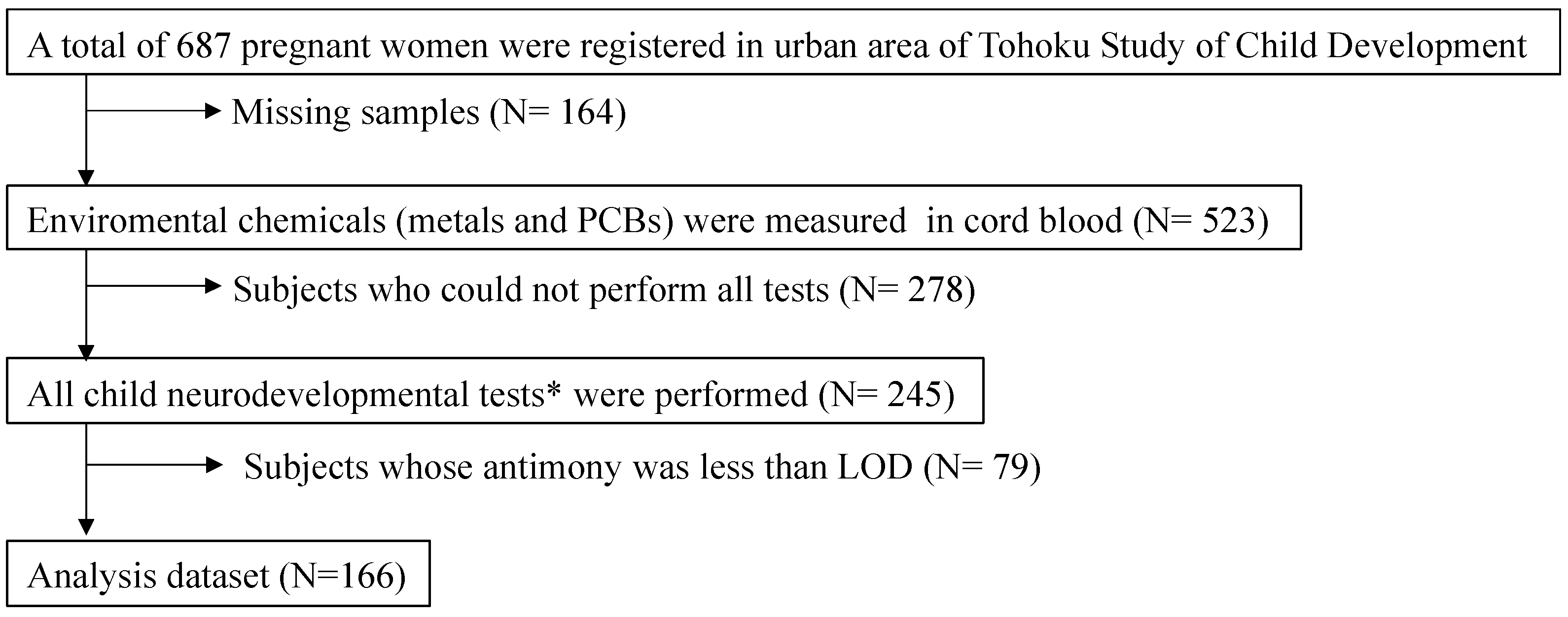
2.1. Study Design, Subjects, and Sampling
2.2. Analytical Methods
2.2.1. Determination of Toxic Metals and Essential Trace Elements
2.2.2. Determination of Polychlorinated Biphenyls
2.2.3. Genomic DNA Extraction and Digestion
2.2.4. LC-MS/MS Analysis for mC/hmC Quantification
2.3. Statistical Analysis
3. Results
4. Discussion
5. Conclusions
Supplementary Materials
Author Contributions
Funding
Institutional Review Board Statement
Informed Consent Statement
Data Availability Statement
Acknowledgments
Conflicts of Interest
References
- Rodier, P.M. Developing brain as a target of toxicity. Environ. Health Perspect. 1995, 103 (Suppl. 6), 73–76. [Google Scholar] [CrossRef] [PubMed] [Green Version]
- Costa, L.G.; Aschner, M.; Vitalone, A.; Syversen, T.; Soldin, O.P. Developmental neuropathology of environmental agents. Annu. Rev. Pharmacol. Toxicol. 2004, 44, 87–110. [Google Scholar] [CrossRef] [PubMed] [Green Version]
- Cedar, H.; Bergman, Y. Programming of DNA methylation patterns. Ann. Rev. Biochem. 2012, 81, 97–117. [Google Scholar] [CrossRef] [PubMed] [Green Version]
- Okamoto, Y.; Yoshida, N.; Suzuki, T.; Shimozawa, N.; Asami, M.; Matsuda, T.; Kojima, N.; Perry, A.C.; Takada, T. DNA methylation dynamics in mouse preimplantation embryos revealed by mass spectrometry. Sci. Rep. 2016, 6, 19134. [Google Scholar] [CrossRef] [Green Version]
- Bell, C.G.; Lowe, R.; Adams, P.D.; Baccarelli, A.A.; Beck, S.; Bell, J.T.; Christensen, B.C.; Gladyshev, V.N.; Heijmans, B.T.; Horvath, S.; et al. DNA methylation aging clocks: Challenges and recommendations. Genome Biol. 2019, 20, 249. [Google Scholar] [CrossRef] [Green Version]
- Stenvinkel, P.; Karimi, M.; Johansson, S.; Axelsson, J.; Suliman, M.; Lindholm, B.; Heimburger, O.; Barany, P.; Alvestrand, A.; Nordfors, L.; et al. Impact of inflammation on epigenetic DNA methylation—A novel risk factor for cardiovascular disease? J. Intern. Med. 2007, 261, 488–499. [Google Scholar] [CrossRef]
- Zhong, J.; Agha, G.; Baccarelli, A.A. The role of DNA methylation in cardiovascular risk and disease: Methodological aspects, study design, and data analysis for epidemiological studies. Circ. Res. 2016, 118, 119–131. [Google Scholar] [CrossRef] [Green Version]
- Bird, A. DNA methylation patterns and epigenetic memory. Genes Dev. 2002, 16, 6–21. [Google Scholar] [CrossRef] [Green Version]
- He, Y.F.; Li, B.Z.; Li, Z.; Liu, P.; Wang, Y.; Tang, Q.; Ding, J.; Jia, Y.; Chen, Z.; Li, L.; et al. Tet-mediated formation of 5-carboxylcytosine and its excision by TDG in mammalian DNA. Science 2011, 333, 1303–1307. [Google Scholar] [CrossRef] [Green Version]
- Tahiliani, M.; Koh, K.P.; Shen, Y.; Pastor, W.A.; Bandukwala, H.; Brudno, Y.; Agarwal, S.; Iyer, L.M.; Liu, D.R.; Aravind, L.; et al. Conversion of 5-methylcytosine to 5-hydroxymethylcytosine in mammalian DNA by MLL partner TET1. Science 2009, 324, 930–935. [Google Scholar] [CrossRef] [Green Version]
- Alvarado-Cruz, I.; Alegria-Torres, J.A.; Montes-Castro, N.; Jimenez-Garza, O.; Quintanilla-Vega, B. Environmental epigenetic changes, as risk factors for the development of diseases in children: A systematic review. Ann. Glob. Health 2018, 84, 212–224. [Google Scholar] [CrossRef] [PubMed] [Green Version]
- Breton, C.V.; Landon, R.; Kahn, L.G.; Enlow, M.B.; Peterson, A.K.; Bastain, T.; Braun, J.; Comstock, S.S.; Duarte, C.S.; Hipwell, A.; et al. Exploring the evidence for epigenetic regulation of environmental influences on child health across generations. Commun. Biol. 2021, 4, 769. [Google Scholar] [CrossRef] [PubMed]
- Martin, E.M.; Fry, R.C. Environmental influences on the epigenome: Exposure—Associated DNA methylation in human populations. Annu. Rev. Public Health 2018, 39, 309–333. [Google Scholar] [CrossRef] [Green Version]
- Perera, F.; Herbstman, J. Prenatal environmental exposures, epigenetics, and disease. Reprod. Toxicol. 2011, 31, 363–373. [Google Scholar] [CrossRef] [PubMed] [Green Version]
- Iwai-Shimada, M.; Satoh, H.; Nakai, K.; Tatsuta, N.; Murata, K.; Akagi, H. Methylmercury in the breast milk of Japanese mothers and lactational exposure of their infants. Chemosphere 2015, 126, 67–72. [Google Scholar] [CrossRef]
- Iwai-Shimada, M.; Kameo, S.; Nakai, K.; Yaginuma-Sakurai, K.; Tatsuta, N.; Kurokawa, N.; Nakayama, S.F.; Satoh, H. Exposure profile of mercury, lead, cadmium, arsenic, antimony, copper, selenium and zinc in maternal blood, cord blood and placenta: The Tohoku Study of Child Development in Japan. Environ. Health Prev. Med. 2019, 24, 35. [Google Scholar] [CrossRef] [Green Version]
- Nakai, K.; Suzuki, K.; Oka, T.; Murata, K.; Sakamoto, M.; Okamura, K.; Hosokawa, T.; Sakai, T.; Nakamura, T.; Saito, Y.; et al. The Tohoku Study of Child Development: A cohort study of effects of perinatal exposures to methylmercury and environmentally persistent organic pollutants on neurobehavioral development in Japanese children. Tohoku J. Exp. Med. 2004, 202, 227–237. [Google Scholar] [CrossRef] [Green Version]
- Nakamura, T.; Nakai, K.; Matsumura, T.; Suzuki, S.; Saito, Y.; Satoh, H. Determination of dioxins and polychlorinated biphenyls in breast milk, maternal blood and cord blood from residents of Tohoku, Japan. Sci. Total Environ. 2008, 394, 39–51. [Google Scholar] [CrossRef]
- Tatsuta, N.; Nakai, K.; Murata, K.; Suzuki, K.; Iwai-Shimada, M.; Yaginuma-Sakurai, K.; Kurokawa, N.; Nakamura, T.; Hosokawa, T.; Satoh, H. Prenatal exposures to environmental chemicals and birth order as risk factors for child behavior problems. Environ. Res. 2012, 114, 47–52. [Google Scholar] [CrossRef]
- Tatsuta, N.; Kurokawa, N.; Nakai, K.; Suzuki, K.; Iwai-Shimada, M.; Murata, K.; Satoh, H. Effects of intrauterine exposures to polychlorinated biphenyls, methylmercury, and lead on birth weight in Japanese male and female newborns. Environ. Health Prev. Med. 2017, 22, 39. [Google Scholar] [CrossRef] [Green Version]
- Tatsuta, N.; Nakai, K.; Kasanuma, Y.; Iwai-Shimada, M.; Sakamoto, M.; Murata, K.; Satoh, H. Prenatal and postnatal lead exposures and intellectual development among 12-year-old Japanese children. Environ. Res. 2020, 189, 109844. [Google Scholar] [CrossRef] [PubMed]
- Suzuki, K.; Nakai, K.; Sugawara, T.; Nakamura, T.; Ohba, T.; Shimada, M.; Hosokawa, T.; Okamura, K.; Sakai, T.; Kurokawa, N.; et al. Neurobehavioral effects of prenatal exposure to methylmercury and PCBs, and seafood intake: Neonatal behavioral assessment scale results of Tohoku study of child development. Environ. Res. 2010, 110, 699–704. [Google Scholar] [CrossRef] [PubMed]
- Tatsuta, N.; Nakai, K.; Sakamoto, M.; Murata, K.; Satoh, H. Methylmercury exposure and developmental outcomes in tohoku study of child development at 18 months of age. Toxics 2018, 6, 49. [Google Scholar] [CrossRef] [Green Version]
- Ministry of the Environment, Japan. Mercury Analysis Manual; Ministry of the Environment, Japan: Tokyo, Japan, 2004.
- Schaller, K.H.; Angerer, J.; Drexler, H. Quality assurance of biological monitoring in occupational and environmental medicine. J. Chromatogr. B Analyt. Technol. Biomed. Life Sci. 2002, 778, 403–417. [Google Scholar] [CrossRef]
- Schisterman, E.F.; Whitcomb, B.W.; Louis, G.M.; Louis, T.A. Lipid adjustment in the analysis of environmental contaminants and human health risks. Environ. Health Perspect. 2005, 113, 853–857. [Google Scholar] [CrossRef] [PubMed] [Green Version]
- Okamoto, Y.; Chou, P.H.; Kim, S.Y.; Suzuki, N.; Laxmi, Y.R.; Okamoto, K.; Liu, X.; Matsuda, T.; Shibutani, S. Oxidative DNA damage in XPC-knockout and its wild mice treated with equine estrogen. Chem. Res. Toxicol. 2008, 21, 1120–1124. [Google Scholar] [CrossRef] [PubMed]
- Okashita, N.; Kumaki, Y.; Ebi, K.; Nishi, M.; Okamoto, Y.; Nakayama, M.; Hashimoto, S.; Nakamura, T.; Sugasawa, K.; Kojima, N.; et al. PRDM14 promotes active DNA demethylation through the ten-eleven translocation (TET)-mediated base excision repair pathway in embryonic stem cells. Development 2014, 141, 269–280. [Google Scholar] [CrossRef] [Green Version]
- Textor, J.; van der Zander, B.; Gilthorpe, M.S.; Liskiewicz, M.; Ellison, G.T. Robust causal inference using directed acyclic graphs: The R package ‘dagitty’. Int. J. Epidemiol. 2016, 45, 1887–1894. [Google Scholar] [CrossRef] [Green Version]
- Kondracki, A.J. Prevalence and patterns of cigarette smoking before and during early and late pregnancy according to maternal characteristics: The first national data based on the 2003 birth certificate revision, United States, 2016. Reprod. Health 2019, 16, 142. [Google Scholar] [CrossRef]
- Scheffers-van Schayck, T.; Tuithof, M.; Otten, R.; Engels, R.; Kleinjan, M. Smoking behavior of women before, during, and after pregnancy: Indicators of smoking, quitting, and relapse. Eur. Addict. Res. 2019, 25, 132–144. [Google Scholar] [CrossRef]
- Ethen, M.K.; Ramadhani, T.A.; Scheuerle, A.E.; Canfield, M.A.; Wyszynski, D.F.; Druschel, C.M.; Romitti, P.A.; National Birth Defects Prevention Study. Alcohol consumption by women before and during pregnancy. Matern Child Health J. 2009, 13, 274–285. [Google Scholar] [CrossRef] [PubMed]
- Nykjaer, C.; Alwan, N.A.; Greenwood, D.C.; Simpson, N.A.; Hay, A.W.; White, K.L.; Cade, J.E. Maternal alcohol intake prior to and during pregnancy and risk of adverse birth outcomes: Evidence from a British cohort. J. Epidemiol. Commu. Health 2014, 68, 542–549. [Google Scholar] [CrossRef] [Green Version]
- Gruzieva, O.; Xu, C.J.; Yousefi, P.; Relton, C.; Merid, S.K.; Breton, C.V.; Gao, L.; Volk, H.E.; Feinberg, J.I.; Ladd-Acosta, C.; et al. Prenatal particulate air pollution and DNA methylation in newborns: An epigenome-wide meta-analysis. Environ. Health Perspect. 2019, 127, 57012. [Google Scholar] [CrossRef]
- Isaevska, E.; Moccia, C.; Asta, F.; Cibella, F.; Gagliardi, L.; Ronfani, L.; Rusconi, F.; Stazi, M.A.; Richiardi, L. Exposure to ambient air pollution in the first 1000 days of life and alterations in the DNA methylome and telomere length in children: A systematic review. Environ. Res. 2021, 193, 110504. [Google Scholar] [CrossRef] [PubMed]
- Lozano, M.; Yousefi, P.; Broberg, K.; Soler-Blasco, R.; Miyashita, C.; Pesce, G.; Kim, W.J.; Rahman, M.; Bakulski, K.M.; Haug, L.S.; et al. DNA methylation changes associated with prenatal mercury exposure: A meta-analysis of prospective cohort studies from PACE consortium. Environ. Res. 2021, 204, 112093. [Google Scholar] [CrossRef]
- Lisanti, S.; Omar, W.A.; Tomaszewski, B.; De Prins, S.; Jacobs, G.; Koppen, G.; Mathers, J.C.; Langie, S.A. Comparison of methods for quantification of global DNA methylation in human cells and tissues. PLoS ONE 2013, 8, e79044. [Google Scholar] [CrossRef] [PubMed] [Green Version]
- Cediel Ulloa, A.; Gliga, A.; Love, T.M.; Pineda, D.; Mruzek, D.W.; Watson, G.E.; Davidson, P.W.; Shamlaye, C.F.; Strain, J.J.; Myers, G.J.; et al. Prenatal methylmercury exposure and DNA methylation in seven-year-old children in the Seychelles Child Development Study. Environ. Int. 2021, 147, 106321. [Google Scholar] [CrossRef]
- Goodrich, J.M.; Sánchez, B.N.; Dolinoy, D.C.; Zhang, Z.; Hernández-Ávila, M.; Hu, H.; Peterson, K.E.; Téllez-Rojo, M.M. Quality control and statistical modeling for environmental epigenetics: A study on in utero lead exposure and DNA methylation at birth. Epigenetics 2015, 10, 19–30. [Google Scholar] [CrossRef] [Green Version]
- Hanna, C.W.; Bloom, M.S.; Robinson, W.P.; Kim, D.; Parsons, P.J.; vom Saal, F.S.; Taylor, J.A.; Steuerwald, A.J.; Fujimoto, V.Y. DNA methylation changes in whole blood is associated with exposure to the environmental contaminants, mercury, lead, cadmium and bisphenol A, in women undergoing ovarian stimulation for IVF. Hum. Reprod. 2012, 27, 1401–1410. [Google Scholar] [CrossRef]
- Miller, R.L.; Yan, Z.; Maher, C.; Zhang, H.; Gudsnuk, K.; McDonald, J.; Champagne, F.A. Impact of prenatal polycyclic aromatic hydrocarbon exposure on behavior, cortical gene expression and DNA methylation of the Bdnf gene. Neuroepigenetics 2016, 5, 11–18. [Google Scholar] [CrossRef] [Green Version]
- Pilsner, J.R.; Hall, M.N.; Liu, X.; Ilievski, V.; Slavkovich, V.; Levy, D.; Factor-Litvak, P.; Yunus, M.; Rahman, M.; Graziano, J.H.; et al. Influence of prenatal arsenic exposure and newborn sex on global methylation of cord blood DNA. PLoS ONE 2012, 7, e37147. [Google Scholar] [CrossRef] [PubMed] [Green Version]
- Tajuddin, S.M.; Amaral, A.F.; Fernández, A.F.; Rodríguez-Rodero, S.; Rodríguez, R.M.; Moore, L.E.; Tardón, A.; Carrato, A.; García-Closas, M.; Silverman, D.T.; et al. Genetic and non-genetic predictors of LINE-1 methylation in leukocyte DNA. Environ. Health Perspect. 2013, 121, 650–656. [Google Scholar] [CrossRef] [PubMed] [Green Version]
- Wright, R.O.; Schwartz, J.; Wright, R.J.; Bollati, V.; Tarantini, L.; Park, S.K.; Hu, H.; Sparrow, D.; Vokonas, P.; Baccarelli, A. Biomarkers of lead exposure and DNA methylation within retrotransposons. Environ. Health Perspect. 2010, 118, 790–795. [Google Scholar] [CrossRef] [PubMed] [Green Version]
- Huang, Y.; Pastor, W.A.; Shen, Y.; Tahiliani, M.; Liu, D.R.; Rao, A. The behaviour of 5-hydroxymethylcytosine in bisulfite sequencing. PLoS ONE 2010, 5, e8888. [Google Scholar] [CrossRef] [Green Version]
- Guerrero-Preston, R.; Goldman, L.R.; Brebi-Mieville, P.; Ili-Gangas, C.; Lebron, C.; Witter, F.R.; Apelberg, B.J.; Hernández-Roystacher, M.; Jaffe, A.; Halden, R.U.; et al. Global DNA hypomethylation is associated with in utero exposure to cotinine and perfluorinated alkyl compounds. Epigenetics 2010, 5, 539–546. [Google Scholar] [CrossRef] [Green Version]
- Herbstman, J.B.; Tang, D.; Zhu, D.; Qu, L.; Sjödin, A.; Li, Z.; Camann, D.; Perera, F.P. Prenatal exposure to polycyclic aromatic hydrocarbons, benzo[a]pyrene-DNA adducts, and genomic DNA methylation in cord blood. Environ. Health Perspect. 2012, 120, 733–738. [Google Scholar] [CrossRef] [Green Version]
- Tellez-Plaza, M.; Tang, W.Y.; Shang, Y.; Umans, J.G.; Francesconi, K.A.; Goessler, W.; Ledesma, M.; Leon, M.; Laclaustra, M.; Pollak, J.; et al. Association of global DNA methylation and global DNA hydroxymethylation with metals and other exposures in human blood DNA samples. Environ. Health Perspect. 2014, 122, 946–954. [Google Scholar] [CrossRef] [Green Version]
- Kurdyukov, S.; Bullock, M. DNA methylation analysis: Choosing the right method. Biology 2016, 5, 3. [Google Scholar] [CrossRef]
- Wu, S.; Hivert, M.F.; Cardenas, A.; Zhong, J.; Rifas-Shiman, S.L.; Agha, G.; Colicino, E.; Just, A.C.; Amarasiriwardena, C.; Lin, X.; et al. Exposure to low levels of lead in utero and umbilical cord blood DNA methylation in project viva: An epigenome-wide association study. Environ. Health Perspect. 2017, 125, 087019. [Google Scholar] [CrossRef] [Green Version]
- Boyle, J.; Yeter, D.; Aschner, M.; Wheeler, D.C. Estimated IQ points and lifetime earnings lost to early childhood blood lead levels in the United States. Sci. Total Environ. 2021, 778, 146307. [Google Scholar] [CrossRef]
- Canfield, R.L.; Henderson, C.R., Jr.; Cory-Slechta, D.A.; Cox, C.; Jusko, T.A.; Lanphear, B.P. Intellectual impairment in children with blood lead concentrations below 10 microg per deciliter. N. Engl. J. Med. 2003, 348, 1517–1526. [Google Scholar] [CrossRef] [PubMed] [Green Version]
- Hwang, Y.H.; Hsiao, C.K.; Lin, P.W. Globally temporal transitions of blood lead levels of preschool children across countries of different categories of human development index. Sci. Total Environ. 2019, 659, 1395–1402. [Google Scholar] [CrossRef] [PubMed]
- Wilhelm, M.; Heinzow, B.; Angerer, J.; Schulz, C. Reassessment of critical lead effects by the German Human Biomonitoring Commission results in suspension of the human biomonitoring values (HBM I and HBM II) for lead in blood of children and adults. Int. J. Hyg. Environ. Health 2010, 213, 265–269. [Google Scholar] [CrossRef] [PubMed]
- US CDC Updates Blood Lead Reference Value for Children. Available online: https://www.cdc.gov/media/releases/2021/p1028-blood-lead.html (accessed on 2 March 2022).


| n = 166 | Mean ± SD Median (P25–P75) | n (%) |
|---|---|---|
| Maternal characteristics | ||
| Maternal age (years) | 31.2 ± 3.8 | |
| Body mass index before pregnancy (kg/m2) | 20.9 ± 2.5 | |
| Smoking habit during pregnancy (smokers, %) | 12 (7.2) | |
| Drinking habit during pregnancy (drinkers, %) | 52 (31.3) | |
| Delivery type (spontaneous, %) | 122 (73.5) | |
| Parity (first, %) | 88 (53.0) | |
| Maternal educational level (graduate high school, %) | 128 (77.1) | |
| Baby characteristics | ||
| Gestational age (weeks) | 39.6 ± 1.3 | |
| Birth weight (g) | 3078.0 ± 329.1 | |
| Sex (boys, %) | 88 (53.0) | |
| Apgar score | 8 (8–9) | |
| Exposure levels in cord blood | ||
| Total PCBs (ng/g-lipid) | 49.6 (30.3–60.5) | |
| Hg (ng/g) | 10.8 (7.0–13.7) | |
| As (ng/mL) | 4.42 (2.69–5.52) | |
| Pb (μg/dL) | 1.06 (0.80–1.27) | |
| Cd (ng/mL) | 0.93 (0.05–1.06) | |
| Sb (ng/mL) | 0.93 (0.41–1.28) | |
| Se (ng/mL) | 185.6 (158.9–210.6) | |
| Cu (ng/mL) | 512.8 (448.5–548.5) | |
| Zn (ng/mL) | 2129.7 (1729.7–2236.6) | |
| DNA methylation status | ||
| mC (ng/100 ng DNA) | 1.11 (1.06–1.15) | |
| hmC (ng/100 ng DNA) | 0.011 (0.010–0.012) |
| n = 166 | mC (ng/100 ng DNA) | hmC (ng/100 ng DNA) |
|---|---|---|
| r | r | |
| Maternal characteristics | ||
| Maternal age (years) | 0.089 | 0.243 * |
| Body mass index before pregnancy (kg/m2) | −0.047 | −0.072 |
| Baby characteristics | ||
| Gestational age (weeks) | 0.033 | −0.096 |
| Birth weight (g) | 0.052 | −0.027 |
| Birth length (cm) | 0.128 | 0.067 |
| Exposure levels in cord blood | ||
| PCBs (ng/g-lipid) | −0.090 | 0.050 |
| Hg (ng/g) | 0.038 | −0.074 |
| As (ng/mL) | −0.058 | −0.123 |
| Pb (μg/dL) | 0.435 ** | 0.155 * |
| Cd (ng/mL) | −0.010 | −0.129 |
| Sb (ng/mL) | 0.288 ** | 0.125 |
| Se (ng/mL) | 0.168 * | 0.008 |
| Cu (ng/mL) | 0.089 | 0.044 |
| Zn (ng/mL) | 0.036 | −0.063 |
| n = 166 | mC (ng/100 ng DNA) | hmC (ng/100 ng DNA) | ||
|---|---|---|---|---|
| Standardized Regression Coefficient, β [95% CI] | p Value | Standardized Regression Coefficient, β [95% CI] | p Value | |
| Exposure markers | ||||
| PCBs (ng/g-lipid) | −0.083 [−0.239, 0.073] | 0.294 | 0.118 [−0.064, 0.300] | 0.203 |
| Hg (ng/g) | 0.110 [−0.033, 0.253] | 0.130 | −0.053 [−0.219, 0.113] | 0.530 |
| As (ng/mL) | −0.085 [−0.224, 0.054] | 0.228 | −0.120 [−0.282, 0.042] | 0.144 |
| Pb (μg/dL) | 0.524 [0.381, 0.666] | <0.0001 | 0.227 [0.061, 0.394] | 0.008 |
| Cd (ng/mL) | −0.056 [−0.195, 0.084] | 0.430 | −0.143 [−0.305, 0.020] | 0.086 |
| Sb (ng/mL) | 0.388 [0.255, 0.521] | <0.0001 | 0.192 [0.037, 0.348] | 0.016 |
| Se (ng/mL) | 0.153 [0.009, 0.297] | 0.038 | 0.042 [−0.126, 0.211] | 0.622 |
| Cu (ng/mL) | −0.001 [−0.167, 0.164] | 0.988 | 0.064 [−0.130, 0.257] | 0.517 |
| Zn (ng/mL) | −0.134 [−0.310, 0.042] | 0.134 | −0.194 [−0.400, 0.011] | 0.064 |
| Possible confounders | ||||
| Maternal age | 0.093 [−0.055, 0.240] | 0.216 | 0.176 [0.003, 0.348] | 0.046 |
| Gestational weeks | −0.031 [−0.172, 0.110] | 0.665 | −0.028 [−0.193, 0.137] | 0.739 |
| Parity | −0.020 [−0.178, 0.139] | 0.808 | 0.057 [−0.128, 0.242] | 0.545 |
| Education levels | 0.156 [−0.015, 0.326] | 0.074 | −0.128 [−0.327, 0.071] | 0.207 |
| BMI before pregnancy | −0.013 [−0.143, 0.117] | 0.848 | −0.057 [−0.209, 0.095] | 0.457 |
| Smoking habit during pregnancy | 0.145 [−0.131, 0.421] | 0.302 | −0.052 [−0.374, 0.271] | 0.752 |
| Drinking habit during pregnancy | −0.046 [−0.184, 0.093] | 0.516 | −0.053 [−0.215, 0.109] | 0.516 |
| Contribution rate, R2 Adjusted R2 | 0.399 0.334 | 0.181 0.093 | ||
Publisher’s Note: MDPI stays neutral with regard to jurisdictional claims in published maps and institutional affiliations. |
© 2022 by the authors. Licensee MDPI, Basel, Switzerland. This article is an open access article distributed under the terms and conditions of the Creative Commons Attribution (CC BY) license (https://creativecommons.org/licenses/by/4.0/).
Share and Cite
Okamoto, Y.; Iwai-Shimada, M.; Nakai, K.; Tatsuta, N.; Mori, Y.; Aoki, A.; Kojima, N.; Takada, T.; Satoh, H.; Jinno, H. Global DNA Methylation in Cord Blood as a Biomarker for Prenatal Lead and Antimony Exposures. Toxics 2022, 10, 157. https://doi.org/10.3390/toxics10040157
Okamoto Y, Iwai-Shimada M, Nakai K, Tatsuta N, Mori Y, Aoki A, Kojima N, Takada T, Satoh H, Jinno H. Global DNA Methylation in Cord Blood as a Biomarker for Prenatal Lead and Antimony Exposures. Toxics. 2022; 10(4):157. https://doi.org/10.3390/toxics10040157
Chicago/Turabian StyleOkamoto, Yoshinori, Miyuki Iwai-Shimada, Kunihiko Nakai, Nozomi Tatsuta, Yoko Mori, Akira Aoki, Nakao Kojima, Tatsuyuki Takada, Hiroshi Satoh, and Hideto Jinno. 2022. "Global DNA Methylation in Cord Blood as a Biomarker for Prenatal Lead and Antimony Exposures" Toxics 10, no. 4: 157. https://doi.org/10.3390/toxics10040157
APA StyleOkamoto, Y., Iwai-Shimada, M., Nakai, K., Tatsuta, N., Mori, Y., Aoki, A., Kojima, N., Takada, T., Satoh, H., & Jinno, H. (2022). Global DNA Methylation in Cord Blood as a Biomarker for Prenatal Lead and Antimony Exposures. Toxics, 10(4), 157. https://doi.org/10.3390/toxics10040157