Article

The Microbial Diversity of Non-Korean Kimchi as Revealed by Viable Counting and Metataxonomic Sequencing

1
Dipartimento di Scienze Agrarie, Alimentari ed Ambientali, Università Politecnica delle Marche, via Brecce Bianche, 60131 Ancona, Italy
2
Department of Agricultural, Forest, and Food Science, University of Turin, Largo Paolo Braccini 2, Grugliasco, 10095 Torino, Italy
3
Istituto Zooprofilattico Sperimentale dell’Umbria e delle Marche, Via Cupa di Posatora 3, 60131 Ancona, Italy
*
Authors to whom correspondence should be addressed.
Foods 2020, 9(11), 1568; https://doi.org/10.3390/foods9111568
Submission received: 14 October 2020 / Revised: 21 October 2020 / Accepted: 26 October 2020 / Published: 29 October 2020

Abstract

:
Kimchi is recognized worldwide as the flagship food of Korea. To date, most of the currently available microbiological studies on kimchi deal with Korean manufactures. Moreover, there is a lack of knowledge on the occurrence of eumycetes in kimchi. Given these premises, the present study was aimed at investigating the bacterial and fungal dynamics occurring during the natural fermentation of an artisan non-Korean kimchi manufacture. Lactic acid bacteria were dominant, while Enterobacteriaceae, Pseudomonadaceae, and yeasts progressively decreased during fermentation. Erwinia spp., Pseudomonas veronii, Pseudomonas viridiflava, Rahnella aquatilis, and Sphingomonas spp. were detected during the first 15 days of fermentation, whereas the last fermentation phase was dominated by Leuconostoc kimchi, together with Weissella soli. For the mycobiota at the beginning of the fermentation process, Rhizoplaca and Pichia orientalis were the dominant Operational Taxonomic Units (OTUs) in batch 1, whereas in batch 2 Protomyces inundatus prevailed. In the last stage of fermentation, Saccharomyces cerevisiae, Candida sake, Penicillium, and Malassezia were the most abundant taxa in both analyzed batches. The knowledge gained in the present study represents a step forward in the description of the microbial dynamics of kimchi produced outside the region of origin using local ingredients. It will also serve as a starting point for further isolation of kimchi-adapted microorganisms to be assayed as potential starters for the manufacturing of novel vegetable preserves with high quality and functional traits.

Graphical Abstract

1. Introduction

Vegetable-based traditional fermented foods are manufactured worldwide but in some countries, they represent an important part of the daily diet. The most popular fermented vegetables include mudhika produced in Africa using cassava (Manihot esculenta) [1]; caxiri, tarubà, and yakupa produced in Latin America through cassava fermentation [2]; and sauerkraut, mainly produced in Central and Eastern European countries with shredded and salted cabbage [3]. Moreover, among the fermented vegetables that are recognized worldwide as unique kickshaws, paocai, jiangshui, miso, natto, tempeh, and kimchi represent further well-known masterpieces of Asian traditions [4].
Among vegetable-based fermented foods typically produced in Asia, kimchi is recognized as the flagship food of Korea [5]. Such a preparation, based on the fermentation of cabbage and/or other vegetables, was probably invented around 4000 years ago [6]. Moreover, during Confucianism, kimchi constituted the core of the ancestral worship table, together with rice and soup [4]. In 2013, the United Nations Educational, Scientific and Cultural Organization (UNESCO) recognized the cultural importance of kimchi and inscribed this Korean traditional preparation of vegetables in the Representative List of The Intangible Cultural Heritage of Humanity.
Kimchi is usually produced using cabbage, radish, and cucumber as main ingredients; moreover, different seasonings, including salts, garlic, leek, red pepper powder, and ginger are used in accordance with local traditions [7]. Actually, more than 200 varieties of kimchi are produced, each characterized by peculiar biochemical, nutritional, and sensory features, which are greatly affected by the raw ingredients (e.g., onion, Korean lettuce, sesame leaves, sweet potato vines, Allium hookeri Thwaites, deodeok, and water dropwort), the preparation method, and even region, seasonality, and cultural traditions [7,8]. Baechu (Chinese cabbage) kimchi is the most popular variety. Traditionally, the secret of making kimchi passed through generations from mothers to daughters [9]. However, as an effect of globalization, individuals and companies worldwide can now access the recipe for kimchi production. It is estimated that the worldwide market for kimchi will reach USD 3850 million in 2024, from USD 3000 million in 2019. Though in Italy kimchi is still far from being popular, various kimchi manufacturers and kimchi suppliers have been operating since the late 2010s.
Mature kimchi possesses unique and flavorful sensory traits, consisting of a combination of fresh, sour, spicy, hot, and sweet tastes [10]. It is generally low in calories and rich in vitamins (A, C, and B complex), various phytochemicals, minerals (calcium, iron, potassium), and dietary fiber [11].
From a microbiological point of view, mature kimchi is a closed ecosystem where complex microbial interactions occur. Kimchi fermentation is an anaerobic process during which the interactions between the microbiota and the raw materials lead to the production of microbial metabolites (e.g., mannitol, lactate, acetate, ethanol, exopolysaccharides, etc.) that strongly characterize the sensory traits of the product [12]. Of note, the production of organic acids from the carbohydrates’ metabolism results in a pH drop to 4.2–4.0 [13], which in turn contributes to the palatability and safety of this fermented vegetable-based food.
To the authors’ knowledge, most of the microbiological studies to date carried out on kimchi deal with the analysis of Korean manufactures, and no scientific data are currently available on non-Asian produced kimchi. A past taxonomic study carried out on Korean kimchi reported the prevalence of lactic acid bacteria as the main fermenting microorganisms, including Leuconostoc, Lactobacillus, Lactococcus, Pediococcus, and Weissella species [12]. More recently, a large-scale targeted metagenomic investigation focusing on the bacterial ecology of kimchi sampled during its natural fermentation showed the occurrence of a more complex and dynamic ecosystem, with the most adapted species prevailing at the end of the fermentation process [10].
Kimchi also represents an important source of candidate bacterial probiotics and protechnological strains that, due to their robustness, are very well adapted to the unfavorable conditions occurring in this food product [14]. A few lactic acid bacteria strains so far isolated from kimchi, such as Lactobacillus acidophilus KFRI342, Lactococcus lactis KC24, L. plantarum LPpnu, and Leuconosctoc mesenteroides LMpnu showed a potential anticancer activity [15,16,17], thus suggesting a possible beneficial effect of the consumption of kimchi on human health.
Unlike bacteria, there is a paucity of available data about the fungal dynamics occurring during kimchi fermentation. As recently reported by Kim et al. [18], in the early stages of fermentation low loads of yeasts can colonize the fermenting vegetable substrate, with a positive impact on the sensory traits of kimchi due to the production of fruity aromatic compounds and a mitigation of kimchi’s acidic and moldy flavors [18]. However, as reported by the same authors, high loads of yeasts (e.g., yeasts forming white colonies) occurring on kimchi’s surface, especially during the late phase of fermentation, can negatively affect the quality of the end product [18].
Given the above premises, the main aim of the present study was to explore the bacterial and fungal dynamics occurring during the natural fermentation of kimchi handcrafted by an artisan Italian manufacturer through conventional microbiological analyses (viable counting) and metataxonomic sequencing.

2. Materials and Methods

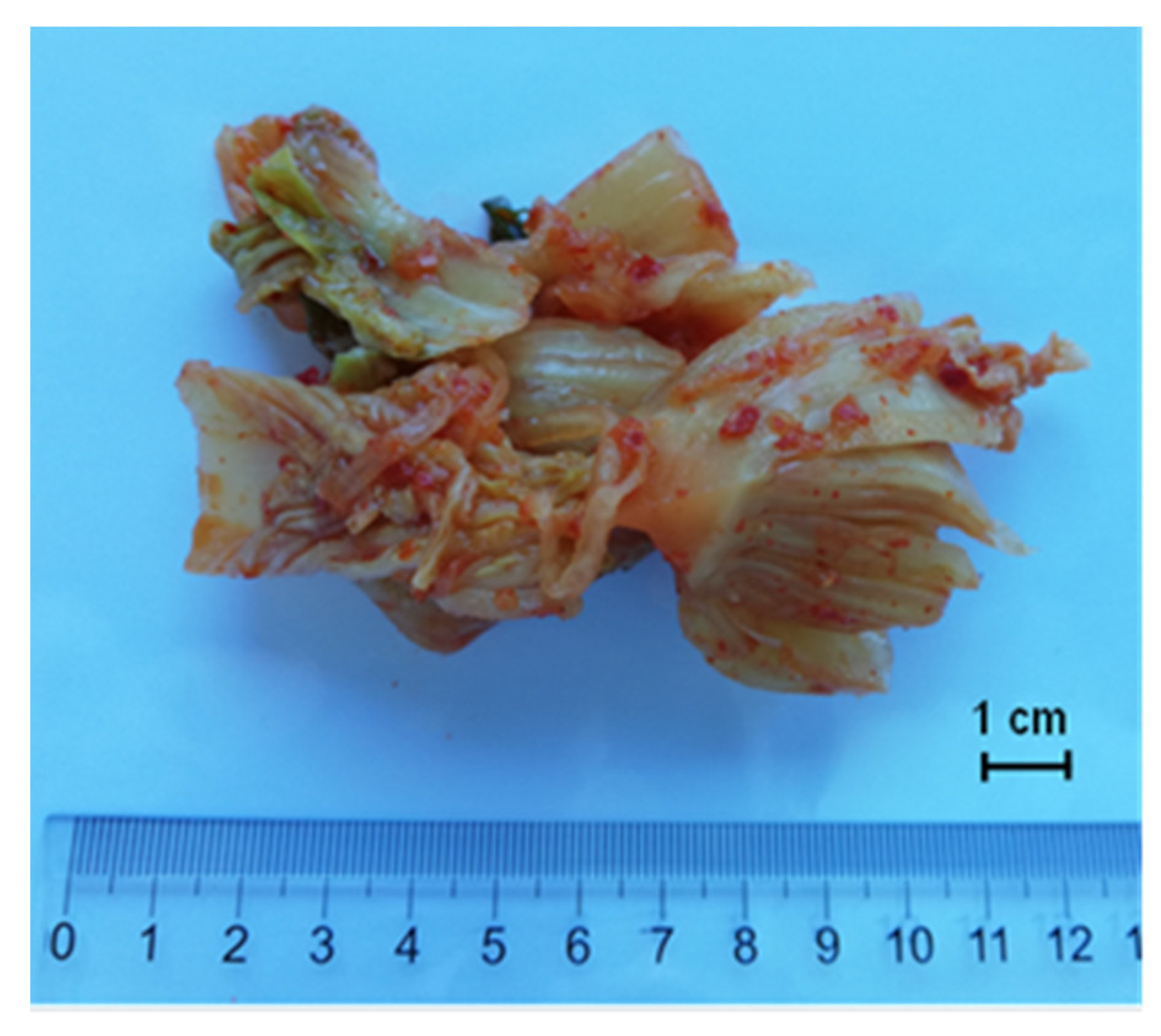
2.1. Kimchi Production

Two independent production batches of kimchi, referred to as batch 1 and batch 2, were prepared by a Korean staff at a small artisan producer located in Santa Maria Nuova (Ancona, Italy) using the following ingredients: Chinese cabbage 60.5 (g 100 g−1), turnip 13.5 (g 100 g−1), water 12.0 (g 100 g−1), onion 4.0 (g 100 g−1), pepper chili powder 2.0 (g 100 g−1), red pepper 2.0 (g 100 g−1), garlic leaves 2.0 (g 100 g−1), spring onion 1.0 (g 100 g−1), carrot 1.0 (g 100 g−1), ginger 0.5 (g 100 g−1), sucrose 1.0 (g 100 g−1), and salt 0.5 (g 100 g−1). All the vegetables were purchased from a local grocer.
Kimchi was produced according to the traditional Korean process described as follows. Briefly, the outer leaves were removed from the Chinese cabbage, trimmed, and then steeped in 10% (w v−1) NaCl for approximately 16–18 h at room temperature. The sauce used for cabbage dressing was obtained by mixing chopped vegetables (turnip, onion, carrot, ginger, spring onion, red peppers, and garlic leaves) with chili pepper powder, water, sugar, and salt. Thereafter, the sauce was stored under refrigerated conditions (5 °C) for approximately 16–18 h. The cabbage was rinsed, and the excess water drained. The sauce was carefully spread over the cabbage leaves. Kimchi was fermented at approximately 5 ± 1 °C for 57 days, up until a fixed pH of 4.2 was reached. Kimchi fermentation was carried out in a plastic box hermetically sealed with a lid. Samples of kimchi (Figure 1) were collected with sterile spoons immediately after preparation (t0) and after 2, 5, 15, 36, 43, 50, and 57 days of fermentation. The samples were transported to the laboratory under refrigerated conditions (4 °C) and processed immediately after arrival.

2.2. pH Determination

pH measurements were performed at the core of each kimchi sample as previously described by Belleggia et al. [19].

2.3. Microbial Counts

Ninety mL of sterile water containing 1 g L−1 bacteriological peptone were added to 10 g aliquots of each sample. The suspensions were homogenized for 2 min at 230 rpm in a Stomacher machine (400 Circulator, International PBI, Milan, Italy). Tenfold serial dilutions were prepared for the enumeration of: (i) mesophilic aerobic bacteria on Plate Count Agar incubated at 30 °C for 48 h; (ii) total mesophilic halophilic aerobic bacteria on Plate Count Agar added with 8% NaCl and incubated at 30 °C for 7 days; (iii) presumptive mesophilic lactobacilli on De Man, Rogosa, and Sharpe (MRS) agar incubated at 30 °C for 48 h; (iv) presumptive mesophilic lactococci on M17 agar incubated at 22 °C for 72 h; (v) halophilic lactobacilli on MRS agar added with 8% NaCl and incubated at 30 °C for 7 days; (vi) halophilic lactococci on M17 agar added with 8% NaCl and incubated at 22 °C for 10 days; (vii) Enterobacteriaceae on Violet Red Bile Glucose Agar (VRBGA) incubated at 37 °C for 24 h; (viii) Pseudomonadaceae enumerated on Pseudomonas Agar Base (PAB) supplemented with Cetrimide-Fucidin-Cephalosporin (CFC) selective supplement (VWR International, Milan, Italy) incubated at 30 °C for 24–48 h; (ix) yeasts counted on Rose Bengal Chloramphenicol Agar (RBCA) incubated at 25 °C for 72–96 h; (x) halophilic yeasts counted on RBCA agar added with 8% NaCl and incubated at 25 °C for 72 h. MRS agar was supplemented with cycloheximide (250 mg L−1) to inhibit the growth of eumycetes.
The results of viable counts were reported as mean value of two biological and three technical replicates expressed as the Log of colony-forming units (cfu) per gram of sample ± standard deviation.
Finally, the presence/absence of Listeria monocytogenes and Salmonella spp. was determined using a MINI VIDAS (Vitek Immunodiagnostic Assay System) apparatus (Biomerieux, Marcy l’Etoile, France) as previously described [19,20].

2.4. RNA Extraction and cDNA Synthesis

The 1.5 mL sample homogenates (10−1 dilution), prepared as described in Section 2.3, were centrifuged at 16,000 rpm for 10 min to obtain cell pellets subsequently covered with RNAlater Stabilization Solution (Ambion, Foster City, CA, USA) and stored at −80 °C until the extraction of RNA using E.Z.N.A. Bacterial RNA Kit (Omega Bio-tek, Norcross, GA, USA). The quantity, purity, and integrity of the extracted RNAs were checked as previously described by Garofalo et al. [21]. cDNA synthesis was performed using SensiFAST cDNA Synthesis Kit for RT-qPCR (Bioline, London, UK).

2.5. Metataxonomic Sequencing and Bioinformatics Analysis

cDNA was used for the amplification of bacterial 16S rRNA (V3/V4 region) [22] and fungal 26S rRNA genes (D1 domain) [23]. The sequencing of the purified PCR amplicons was performed in a MiSeq instrument in a 2 × 250 bp configuration, while QIIME v. 1.9 [24] was used for the analysis of the obtained reads. For bacteria, after Operational Taxonomic Units (OTUs) clustering at 99% of similarity, centroid sequences were mapped against the Greengenes 16S rRNA gene database, while for eumycetes the in-house database from Mota-Gutierrez et al. [23] was used. Taxonomic assignments were double-checked using BLAST suite tools. Chloroplast and mitochondria sequences were removed from the data sets. The OTUs table was rarefied at the lowest number of sequence/samples displaying the highest taxonomic resolution.

2.6. Data Analysis

The alpha diversity index was calculated by the VEGAN package of R. Diversity index, and OTUs table were used in R to find statistically significant differences in the samples as a function of the fermentation time. Principal Component Analysis (PCA), aimed at exploring relationships between experimental variables and detecting possible sample clusters, and one-way ANOVA analysis, used to analyze the effect of ripening time on the dependent variables for each batch separately, were performed as described by Belleggia et al. [19]

3. Results

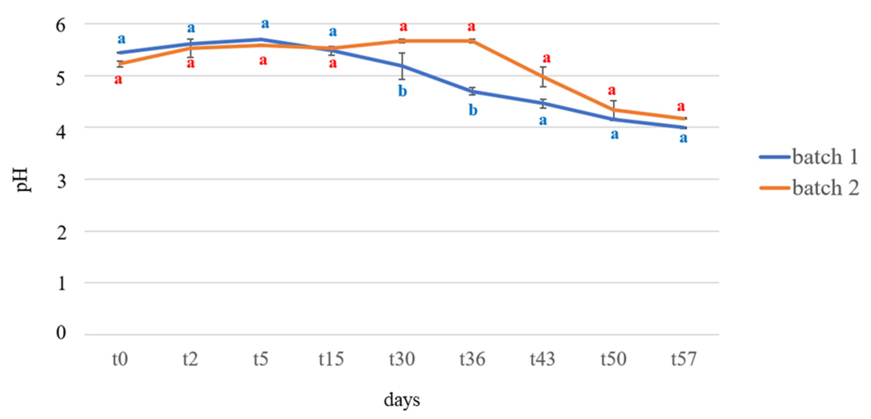
3.1. pH Determination

The results of pH measurements are shown in Figure 2. The values detected in the two analyzed batches had a similar trend, although kimchi produced in batch 1 was characterized by a faster pH reduction than batch 2. In more detail, at t0, the pH values were 5.44 ± 0.01 and 5.22 ± 0.06 for batch 1 and batch 2, respectively. A significant drop in pH was observed between t30 and t36 for batch 1, whereas a more progressive reduction was observed in batch 2, where a drop in pH was detected between t36 and t43. At t57 no significant differences were detected between the pH values of both batches, which tested at 3.99 ± 0.01 and 4.17 ± 0.01 for batch 1 and batch 2, respectively.

3.2. Microbiological Analyses

The results of viable counts performed on the two batches of kimchi are reported in Table 1.
In more detail, the counts of mesophilic aerobic bacteria showed the same trend in both batches, with no significant differences between them.
For halophilic mesophilic aerobic bacteria, as well, at t57 no significant differences in both the analyzed batches were observed.
Regarding presumptive lactobacilli and presumptive halophilic lactobacilli, a progressive increase in the load of these microorganisms was seen during fermentation; the highest counts were observed from t36 to t50 in batch 1 and from t43 to t57 in batch 2.
Regarding presumptive lactococci and presumptive halophilic lactococci, the counts in samples of batches 1 and 2 showed no significant differences at t57.
Low Enterobacteriaceae counts were detected, with no significant differences between the two batches from t43 to t57.
As for Pseudomonadaceae, from t36 to t57, they were <1.0 Log cfu g−1 in both batches.
Regarding yeasts and halophilic yeasts, counts < 1.0 Log cfu g−1 were detected in both batches at t57.
Finally, L. monocytogenes or Salmonella spp. were never detected in 25 g of product, irrespective of the sampling time and the production batch.

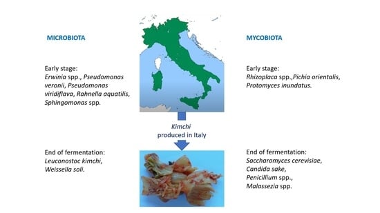
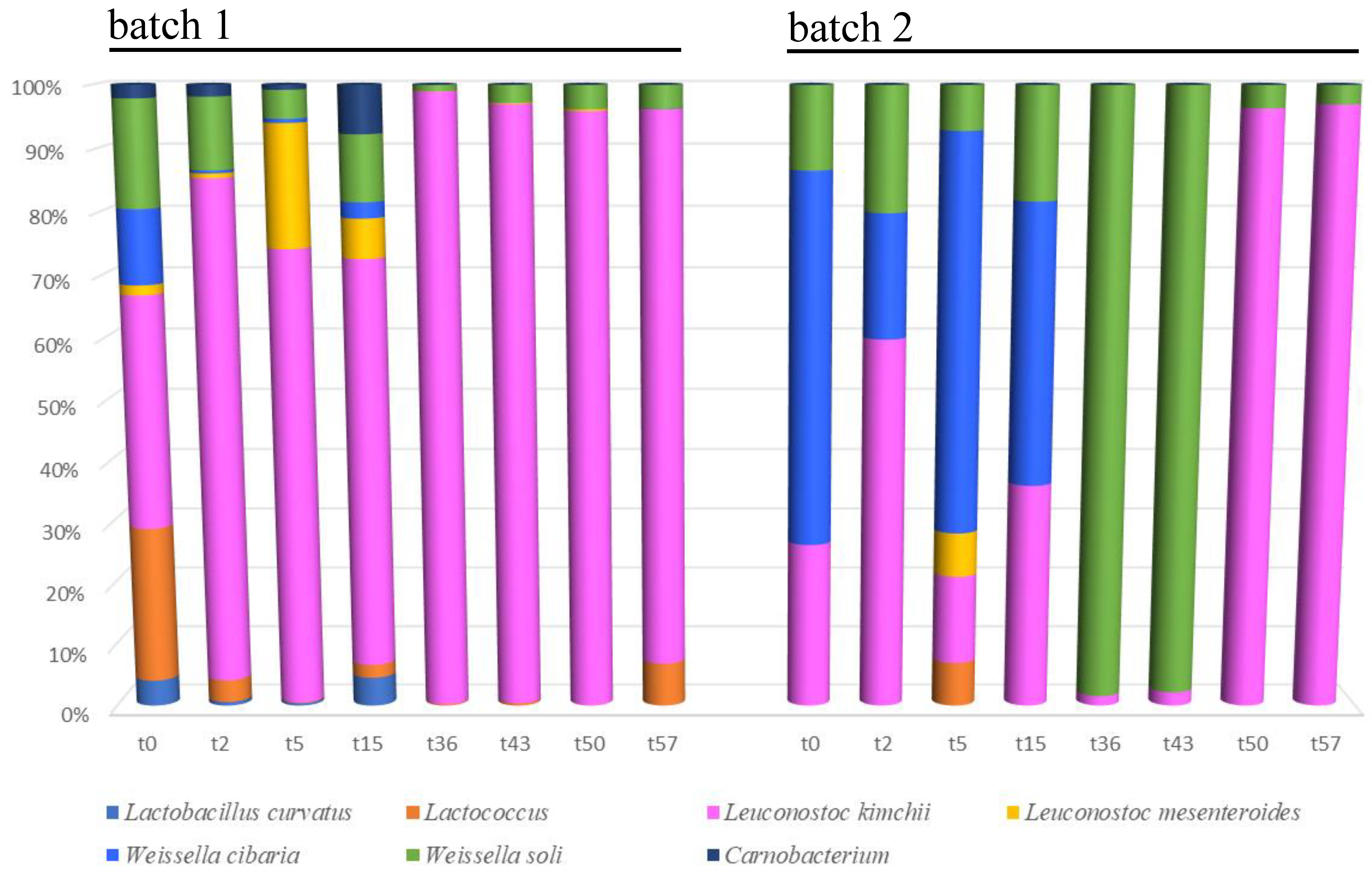
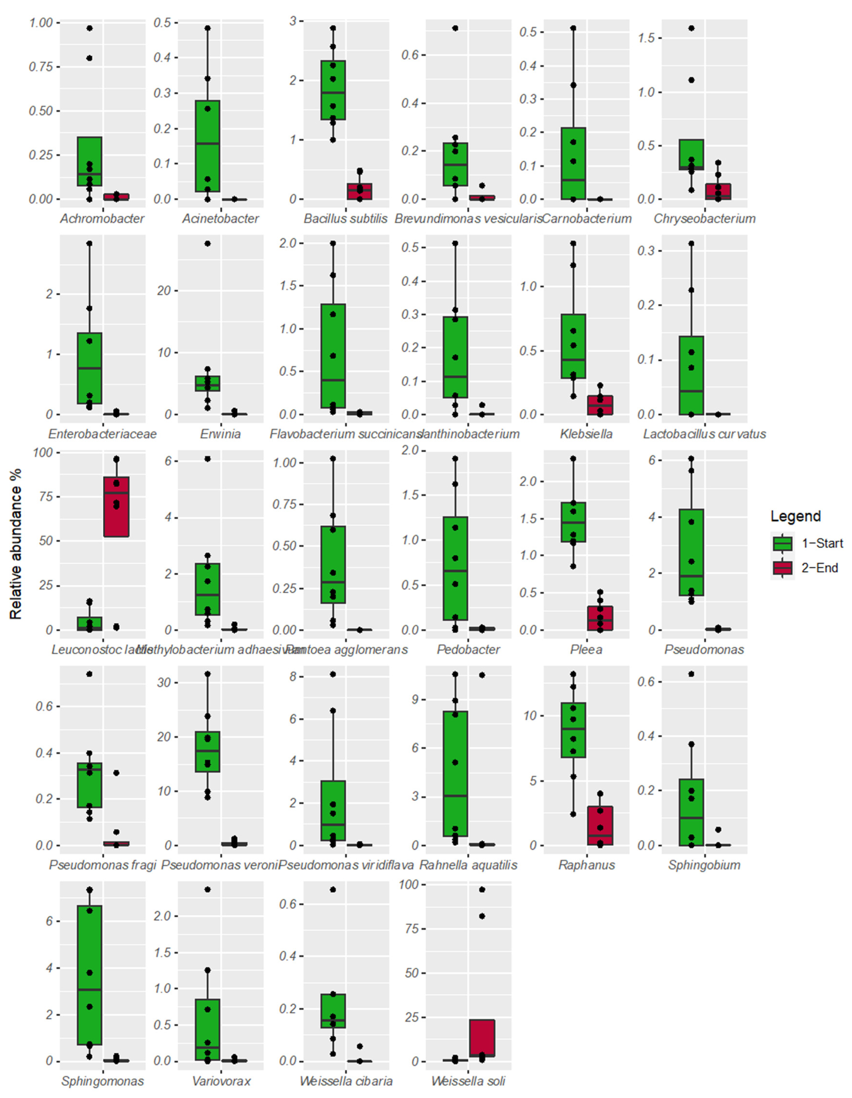
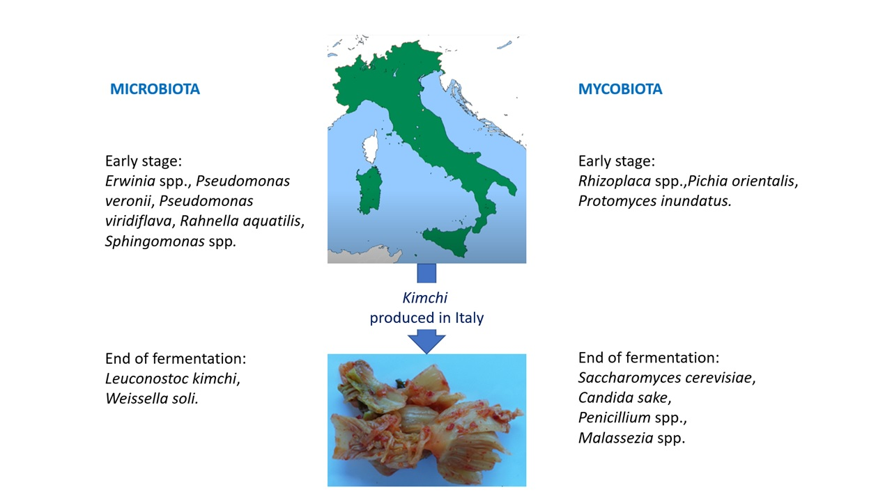
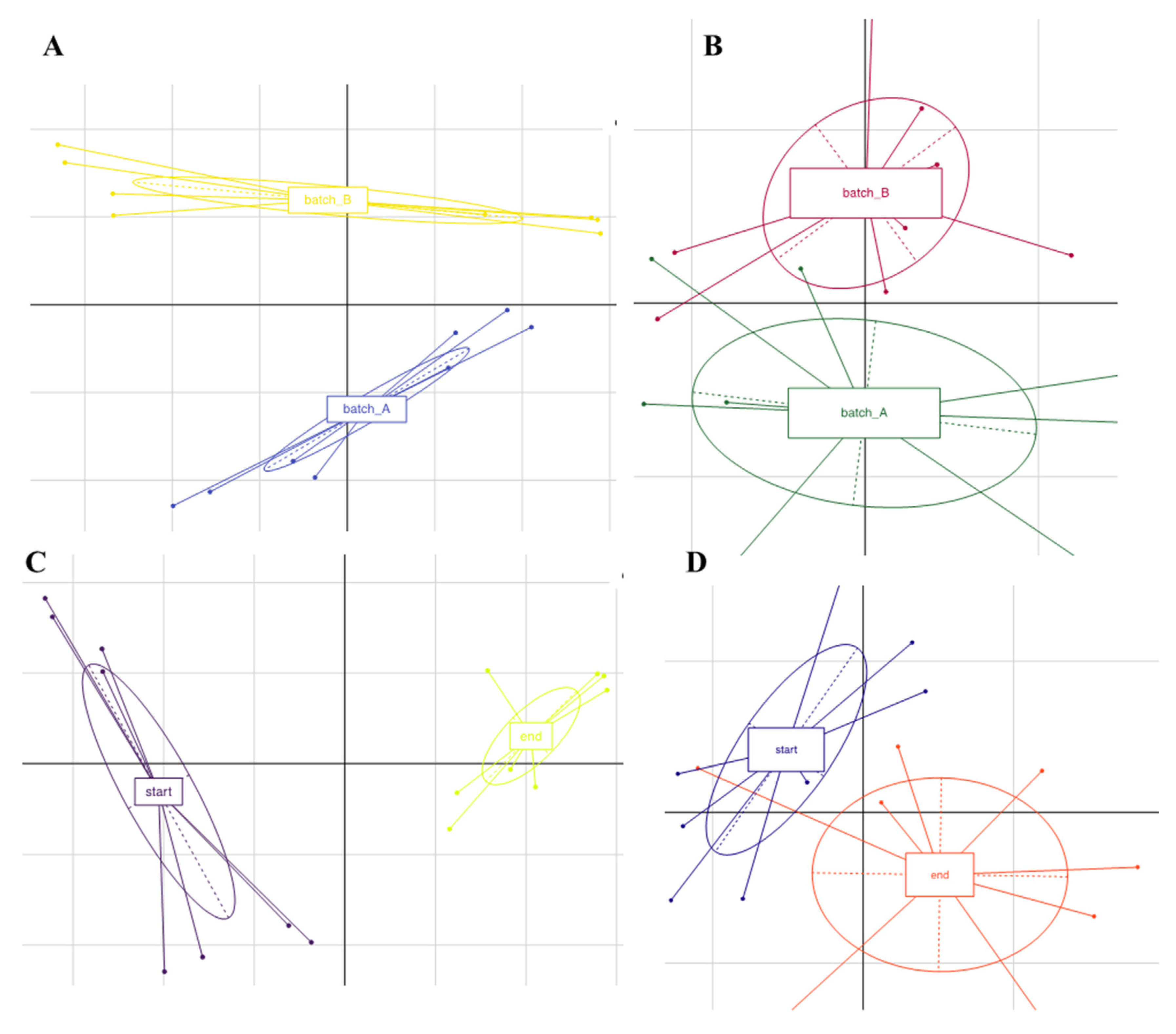
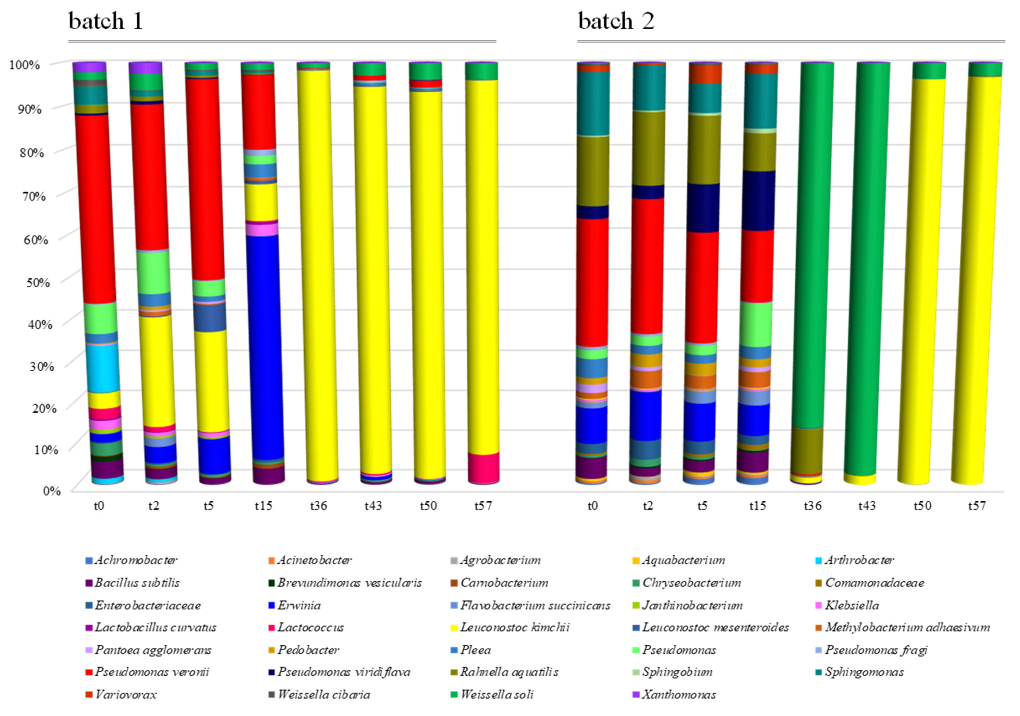
3.3. Microbiota Diversity

The total number of sequences used reach 94,353 reads, with an average value of 5585 reads/sample and a mean sequence length of 460 bp. Sample coverage calculation showed a satisfactory coverage for all the samples (>96%), whereas alpha diversity index did not show any significant difference as a function of the batch. Principal component analysis based on OTU table revealed a clear separation of the samples as a function of the batch (Figure 3, panel A). Samples analyzed at the beginning of the fermentation clustered together and were well separated from the samples at the end of this process (Figure 3, panel C). The microbiota composition at the highest taxonomic level (Figure 4) showed that, especially in batch 1, Leuconostoc kimchii was the dominant OTU after five days of fermentation (reaching almost 80% of the relative abundance up until the end of fermentation). Moreover, in batch 2, Leuconostoc kimchii was the dominant OTU only in the last two sampling points (reaching 90% of the relative abundance). Several spoilage microorganisms were identified in both the batches. In more detail, the presence of Erwinia spp. together with Pseudomonas veronii, Pseudomonas viridiflava, Rahnella aquatilis, and Sphingomonas spp. was observed during the first 15 days of fermentation. Erwinia spp. was predominant in batch 1 after 15 days of fermentation attesting at 28% of the relative abundance, whereas P. veronii was predominant in batch 1 in the first 15 days of fermentation (attesting at 25% and 16% of the relative abundance in batch 1 and 2, respectively). P. viridiflava, Sphingomonas spp., and R. aquatilis were predominant in batch 2 during the first 15 days of fermentation (with a relative abundance of 5%, 6% and 9%, respectively) (Figure 4). In addition, a high presence of Weissella soli was observed in batch 2 after 36 and 46 days of fermentation, reaching 90% of the relative abundance. Figure 5 shows the relative abundance of the sole lactic acid bacteria OTUs detected by sequencing in the two analyzed batches of kimchi.
By comparing the relative abundance of the OTUs between the beginning and the end of fermentation, only Weissella soli and Leuconostoc kimchii appeared to be associated with the end of fermentation (FDR < 0.05), whereas the other OTUs (including the minor fraction with <1% of the relative species abundance) were associated with the early step of fermentation (Figure 6).

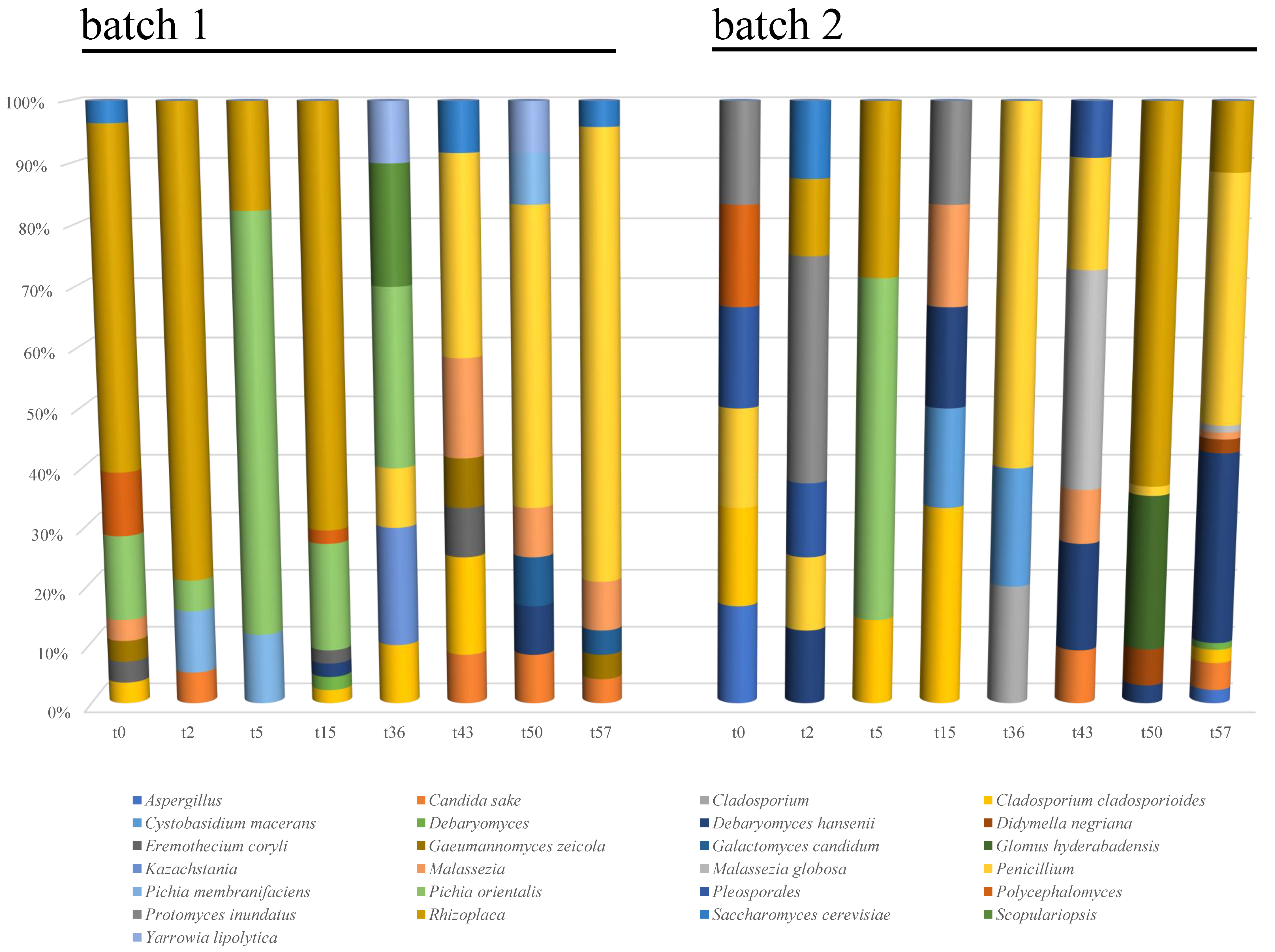
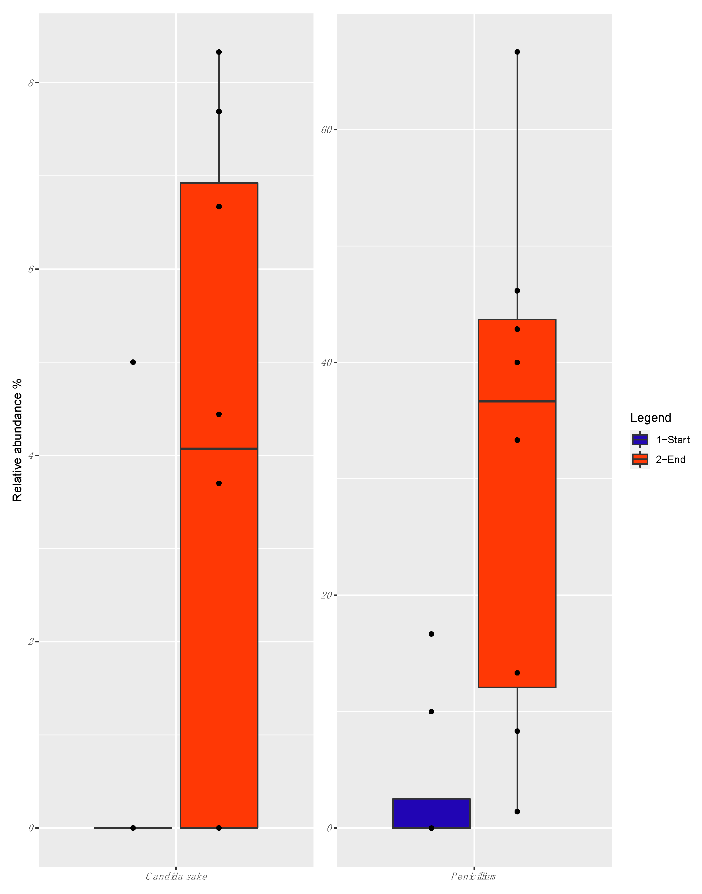
3.4. Mycobiota Diversity

The total number of sequences used for the downstream analysis reached 634,252 reads, with an average value of 40,910 reads/sample and a mean sequence length of 395 bp. Overall, a satisfactory sample coverage was showed by all the samples (>96%). Alpha diversity indexes were not significantly different according to time or batch.
PCA based on OTUs relative abundance of fungal taxa revealed two clusters separated according to batch and fermentation time (Figure 3, panel B and D, respectively).
The mycobiota composition at the highest taxonomic level (Figure 7) showed an evolution across time, in both the analyzed batches, with significant differences between them. In batch 1, Rhizoplaca and Pichia orientalis were the dominant OTUs in the first 15 days of fermentation (with approximately 65% and 17% of the relative abundance, respectively), whereas, at the end of fermentation, Penicillium, Candida sake, Malassezia, and Saccharomyces cerevisiae were the most abundant taxa, reaching 67, 8, 7, and 3% of the relative species abundance, respectively.
In batch 2, a stable occurrence of Penicillium spp. was detected, especially at the end of the fermentation (when it reached 40% of the relative abundance), together with Rhizoplaca spp. (58% and 11% of the relative abundance, after 50 and 57 days, respectively) (Figure 7). Protomyces inundatus was predominant after 2 days of fermentation, with 30% relative abundance, whereas Glomus hyderabadensis and Debaromyces hansenii were predominant after 50 and 57 days of fermentation, reaching 24% and 31% of the relative abundance, respectively. Malassezia and Candida sake were also detected as minority taxa (<10% of the relative abundance) at the end of fermentation.
When the relative abundance of the OTUs at the beginning and the end of the fermentation were compared, only Penicillium and Candida sake appeared to be associated with the end of fermentation (FDR < 0.05) (Figure 8).

4. Discussion

As reviewed by Chang [25], kimchi is produced through a spontaneous fermentation, generally lasting from a few weeks (e.g., four) to months (e.g., three); this fermentation process is mainly mediated by lactic acid bacteria naturally occurring in the raw materials and the production environment.
Regarding the samples of kimchi analyzed in the present study, pH values detected in both batches at t0 were in accordance with those reported for Korean kimchi at the beginning of fermentation, attesting between 5.0 and 5.4, depending on the raw material used [10,12]. However, a different evolution of this parameter was seen during the fermentation with respect to Korean kimchi, which reaches a pH of ~4.0 as early as 4 weeks after fermentation [10,26]. Indeed, in the samples herein analyzed, a pH value of about 4.0 was reached after 57 days of fermentation. It is noteworthy that the type of raw material and its natural contamination can strongly affect the evolution of pH during kimchi fermentation; in this regard, Song et al. [27] have recently reported different pH values for mature cabbage-, garlic-, ginger-, and red pepper-origin kimchi. Interestingly, the pH values detected in the kimchi manufacture under study were in accordance with those reported for cabbage-kimchi.
As a general trend, viable counts suggested the establishment of an active microbial community composed mainly of mesophilic aerobes and lactic acid bacteria; in contrast, progressively decreasing counts of Enterobacteriaceae, Pseudomonadaceae, and yeasts were seen during kimchi fermentation.
Regarding viable counts of total mesophilic aerobes, enumerated on growth media with or without the addition of 3% (w/v) NaCl, similar trends were observed between the two analyzed batches. In more detail, almost stable loads attesting at 5 Log cfu g−1 were seen during the whole fermentation period. The values detected at t0 were in accordance with those reported by Lee et al. [10] in the raw materials used for kimchi manufactured in Gyeong-gi province of Korea. By contrast, the counts of total mesophilic aerobes were lower than those reported in Korean kimchi by Hong et al. [28], attesting at >8 Log cfu g−1 after 9 days of fermentation. Interestingly, Hong et al. [28] reported a linear association between the fermentation temperature and loads of total mesophilic aerobes, with a slowdown of the microbial growth at 4 °C.
Regarding lactic acid bacteria counts, two different trends were seen in the two production batches, feasibly due to the differences occurring in the microbiota composition of the vegetables used as ingredients or the microbial interactions established during fermentation. In more detail, in batch 1 the load of lactobacilli progressively increased up until day 36, when it reached a maximum mean value of 6 Log cfu g−1. In batch 2, the load of lactobacilli remained almost stable during the early stage of fermentation (first 15 days), which was followed by a rapid increase. Markedly higher viable cell counts of lactobacilli, attesting at about 9 Log cfu g−1 were reported by Hong et al. [28] in a previous research dealing with the microbial dynamics of kimchi during its natural fermentation. Lactic acid bacteria counts detected in the present study were also lower that those reported by Lee et al. [10] in 88 kimchi manufactures sampled from six provinces (Chungcheong, Gangwon, Gyeonggi, Gyeongsang, Jeolla, and Jeju) in South Korea.
In accordance with lactic acid bacteria increase, in both production batches a progressive decrease of Enterobacteriaceae counts was seen, with a complete disappearance of the latter microorganisms at days 36 and 50 in batches 1 and 2, respectively. As previously found in numerous fermented vegetables, the acidity due to accumulation of organic acids produced by lactic acid bacteria was mainly responsible for the inhibition and death of Enterobacteriaceae [29,30,31,32]. Members of this family are acknowledged as indicators of process hygiene, hence, their load in foods should be carefully assessed [33].
Relatively high counts of Pseudomonadaceae were detected at early stages of fermentation (from day 0 to 15). This finding agrees well with the results reported by Park et al. [34] about the possible dominance of pseudomonads during the first stages of kimchi fermentation. The counts of Pseudomonadaceae were also in accordance with those reported by Wouters et al. [35] in raw materials used for the production of spontaneously fermented leek, attesting at about 5 Log cfu g−1 and progressively disappearing during the first week of fermentation.
Finally, viable counts of yeasts on Rose Bengal Agar without or with 3% (w/v) NaCl showed a similar decreasing trend in both production batches, with values below the limit of detection at the late stage of fermentation. Data herein obtained are in accordance with the occurrence of yeasts in kimchi already reported by Kim et al. [18], whereas a quite different picture emerged from the study of Jeong et al. [26], who reported an increase in the load of Saccharomyces spp. during fermentation of kimchi, from day 19 up until day 45. Moreover, Chang et al. [36] reported the isolation of yeasts such as Pichia spp. from mature kimchi. Since yeasts are halotolerant and acidophilic microorganisms that are frequently isolated from acidic fermented vegetables and brine [14], further research is needed to better elucidate the scarce occurrence of yeasts in the kimchi manufacture herein analyzed.
The results of metataxonomic analyses highlighted the presence of vegetable-associated bacteria with a progressive reduction of spoilage taxa counterbalanced by a progressive increase of pro-technological taxa mainly ascribed to the lactic acid bacteria group, which dominated from t36 to t57.
Regarding OTUs that initially characterized the analyzed kimchi, Pseudomonas species were detected in samples collected from both batches from t0 to t15. Pseudomonas species had already been detected by Lee et al. [8] in samples of Korean kimchi; moreover, as reported by Song et al. [27], pseudomonads were among the dominating taxa during the early fermentation of cabbage-origin Korean kimchi. Interestingly, in the present study, the decrease of the relative abundance of Pseudomonas spp. during fermentation was in agreement with the progressive decrease of viable counts of pseudomonads, thus confirming the profitability of combining culture-dependent with culture-independent methods. Regarding the occurrence of P. veronii, this species was first isolated from natural mineral waters [37] and more recently found as part of the core microbiota of fresh-cut produce processing facilities, together with P. viridiflava [38]. Moreover, both P. veronii and P. viridiflava were reported to be the causative agents of soft rot in carrot [39], thus suggesting carrots as a potential source of contamination by these two species in the analyzed kimchi.
During the early stage of kimchi fermentation (from t0 to t15), R. aquatilis was abundantly detected in samples from batch 2. This epiphytic bacterium can be frequently isolated from water, plant leaves and fruit, soil, foods as well as non-environmental samples such as blood, bronchial washings, wounds, and urine [40]. Rahnella species have already been detected in the microbiota of 25 cabbage-origin kimchi samples from traditional Korean temples that produce traditional temple style food [41]. These microorganisms were also detected in a large-scale metagenomics study carried out on 88 Korean kimchi sampled during fermentation [10], thus confirming the adaptation of this bacterial genus to the kimchi environment.
Regarding the presence of Erwinia spp., this bacterial genus, which is included in the Enterobacteriaceae family, was detected until t15. Erwinia encompasses species with cellulolytic and pectolytic activities that produce the soft rot of potato tubers [42]. The occurrence of Erwinia spp. in the early stage of fermentation of Korean white kimchi was already reported by Park et al. [43], whereas the same genus was also found by Park et al. [34] in 10 industrial-scale batches of Korean kimchi.
As for the presence of Sphingomonas spp., more than 100 species belonging to this bacterial genus have already been isolated from various environmental sources, including phyllosphere, rhizosphere, and plant roots [44]. Moreover, this genus was detected by Jung et al. [45] during the early stage of Korean kimchi fermentation [45].
Between t15 and t36, a clear shift in the relative abundance of the microbial species emerged, with an almost complete replacement of spoilage species with lactic acid bacteria, mainly represented by W. soli and L. kimchi. This finding suggests the establishment during fermentation of unfavorable environmental conditions for the survival of undesired bacteria.
Regarding W. soli, principally detected in samples from batch 2, this lactic acid bacteria species belonging to the Leuconostoccaceae family was first isolated from the soil environment by Magnusson et al. [46]. W. soli possesses a heterofermentative metabolism that, from glucose fermentation, produces lactic acid, CO2, ethanol, and/or acetate. Moreover, some Weissella strains, including Weissella cibaria, also detected in this study, were found to produce bacteriocins, such as weissellin, weissellicin 110, and weissellicin D, L., M and Y [47]. Although several Weissella species were abundantly detected in Korean kimchi [10], to the authors’ knowledge W. soli has rarely been isolated from this food product, where it was found as a minor species by Jeong et al. [26]. Although there is a lack of knowledge on the role of W. soli in kimchi, it is noteworthy that Weissella strains isolated from this fermented vegetable have recently shown potential prophylactic properties and probiotic features, being able to tolerate artificial gastric juice and bile salts and showing a high binding capacity for intestinal epithelial cells [48].
Leuconostoc kimchii was found to dominate in both analyzed batches at the end of fermentation (from t50 to t57). This microorganism was first isolated from kimchi manufactured in Korea, thus suggesting its strong adaptation to this fermented vegetable [49,50,51]. The use of L. kimchi strains as starter cultures for kimchi production was previously evaluated thanks to the potential health benefits for the consumers (e.g., lipid metabolism regulation) [51]. Interestingly, a recent study carried out on diet-induced obese mice reported that in animals administered with L. kimchii GJ2 isolated from Korean kimchi a significant decrease in hepatic triglyceride and fatty acid content was observed [52]. Moreover, Jo et al. [53] reported that kimchi fermented with the strain L. kimchii GJ2 provided efficient cholesterol-lowering effects in rats fed with a high-fat and high-cholesterol diet, thus suggesting the suitable use of this strain for the production of kimchi with functional features. Consistent with the well-known exopolysaccharides (EPS)-producing capabilities of leuconostocs, the production of dextran and levan with potential prebiotic activity by L. kimchi was reported by Schleifer [54] and Torres-Rodríguez et al. [55]. More recently, Rizzello et al. [56] have characterized the EPS production ability and other technological features of a L. kimchi strain for its exploitation as a starter culture for legumes fermentation, thus suggesting the potential suitability of this species for the manufacturing of novel vegetable-based fermented foods.
The metataxonomic analyses applied directly to the food matrix have allowed major and minor fungal taxa to be also detected, thus representing a further step toward the knowledge of the mycobiota of kimchi. As a general trend, Rhizoplaca spp. and P. orientalis were found to dominate the early stage of kimchi fermentation.
Rhizoplaca is a genus of foliose lichenized fungi (e.g., Rhizoplaca melanophthalma) in the family Lecanoraceae, including several morphologically distinct species that are geographically and ecologically widespread [57].
As for the presence of P. orientalis (Syn. Issatchenkia orientalis), yeasts in the genus Pichia were already isolated from mature kimchi characterized by low pH values [36]. In this regard, Pichia kluyveri was detected by Chang et al. [36] in Korean kimchi, whereas the osmotolerant yeast Pichia guilliermondii was detected in waste brine generated from kimchi production [58]. As reported by Chang et al. [36], the growth of P. orientalis is generally suppressed during kimchi fermentation, thus explaining its dominance in the sole early stage fermentation in both batches herein analyzed.
In the present study, Penicillium spp., S. cerevisiae, and C. sake were associated with the late stage of fermentation. However, the low load of eumycetes (<1 Log cfu g−1) found in both analyzed batches at the end of fermentation suggests the occurrence of the detected taxa at very low levels or in the state of viable but not culturable cells.
Interestingly, the occurrence of both Saccharomyces and Candida species has already been reported by Jeong et al. [26] during the late stage of fermentation of dongchimi, the traditional Korean watery kimchi. As suggested by Chang et al. [36], these yeasts can grow in kimchi at pH values between 4.0 and 7.0; moreover, white colony-forming yeasts, such as C. sake and Debaryomyces spp., were also detected in packed kimchi stored in refrigerated conditions [18], thus confirming their adaptation to cold environments.

5. Conclusions

Kimchi is one of the food products that best represent the tradition of the Korean cuisine and possess functional and health-promoting features related mainly to the metabolic activity of lactic acid bacteria. Based on the overall results herein collected, the non-Korean kimchi manufacture analyzed was characterized by microbial populations and dynamics that greatly overlapped those of more well-known Korean “relatives.” Indeed, typical kimchi-associated lactic acid bacteria species were surprisingly detected as dominant taxa in both batches analyzed, thus confirming their high adaptation to kimchi raw materials and production process. As already revealed by the available scientific literature, lactic acid bacteria isolated from kimchi can likely be used as starter cultures to produce novel functional fermented vegetables. As a matter of fact, lactic acid bacteria occurring in kimchi are well adapted to salty environments, and this feature can be extremely advantageous for their exploitation as starters for the fermentation of vegetables in brine. Given this premise, the knowledge gained in the present study represents a step forward in the description of the microbial dynamics of kimchi produced outside the region of origin using local ingredients. At this regard, the results collected compared to those available in the scientific literature for Korean kimchi manufactures seem to suggest a neatly higher effect of process parameters rather than the adventitious microbial populations on the shaping of mature kimchi microbiota. The evidences emerged from the present study will also serve as a starting point for further isolation of vegetable-adapted lactic acid bacteria to be assayed as potential starters for the manufacturing of novel vegetable preserves with high quality and functional traits. The present study also highlighted the presence of fungal taxa, whose contribution to the sensory traits of kimchi must be further investigated.

Author Contributions

Conceptualization, A.O. and L.A.; software, I.F.; formal analysis, A.M., I.F., V.M., M.R.C., D.O., C.B., G.T., L.B., F.C. and R.M.; investigation, A.O.; resources, A.O. and L.A.; data curation, A.M.; writing—original draft preparation, L.A., A.O. and A.M.; writing—review and editing, A.O. and L.A.; funding, L.A.; visualization, L.C. All authors have read and agreed to the published version of the manuscript.

Funding

This research was funded by the Marche Region under the PSR2014–2020 Progamme “Misura 16.1–Sostegno per la costituzione e la gestione dei gruppi operativi del PEI in materia di produttività e sostenibilità dell’agricoltura Azione 2-Fase di gestione del G.O. e realizzazione del Piano di Attività” project: “New vegetable organic preserves from autochthonous sea fennel organic crop (Crithmum maritimum L.)” and the POR Marche FSE 2014/2020 Progetto “Dottorato Innovativo”–Borse di studio per Dottorato di ricerca per l’innovazione del sistema regionale-Edizione Anno Accademico 2018/2019.

Acknowledgments

The authors wish to thank Lee Joo Hee of Il Torrione Korean cuisine restaurant, Santa Maria Nuova (AN), Italy for his valuable support for production and providing of kimchi

Conflicts of Interest

The authors declare no conflict of interest.

References

  1. Misihairabgwi, J.; Cheikhyoussef, A. Traditional fermented foods and beverages of Namibia. J. Ethn. Foods 2017, 4, 145–153. [Google Scholar] [CrossRef]
  2. Ramos, C.L.; Schwan, R.F. Technological and nutritional aspects of indigenous Latin America fermented foods. Curr. Opin. Food Sci. 2017, 13, 97–102. [Google Scholar] [CrossRef]
  3. Majcherczyk, J.; Surówka, K. Effects of onion or caraway on the formation of biogenic amines during sauerkraut fermentation and refrigerated storage. Food Chem. 2019, 298, 125083. [Google Scholar] [CrossRef] [PubMed]
  4. Jaehae, L.; Kyeongsoon, H.; Chaelin, P.; Ilgwon, K.; Jeongwon, K.; Dukno, Y.; Montanari, M.; Naomichi, I.; Cwiertka, K.J.; Younghee, S.; et al. Humanistic Understanding of Kimchi and Kimjang Culture, 1st ed.; World Institute of Kimchi: Gwangju, Korea, 2015. [Google Scholar]
  5. Patra, J.K.; Das, G.; Paramithiotis, S.; Shin, H.S. Kimchi and other widely consumed traditional fermented foods of Korea: A review. Front. Microbiol. 2016, 7, 1493. [Google Scholar] [CrossRef] [PubMed]
  6. Jang, D.-J.; Chung, K.R.; Yang, H.J.; Kim, K.-S.; Kwon, D.Y. Discussion on the origin of kimchi, representative of Korean unique fermented vegetables. J. Ethn. Foods 2015, 2, 126–136. [Google Scholar] [CrossRef] [Green Version]
  7. Jung, J.Y.; Lee, S.H.; Jeon, C.O. Kimchi microflora: History, current status, and perspectives for industrial kimchi production. App. Microbiol. Biotechnol. 2014, 98, 2385–2393. [Google Scholar] [CrossRef]
  8. Lee, H.-W.; Yoon, S.-R.; Kim, S.-J.; Lee, H.M.; Lee, J.Y.; Lee, J.-H.; Kim, S.H.; Ha, J.-H. Identification of microbial communities, with a focus on foodborne pathogens, during kimchi manufacturing process using culture-independent and -dependent analyses. LWT Food Sci. Technol. 2017, 81, 153–159. [Google Scholar] [CrossRef]
  9. Chung, H.-K.; Shin, D.; Chung, K.R.; Choi, S.Y.; Woo, N. Recovering the royal cuisine in Chosun Dynasty and its esthetics. J. Ethn. Foods 2017, 4, 242–253. [Google Scholar] [CrossRef]
  10. Lee, M.; Song, J.H.; Jung, M.Y.; Lee, S.H.; Chang, J.Y. Large-scale targeted metagenomics analysis of bacterial ecological changes in 88 kimchi samples during fermentation. Food Microbiol. 2017b, 66, 173–183. [Google Scholar] [CrossRef]
  11. Cheigh, H. Production, characteristics and health functions of kimchi. Acta Hortic. 1999, 483, 405–420. [Google Scholar] [CrossRef]
  12. Jung, J.Y.; Lee, S.H.; Kim, J.M.; Park, M.S.; Bae, J.-W.; Hahn, Y.; Madsen, E.L.; Jeon, C.O. Metagenomic analysis of kimchi, a traditional Korean fermented food. App. Environ. Microbiol. 2011, 77, 2264–2274. [Google Scholar] [CrossRef] [Green Version]
  13. Lee, M.-E.; Jang, J.-Y.; Lee, J.-H.; Park, H.-W.; Choi, H.-J.; Kim, T.-W. Starter cultures for kimchi fermentation. J. Microbiol. Biotechnol. 2015, 25, 559–568. [Google Scholar] [CrossRef]
  14. Torres, S.; Verón, H.; Contreras, L.; Isla, M.I. An overview of plant-autochthonous microorganisms and fermented vegetable foods. Food Sci. Human Wellness 2020, 9, 112–123. [Google Scholar] [CrossRef]
  15. Chang, J.-H.; Shim, Y.Y.; Cha, S.-K.; Chee, K.M. Probiotic characteristics of lactic acid bacteria isolated from kimchi. J. Appl. Microbiol. 2010, 109, 220–230. [Google Scholar] [CrossRef] [PubMed]
  16. Lee, N.-K.; Han, K.J.; Son, S.-H.; Eom, S.J.; Lee, S.-K.; Paik, H.-D. Multifunctional effect of probiotic Lactococcus lactis KC24 isolated from kimchi. LWT Food Sci. Technol. 2015b, 64, 1036–1041. [Google Scholar] [CrossRef]
  17. Lee, K.-H.; Bong, Y.-J.; Lee, H.A.; Kim, H.-Y.; Park, K.-Y. Probiotic effects of Lactobacillus plantarum and Leuconostoc mesenteroides isolated from kimchi. J. Korean Soc. Food Sci. Nutr. 2016, 45, 12–19. [Google Scholar] [CrossRef]
  18. Kim, M.-J.; Lee, H.-W.; Kim, J.Y.; Kang, S.E.; Roh, S.W.; Hong, S.W.; Yoo, S.R.; Kim, T.-W. Impact of fermentation conditions on the diversity of white colony-forming yeast and analysis of metabolite changes by white colony-forming yeast in kimchi. Food Res. Int. 2020, 136, 109315. [Google Scholar] [CrossRef] [PubMed]
  19. Belleggia, L.; Milanović, V.; Ferrocino, I.; Cocolin, L.; Naceur Haouet, M.; Scuota, S.; Maoloni, A.; Garofalo, C.; Cardinali, F.; Aquilanti, L.; et al. Is there any still undisclosed biodiversity in Ciauscolo salami? A new glance into the microbiota of an artisan production as revealed by high-throughput sequencing. Meat Sci. 2020, 165, 108128. [Google Scholar] [CrossRef] [Green Version]
  20. Haouet, M.N.; Altissimi, M.S.; Mercuri, M.L.; Baldassarri, C.; Osimani, A.; Clementi, F.; Ortenzi, R. Evaluation of the safety of Milano-type dry fermented sausages produced by a fast drying technology. Ital. J. Food Sci. 2017, 29, 1–9. [Google Scholar]
  21. Garofalo, C.; Bancalari, E.; Milanović, V.; Cardinali, F.; Osimani, A.; Sardaro, M.L.S.; Bottari, B.; Bernini, V.; Aquilanti, L.; Clementi, F.; et al. Study of the bacterial diversity of foods: PCR-DGGE versus LH-PCR. Int. J. Food Microbiol. 2017, 242, 24–36. [Google Scholar] [CrossRef]
  22. Klindworth, A.; Pruesse, E.; Schweer, T.; Peplies, J.; Quast, C.; Horn, M.; Glöckner, F.O. Evaluation of general 16S ribosomal RNA gene PCR primers for classical and next-generation sequencing-based diversity studies. Nucleic Acids Res. 2013, 41, e1. [Google Scholar] [CrossRef] [PubMed]
  23. Mota-Gutierrez, J.; Ferrocino, I.; Rantsiou, K.; Cocolin, L. Metataxonomic comparison between internal transcribed spacer and 26S ribosomal large subunit (LSU) rDNA gene. Int. J. Food Microbiol. 2019, 290, 132–140. [Google Scholar] [CrossRef] [PubMed] [Green Version]
  24. Caporaso, J.G.; Kuczynski, J.; Stombaugh, J.; Bittinger, K.; Bushman, F.D.; Costello, E.K.; Fierer, N.; Peña, A.G.; Goodrich, J.K.; Gordon, J.I.; et al. QIIME allows analysis of highthroughput community sequencing data. Nat. Methods 2010, 7, 335–336. [Google Scholar] [CrossRef] [PubMed] [Green Version]
  25. Chang, H.C. Healthy and safe Korean traditional fermented foods: Kimchi and chongkukjang. J. Ethn. Foods 2018, 5, 161–166. [Google Scholar] [CrossRef]
  26. Jeong, S.H.; Jung, J.Y.; Lee, S.H.; Jin, H.M.; Jeon, C.O. Microbial succession and metabolite changes during fermentation of dongchimi, traditional Korean watery kimchi. Int. J. Food Microbiol. 2013, 164, 46–53. [Google Scholar] [CrossRef]
  27. Song, H.S.; Whon, T.W.; Kim, J.; Lee, S.H.; Kim, J.Y.; Kim, Y.B.; Choi, H.-J.; Rhee, J.-K.; Roh, S.W. Microbial niches in raw ingredients determine microbial community assembly during kimchi fermentation. Food Chem. 2020, 318, 126481. [Google Scholar] [CrossRef]
  28. Hong, Y.; Yang, H.-S.; Chang, H.-C.; Kim, H.-Y. Comparison of bacterial community changes in fermenting kimchi at two different temperatures using a denaturing gradient gel electrophoresis analysis. J. Microbiol. Biotechnol. 2013, 23, 76–84. [Google Scholar] [CrossRef] [Green Version]
  29. Yamani, M.I. Fermentation of brined tunrnip roots using Lactobacillus plantarum and Leuconostoc mesenteroides starter cultures. World J. Microbiol. Biotechnol. 1993, 9, 176–179. [Google Scholar] [CrossRef]
  30. Hurtado, A.; Reguant, C.; Esteve-Zarzoso, B.; Bordons, A.; Rozès, N. Microbial population dynamics during the processing of Arbequina table olives. Food Res. Int. 2008, 41, 738–744. [Google Scholar] [CrossRef]
  31. Tamang, B.; Tamang, J.P. In situ fermentation dynamics during production of gundruk and khalpi, ethnic fermented vegetables products of the Himalayas. Indian J. Microbiol. 2010, 50, 93–98. [Google Scholar] [CrossRef] [Green Version]
  32. Wouters, D.; Bernaert, N.; Conjaerts, W.; Van Droogenbroeck, B.; De Loose, M.; De Vuyst, L. Species diversity, community dynamics, and metabolite kinetics of spontaneous leek fermentations. Food Microbiol. 2013, 33, 185–196. [Google Scholar] [CrossRef]
  33. Osimani, A.; Milanović, V.; Cardinali, F.; Garofalo, C.; Clementi, F.; Pasquini, M.; Riolo, P.; Ruschioni, S.; Isidoro, N.; Loreto, N.; et al. The bacterial biota of laboratory-reared edible mealworms (Tenebrio molitor L.): From feed to frass. Int. J. Food Microbiol. 2018, 272, 49–60. [Google Scholar] [CrossRef]
  34. Park, E.-J.; Chun, J.; Cha, C.-J.; Park, W.-S.; Jeon, C.O.; Bae, J.-W. Bacterial community analysis during fermentation of ten representative kinds of kimchi with barcoded pyrosequencing. Food Microbiol. 2012, 30, 197–204. [Google Scholar] [CrossRef]
  35. Wouters, D.; Grosu-Tudor, S.; Zamfir, M.; De Vuyst, L. Bacterial community dynamics, lactic acid bacteria species divesity and metabolite kinetics of traditional Romanian vegetable fermentations. J. Sci. Food Agric. 2013, 93, 749–760. [Google Scholar] [CrossRef]
  36. Chang, H.-W.; Kim, K.-H.; Nam, Y.-D.; Roh, S.W.; Kim, M.-S.; Jeon, C.O.; Oh, H.-M.; Bae, J.-W. Analysis of yeast and archaeal population dynamics in kimchi using denaturing gradient gel electrophoresis. Int. J. Food Microbiol. 2008, 126, 159–166. [Google Scholar] [CrossRef] [PubMed]
  37. Elomari, M.; Coroler, L.; Hoste, B.; Gillis, M.; Izard, D.; Leclerc, H. DNA relatedness among Pseudomonas strains isolated from natural mineral waters and proposal of Pseudomonas veronii sp. nov. Int. J. Syst. Evol. Microbiol. 1996, 46, 1138–1144. [Google Scholar] [CrossRef] [PubMed] [Green Version]
  38. Gu, G.; Ottesen, A.; Bolten, S.; Wang, L.; Luo, Y.; Rideout, S.; Lyu, S.; Nou, X. Impact of routine sanitation on the microbiomes in a fresh produce processing facility. Int. J. Food Microbiol. 2019, 294, 31–41. [Google Scholar] [CrossRef] [PubMed] [Green Version]
  39. Kahala, M.; Blasco, L.; Joutsjoki, V. Molecular characterization of spoilage bacteria as a means to observe the microbiological quality of carrot. J. Food Prot. 2012, 75, 523–532. [Google Scholar] [CrossRef] [PubMed]
  40. Calvo, J.; Calvente, V.; de Orellano, M.E.; Benuzzi, D.; Sanz de Tosetti, M.I. Biological control of postharvest spoilage caused by Penicillium expansum and Botrytis cinerea in apple by using the bacterium Rahnella aquatilis. Int. J. Food Microbiol. 2007, 113, 251–257. [Google Scholar] [CrossRef]
  41. Lee, M.; Song, J.H.; Park, J.M.; Chang, J.Y. Bacterial diversity in Korean temple kimchi fermentation. Food Res. Int. 2019, 126, 108592. [Google Scholar] [CrossRef]
  42. Ragaert, P.; Devlieghere, F.; Debevere, J. Role of microbiological and physiological spoilage mechanisms during storage of minimally processed vegetables. Postharvest Biol. Technol. 2017, 44, 185–194. [Google Scholar] [CrossRef]
  43. Park, S.; Ji, Y.; Park, H.; Lee, K.; Park, H.; Beck, B.R.; Shin, H.; Holzapfel, W.H. Evaluation of functional properties of lactobacilli isolated from Korean white kimchi. Food Control. 2016, 69, 5–12. [Google Scholar] [CrossRef]
  44. Menon, R.R.; Kumari, S.; Kumar, P.; Verma, A.; Krishnamurthi, S.; Rameshkumar, N. Sphingomonas pokkalii sp. nov., a novel plant associated rhizobacterium isolated from a saline tolerant pokkali rice and its draft genome analysis. Syst. App. Microbiol. 2019, 42, 334–342. [Google Scholar] [CrossRef]
  45. Jung, M.Y.; Kim, T.-W.; Lee, C.; Kim, J.Y.; Song, H.S.; Kim, Y.B.; Ahn, S.W.; Kim, J.S.; Roh, S.W.; Lee, S.H. Role of jeotgal, a Korean traditional fermented fish sauce, in microbial dynamics and metabolite profiles during kimchi fermentation. Food Chem. 2018, 265, 135–143. [Google Scholar] [CrossRef] [PubMed]
  46. Magnusson, J.; Jonsson, H.; Schnürer, J.; Roos, S. Weissella soli sp. nov., a lactic acid bacterium isolated from soil. Int. J. Syst. Evol. Microbiol. 2002, 52, 831–834. [Google Scholar] [CrossRef] [PubMed]
  47. Kim, E.; Cho, Y.; Lee, Y.; Han, S.-K.; Kim, C.-G.; Choo, D.-W.; Kim, Y.-R.; Kim, H.-Y. A proteomic approach for rapid identification of Weissella species isolated from Korean fermented foods on MALDI-TOF MS supplemented with an in-house database. Int. J. Food Microbiol. 2017, 243, 9–15. [Google Scholar] [CrossRef]
  48. Yu, H.-S.; Jang, H.J.; Lee, N.-K.; Paik, H.-D. Evaluation of the probiotic characteristics and prophylactic potential of Weissella cibaria strains isolated from kimchi. LWT 2019, 112, 108229. [Google Scholar] [CrossRef]
  49. Oh, H.-M.; Cho, Y.-J.; Kim, B.K.; Roe, J.-H.; Kang, S.-O.; Nahm, B.H.; Jeong, G.; Han, H.-U.; Chun, J. Complete genome sequence analysis of Leuconostoc kimchii IMSNU 11154. J. Bacteriol. 2010, 192, 3844–3845. [Google Scholar] [CrossRef] [Green Version]
  50. Lee, S.H.; Jung, J.Y.; Lee, S.H.; Jeon, C.O. Complete genome sequence of Leuconostoc kimchii strain C2, isolated from Kimchi. J. Bacteriol. 2011, 193, 5548. [Google Scholar] [CrossRef] [Green Version]
  51. Jung, J.Y.; Jeong, J.-W.; Lee, S.-Y.; Jin, H.M.; Choi, H.W.; Ryu, B.-G.; Han, S.-S.; Kang, H.K.; Chung, E.J.; Choi, K.-M. Complete genome sequence of Leuconostoc kimchii strain NKJ218, isolated from homemade kimchi. Microbiol. Resour. Announc. 2019, 8, e00367-19. [Google Scholar] [CrossRef] [Green Version]
  52. Choi, B.-R.; Kwon, E.-Y.; Kim, H.-J.; Choi, M.-S. Role of synbiotics containing D-allulose in the alteration of body fat and hepatic lipids in diet-induced obese mice. Nutrients 2018, 10, 1797. [Google Scholar] [CrossRef] [PubMed] [Green Version]
  53. Jo, S.Y.; Choi, E.A.; Lee, J.J.; Chang, H.C. Characterization of starter kimchi fermented with Leuconostoc kimchii GJ2 and its cholesterol-lowering effects in rats fed a high-fat and high-cholesterol diet. J. Sci. Food Agric. 2015, 95, 2750–2756. [Google Scholar] [CrossRef] [PubMed]
  54. Schleifer, K.H. Leuconostocaceae fam. nov. Bergey’s Man. Syst. Archaea Bact. 2019. [Google Scholar] [CrossRef]
  55. Torres-Rodríguez, I.; Rodríguez-Alegría, M.E.; Miranda-Molina, A.; Giles-Gómez, M.; Conca Morales, R.; López-Munguía, A.; Bolívar, F.; Escalante, A. Screening and characterization of extracellular polysaccharides produced by Leuconostoc kimchii isolated from traditional fermented pulque beverage. Springerplus 2014, 3, 583. [Google Scholar] [CrossRef] [Green Version]
  56. Rizzello, C.G.; Coda, R.; Wang, Y.; Verni, M.; Kajala, I.; Katina, K.; Laitila, A. Characterization of indigenous Pediococcus pentosaceus, Leuconostoc kimchii, Weissella cibaria and Weissella confusa for faba bean bioprocessing. Int. J. Food Microbiol. 2019, 302, 24–34. [Google Scholar] [CrossRef]
  57. Leavitt, S.D.; Fankhauser, J.D.; Leavitt, D.H.; Porter, L.D.; Johnson, L.A.; St. Clair, L.L. Complex patterns of speciation in cosmopolitan “rock posy” lichens–Discovering and delimiting cryptic fungal species in the lichen-forming Rhizoplaca melanophthalma species-complex (Lecanoraceae, Ascomycota). Mol. Phylogenetics Evol. 2011, 59, 587–602. [Google Scholar] [CrossRef]
  58. Choi, M.H.; Park, Y.H. Growth of Pichia guilliermondii A9, an osmotolerant yeast, in waste brine generated from kimchi production. Bioresour. Technol. 1999, 70, 231–236. [Google Scholar] [CrossRef]
Figure 1. Ready-to-eat cabbage-origin kimchi.
Foods 09 01568 g001
Figure 2. Results of pH measurements of two kimchi manufactures (batch 1 and batch 2) during fermentation. Values are expressed as means ± standard deviation. For each sampling time, means with different letters are significantly different (p < 0.05).
Foods 09 01568 g002
Figure 3. Principal Component Analysis (PCA) based on Operational Taxonomic Units (OTUs) relative abundance of kimchi samples for bacteria (panel A) and fungi (panel B) grouped according to the batch, or according to: (i) the fermentation period; (ii) beginning of fermentation: t0, t2, t5, t15; (iii) end of fermentation: t36, t43, t50, t57, for bacteria (panel C) and fungi (panel D).
Foods 09 01568 g003
Figure 4. Relative abundance of bacterial Operational Taxonomic Units (OTUs) detected by sequencing in the analyzed kimchi batches. Samples are grouped according to batch (1 and 2) and labeled according to fermentation time.
Foods 09 01568 g004
Figure 5. Relative abundance of lactic acid bacteria Operational Taxonomic Units (OTUs) detected by sequencing in the analyzed kimchi batches. Samples are grouped according to batch (1 and 2) and labeled according to fermentation time.
Foods 09 01568 g005
Figure 6. Boxplots showing the relative abundance of bacterial Operational Taxonomic Units (OTUs) between the beginning of fermentation: t0, t2, t5, t15, and the end of fermentation: t36, t43, t50, t57. Boxes represent the interquartile range (IQR) between the first and third quartiles, and the line inside represents the median (2nd quartile).
Foods 09 01568 g006
Figure 7. Relative abundance of fungal Operational Taxonomic Units (OTUs) detected by sequencing in the analyzed kimchi batches. Samples are grouped according to batch (1 and 2) and labeled according to fermentation time.
Foods 09 01568 g007
Figure 8. Boxplots showing the relative abundance of fungal Operational Taxonomic Units (OTUs) between the beginning of fermentation: t0, t2, t5, t15, and the end of fermentation: t36, t43, t50, t57. Boxes represent the interquartile range (IQR) between the first and third quartiles, and the line inside represents the median (2nd quartile).
Foods 09 01568 g008
Table 1. Results of viable counting (Log cfu g−1) of bacteria and eumycetes in kimchi during fermentation.
BatchSampling Time (Days)Mesophilic Aerobic BacteriaHalophilic Mesophilic Aerobic BacteriaPresumptive LactobacilliPresumptive Halophilic LactobacilliPresumptive LactococciPresumptive Halophilic LactococciEnterobacteriaceaeYeastsHalophilic YeastsPseudomonadaceae
1t05.2 ± 0.1 a5.3 ± 0.1 a3.5 ± 0.1 b3.5 ± 0.1 b5.0 ± 0.1 a4.9 ± 0.0 a1.7 ± 0.3 b1.3 ± 0.0 a<1.0 b4.5 ± 0.2 a
t25.3 ± 0.0 a5.3 ± 0.1 a3.6 ± 0.5 b3.5 ± 0.5 b5.0 ± 0.1 a5.1 ± 0.1 a2.3 ± 0.6 b1.4 ± 0.1 a1.4 ± 0.1 a5.2 ± 0.1 a
t55.2 ± 0.0 a5.2 ± 0.0 a4.2 ± 0.0 b4.2 ± 0.0 b5.0 ± 0.1 a5.1 ± 0.2 a3.5 ± 0.5 a1.1 ± 0.1 a1.2 ± 0.3 a4.8 ± 0.3 a
t155.3 ± 0.1 a5.3 ± 0.0 a4.8 ± 0.2 b4.8 ± 0.1 b5.2 ± 0.0 a4.8 ± 0.1 a3.4 ± 0.2 a<1.0 b<1.0 b5.1 ± 0.1 a
t366.3 ± 0.1 a5.9 ± 0.2 a6.3 ± 0.0 a6.2 ± 0.2 a5.0 ± 0.2 a4.9 ± 0.2 a<1.0 c<1.0 b<1.0 b<1.0 b
t435.6 ± 0.0 a5.3 ± 0.0 a6.3 ± 0.1 a6.4 ± 0.1 a5.0 ± 0.2 a5.1 ± 0.1 a<1.0 c<1.0 b<1.0 b<1.0 b
t505.5 ± 0.0 a5.4 ± 0.0 a6.2 ± 0.2 a5.6 ± 0.1 a4.9 ± 0.3 a5.0 ± 0.1 a<1.0 c<1.0 b<1.0 b<1.0 b
t575.4 ± 0.0 a5.3 ± 0.0 a4.6 ± 0.1 b4.7 ± 0.1 b5.0 ± 0.2 a5.0 ± 0.1 a<1.0 c<1.0 b<1.0 b<1.0 b
2t05.7 ± 0.3 a5.6 ± 0.2 a2.1 ± 0.2 c2.4 ± 0.0 b5.5 ± 0.3 a5.4 ± 0.1 a2.8 ± 0.2 a2.5 ± 0.1 a1.9 ± 0.0 a4.9 ± 0.1 a
t25.3 ± 0.1 a5.3 ± 0.0 a1.5 ± 0.1 c1.4 ± 0.2 b5.2 ± 0.1 a5.1 ± 0.2 a2.0 ± 0.2 a2.1 ± 0.0 a1.7 ± 0.0 a4.2 ± 0.0 a
t55.2 ± 0.1 a5.2 ± 0.1 a1.2 ± 0.0 c1.2 ± 0.1 b5.0 ± 0.1 a4.8 ± 0.1 a1.8 ± 0.1 a1.5 ± 0.2 b1.0 ± 0.2 b4.9 ± 0.0 a
t155.1 ± 0.1 a4.8 ± 0.1 a2.0 ± 0.1 c1.5 ± 0.1 b4.9 ± 0.0 a4.4 ± 0.0 a2.4 ± 0.3 a1.0 ± 0.0 b< 1.0 b4.9 ± 0.0 a
t365.1 ± 0.1 a5.1 ± 0.2 a4.8 ± 0.7 b2.5 ± 0.8 b4.8 ± 0.0 a4.3 ± 0.4 a1.6 ± 0.6 a<1.0 c<1.0 b<1.0 b
t434.9 ± 0.0 a4.8 ± 0.2 a7.3 ± 0.2 a4.2 ± 0.3 a4.5 ± 0.3 a4.4 ± 0.3 a<1.0 c<1.0 c<1.0 b<1.0 b
t504.9 ± 0.0 a4.7 ± 0.0 a7.3 ± 0.0 a5.0 ± 0.4 a4.6 ± 0.1 a4.6 ± 0.0 a<1.0 c<1.0 c<1.0 b<1.0 b
t574.9 ± 0.1 a4.9 ± 0.1 a6.4 ± 0.5 a4.3 ± 0.1 a4.2 ± 0.1 a4.2 ± 0.1 a<1.0 c<1.0 c<1.0 b<1.0 b
Note: cfu, colony-forming units; Values are expressed as means ± standard deviation; For each batch, within each column, means with different superscript letters are significantly different (p < 0.05).
Publisher’s Note: MDPI stays neutral with regard to jurisdictional claims in published maps and institutional affiliations.

