Article

Antibiofilm Activity of Amomum tsaoko Essential Oil on Staphylococcus aureus and Its Application in Pork Preservation

School of Life Sciences, Shanghai University, Shanghai 200444, China
*
Authors to whom correspondence should be addressed.
Foods 2025, 14(4), 662; https://doi.org/10.3390/foods14040662
Submission received: 15 January 2025 / Revised: 31 January 2025 / Accepted: 13 February 2025 / Published: 15 February 2025
(This article belongs to the Section Food Packaging and Preservation)

Abstract

:
Staphylococcus aureus (S. aureus) can contaminate food by forming biofilms, leading to significant food safety concerns. Amomum tsaoko essential oil (AEO) has been shown to be an effective plant-derived antibacterial agent. This study investigated the antibiofilm activity of AEO and evaluated its potential benefit in pork preservation. The results showed that AEO solution (2 mg/mL) can effectively remove the biofilm of S. aureus on food contact materials, achieving a removal rate of over 90%. Scanning electron microscopy (SEM) revealed that the S. aureus biofilm structure was disrupted after treatment with AEO. Meanwhile, AEO treatment significantly reduced the initial formation of S. aureus biofilms and extracellular polymeric substance (EPS) production. In addition, AEO down-regulated the expression of key biofilm-associated genes, including icaA, icaB, agrA, cidA, cidB, and cidC, thereby regulating formation. AEO also exhibited significant antibiofilm activity in pork preservation, effectively controlling key indicators associated with pork spoilage. This study revealed the potential of AEO in food preservation, demonstrating its ability to disrupt S. aureus biofilms by inhibiting initial formation, reducing the release of EPS secretion, and regulating the expression of biofilm-associated genes.

1. Introduction

Staphylococcus aureus (S. aureus) is a foodborne pathogen that not only causes food contamination but also poses potential risks to human health. It is frequently found in protein-rich foods like poultry products, dairy products, and pastry products, contributing to food contamination [1,2,3,4]. The bacterium S. aureus can survive in food and even adhere to surfaces of food processing equipment, such as stainless steel, glass, and plastic, where it forms biofilms [5]. Biofilms consist of a dense network structure of self-produced autocrine EPSs [6]. Compared to planktonic bacteria, biofilm-forming cells are more resistant to removal. Biofilm has become a problematic matter across various food industries, such as beer production, fish processing, and pork production [7,8]. Consequently, reducing the hazards posed by biofilms has become an important goal for the food industry.
Methods to prevent biofilm contamination include physical sterilization and chemical preservatives [9,10]. Nevertheless, in everyday applications, physical sterilization methods are not appropriate for most types of foods because of the significant equipment requirements. Additionally, the environmental issues and the potential for bacterial resistance linked to the use of chemical preservatives to combat biofilm cannot be overlooked [11]. Therefore, environmentally friendly plant extracts are gaining recognition as promising antibiofilm agents [12,13]. Alongside certain polysaccharides and polyphenols extracted from plants, the antibiofilm activity of plant essential oils (EOs) has been widely studied [14]. EOs are intricate blends of volatile compounds, predominantly obtained through hydrodistillation from aromatic plants [15]. They mainly consist of terpenoids, hydrocarbons and phenols, which frequently demonstrate potent antibacterial and antioxidant activities [16,17]. Previous studies have found that oregano and Litsea cubeba can significantly reduce the amount of S. aureus [18,19], while cinnamon essential oil can significantly inhibit Pseudomonas aeruginosa [20]. EOs have been identified as a potential natural preservative source, and have garnered significant attention from researchers.
Amomum tsaoko essential oil (AEO) is a viscous liquid derived from the fruit of Amomum tsaoko, which is commonly found in southwest China and is recognized as a popular commercial spice utilized as a food additive [21]. AEO has demonstrated high efficiency against various foodborne pathogens and spoilage microorganisms. AEO exhibits inhibitory effects against both Gram-positive bacteria and Gram-negative bacteria [22]. The antibacterial activity of AEO against bacteria is primarily attributed to its ability to disrupt cell membrane integrity and alter cellular morphology. The leakage of alkaline phosphatase from Listeria monocytogenes treated with AEO indicates that AEO can also disrupt the bacterial cell wall structure [23]. Our previous research identified more than 100 components of AEO through GC-MS, such as 1,8-cineole, α-pinene, (E)-dec-2-enal, and citral, which exhibited significant inhibitory effects against S. aureus [24]. Notably, AEO demonstrates a notable ability to inhibit S. aureus at 0.20 mg/mL, proving more effective than other essential oils [25]. While some studies have examined the antibacterial properties of AEO, its potential antibiofilm activity remain unclear, and the application potential of AEO in food preservation still needs to be further investigated.
This study determined the efficacy of AEO in removing S. aureus biofilm. Additionally, the mechanism of antibiofilm activity of AEO was explored by analyzing changes in micromorphology, initial adhesion, EPS content, and the expression of genes associated with biofilm formation in S. aureus. Finally, AEO was applied to fresh pork to assess its ability to delay spoilage. This study aims to provide valuable knowledge regarding the potential application of AEO and the creation of novel antibiofilm strategies in food preservation.

2. Materials and Methods

2.1. Chemicals and Bacterial Culture

Anhydrous sodium sulfate, dimethyl sulfoxide, Tween 80, ethanol, acetone, glutaraldehyde, and formaldehyde were purchased from China National Pharmaceutical Group Chemical Reagent Co., Ltd., in Shanghai, China, and were of analytical grade. Crystal violet, 3-(4,5-dimethyl-2-thiazolyl)-2,5-diphenyl-2-H-tetrazolium bromide (MTT), Luria–Bertani (LB), Trypticase Soy Broth (TSB), and Kana were purchased from Beyotime Biotechnology Inc. in Shanghai, China.
The S. aureus strain (ATCC 6538) was obtained from the Food Quality and Safety Control Laboratory at Shanghai University (Shanghai, China) and was cultured in LB liquid medium at 37 °C until it reached the logarithmic growth phase.

2.2. Essential Oil Extraction

Dried Amomum tsaoko samples were purchased from supermarket (ParknShop, Yunnan, China), and stored at 20 °C, shielded from light. The AEO was obtained via the steam distillation method. Specifically, the fruits of Amomum tsaoko were ground into a fine powder and filtered through a 40-mesh sieve. Then, 50 g of the Amomum tsaoko powder was added to 400 mL of distilled water and subjected to steam distillation for 4 h. The AEO was filtered using a 0.22 µm organic filter and dried using anhydrous sodium sulfate. The AEO was diluted to the appropriate concentration with 0.1% Tween 80 and solvent containing 1% dimethyl sulfoxide. Finally, the extracted AEO was stored in brown glass bottles at 4 °C for future analysis.

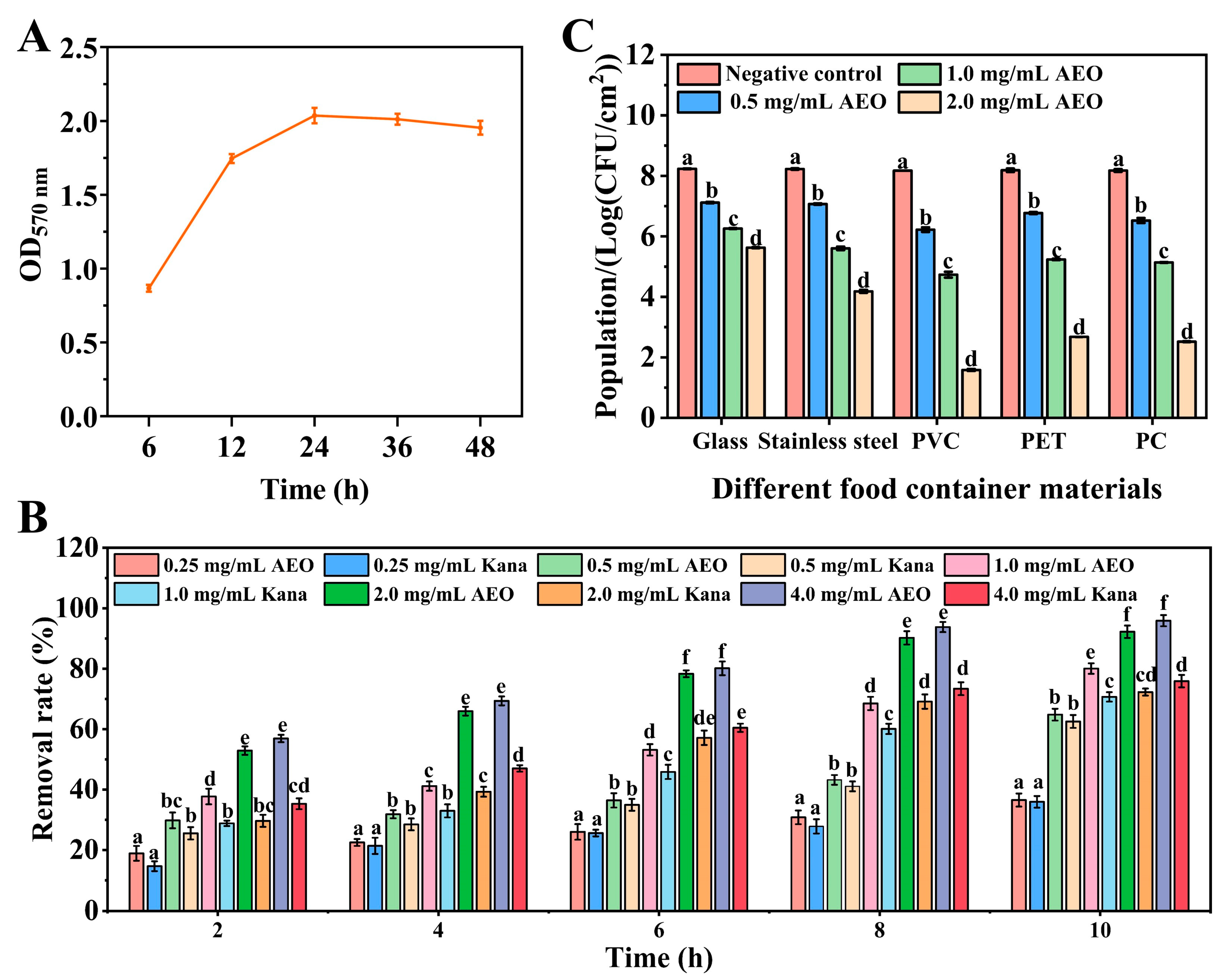
2.3. The Biofilm-Forming Ability of S. aureus

The ability to form biofilms was assessed using the crystal violet staining method [26]. Briefly, a bacterial suspension (108 CFU/mL, 40 μL) was added to a 96-well plate that contained 160 μL of sterile TSB medium. This was incubated for 6, 12, 24, 36, and 48 h, respectively. After the incubation periods, each well’s biofilm was treated with 1% (v/v) crystal violet for a duration of 15 min. After washing with sterile water, an ethanol–acetone solution (4:1, v/v) was used for decolorization, and the optical density at 570 nm was measured using a multi-mode microplate reader (SpectraMax® iD5, Molecular Devices, Sunnyvale, CA, USA).

2.4. The Removal Effect of AEO on S. aureus Biofilm

The removal effect of AEO on the biofilm of S. aureus was evaluated using the MTT assay. A suspension of S. aureus (108 CFU/mL, 50 μL) was combined with 150 µL of sterile TSB into a 96-well plate and cultivated at 37 °C for 24 h. Subsequently, AEO was added at different concentrations. Kana is a commonly used antibiotic, and was used as a positive control. All groups were placed at 37 °C for 10 h. After staining with MTT, the OD570nm value was measured.
Removal   rate   % = [ ( OD Control     OD Sample ) / OD Control ]   × 100

2.5. Effect of AEO on S. aureus Biofilm on the Surface of Different Food Container Materials

Biofilms were cultured on sterilized glass sheets, plastic sheets (PET, PC, and PVC), and stainless steel with a size of φ1 cm. The biofilms were then treated with AEO at concentrations of 0.5, 1.0, and 2.0 mg/mL AEO for 8 h. Following treatment, the sheets were subjected to ultrasonication (100 W, 15 min) in phosphate-buffer solution (PBS). Then, the samples were incubated at 37 °C for 24 h and the removal effect was assessed by counting the total number of colonies.

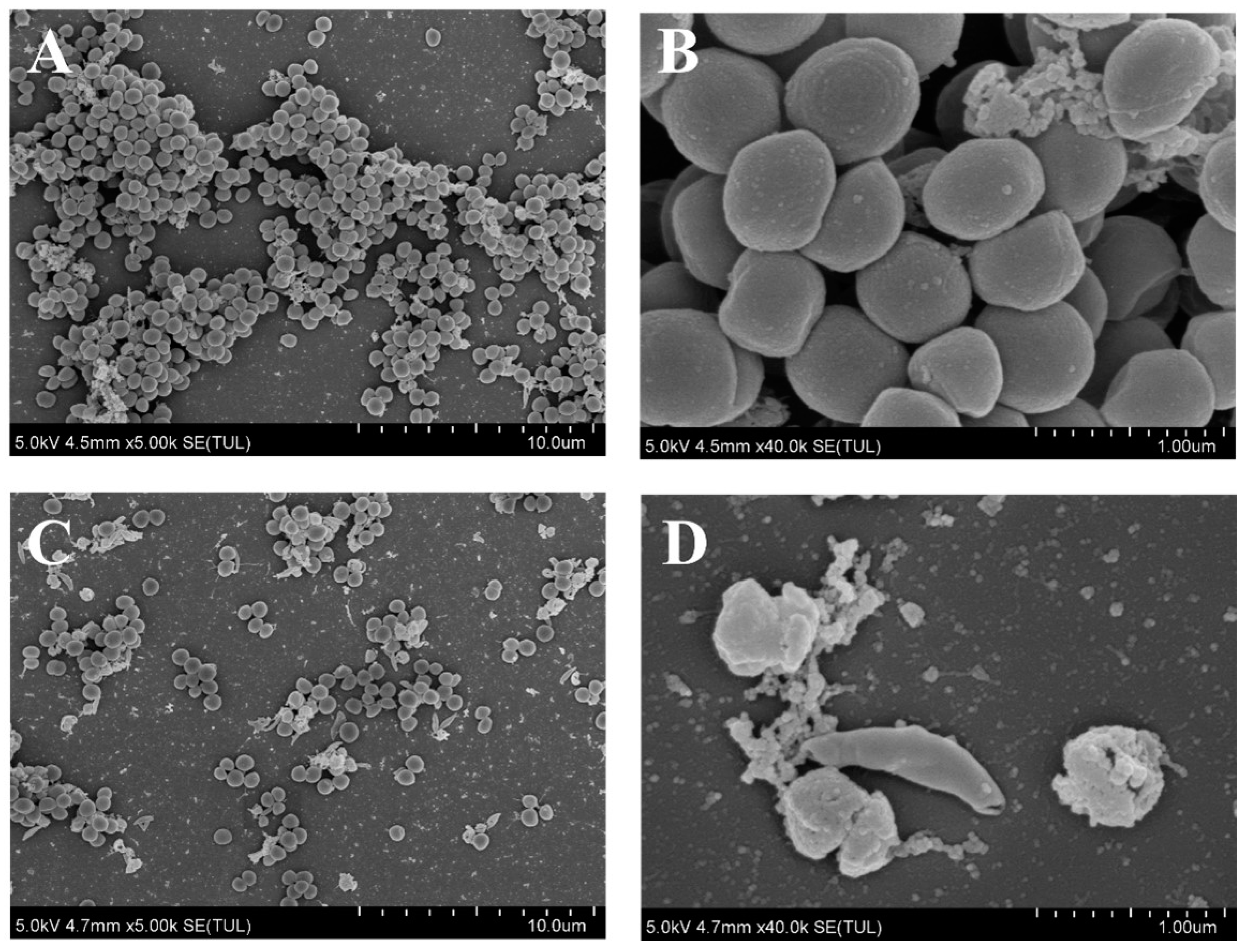
2.6. SEM Analysis of S. aureus Treated with AEO

Following Section 2.4, biofilms were cultivated on sterile coverslips (2 × 2 cm2) and subsequently treated with AEO (2 mg/mL) for 8 h at 37 °C. A control solution containing 0.1% (v/v) Tween 80 and 1% (v/v) dimethyl sulfoxide was utilized. The biofilms were rinsed with PBS and then fixed in a 2.5% glutaraldehyde solution at 4 °C for 24 h. After three washes with PBS, the samples underwent dehydration using a gradient of ethanol (50%, 70%, 80%, 90%, and 100%). Subsequently, critical-point drying was performed, and the samples were immediately coated with a thin layer of gold through sputter-coating. The biofilm was visualized under SEM (SU8010, Hitachi Ltd., Tokyo, Japan) at magnifications of 5000× and 40,000×, respectively.

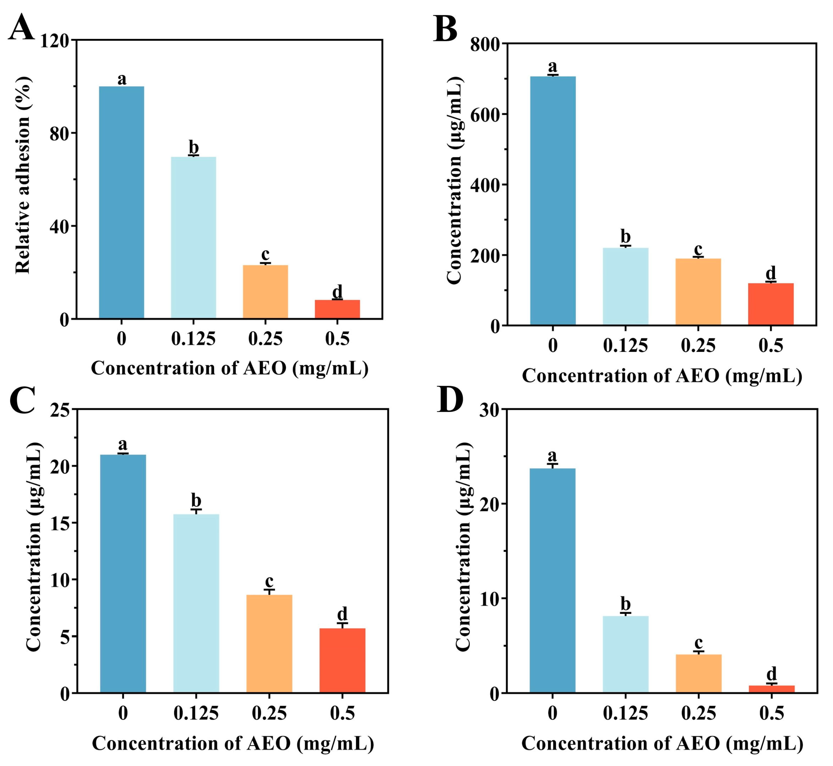
2.7. Impact of AEO on the Initial Adhesion of S. aureus in Biofilm Formation

This was carried out according to the method of Cui et al. [27], with modifications. A suspension of S. aureus (108 CFU/mL) was inoculated in sterile TSB medium along with AEO (0.125, 0.25 and 0.5 mg/mL) and cultivated at 37 °C until the absorbance at 600 nm (OD600nm) reached 0.5. Afterwards, the culture medium was removed by centrifugation and the cells were washed with PBS and then resuspended in PBS. Each group was incubated at 37 °C for 1 h. The samples were fixed using 25% formaldehyde and stained with crystal violet. The absorbance was subsequently measured at 570 nm to evaluate the adhesion, with the relative adhesion rate calculated according to the following formula:
Relative   adhesion   rate   ( % ) = OD Treatment / OD Control   ×   100

2.8. Effect of AEO on the Release of EPS

In accordance with Section 2.3, sterile coverslips were immersed in TSB medium containing a S. aureus suspension and AEO was added at concentrations of 2.7 as mentioned above for processing. Each group was cultured at 37 °C for 2 d, the culture medium was removed, and the wells were washed three times with sterile PBS to discard floating bacteria. Next, PBS was added and a sonicator used to disrupt the biofilm, applying 100 W of ultrasound treatment for 5 min. The resulting bacterial suspension was centrifuged at 5000 rpm for 10 min, and the supernatant filtered through a 0.22 μm membrane to analyze the extracellular matrix content. Extracellular protein levels were measured with the Coomassie Brilliant Blue method. Extracellular polysaccharides were quantified using the phenol-sulfuric acid method [28], and a colorimetric analysis was conducted at 595 nm. The quantity of extracellular DNA (eDNA) was assessed using a bacterial genomic DNA extraction kit (Beyotime, Shanghai, China), with absorbance readings taken at 260 nm [29].

2.9. Effects of AEO on the Relative Expression of Biofilm-Formation-Related Genes in S. aureus

RT-PCR was utilized to evaluate the impact of AEO on the relative expression levels of genes involved intracellular adhesin genes (ica A, ica B), the auxiliary gene regulation gene (agr A), and the programmed cell death system operon genes (cidA, cidB and cidC). The sequences of the primers are listed in Table 1. Following treatment of the S. aureus biofilm with different concentrations of AEO according to the method described in Section 2.8, total RNA was extracted using a Bacteria Total RNA Isolation Kit (Yeasen Biotechnology (Shanghai) Co., Ltd., Shanghai, China). The template complementary DNA (cDNA) was synthesized via reverse transcription using the ATGScript® RT Mix for qPCR kit (ATG Biotechnology Co., Ltd., Nanjing, China) (Table S1). RT-PCR was performed by Hieff® qPCR SYBR Green Master Mix (No Rox) (Yeasen Biotechnology (Shanghai) Co., Ltd., Shanghai, China) (Table S2), with 16S rRNA acting as the internal reference gene. The relative expression levels of the genes were calculated using the comparative threshold cycle (2−ΔΔCT) method.

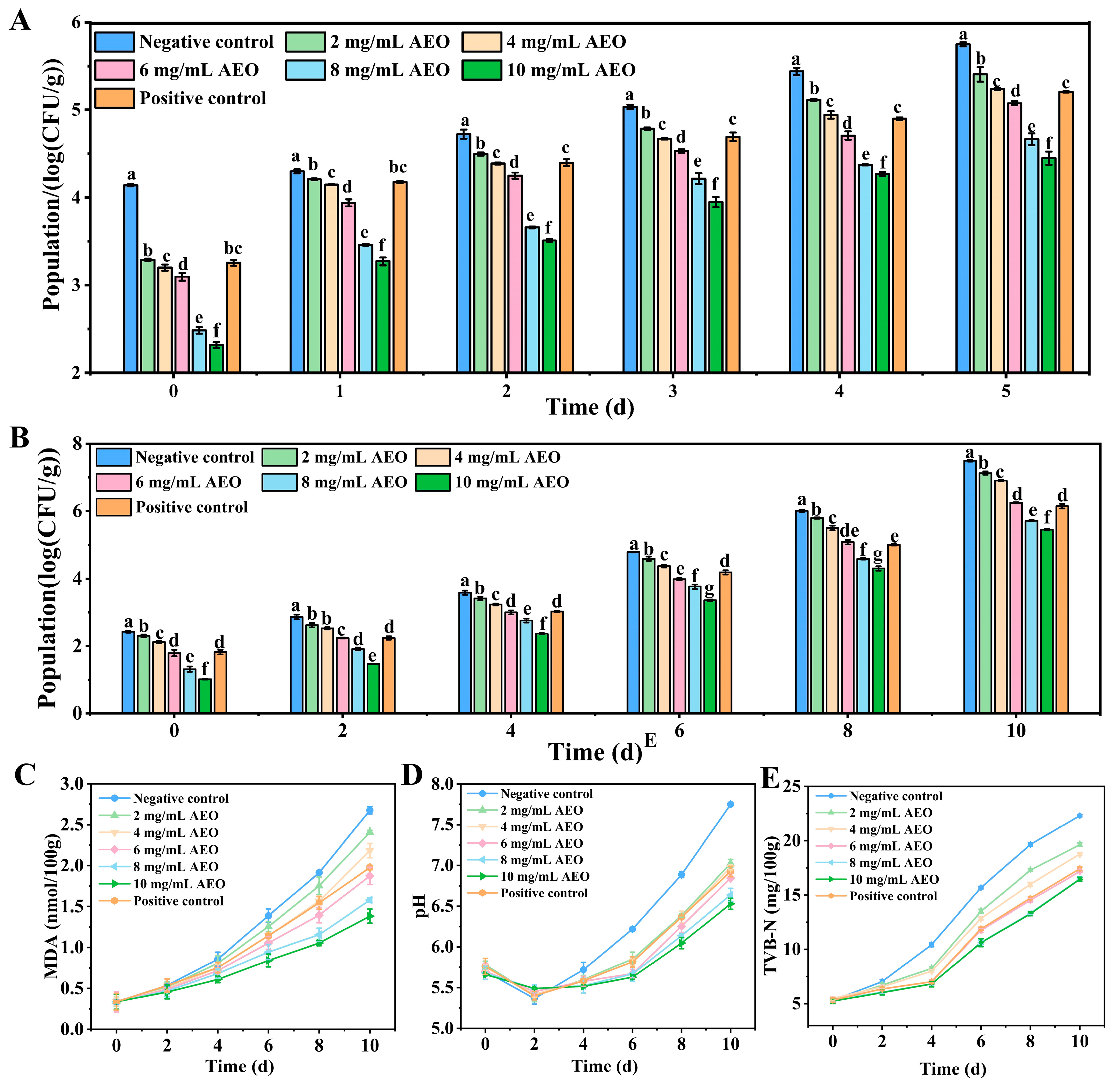
2.10. The Effect of AEO on the Removal of S. aureus Biofilm on Fresh Pork

Fresh pork tenderloin samples were obtained from a local fresh food market in Shanghai, China. Prior to cutting the samples into 2 × 2 × 1.5 cm cubes, the intracellular muscle fat and subcutaneous layers were trimmed away. Each group was irradiated under ultraviolet light (254 nm) for 30 min to eliminate natural microbial colonies, and then placed in a bacterial suspension (104 CFU/mL) at 4 °C for 12 h. Subsequently, the samples were washed with PBS and allowed to dry naturally, after which they were maintained at 4 °C for 48 h to facilitate biofilm formation. The experimental groups were subsequently immersed in AEO at concentrations of 2–10 mg/mL for 30 s. The negative control consisted of a solution containing 0.1% (v/v) Tween 80 and 1% (v/v) dimethyl sulfoxide, while a Kana solution (0.05 mg/mL) was used as the positive control. The number of colonies was evaluated every day until the fifth day.

2.11. Effect of AEO on the Storage Quality of Fresh Pork

Pork is susceptible to contamination by foodborne pathogens, which can lead to spoilage and deterioration. The pork tenderloin samples were sliced into filets measuring 2 × 2 × 1.5 cm and randomly assigned to seven groups (n = 5). The experimental groups were treated with different concentrations of AEO for 30 s. The samples were stored at 4 °C in the refrigerator for 10 d, with a sample taken every 48 h. The samples were blended with distilled water in a 1:10 (w/v) ratio. The resulting homogenates were then centrifuged at 6000 rpm for 10 min to obtain the supernatant for subsequent experiments. The total viable count (TVC) was determined using the plate counting technique, while the detection of thiobarbituric acid reactive substances (TBARS) was carried out following the spectrophotometric method. The pH value of the samples was determined with a pH meter (Mettler Toledo Inc., Greifensee, Switzerland) and the microdiffusion method was used to determine the total volatile basic nitrogen (TVB-N) [30].

2.12. Statistical Analysis

The experimental results were expressed as mean ± standard deviation, with each experiment conducted in triplicate. The collected data were analyzed using one-way analysis of variance (ANOVA) in SPSS version 22.0, where a p-value of less than 0.05 was considered a statistically significant difference. Graphs were generated using Origin 2019 b software (Northampton City, MA, USA).

3. Results

3.1. The Removal Effect of AEO on Biofilms of S. aureus

The findings demonstrate that S. aureus biofilms primarily formed within 0–24 h under the experimental culture conditions (Figure 1A). Therefore, the biofilm culture time for S. aureus was established at 24 h for subsequent experiments. Changes in the biofilm after treatment with different concentrations of AEO are shown in Figure 1B. As the treatment time and AEO concentration increased, the number of remaining biofilms consistently decreased. In the low concentration groups (below 0.5 mg/mL), the removal rate of biofilm was similar to that observed with an equivalent concentration of Kana solution (p > 0.05). When the concentration of AEO was above 1 mg/mL, AEO showed a significantly better performance compared to the Kana solution (p < 0.05). Specifically, the removal rates of biofilm treated with 2 mg/mL and 4 mg/mL AEO treatment groups reached 92.27% and 95.94%, respectively, after 10 h. The results indicated that AEO is effective in removing S. aureus biofilm.

3.2. The Effect of AEO on the Removal of S. aureus Biofilm on Different Food Container Materials

To demonstrate the feasibility of AEO in food applications, we investigated its removal effect on S. aureus biofilm from various food container materials (Figure 1C). The colony count in the negative control group remained stable and was consistent with the results of the blank control, suggesting that the negative control had no removal effect on biofilms. In the AEO treatment groups, the efficacy of biofilm removal was found to be directly proportional to the concentration of AEO applied. Notably, AEO at a concentration of 2 mg/mL demonstrated a significantly higher removal effect on different food contact surfaces, achieving a biofilm removal rate as high as 99.99%, while the control group had a higher content of biofilm. These results indicate that AEO exhibits a strong capacity to eliminate S. aureus biofilm from the surfaces of various food container materials, suggesting its potential as an effective biofilm remover on surfaces in food applications.

3.3. Effect of AEO on the Micromorphology of S. aureus Biofilm

Research suggests that the biofilm in the control group firmly adhered to the surface of the glass slides, forming a dense biofilm structure, with bacterial cells appearing spherical (Figure 2A,B). In contrast, following treatment with AEO, the distance between bacterial cells increased, and the biofilm became dispersed. Additionally, some bacterial cell surfaces exhibited signs of collapse and roughness (Figure 2C,D). These findings indicate that AEO treatment effectively disrupts the micromorphology structure of S. aureus biofilm, achieving a removal effect.

3.4. Effect of AEO on the Initial Adhesion of S. aureus Biofilm Formation

Bacterial initial adhesion affects the formation of biofilms. The adhesion rate in the control group was relatively high, while the adhesion rate in the AEO treatment group significantly decreased (p < 0.05) (Figure 3A). As the concentration of AEO increased, the relative adhesion rate of S. aureus biofilm formation exhibited a continuous decrease. After treatment with 0.5 mg/mL AEO, the initial adhesion of S. aureus was effectively suppressed, with the adhesion rate reduced to 8.23%. The above study indicates that AEO can reduce the biofilm capacity of S. aureus by influencing its initial adhesion.

3.5. Effect of AEO on the Release of EPS

The resilience of biofilms to extreme conditions is largely due to the presence of EPSs, which primarily comprise the extracellular protein, extracellular polysaccharide, and eDNA [31]. As shown in Figure 3B–D, a significant decrease in EPSs was observed in a dose-dependent manner following treatment with AEO at concentrations ranging from 0.125 mg/mL to 0.5 mg/mL (p < 0.05). After treatment with 0.5 mg/mL AEO, the EPS levels in the S. aureus biofilm were significantly reduced compared to the control group (p < 0.05). Specifically, the extracellular protein, extracellular polysaccharide, and eDNA content decreased by 82.99%, 72.91%, and 96.62%, respectively. These findings suggest that AEO effectively inhibits the release of EPSs.

3.6. Effects of AEO on Relative Expression of Biofilm-Formation-Related Genes of S. aureus

Bacteria that lack polysaccharide intercellular adhesion (PIA) show a marked reduction in their ability to stick to one another at later stages, ultimately preventing biofilm formation [32]. The production of PIA is regulated by the ica operon, which includes the icaA and icaB gene clusters, as well as the agrA gene, all of which are associated with the early stages of biofilm formation (Figure 4A). With increasing concentrations of AEO, the expression levels of these genes significantly decreased (p < 0.05). The cidABC genes primarily regulate programmed bacterial cell death and influence the release of eDNA [33]. After treatment with 0.25 mg/mL AEO, the relative expression levels of cidA, cidB, and cidC decreased by 81%, 55%, and 72%, respectively. Additionally, AEO treatment down-regulated the expression levels of the atlA, sarA, and srtA genes (Figure S1). These results indicated that AEO would reduce the expression of the icaA, icaB, agrA, cidA, cidB, and cidC genes, thereby affecting biofilm formation (Figure 4B).

3.7. Effect of AEO on the Removal of S. aureus Biofilm on Fresh Pork

The bacterial count on the surface of the pork in the negative control group increased significantly after 5 d of storage at 4 °C (Figure 5A). In contrast, bacterial growth in the AEO treatment group was suppressed, with the level of inhibition corresponding to the concentration of AEO applied. Notably, the biofilm removal rate after treatment with 4 mg/mL AEO was similar to that of the Kana treatment group (p > 0.05). These results indicate that AEO can effectively remove S. aureus biofilm from the surface of pork.

3.8. Effect of AEO on Storage Quality of Fresh Pork

After 10 d of storage, the fresh pork treated with AEO and Kana still retained a reddish color, while the control group had turned pale (Figure S3). The primary indicators used to assess the freshness of pork products include TVC, TBARS, pH, and TVB-N. The results of the TVC analysis are shown in Figure 5B, and demonstrate that the colony counts in various sample groups steadily increased as the storage duration lengthened. However, pork treated with AEO demonstrated a significant inhibition of TVC growth. Notably, the 6 mg/mL AEO treatment group showed comparable inhibitory effects on colony formation to the Kana treatment group (p > 0.05). In summary, the application of AEO can effectively delay the changes in the TVC of pork.
TBARS serves as a standard index for assessing lipid oxidative byproducts, mainly MDA [34], as shown in Figure 5C. An increase in MDA levels was observed across all samples as the storage time extended. The inhibitory effect of the positive control was comparable in the 4–6 mg/mL AEO treatment groups. By 10 d of storage, compared to the control group, the MDA content in the AEO treatment group was significantly reduced (p < 0.05). These results indicate that AEO effectively inhibits lipid oxidation in pork during prolonged storage.
The pH levels of pork samples treated with AEO showed a more gradual increase compared to the control group (Figure 5D). During the storage process, the negative control group displayed the most rapid rise in pH values of the pork samples. Initially, the inhibitory effect of AEO increased with higher treatment concentrations, but subsequently plateaued. Notably, AEO treatment exhibited stronger inhibition at concentrations above 6 mg/mL compared to the positive control group.
TVB-N was utilized to assess the relative freshness of the pork throughout the storage period (Figure 5E). As storage time increased, the elevation of TVB-N was notably more obvious in the negative control group, whereas the AEO treatment groups significantly suppressed the elevation in TVB-N levels. Specifically, in the 6 mg/mL, 8 mg/mL, and 10 mg/mL AEO groups, the TVB-N content remained below 15 mg/100 g even after 8 d of storage. The above study demonstrates that AEO significantly inhibits the decay of pork, thereby extending its shelf life to 8 d.

4. Discussion

S. aureus is a prevalent foodborne pathogen that can cause the development of biofilms on food processing surfaces, posing a considerable risk for cross-contamination in foods [35]. Once a biofilm has formed, its unique spatial structure makes it challenging to remove, exhibiting enhanced environmental adaptability. Additionally, the widespread use of chemical preservatives in the food industry has led to an increasing prevalence of antibiotic-resistant bacteria. Therefore, controlling biofilms has become a focal point in food safety research.
This study demonstrated that AEO possesses significant antibiofilm activity against S. aureus, effectively removing biofilms from various food container materials, including glass, stainless steel, and plastic. After treatment with 2 mg/mL AEO, the biofilm removal rate on PVC reached an impressive 99.99%, slightly higher than those of many biologically active compounds isolated from plants [36,37]. The aggregation and stacking of colonies can be used to form dense three-dimensional structures known as biofilms [38]. SEM clearly demonstrated that AEO significantly disrupts the structure of the biofilm. Following AEO treatment, a reduction in the number of bacterial cells was observed, along with a dispersion of the biofilm structure. Additionally, some bacterial cell surfaces collapsed and became rough, likely due to the exudation of cellular content. This aligns with earlier studies suggesting that different antibiofilm agents can dismantle the intricate structure of biofilms and break apart microcolonies [39].
Biofilm formation and initial colonization require adhesion to the surface of the substrate. Following the initial bacterial adhesion, bacterial proliferation and EPS production rapidly cause the formation of biofilms [40]. This study explored the potential of AEO to decrease the initial adhesion capacity of S. aureus, consequently reducing its biofilm-forming ability. During the cell proliferation stage, the production of EPSs contributes to the integrity of the biofilm structure, which primarily consists of extracellular proteins, extracellular polysaccharides, and eDNA [6]. Extracellular protein is the most abundant substance within biofilm EPSs and plays an important role in biofilm formation and stability [41]. Extracellular polysaccharides have several roles, such as facilitating adhesion to surfaces, building and stabilizing biofilm structures, and safeguarding cells against antibacterial treatments [28]. eDNA, which originates from bacterial lysis and bacterial secretion, is essential for initial attachment and biofilm formation [42]. This study demonstrates that AEO can significantly inhibit the secretion of EPS of S. aureus, corroborating the results regarding its initial adhesion capacity, thereby suggesting that AEO influences biofilm formation. This is consistent with conclusions drawn by Zhang et al. [43], which demonstrated that 3,3′-diindolylmethane can prevent the production of EPS by S. aureus. These results imply that AEO may inhibit the formation of S. aureus biofilms by reducing EPS release, thereby compromising the integrity of the biofilm’s three-dimensional structure.
Biofilm formation can be categorized into early, middle, and late stages, each involving the expression and regulation of numerous genes [44]. This study demonstrated that AEO down-regulates the expression of the icaA, icaB, agrA, cidA, cidB, and cidC genes. The early stages of biofilm formation are divided into two phases: the reversible and irreversible adhesion phases. PIA is crucial for promoting reversible adhesion, while quorum sensing (QS) regulates irreversible adhesion [45]. The icaA/B genes are responsible for regulating the transfer of N-acetylglucamide (GlcNAc) to the growing PIA chain and for deacetylating PIA to enhance its adhesion properties [46]. Bacteria deficient in PIA exhibit a marked reduction in their ability to adhere to one another, thereby failing to form biofilms. The QS system is an important signaling pathway for biofilm formation, effectively inhibiting biofilm generation by inhibiting the production of signal molecules and interfering with their transmission [47]. AEO influences biofilm formation by regulating the transcription level of genes within the Agr system. The lipopeptide fengycin has been shown to inhibit S. aureus by interfering with the Agr system, which aligns with the results of this study [48]. The cidA, cidB, and cidC genes exhibit similar functions to perforin, mainly regulating programmed cell death, bacterial lysis, and the release of eDNA [49]. The results showed that AEO down-regulates the expression of the icaA and icaB genes, with the proteins encoded by these genes inhibiting the synthesis of PIA and subsequently affecting biofilm formation. Furthermore, AEO also influences biofilm formation by downregulating the expression of the agrA gene. Overall, AEO inhibits the expression of the icaA, icaB, cidA, cidB, and cidC genes. This also suggests that AEO reduces biofilm activity by inhibiting the expression of genes related to extracellular polysaccharides and extracellular DNA in EPS.
To assess the effective antibiofilm activity of AEO, its application in pork preservation was further investigated. This study verified that AEO can effectively reduce S. aureus biofilm on the surface of pork. After 10 d of storage at 4 °C, bacterial growth in the AEO-treated group was significantly inhibited, with the level of inhibition correlating positively with the concentration of AEO used. The TVC, TBARS, pH, and TVB-N are key indicators used to indicate the freshness of pork. The TBARS level, which indicates the extent of lipid oxidation, rises as unsaturated fatty acids in pork samples decompose and oxidize. Variations in pH can occur due to the enzymatic activity of microorganisms and the autolysis of internal enzymes, which lead to the production of nitrogenous compounds. An increase in TVB-N is linked to the breakdown and metabolism of proteins and other non-protein nitrogen compounds [50]. Therefore, the enhancement of these four indicators is inversely proportional to the freshness of the pork. AEO effectively reduced the growth of TVC during pork storage and slowed down the increases in TBARS, pH, and TVB-N values, extending the shelf life of the pork by up to 8 d. In comparison to the negative control group, AEO demonstrated positive effects on TVC, TBARS, pH, and TVB-N. The essential oil solvent used may not be suitable for applications in food. Future efforts could consider combining techniques such as chitosan or nanoemulsion to enhance food safety while further improving its preservation effectiveness. Subsequently, the sensory evaluation results also showed that consumers still have a high acceptance of pork treated with AEO (Table S3). These findings suggest that AEO holds promising potential for application in food preservation.

5. Conclusions

AEO exhibited significant antibiofilm activity, effectively removing S. aureus biofilms attached to food materials. SEM revealed that AEO achieved a better removal effect by disrupting the S. aureus biofilm structure. AEO influenced the early colonization of bacteria on the surface of the product and dispersed EPSs, thereby affecting the formation of S. aureus biofilms. Furthermore, AEO inhibited the transcription levels of genes associated with EPS synthesis (icaA, icaB, cidA, cidB, and cidC) as well as the quorum-sensing-related gene (agrA). In addition, the application experiments demonstrated that AEO could effectively remove S. aureus biofilms on the surface of pork and extend its shelf life. This study enhanced our understanding of AEO antibiofilm activity and offers a new green preservation method for its application in food preservation.

Supplementary Materials

The following supporting information can be downloaded at https://www.mdpi.com/article/10.3390/foods14040662/s1, Table S1. Reverse transcription reaction system; Table S2. The cycle conditions used for real-time qRT-PCR; Table S3. Sensory evaluation of fresh pork; Figure S1. Effect of AEO on expression levels of genes atlA, sarA and srtA; Figure S2. Weight loss and color analysis; Figure S3. Photographs of different treatment groups stored for 0 d and 10 d.

Author Contributions

Z.Y.: Investigation, methodology, data curation, writing—original draft. J.G.: formal analysis, visualization, writing—review and editing.; Q.C.: conceptualization.; S.W.: data curation, validation. Z.Q.: methodology, writing—review and editing, validation, funding acquisition. H.G.: supervision, funding acquisition. All authors have read and agreed to the published version of the manuscript.

Funding

This research was funded by the National Natural Science Foundation of China, grant numbers 32272390 and 32172160.

Institutional Review Board Statement

Not applicable.

Informed Consent Statement

Not applicable.

Data Availability Statement

The original contributions presented in this study are included in the article/Supplementary Materials. Further inquiries can be directed to the corresponding authors.

Conflicts of Interest

The authors declare that the research was conducted in the absence of any commercial or financial relationships that could be construed as potential conflicts of interest.

Abbreviations

The following abbreviations are used in this manuscript:
S. aureusStaphylococcus aureus S. aureus
AEOAmomum tsaoko essential oil
SEMScanning electron microscopy
EPSExtracellular polymeric substances
EOsEssential oils
TVCTotal viable count
TBARSThiobarbituric acid reactive substances
TVB-NTotal volatile basic nitrogen

References

  1. Gunjan; Vidic, J.; Manzano, M.; Raj, V.S.; Pandey, R.P.; Chang, C.M. Comparative meta-analysis of antimicrobial resistance from different food sources along with one health approach in Italy and Thailand. One Health 2023, 16, 100477. [Google Scholar] [CrossRef]
  2. Zhang, P.; Wang, P.; Fu, X.; Xu, X.; Ruan, F.; Wang, T.; Chang, G.; Wan, Y.; Zhang, Y.; Wang, X. Prevalence and characterization of Staphylococcus aureus in raw eggs and it’s growth and enterotoxin a production in egg contents. LWT 2023, 174, 114379. [Google Scholar] [CrossRef]
  3. G. Abril, A.; G. Villa, T.; Barros-Velázquez, J.; Cañas, B.; Sánchez-Pérez, A.; Calo-Mata, P.; Carrera, M. Staphylococcus aureus exotoxins and their detection in the dairy industry and mastitis. Toxins 2020, 12, 537. [Google Scholar] [CrossRef] [PubMed]
  4. Nero, L.A.; Botelho, C.V.; Sovinski, A.I.; Grossi, J.L.; Call, D.R.; Dos Santos Bersot, L. Occurrence and distribution of antibiotic-resistant Staphylococcus aureus in a Brazilian pork production Chain. J. Food Prot. 2022, 85, 973–979. [Google Scholar] [CrossRef]
  5. Sullivan, D.J.; Azlin-Hasim, S.; Cruz-Romero, M.; Cummins, E.; Kerry, J.P.; Morris, M.A. Antimicrobial effect of benzoic and sorbic acid salts and nano-solubilisates against Staphylococcus aureus, Pseudomonas fluorescens and chicken microbiota biofilms. Food Control 2020, 107, 106786. [Google Scholar] [CrossRef]
  6. Xu, Z.; Xie, J.; Soteyome, T.; Peters, B.M.; Shirtliff, M.E.; Liu, J.; Harro, J.M. Polymicrobial interaction and biofilms between Staphylococcus aureus and Pseudomonas aeruginosa: An underestimated concern in food safety. Curr. Opin. Food Sci. 2019, 26, 57–64. [Google Scholar] [CrossRef]
  7. Ghosh, S.; Sarkar, T.; Chakraborty, R. Formation and development of biofilm an alarming concern in food safety perspectives. Biocatal. Agric. Biotechnol. 2021, 38, 102210. [Google Scholar] [CrossRef]
  8. Wagner, E.M.; Pracser, N.; Thalguter, S.; Fischel, K.; Rammer, N.; Pospisilova, L.; Alispahic, M.; Wagner, M.; Rychli, K. Identification of biofilm hotspots in a meat processing environment: Detection of spoilage bacteria in multi-species biofilms. Int. J. Food Microbiol. 2020, 328, 108668. [Google Scholar] [CrossRef]
  9. Cha, M.Y.; Ha, J.W. Low-energy X-ray irradiation effectively inactivates major foodborne pathogen biofilms on various food contact surfaces. Food Microbiol. 2022, 106, 104054. [Google Scholar] [CrossRef] [PubMed]
  10. Abi Assaf, J.; Holden, E.R.; Trampari, E.; Webber, M.A. Common food preservatives impose distinct selective pressures on Salmonella Typhimurium planktonic and biofilm populations. Food Microbiol. 2024, 121, 104517. [Google Scholar] [CrossRef]
  11. Li, Q.; Liu, L.; Guo, A.; Zhang, X.; Liu, W.; Ruan, Y. Formation of multispecies biofilms and their resistance to disinfectants in food processing environments: A review. J. Food Prot. 2021, 84, 2071–2083. [Google Scholar] [CrossRef] [PubMed]
  12. Furquim Dos Santos Cardoso, V.; Amaral Roppa, R.H.; Antunes, C.; Silva Moraes, A.N.; Santi, L.; Konrath, E.L. Efficacy of medicinal plant extracts as dental and periodontal antibiofilm agents: A systematic review of randomized clinical trials. J. Ethnopharmacol. 2021, 281, 114541. [Google Scholar] [CrossRef] [PubMed]
  13. Kunová, S.; Sendra, E.; Haščík, P.; Vuković, N.L.; Vukić, M.D.; Hsouna, A.B.; Mnif, W.; Kačániová, M. Microbiological quality of deer meat treated with essential oil Litsea cubeba. Animals 2022, 12, 2315. [Google Scholar] [CrossRef]
  14. Jannat, B.; Mirza Alizadeh, A.; Farshi, P.; Dadgarnejad, M.; Hosseini, H.; Hashempour-Baltork, F.; Jafari, S.M. Anti-biofilm activity of essential oils in fruit and vegetable: A systematic review. Food Control 2023, 152, 109875. [Google Scholar] [CrossRef]
  15. Abdessemed, M.; Bouacida, S.; Turki, M.; Ben Haj Koubaier, H.; Omrani, S.; Allouache, R.; Bouzouita, N.; Karoui, R.; Snoussi, A. Chemical characterization and biological activities evaluation of Myrtus communis L. essential Oil extraction by-product towards circular economy and sustainability. Foods 2024, 13, 2211. [Google Scholar] [CrossRef] [PubMed]
  16. Bhavaniramya, S.; Vishnupriya, S.; Al-Aboody, M.S.; Vijayakumar, R.; Baskaran, D. Role of essential oils in food safety: Antimicrobial and antioxidant applications. GOST 2019, 2, 49–55. [Google Scholar] [CrossRef]
  17. Mishra, A.P.; Devkota, H.P.; Nigam, M.; Adetunji, C.O.; Srivastava, N.; Saklani, S.; Shukla, I.; Azmi, L.; Shariati, M.A.; Melo Coutinho, H.D.; et al. Combination of essential oils in dairy products: A review of their functions and potential benefits. LWT 2020, 133, 110116. [Google Scholar] [CrossRef]
  18. Cui, H.; Zhang, C.; Li, C.; Lin, L. Antibacterial mechanism of oregano essential oil. Ind. Crops Prod. 2019, 139, 111498. [Google Scholar] [CrossRef]
  19. Hu, W.; Li, C.; Dai, J.; Cui, H.; Lin, L. Antibacterial activity and mechanism of Litsea cubeba essential oil against methicillin-resistant Staphylococcus aureus (MRSA). Ind. Crops Prod. 2019, 130, 34–41. [Google Scholar] [CrossRef]
  20. Hou, F.; Chen, X.; Yi, F.; Song, L.; Zhan, S.; Han, X.; Zhang, L.; Li, F.; Wang, X.; Liu, Z. Antibacterial and antibiofilm properties of cinnamon essential oil on Pseudomonas tolaasii and application of potato starch/CEO active pads in preservation of mushroom (Agaricus bisporus). Food Control 2024, 165, 110705. [Google Scholar] [CrossRef]
  21. Hazitai, A.; Fei, H.-L.; Zhu, C.-Y.; Li, R.-X.; Zhang, L.-X.; Fan, P.-F. Cardamom (Amomum tsaoko) agroforest is important habitat for skywalker hoolock gibbon (Hoolock tianxing) in Mt. Gaoligong, Yunnan, China. Glob. Ecol. Conserv. 2024, 54, e03129. [Google Scholar] [CrossRef]
  22. Thinh, B.B.; Chac, L.D.; Hanh, D.H.; Korneeva, A.A.; Hung, N.; Igoli, J.O. Effect of extraction method on yield, chemical composition and antimicrobial activity of essential oil from the fruits of Amomum villosum var. xanthioides. J. Essent. Oil Bear. Plants 2022, 25, 28–37. [Google Scholar] [CrossRef]
  23. Tang, Z.; Zhao, M.; Han, Y.; Gao, L.; Chen, J.; Chen, W.; Li, Y. Study on antibacterial activity of Amomum tsao-ko extracts against bacillus subtilis and Listeria monocytogenes and preliminary investigation of its antibacterial mchanism. IOP Conf. Ser. Earth Environ. Sci. 2020, 512, 012080. [Google Scholar] [CrossRef]
  24. Li, W.; Li, J.; Qin, Z.; Wang, Y.; Zhao, P.; Gao, H. Insights into the composition and antibacterial activity of Amomum tsao-ko essential oils from different regions based on GC-MS and GC-IMS. Foods 2022, 11, 1402. [Google Scholar] [CrossRef] [PubMed]
  25. Guo, J.; Li, W.; Wan, S.; Zhou, J.; Qin, Z.; Gao, H. Antibacterial activity of Amomum tsaoko essential oil and its interaction with Staphylococcus aureus. LWT 2024, 191, 115700. [Google Scholar] [CrossRef]
  26. Andersen, J.B.; Rybtke, M.; Tolker-Nielsen, T. The dynamics of biofilm development and dispersal should be taken into account when quantifying biofilm via the crystal violet microtiter plate assay. Biofilm 2024, 8, 100207. [Google Scholar] [CrossRef] [PubMed]
  27. Cui, H.; Zhang, C.; Li, C.; Lin, L. Inhibition mechanism of cardamom essential oil on methicillin-resistant Staphylococcus aureus biofilm. LWT 2020, 122, 109057. [Google Scholar] [CrossRef]
  28. Zhang, C.; Li, C.; Abdel-Samie, M.A.; Cui, H.; Lin, L. Unraveling the inhibitory mechanism of clove essential oil against Listeria monocytogenes biofilm and applying it to vegetable surfaces. LWT 2020, 134, 110210. [Google Scholar] [CrossRef]
  29. Liu, J.; Wu, S.; Feng, L.; Wu, Y.; Zhu, J. Extracellular matrix affects mature biofilm and stress resistance of psychrotrophic spoilage Pseudomonas at cold temperature. Food Microbiol. 2023, 112, 104214. [Google Scholar] [CrossRef] [PubMed]
  30. Ruan, C.; Zhang, Y.; Sun, Y.; Gao, X.; Xiong, G.; Liang, J. Effect of sodium alginate and carboxymethyl cellulose edible coating with epigallocatechin gallate on quality and shelf life of fresh pork. Int. J. Biol. Macromol. 2019, 141, 178–184. [Google Scholar] [CrossRef] [PubMed]
  31. Kang, S.; Yang, Y.; Hou, W.; Zheng, Y. Inhibitory effects of lactobionic acid on biofilm formation and virulence of Staphylococcus aureus. Foods 2024, 13, 2781. [Google Scholar] [CrossRef] [PubMed]
  32. Nguyen, H.T.T.; Nguyen, T.H.; Otto, M. The staphylococcal exopolysaccharide PIA-Biosynthesis and role in biofilm formation, colonization, and infection. Comput. Struct. Biotechnol. J. 2020, 18, 3324–3334. [Google Scholar] [CrossRef]
  33. Maggio, F.; Rossi, C.; Serio, A.; Chaves-Lopez, C.; Casaccia, M.; Paparella, A. Anti-biofilm mechanisms of action of essential oils by targeting genes involved in quorum sensing, motility, adhesion, and virulence: A review. Int. J. Food Microbiol. 2025, 426, 110874. [Google Scholar] [CrossRef] [PubMed]
  34. Bernardo, P.; Fernandes, M.J.; Fernandes, M.H.; Teixeira, M.P.; Alfaia, C.M.; Serrano, C.; Patarata, L.; Fraqueza, M.J. Salt reduction strategies for dry cured meat products: The use of KCl and microencapsulated spices and aromatic plant extracts. Meat Sci. 2025, 221, 109719. [Google Scholar] [CrossRef]
  35. Campana, R.; Casettari, L.; Fagioli, L.; Cespi, M.; Bonacucina, G.; Baffone, W. Activity of essential oil-based microemulsions against Staphylococcus aureus biofilms developed on stainless steel surface in different culture media and growth conditions. Int. J. Food Microbiol. 2017, 241, 132–140. [Google Scholar] [CrossRef] [PubMed]
  36. Qian, W.; Liu, M.; Fu, Y.; Zhang, J.; Liu, W.; Li, J.; Li, X.; Li, Y.; Wang, T. Antimicrobial mechanism of luteolin against Staphylococcus aureus and Listeria monocytogenes and its antibiofilm properties. Microb. Pathog. 2020, 142, 104056. [Google Scholar] [CrossRef] [PubMed]
  37. Erhabor, R.C.; Erhabor, J.O.; Nkadimeng, S.M.; McGaw, L.J. In vitro antimicrobial, antibiofilm and antioxidant activities of six South African plants with efficacy against selected foodborne pathogens. S. Afr. J. Bot. 2022, 146, 643–652. [Google Scholar] [CrossRef]
  38. Cui, H.; Li, H.; Abdel-Samie, M.A.; Surendhiran, D.; Lin, L. Anti-Listeria monocytogenes biofilm mechanism of cold nitrogen plasma. Innov. Food Sci. Emerg. Technol. 2021, 67, 102571. [Google Scholar] [CrossRef]
  39. Wu, X.; Ma, G.L.; Chen, H.W.; Zhao, Z.Y.; Zhu, Z.P.; Xiong, J.; Yang, G.X.; Hu, J.F. Antibacterial and antibiofilm efficacy of the preferred fractions and compounds from Euphorbia humifusa (herba euphorbiae humifusae) against Staphylococcus aureus. J. Ethnopharmacol. 2023, 306, 116177. [Google Scholar] [CrossRef]
  40. Lin, L.; Zhang, P.; Chen, X.; Hu, W.; Abdel-Samie, M.A.; Li, C.; Cui, H. Inhibition of Staphylococcus aureus biofilms by poly-L-aspartic acid nanoparticles loaded with Litsea cubeba essential oil. Int. J. Biol. Macromol. 2023, 242, 124904. [Google Scholar] [CrossRef]
  41. Frassinetti, S.; Falleni, A.; Del Carratore, R. Effect of itraconazole on Staphylococcus aureus biofilm and extracellular vesicles formation. Microb. Pathog. 2020, 147, 104267. [Google Scholar] [CrossRef]
  42. Banerji, R.; Mahamune, A.; Saroj, S.D. Aqueous extracts of spices inhibit biofilm in Listeria monocytogenes by down regulating release of eDNA. LWT 2022, 154, 112566. [Google Scholar] [CrossRef]
  43. Zhang, H.; Guo, X.; Tian, L.; Wang, N.; Li, Y.; Kushmaro, A.; Marks, R.; Sun, Q. Antibiofilm activity of 3,3′-diindolylmethane on Staphylococcus aureus and its disinfection on common food-contact surfaces. Food Sci. Hum. Wellness 2022, 11, 1222–1232. [Google Scholar] [CrossRef]
  44. Li, H.; Li, C.; Ye, Y.; Cui, H.; Lin, L. Inhibition mechanism of cyclo (L-Phe-L-Pro) on early stage Staphylococcus aureus biofilm and its application on food contact surface. Food Biosci. 2022, 49, 101968. [Google Scholar] [CrossRef]
  45. Liu, M.; Wu, X.; Li, J.; Liu, L.; Zhang, R.; Shao, D.; Du, X. The specific anti-biofilm effect of gallic acid on Staphylococcus aureus by regulating the expression of the ica operon. Food Control 2017, 73, 613–618. [Google Scholar] [CrossRef]
  46. Formosa-Dague, C.; Feuillie, C.; Beaussart, A.; Derclaye, S.; Kucharíková, S.; Lasa, I.; Van Dijck, P.; Dufrêne, Y.F. Sticky matrix: Adhesion mechanism of the Staphylococcal Polysaccharide intercellular adhesin. ACS Nano 2016, 10, 3443–3452. [Google Scholar] [CrossRef] [PubMed]
  47. Gless, B.H.; Bojer, M.S.; Peng, P.; Baldry, M.; Ingmer, H.; Olsen, C.A. Identification of autoinducing thiodepsipeptides from staphylococci enabled by native chemical ligation. Nat. Chem. 2019, 11, 463–469. [Google Scholar] [CrossRef] [PubMed]
  48. Piewngam, P.; Zheng, Y.; Nguyen, T.H.; Dickey, S.W.; Joo, H.S.; Villaruz, A.E.; Glose, K.A.; Fisher, E.L.; Hunt, R.L.; Li, B.; et al. Pathogen elimination by probiotic Bacillus via signalling interference. Nature 2018, 562, 532–537. [Google Scholar] [CrossRef]
  49. Tao, J.; Yan, S.; Zhou, C.; Liu, Q.; Zhu, H.; Wen, Z. Total flavonoids from Potentilla kleiniana Wight et Arn inhibits biofilm formation and virulence factors production in methicillin-resistant Staphylococcus aureus (MRSA). J. Ethnopharmacol. 2021, 279, 114383. [Google Scholar] [CrossRef] [PubMed]
  50. Wan, Y.; Wang, X.; Yang, L.; Li, Q.; Ma, L.; Bai, T.; Zheng, X.; Wang, X. Anti Staphylococcus aureus activity of shikonin @ chitosan nanoemulsion and its effect on the storage quality of pork. Food Control 2024, 158, 110203. [Google Scholar] [CrossRef]
Figure 1. The removal effect of AEO on S. aureus biofilm. (A) Biofilm formation ability of S. aureus. (B) The removal effect of AEO on S. aureus biofilm on the 96-well plate. Kana solution was used as a positive control. (C) Effect of AEO on the biofilm removal for different food materials. A mixture of 0.1% (v/v) Tween 80 and 1% (v/v) dimethyl sulfoxide was used as the negative control. The experimental results are presented as the mean ± standard deviation (n = 3). Different letters indicate significant differences between groups (p < 0.05).
Foods 14 00662 g001
Figure 2. SEM of S. aureus biofilm formed on the coverslips. (A,B) Scanning electron microphotographs of untreated S. aureus biofilms. (C,D) Scanning electron microphotographs of S. aureus biofilm treated with AEO (2 mg/mL). Images were taken at 5000× and 40,000× magnification.
Foods 14 00662 g002
Figure 3. Effect of AEO on biofilm formation. Effect of AEO on initial adhesion (A), extracellular protein (B), extracellular polysaccharide (C), and eDNA (D) of S. aureus biofilm. A mixture of 0.1% (v/v) Tween 80 and 1% (v/v) dimethyl sulfoxide was used as the control (zero point). Different letters indicate significant differences between groups (p < 0.05).
Foods 14 00662 g003
Figure 4. Effect on expression of genes related to S. aureus biofilm formation. (A) The related gene expression levels as assessed using qRT-PCR. Control: A mixture of 1% (v/v) dimethyl sulfoxide and 0.1% (v/v) Tween 80. (B) The regulatory process of related genes (orange arrows indicate down-regulation of genes). PIA: Polysaccharide intercellular adhesion; QS: quorum sensing; eDNA: extracellular DNA. Different letters indicate significant differences between groups (p < 0.05).
Foods 14 00662 g004
Figure 5. Effect of AEO on fresh pork preservation. (A) AEO removal effect on S. aureus biofilm on fresh pork surfaces. Effect of AEO on TVC (B), TBARS (C), pH (D), and TVB-N (E). All pork samples were stored at 4 °C for 10 d. Negative control: A mixture of 0.1% (v/v) Tween 80 and 1% (v/v) dimethyl sulfoxide. Positive control: Kana solution (0.05 mg/mL). Different letters indicate significant differences between groups (p < 0.05).
Foods 14 00662 g005
Table 1. The primers used for RT-qPCR.
GeneSequence (5’-3’)
icaAForward: AGTGCAGTTGTCGATGTTGGCTAC
Reverse: ACACATGGCAAGCGGTTCATACTT
icaBForward: TTTGAAACACATACCCACGATTTGC
Reverse: TTGGAGTTCGGAGTGACTGCTT
agrAForward: CTGATAATCCTTATGAGGTGCTTGA
Reverse: AGGTAAGTTCACTGTGACTCGTA
cidAForward: CTTGGGTAGAAGACGGTGCAAACTT
Reverse: AGCGTAATTTCGGAAGCAACATCCA
cidBForward: AGCCGCAGTAGGTATCGAAGTGT
Reverse: CTAGTGCTTTAGCTGTGCCAAATGC
cidCForward: GGTACAATGGGTTGCGGTCTTCC
Reverse: ACCTTTACCACCTGCTGCCTCA
16S rRNAForward: TGTTCTCAGTTCGGATTGTA
Reverse: ATACGGCTACCTTGTTACG
Disclaimer/Publisher’s Note: The statements, opinions and data contained in all publications are solely those of the individual author(s) and contributor(s) and not of MDPI and/or the editor(s). MDPI and/or the editor(s) disclaim responsibility for any injury to people or property resulting from any ideas, methods, instructions or products referred to in the content.

