Article

In Vitro Assessment of the Prebiotic Effects of Poria Cocos Polysaccharides Using Fecal Microbiota from Normal-Weight and Obese Children

1
Department of Pediatric Endocrinology and Inherited Metabolic Diseases, Children’s Hospital of Fudan University, Shanghai 201102, China
2
Shanghai Innovation Center of TCM Health Service, Shanghai University of Traditional Chinese Medicine, Shanghai 201203, China
3
State Key Laboratory of Integration and Innovation of Classic Formula and Modern Chinese Medicine, Shanghai University of Traditional Chinese Medicine, Shanghai 201203, China
4
Section of Endocrinology, Internal Medicine, School of Medicine, Yale University, New Haven, CT 06510, USA
*
Authors to whom correspondence should be addressed.
Foods 2025, 14(23), 4077; https://doi.org/10.3390/foods14234077
Submission received: 3 November 2025 / Revised: 19 November 2025 / Accepted: 25 November 2025 / Published: 27 November 2025
(This article belongs to the Section Food Biotechnology)

Abstract

Poria cocos polysaccharides (PCP) are recognized as potential prebiotics with documented metabolic benefits in adults. However, their impact on the gut microbiota of children remains unclear. This study aimed to evaluate the effects of PCP versus inulin (INL) on the gut microbiota and bacterial metabolites in normal-weight children (CON) and obese children (OB). In vitro fermentation was conducted using fecal samples pooled from five normal-weight children and five obese children, respectively. The samples were incubated with PCP, INL, or a blank control under anaerobic conditions at 37 °C for 24 h. After fermentation, the effects of PCP and INL on gut microbiota were evaluated using 16S rRNA sequencing. Bacteria-derived metabolites were measured using targeted metabolic profiling. Single-strain validation was performed to confirm effects on key bacterial taxa. PCP supplementation promoted the growth of Bifidobacterium and Limosilactobacillus in both normal-weight and obese children, accompanied by a rise in acetic acid production, particularly in normal-weight children. Compared to INL, PCP showed similar but slightly weaker effects on Bifidobacterium growth and short-chain fatty acids (SCFAs) production, but more strongly stimulated Limosilactobacillus growth. Notably, PCP also stimulated the production of indolelactic acid in both obese and normal-weight children. Correlation analysis indicated that Bifidobacterium and Limosilactobacillus were positively associated with acetic acid, lactic acid, and indolelactic acid, and negatively associated with tryptophan. Single-strain fermentation supported the community-level findings. PCP and INL both modulate gut microbiota and metabolic profiles in children, with PCP demonstrating a distinct prebiotic profile. Notably, PCP increased health-associated metabolites such as acetic acid and indolelactic acid, which are implicated in gut barrier support, immune modulation, and metabolic regulation. These findings suggest PCP may be considered a functional food component for supporting gut health in children, warranting further research.

1. Introduction

Wolfiporia extensa (syn. Poria cocos), known as “Fuling” in China, is a fungal species widely used in functional foods and traditional Chinese medicine [1]. Studies have demonstrated that Wolfiporia extensa is rich in polysaccharides and triterpenoids [2,3]. Poria cocos polysaccharides (PCP) are the principal constituents of Wolfiporia extensa, constituting approximately 70–90% of the dry sclerotial biomass [4,5]. Further studies have revealed that PCP have broad biological activities, such as anti-inflammatory, immunomodulatory, antioxidant, tumor suppression, and organ protection, particularly in the liver and kidney [3,4,6,7,8].
Gut microbiota, composed of trillions of microbes in the human colon, plays an important role in maintaining host homeostasis [9]. The gut microbiota generates a wide range of metabolites of multiple chemical categories, including lipopolysaccharides, short-chain fatty acids (SCFAs), bile acids, tryptophan metabolites, and indole derivatives [10]. They act as key molecular mediators that link microbial activities to host metabolism, immunity, and disease [11]. Recent studies have indicated that active components of functional foods could modulate the composition and function of gut microbiota to maintain metabolic health [10,12,13,14]. For example, Ge et al. [15] reported that polysaccharides from coix seed regulated the composition of gut microbiota by increasing the relative abundance of Bifidobacterium, Limosilactobacillus, and Collinsella. In addition, these polysaccharides also facilitated the production of SCFAs.
The prevalence of childhood obesity has increased rapidly in recent years, representing a major global public health challenge [16]. Accumulating evidence indicates that dietary factors influence childhood obesity both directly and indirectly, through the modulation of gut microbiota, which plays an important role in obesity development [17,18]. Compared to adults, children exhibit distinct gut microbiota profiles [19]. According to Radjabzadeh et al. [20], children had notably lower diversity than adults. Additionally, they observed a higher relative abundance of the genus Bacteroides in children, while the genus Blautia was more abundant among adults. These developmental differences in key genera mean that findings from adult studies cannot be directly extrapolated to pediatric populations. In addition, obese children show significant alterations in gut microbiota relative to their normal-weight peers [17]. Previous studies have shown that PCP can enhance the relative abundance of beneficial bacteria, such as Lactobacillus and Bifidobacterium, as well as increase SCFA concentrations in normal-weight adults [2]. Nevertheless, the effects of PCP on the gut microbiota of children, especially those with obesity, have not yet been reported.
Here, we outline the experimental design, rationale, and protocols developed for the in vitro fermentation study assessing the effects of PCP and inulin (INL) on the gut microbiota and metabolic profiles of children. The study utilized fecal samples from two distinct groups: normal-weight children (CON) and obese children (OB). To our knowledge, this study is the first to examine how PCP influence the gut microbiota and metabolites of obese children. The findings are anticipated to provide important evidence for the potential use of PCP in child-oriented functional foods.

2. Materials and Methods

2.1. Children’s Research Subjects

Ten children, consisting of 5 normal-weight children and 5 obese children, were enrolled. Obesity was defined according to the age- and sex-specific BMI reference cutoffs for overweight and obesity in Chinese children and adolescents (2–18 years), which were recommended by the Working Group on Obesity in China (WGOC) [21]. Subjects were excluded from taking antibiotics, prebiotics, or other microbiota-modulating drugs within one month. Subjects with acute or chronic gastrointestinal diseases were also not enrolled.

2.2. Laboratory Measurements

In this study, participants’ FBG, FINS, ALT, AST, SCR, UA, TC, TG, HDL-C and LDL-C were measured in the Clinical Laboratory of the Children’s Hospital of Fudan University.

2.3. Materials and Equipment

A commercial water-soluble PCP product (80% purity, S25272) was obtained from Yuanye Biotechnology Co., Ltd., Shanghai, China. INL was procured from Fuxuan Biotechnology Co., Ltd., Jinhua, Zhejiang, China. MPYG medium (HB8931), MRS medium (HB0384-1), anaerobic packages (HBYY001), and anaerobic bags (HBYY007) were purchased from Hopebio, Qingdao, Shandong, China. L-cysteine was obtained from Damas-Beta, Shanghai, China. The anaerobic workstation (Concept 400) was obtained from The Baker Company, Sanford, ME, USA. The refrigerated centrifuge (Z216MK) was procured from Hermle, Wehingen, BW, Germany. The Stratus plate reader was purchased from Cerillo, Charlottesville, VA, USA.

2.4. Monosaccharide Analysis of PCP

Monosaccharide composition was determined by trifluoroacetic acid (TFA; ANPEL, Shanghai, China) hydrolysis followed by ion chromatography. PCP samples were hydrolyzed with 2 M TFA at 121 °C for 2 h. Samples were dried under a nitrogen stream, washed with methanol (ANPEL), and evaporated to dryness. The washing procedure was repeated 2–3 times. The residue was dissolved in deionized water and filtered through 0.22-μm microporous filter for measurement.
The sample extracts were analyzed by high-performance anion-exchange chromatography with pulsed amperometric detection on a Dionex ICS-5000+ system (Thermo Fisher Scientific, Waltham, MA, USA). Separation was carried out on a CarboPac™ PA20 column (3 × 150 mm, 10 μm; Dionex, Sunnyvale, CA, USA) with a flow rate of 0.5 mL/min and an injection volume of 5 μL. The mobile phases consisted of deionized water (A), 0.1 M NaOH (B), and 0.1 M NaOH (Sigma-Aldrich, St. Louis, MO, USA) containing 0.2 M sodium acetate (Sigma-Aldrich) (C). The gradient program was as follows: 0–26 min, a linear increase from A/B/C = 95/5/0 (v/v) to 85/5/10 (v/v); 26–42 min, held at 85/5/10 (v/v); 42.0–42.1 min, changed to 60/0/40 (v/v); 42.1–52.0 min, maintained at 60/40/0 (v/v); 52.0–52.1 min, returned to 95/5/0 (v/v); and 52.1–60 min, equilibrated at 95/5/0 (v/v). Monosaccharides were identified by comparing retention times with authentic standards (Sigma-Aldrich), and quantification was performed using calibration curves derived from standard solutions. Chromatographic data were collected and processed using Chromeleon 7.2 CDS software (Thermo Fisher Scientific), and quantitative results were exported for further statistical analysis.

2.5. Collection of Fecal Samples and In Vitro Fermentation

Fecal samples from normal-weight and obese children were collected and immediately placed into 5 mL sterile cryogenic storage tubes, which were then transferred into anaerobic bags containing pre-anaerated packages. The samples were subsequently stored at −80 °C until further use for in vitro fermentation. Frozen fecal samples were thawed on ice, and fecal samples from children in the CON and OB groups were pooled separately to reduce inter-individual variation. The pooled samples were then mixed with phosphate-buffered saline (pH 7.2) to prepare a 10% (w/v) suspension, and subsequently vortexed until no visible large fecal particles were seen. The mixture was centrifuged at 2000× g for 10 min. The supernatants were transferred into sterile centrifuge tubes.
Previous studies have demonstrated that a concentration of 1% (w/v) PCP and INL is commonly used in in vitro fermentation models to ensure sufficient substrate to assess gut microbiota activity [2,22]. Accordingly, fermentation was carried out in MPYG medium with either PCP or INL at 1% (w/v) to establish the PCP group and the INL group. The MYPG medium without PCP or INL served as the blank group (BLK group). Each medium (PCP-containing, INL-containing, or BLK) was then mixed with fecal suspension at a ratio of 19:1 (v/v) and incubated at 37 °C for 24 h. After centrifugation at 9000× g for 5 min, the supernatants were filtered through a 0.22-μm membrane. Both supernatants and sediments were preserved at −80 °C until use.

2.6. In Vitro Culture of Single Strains

Bifidobacterium pseudocatenulatum ATCC27919, Limosilactobacillus reuteri ATCC23272, and Enterococcus faecium ATCC19433 were purchased from Biobw Biotechnology Co., Ltd., Beijing, China. These strains were activated in MRS broth containing 0.05% L-cysteine at 37 °C for 24 h. Following activation, the cultures were divided into three groups based on supplementation: the PCP group (MRS supplemented with 1% [v/v] PCP), the INL group (MRS supplemented with 1% [v/v] INL), and the BLK group (MRS without supplementation, serving as the blank control). After 24 h of co-culture, the optical density at 600 nm (OD600) was measured.

2.7. DNA Extraction,16S rRNA Gene Sequencing

Microbial DNA was extracted from samples using the FastPure Stool DNA Isolation Kit (MJYH, Shanghai, China). DNA integrity and concentration were verified by agarose gel electrophoresis and NanoDrop 2000 (Thermo Fisher Scientific). The V3–V4 region of the 16S rRNA gene was amplified using TransStart Fastpfu DNA Polymerase (AP221-02, TransGen Biotech, Beijing, China). The amplification employed 338F (5′-ACTCCTACGGGAGGCAGCAG-3′) and 806R (5′-GGACTACHVGGGTWTCTAAT-3′) primer pairs on an ABI GeneAmp® 9700 Thermal Cycler (Applied Biosystems, Foster City, CA, USA). PCR products were purified using the PCR Clean-Up Kit (YuHua, Shanghai, China), quantified using Qubit 4.0 (Thermo Fisher Scientific). Purified amplicons were pooled in equimolar amounts and sequenced on the Illumina NextSeq 2000 platform (Illumina, San Diego, CA, USA) by Majorbio Bio-Pharm Technology Co., Ltd. (Shanghai, China).
Raw reads were processed with fastp (v0.19.6), where reads were trimmed when the average quality score fell below Q20 within a 50 bp sliding window, and reads <50 bp or containing ambiguous nucleotides were removed. The resulting high-quality reads were then merged using FLASH (v1.2.7). High-quality sequences were clustered into operational taxonomic units (OTUs) at a 97% similarity threshold with UPARSE (v7.1). Taxonomic classification of representative sequences was conducted with the RDP Classifier (v2.2) against the SILVA v138 database, applying a confidence cutoff of 0.7. To standardize sequencing depth across samples, all datasets were rarefied to 20,000 reads, with Good’s coverage exceeding 99%.

2.8. Targeted Metabolomic Profiling

Supernatants from fermentation were thawed on ice, lyophilized to remove residual water, and then transferred to the Eppendorf epMotion Workstation (Eppendorf Inc., Hamburg, Germany). A total of 120 μL of ice-cold methanol (Thermo Fisher Scientific) containing partial internal standards was automatically added to each sample. The mixture was vortexed thoroughly and then centrifuged at 4000× g for 30 min. The clear supernatants were returned back to the workstation. Then, 30 μL of the supernatant was transferred to a clean 96-well plate, and 20 μL of freshly prepared derivatization reagents (Merck KGaA, Darmstadt, Germany) was added to each well. After derivatization, 330 μL of ice-cold methanol was added to dilute the samples. The samples were then stored at −20 °C for 20 min and centrifuged again at 4000× g at 4 °C for 30 min before analysis.
Metabolite analysis was performed on an ACQUITY UPLC–MS/MS system (Waters Corp., Milford, MA, USA) equipped with a BEH C18 analytical column (2.1 × 100 mm, 1.7 μm; Waters Corp.) and a VanGuard pre-column (2.1 × 5 mm, 1.7 μm; Waters Corp.). The column and auto-sampler temperatures were maintained at 40 °C and 10 °C, respectively. The flow rate was set to 0.4 mL/min, and the injection volume was 5 μL. Mobile phases consisted of water with 0.1% formic acid (Sigma-Aldrich) (A) and acetonitrile/isopropanol (Thermo Fisher Scientific) (70:30, v/v) (B), with a gradient elution program as follows: 0–1 min (5% B), 1–11 min (5–78% B), 11–13.5 min (78–95% B), 13.5–14 min (95–100% B), 14–16 min (100% B), 16–16.1 min (100–5% B), and 16.1–18 min (5% B).
Raw UPLC-MS/MS data were analyzed using the TMBQ software (v1.0, Metabo-Profile, Shanghai, China) to perform peak identification, integration, calibration, and quantitation for each metabolite [20].

2.9. Statistical Analysis of Data

All data were presented as the mean ± standard deviation (SD). Statistical analyses were performed using GraphPad Prism software (v10.1.2, USA) and IBM SPSS Statistics (v25, USA). For comparisons between two groups, Student’s t-test or the Mann–Whitney U test was applied depending on data distribution. For three groups, two-way ANOVA was used, followed by Tukey’s post hoc test. Statistical significance was established at the thresholds of p < 0.05 (*), p < 0.01 (**), and p < 0.001 (***). Bioinformatic analysis was conducted using the OECloud platform (https://cloud.oebiotech.com, accessed on 28 September 2025; OE Biotech, Shanghai, China).

3. Results

3.1. Demographic and Clinical Characteristics of Subjects

The demographic and clinical characteristics of the participants are listed in Table S1. The age and sex distributions were comparable between the two groups. In comparison to the CON group, the OB group exhibited significantly higher BMIs (p < 0.01). In addition, ALT and TG levels were significantly elevated in the OB group (p < 0.01) (Table S1).

3.2. Monosaccharide Composition of Poria Cocos Polysaccharides

The structural features of PCP, including monosaccharide composition, molecular weight, chain conformation, glycosyl linkage type, and branch structure, have been extensively characterized in published literature [23,24]. Given this established body of structural knowledge, the analytical determination performed in the current study on the commercial PCP product focused on verifying its identity and quality control. This was achieved by determining the monosaccharide composition using ion chromatography. The result showed that PCP was mainly composed of four monosaccharide constituents, among which glucose was the primary constituent, accounting for approximately 89.74% of the total sugar content. The other identified components included fucose, galactose, and mannose (Figure S1 and Table S2). This result verified that the commercial product possessed the characteristic saccharide profile established in the literature for PCP [23,24].

3.3. Effect of PCP on the Gut Microbiota of Normal-Weight and Obese Children

In this study, 16S rRNA gene sequencing was used to assess the impact of PCP on the gut microbiota of normal-weight and obese children. Alpha diversity analysis of the fermentation system was assessed and presented using ACE (Abundance-based Coverage Estimator) and Shannon indices in Figure 1. In the OB group, PCP treatment increased the ACE index, but there were no statistically significant differences in either the CON or OB group (Figure 1b). Shannon diversity index increased significantly in the CON group but decreased in the OB group after PCP treatment (p < 0.001) (Figure 1c). Principal coordinate analysis (PCoA) based on Bray–Curtis distances was performed in Figure 1a. The first two principal components explained 71.99% and 19.44% of the variance, respectively (Figure 1a). The PCoA results showed that the CON and OB groups were well separated, indicating distinct microbial community structures between the CON and OB groups. In addition, in both the CON and OB groups, PCP and INL treatments were separated from BLK, indicating that PCP and INL treatments shaped the gut microbiota structure.
To further characterize these differences, we analyzed the bacterial composition of each group at the phylum and genus levels. At the phylum level, Bacillota, Pseudomonadota, Actinomycetota, Bacteroidota dominated after fermentation. Compared to BLK, PCP, and INL treatments increased the ratio of Actinomycetota and decreased ratio of Bacillota in both the CON and OB groups. INL treatment showed a greater increase in Actinomycetota than PCP treatment (Figure 1d). At the genus level, the microbial community after fermentation was predominantly composed of Bifidobacterium, Enterococcus, Escherichia-Shigella, and Limosilactobacillus (Figure 1e).
Linear Discriminant Analysis Effect Size (LEfSe) analysis was then conducted separately in the CON and OB groups (Figure 1f–g). Bifidobacterium was consistently enriched in INL treatment under both the CON and OB groups. Limosilactobacillus was specifically enriched in PCP treatment of the CON group, whereas PCP treatment in the OB group showed enrichment of the related order Lactobacillales. In addition, the BLK of the CON group was characterized by enrichment of the family Enterococcaceae. In contrast, in the OB group, PCP treatment promoted the enrichment of Enterococcus together with Enterococcaceae. Relative abundance analysis was consistent with the LEfSe analysis (Figure 1h–j). In the CON group, PCP treatment and INL treatment significantly increased the relative abundance of Bifidobacterium and Limosilactobacillus (p < 0.001). INL treatment showed a higher increase in the relative abundance of Bifidobacterium, whereas PCP treatment elevated the relative abundance of Limosilactobacillus (p < 0.001). In addition, PCP and INL treatments both significantly decreased the relative abundance of Enterococcus (p < 0.001). In the OB group, PCP and INL treatments increased the relative abundance of Bifidobacterium and Limosilactobacillus. However, compared to BLK, both PCP and INL increased the relative abundance of Enterococcus in the OB group (p < 0.001).

3.4. Impact of PCP on Targeted Metabolites

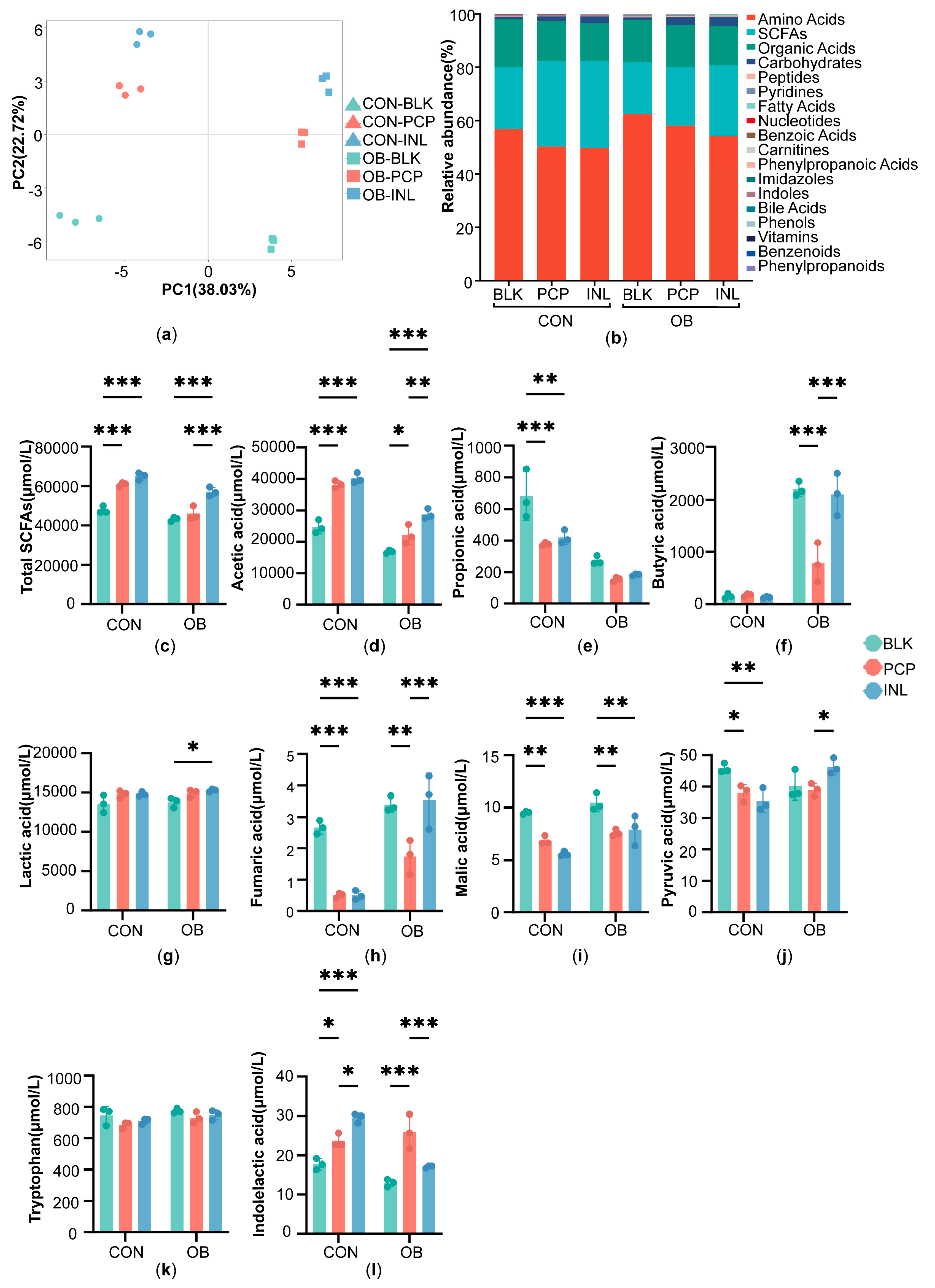
Since shifts in gut microbes are often associated with metabolic changes, we used targeted profiling of about 300 metabolites to assess the PCP’s functional consequences. Partial least squares discriminant analysis (PLS-DA) revealed a clear separation between the CON and OB groups. In both the CON and OB groups, PCP and INL treatments were well separated from BLK, indicating that PCP and INL could affect microbiota metabolism notably (Figure 2a). The detected metabolites mainly consisted of amino acids and their derivatives, SCFAs, organic acids, and carbohydrates (Figure 2b).
The results of SCFAs after fermentation are presented in Figure 2c–f. In the CON group, PCP and INL treatments raised the acetic acid and total SCFAs compared to BLK (p < 0.001), reduced propionic acid (PCP: p < 0.001, INL: p < 0.01), and caused only slight changes in butyric acid. Notably, the levels of acetic, propionic, and butyric acids, as well as total SCFAs, were comparable between PCP and INL treatments, suggesting that these interventions exerted similar overall effects on SCFA production. In the OB group, both PCP and INL also increased the acetic acid (PCP: p < 0.05, INL: p < 0.001) and total SCFAs (INL: p < 0.001) levels relative to BLK, accompanied by reductions in both propionic and butyric acids. PCP treatment increased acetic acid and total SCFA levels in the OB group, demonstrating its effectiveness in modulating SCFA production, although was weaker compared to INL. In addition to SCFAs, several organic acids were also determined (Figure 2g–j). In the CON and OB groups, lactic acid increased after the PCP and INL treatments compared to BLK, whereas PCP treatment significantly reduced the levels of fumaric acid (CON: p < 0.001, OB: p < 0.01), malic acid (p < 0.01), and pyruvic acid (CON: p < 0.05).
With respect to tryptophan metabolism, the tryptophan level saw a decrease in both the CON and OB groups (Figure 2k). Meanwhile, indolelactic acid, a microbial-derived metabolite of tryptophan, was significantly elevated after PCP treatment in both the CON and OB groups (CON: p < 0.05, OB: p < 0.001) (Figure 2l).

3.5. Correlation Between Gut Microbiota Composition and Targeted Metabolites

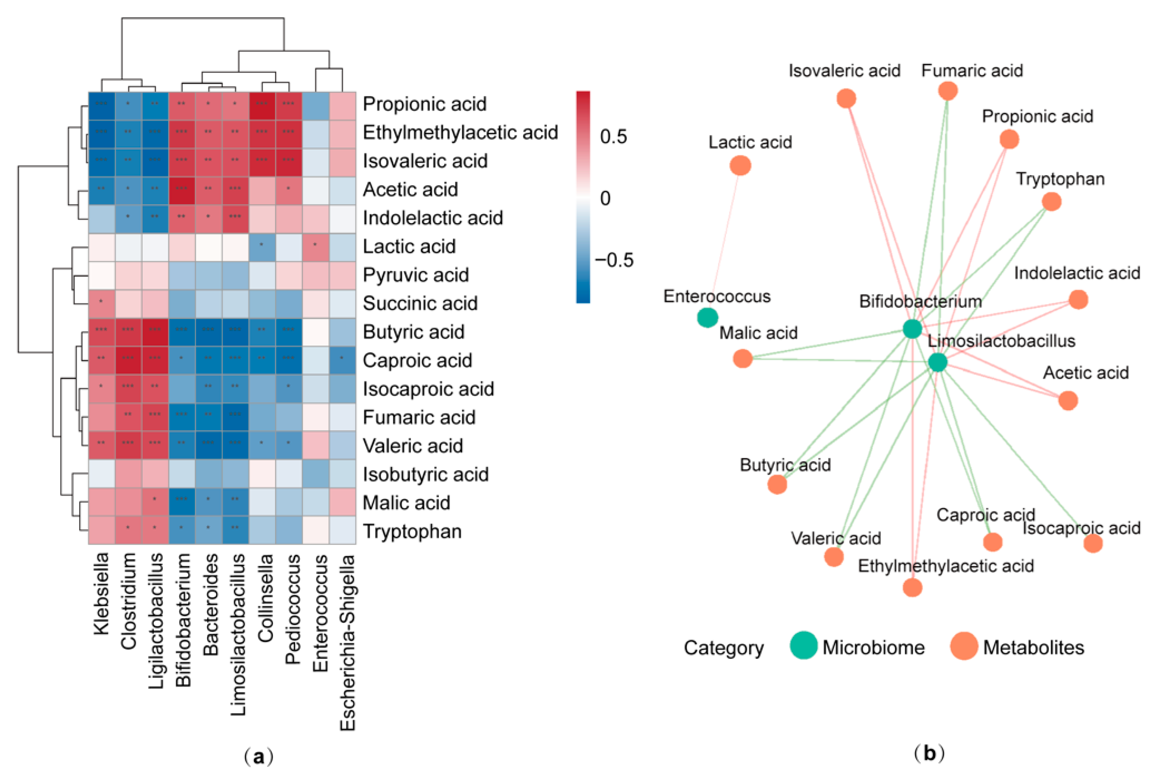
Spearman correlation analysis between the relative abundance of the top ten gut microbiota and the concentration of metabolites was performed using the comprehensive dataset encompassing all groups (OB and CON) (Figure 3). Bifidobacterium and Limosilactobacillus were positively linked to acetic, ethylmethylacetic, propionic, and indolelactic acids, but negatively associated with butyric acid, certain other organic acids, and tryptophan. In addition, Enterococcus exhibited a positive correlation with lactic acid. Collectively, these findings revealed a strong link between gut microbiota and the generation of SCFAs and other metabolites, demonstrating that PCP can affect the gut microbiota composition and associated metabolic outputs.

3.6. Effects of PCP in Single-Strain Cultures

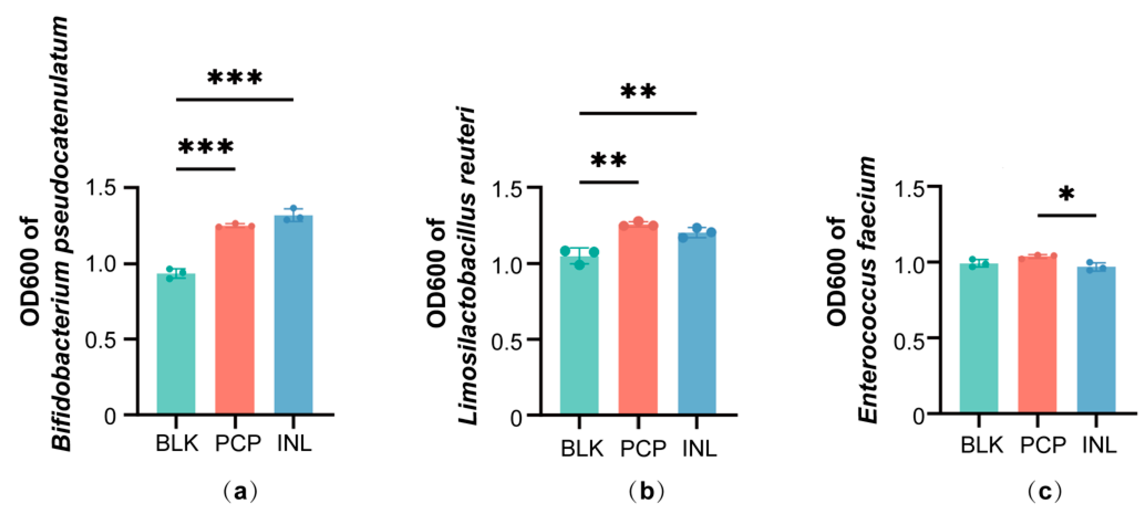
16S rRNA sequencing identified shifts at the genus level after PCP treatment. To further validate these findings, we selected representative species for single-strain fermentation. The results showed that compared with BLK, both PCP and INL significantly promoted the growth of Bifidobacterium pseudocatenulatum (p < 0.001) and Limosilactobacillus reuteri (p < 0.01) after 24 h of co-culture (Figure 4a,b). For Bifidobacterium pseudocatenulatum, INL showed a slightly higher trend than PCP. In contrast, for Limosilactobacillus reuteri, PCP tended to promote greater growth than INL. In addition, Enterococcus faecium exhibited a slight increase after PCP treatment compared to BLK (Figure 4c).

4. Discussion

This study examined how PCP affected gut microbiota in normal-weight and obese children using in vitro fermentation. Our results indicated that PCP exerted significant regulatory effects on gut microbial composition and metabolic activities, highlighting its potential to contribute to microbiota balance and promote metabolic well-being in children.
In the CON group, the enrichment of Bifidobacterium and Limosilactobacillus was observed after PCP treatment, in agreement with previous studies in healthy adults showing that PCP enhanced these genera [2]. The genera are producers of SCFAs, and are involved in immune and barrier regulation, which may partially explain the observed metabolic shifts [25,26,27,28]. The reduction in Escherichia-Shigella is consistent with studies showing that increased SCFA levels and lower pH suppress Enterobacteriaceae such as Escherichia-Shigella [29]. Functionally, acetic acid not only represents a key end-product of Bifidobacterium and Limosilactobacillus metabolism but can also be cross-fed to butyrate-producing bacteria, thereby further contributing to gut health [30]. Interestingly, the marked reduction in intermediate metabolites such as fumaric acid, malic acid, and pyruvic acid has rarely been reported in PCP studies. Previous PCP studies have focused primarily on SCFA end-products [2]. The changes of intermediate metabolites suggested that PCP redirected microbial metabolism from incomplete fermentation pathways toward the highly efficient core anaerobic microbial fermentation pathways. This led to promoting the production of primary fermentation acids rather than the buildup of intermediate metabolites.
In the OB group, PCP also showed beneficial effects, though less pronounced than in the CON group. Da Silva et al. [31] reported that children with obesity had reduced gut microbiota diversity, and at the genus level, obese children also exhibited a significantly lower abundance of Bifidobacterium. According to the framework of Patlok et al. [32], genera with higher initial abundance expand more strongly when fiber is supplied, suggesting that the lower baseline level of PCP-responsive genera in obese children contributed to their attenuated response. PCP consistently promoted Bifidobacterium, indicating that its prebiotic effect on this genus showed stability across host conditions. Interestingly, Enterococcus decreased in the CON group but expanded in the OB group after PCP treatment, likely due to baseline community differences: in the CON group, expansion of Bifidobacterium and Limosilactobacillus limited Enterococcus, whereas in the OB group, Enterococcus more effectively utilized PCP-derived substrates [27]. Enterococcus is often considered an opportunistic pathogen and can be associated with dysbiosis and inflammation. However, Enterococcus also participates in lactic acid production and possesses anti-oxidant properties, indicating that its expansion may represent a functional adaptation in obese children [28,33,34]. Another intriguing finding is that ACE and Shannon diversity indices showed opposite trends after the PCP and INL treatments. This outcome suggests that while PCP successfully diversified the community by increasing the total number of unique species (richness, measured by ACE), it also strongly favored the expansion and dominance of a few specific beneficial taxa, thereby reducing the overall mathematical evenness (measured by Shannon) [35]. The differences between the CON and OB groups highlighted the complexity of the microbiota community in obese children. Overall, PCP maintained its prebiotic effect in the OB group by promoting Bifidobacterium and acetic acid, although with lower efficiency under this microbial background.
Interestingly, one metabolic feature was enhanced by PCP in both the CON and OB groups. PCP fermentation reduced tryptophan and raised indolelactic acid, an indole derivative produced from tryptophan mainly by Bifidobacterium and Limosilactobacillus [36,37,38]. Indolelactic acid functions as a natural ligand of the aryl hydrocarbon receptor (AhR). By activating AhR signaling, indolelactic acid has been shown to strengthen intestinal barrier integrity and promote mucosal immune regulation [39]. Because obesity is often characterized by the reduced production of beneficial tryptophan-derived metabolites, the PCP-induced increase in indolelactic acid suggests that PCP may help restore this impaired pathway and provide benefits beyond SCFA-mediated effects [40,41].
A seemingly counterintuitive finding was the reduction in propionate and the lack of butyrate production in the CON group, coupled with a negative correlation between Bifidobacterium and butyric acid across all samples. Similarly, butyrate production was reduced after PCP treatment in the OB group. Similar reductions in butyric acid have also been reported in other in vitro PCP fermentation studies [2]. This likely reflects the limitation of the in vitro batch fermentation model, which does not mimic the human colon’s complex bacterial cross-feeding. Rapid fermentation by bacteria like Bifidobacterium and Limosilactobacillus can lead to acetic and lactic acid accumulation in a 24 h closed-batch system. This buildup lowers the pH and inhibits the growth and metabolic activity of the more sensitive, butyrate-producing bacteria. In addition, −80 °C storage may further reduce the viability of butyrate-producing bacteria. Bircher et al. [42] reported that butyrate-producing species, such as Roseburia intestinalis and Eubacterium hallii, exhibit extremely low recovery rates after −80 °C storage. Although the reduced recovery of butyrate-producing bacteria during −80 °C storage may have lowered the absolute butyrate levels, this systematic effect applied equally across all treatments and does not affect the comparative conclusions. Therefore, the model likely captures the primary fermentation surge but fails to replicate the stable, continuous environment of the colon where these cross-feeding pathways would continuously convert acetate into butyrate [43].
In addition, at the community level, INL more effectively promoted Bifidobacterium, whereas PCP more strongly increased Limosilactobacillus. These findings are consistent with prior evidence showing that inulin exerts a characteristic bifidogenic effect and is preferentially utilized by Bifidobacterium, while PCP can be metabolized by Limosilactobacillus and has been shown to enhance their growth or activity [3,44]. Although direct comparisons between INL and PCP remain scarce, our observations align with these substrate preference trends. Single-strain fermentations further confirmed the community-level findings. With respect to SCFA production, INL exerted stronger effects than PCP. Beyond SCFAs, differences were also observed in tryptophan metabolism: INL induced a higher indolelactic acid level in the CON group, while PCP more strongly enhanced the indolelactic acid level in the OB group. Our findings show that INL and PCP have largely similar, yet possibly complementary effects, likely operating through overlapping mechanisms with minor differences in microbiota and metabolic pathways.

5. Conclusions

In summary, PCP exhibited clear prebiotic potential by modulating gut microbiota and regulating key metabolites in children. These findings support its potential application as a functional food intervention to improve gut health in both obese and normal-weight children. The benefits appeared more pronounced in normal-weight children, suggesting that differences associated with health status may influence the response. This is a preliminary in vitro finding of PCP’s effects. However, the findings must be interpreted with caution. The use of a small, (n= 5 per group), pooled in vitro model limits the statistical power and prevents analysis of inter-individual variation. A further limitation is the use of 16S rRNA gene sequencing, which detects DNA from both viable and non-viable cells. This method cannot confirm whether the detected microbes were metabolically active at the time of sampling. In vivo studies are required to confirm safety and efficacy, particularly in the obese pediatric population. Future studies should also investigate whether personalized interventions, including the potential combination of PCP with probiotics, could optimize its prebiotic efficacy, particularly in obese children.

Supplementary Materials

The following supporting information can be downloaded at: https://www.mdpi.com/article/10.3390/foods14234077/s1, Table S1: Characteristics of subjects. Table S2: Content of monosaccharides in PCP. Figure S1: Monosaccharide content in PCP.

Author Contributions

Conceptualization, R.-R.W., F.-H.L. and D.-Y.Q.; methodology, R.-R.W., D.-Y.Q., X.-Q.L.; formal analysis, D.-Y.Q.; investigation, D.-Y.Q., X.-Q.L., Y.L., X.C., W.-N.Z., X.Y.; resources, R.-R.W., F.-H.L.; data curation, D.-Y.Q.; writing—original draft preparation, D.-Y.Q.; writing—review and editing, R.-R.W., F.-H.L., D.-Y.Q.; visualization, D.-Y.Q.; supervision, R.-R.W., F.-H.L.; project administration, R.-R.W., F.-H.L.; funding acquisition, F.-H.L. All authors have read and agreed to the published version of the manuscript.

Funding

This research was funded by the National Key Research and Development Program of China (2021YFC2701900).

Institutional Review Board Statement

The study protocol was approved by the Ethics Committee of Children’s Hospital of Fudan University (approval no. [2022]04, 28 April 2024).

Informed Consent Statement

Informed consent was obtained from the parents or legal guardians of all participating children.

Data Availability Statement

The original contributions presented in this study are included in the article/Supplementary Materials. Further inquiries can be directed to the corresponding authors.

Conflicts of Interest

The authors declare no conflicts of interest.

Abbreviations

The following abbreviations are used in this manuscript:
ACEAbundance-based Coverage Estimator
AhRAryl hydrocarbon receptor
ALTAlanine aminotransferase
ASTAspartate aminotransferase
BMIBody mass index
FBGFasting blood glucose
FINSFasting insulin
HDL-CHigh-density lipoprotein cholesterol
HOMA-βHomeostasis model assessment of β-cell function
HOMA-IRHomeostasis model assessment of insulin resistance
INLInulin
LDL-CLow-density lipoprotein cholesterol
LEfSeLinear Discriminant Analysis Effect Size
OTUsOperational taxonomic units
PCoAPrincipal coordinate analysis
PCPPoria cocos polysaccharides
PLS-DAPartial least squares discriminant analysis
SCFAsShort-chain fatty acids
SCRSerum creatinine
SDStandard deviation
TCTotal cholesterol
TFATrifluoroacetic acid
TGTriglycerides
UAUric acid
WGOCWorking Group on Obesity in China

References

  1. Zhao, M.; Guan, Z.; Tang, N.; Cheng, Y. The Differences between the Water- and Alkaline-Soluble Poria Cocos Polysaccharide: A Review. Int. J. Biol. Macromol. 2023, 235, 123925. [Google Scholar] [CrossRef] [PubMed]
  2. Zhou, X.; Li, Y.; Yang, Y.; Wei, L.; Wang, C.; Xu, J.; Song, J.; Liu, S.; Bai, J.; Suo, H. Regulatory Effects of Poria Cocos Polysaccharides on Gut Microbiota and Metabolites: Evaluation of Prebiotic Potential. NPJ Sci. Food 2025, 9, 53. [Google Scholar] [CrossRef] [PubMed]
  3. Li, X.; Li, X.; Liu, L.; Zhao, J.; Zhang, Y.; Chen, L. Antioxidant Activity and Metabolic Profile Changes in Poria Cocos Fermented by Breast-Milk-Derived Limosilactobacillus Reuteri HM-R28. Front. Nutr. 2025, 12, 1625875. [Google Scholar] [CrossRef] [PubMed]
  4. Duan, Y.; Huang, J.; Sun, M.; Jiang, Y.; Wang, S.; Wang, L.; Yu, N.; Peng, D.; Wang, Y.; Chen, W.; et al. Poria Cocos Polysaccharide Improves Intestinal Barrier Function and Maintains Intestinal Homeostasis in Mice. Int. J. Biol. Macromol. 2023, 249, 125953. [Google Scholar] [CrossRef]
  5. Ng, C.Y.J.; Lai, N.P.Y.; Ng, W.M.; Siah, K.T.H.; Gan, R.-Y.; Zhong, L.L.D. Chemical Structures, Extraction and Analysis Technologies, and Bioactivities of Edible Fungal Polysaccharides from Poria Cocos: An Updated Review. Int. J. Biol. Macromol. 2024, 261, 129555. [Google Scholar] [CrossRef]
  6. Deng, L.; Huang, G. Preparation, Structure and Application of Polysaccharides from Poria Cocos. RSC Adv. 2024, 14, 31008–31020. [Google Scholar] [CrossRef]
  7. Wan, J.; Wang, F.; Xiao, Y.; Cheng, Y.; Zheng, S.; Jiang, Q.; Tan, B.; Li, X.; Chen, J.; Liao, S. Poria Cocos Polysaccharide Alleviates Dextran Sulphate Sodium-Induced Ulcerative Colitis in Mice by Modulating Intestinal Inflammatory Responses and Microbial Dysbiosis. Int. J. Biol. Macromol. 2024, 283, 137450. [Google Scholar] [CrossRef]
  8. Sun, M.; Yao, L.; Yu, Q.; Duan, Y.; Huang, J.; Lyu, T.; Yu, N.; Peng, D.; Chen, W.; Wang, Y.; et al. Screening of Poria Cocos Polysaccharide with Immunomodulatory Activity and Its Activation Effects on TLR4/MD2/NF-κB Pathway. Int. J. Biol. Macromol. 2024, 273, 132931. [Google Scholar] [CrossRef]
  9. Chandrasekaran, P.; Weiskirchen, S.; Weiskirchen, R. Effects of Probiotics on Gut Microbiota: An Overview. Int. J. Mol. Sci. 2024, 25, 6022. [Google Scholar] [CrossRef]
  10. Zuo, W.-F.; Pang, Q.; Yao, L.-P.; Zhang, Y.; Peng, C.; Huang, W.; Han, B. Gut Microbiota: A Magical Multifunctional Target Regulated by Medicine Food Homology Species. J. Adv. Res. 2023, 52, 151–170. [Google Scholar] [CrossRef]
  11. Wang, J.; Zhu, N.; Su, X.; Gao, Y.; Yang, R. Gut-Microbiota-Derived Metabolites Maintain Gut and Systemic Immune Homeostasis. Cells 2023, 12, 793. [Google Scholar] [CrossRef]
  12. Wei, Y.; Jiang, Y.; Liu, M.; Zhang, X.; Guo, S.; Su, S.; Pan, Y.; Qiu, Z.; Zhang, X.; Duan, J.; et al. Chemical Structures of Lycii Fructus Polysaccharides Tailored the Gut Microbiota Composition of Aged Caenorhabditis elegans. Food Hydrocolloid. 2025, 169, 111608. [Google Scholar] [CrossRef]
  13. Bai, Y.; Zhou, Y.; Zhang, R.; Chen, Y.; Wang, F.; Zhang, M. Gut Microbial Fermentation Promotes the Intestinal Anti-Inflammatory Activity of Chinese Yam Polysaccharides. Food Chem. 2023, 402, 134003. [Google Scholar] [CrossRef] [PubMed]
  14. Zhou, M.; Zhang, Y.; Song, Z.; Tang, S.; Liu, Z.; Pang, M.; Zhang, D.; Wu, X.; Yu, X.; Wang, P.; et al. Enhanced Bioactivity of Honeysuckle-Cassia Seeds Extracts through Lactobacillus Acidophilus and Bacillus Subtilis Co-Fermentation: Impact on Alcoholic Liver Disease and Gut Microbiota. Food Chem. 2025, 486, 144463. [Google Scholar] [CrossRef] [PubMed]
  15. Ge, Q.; Hou, C.-L.; Rao, X.-H.; Zhang, A.-Q.; Xiao, G.-M.; Wang, L.-Y.; Jin, K.-N.; Sun, P.-L.; Chen, L.-C. In Vitro Fermentation Characteristics of Polysaccharides from Coix Seed and Its Effects on the Gut Microbiota. Int. J. Biol. Macromol. 2024, 262, 129994. [Google Scholar] [CrossRef]
  16. Lister, N.B.; Baur, L.A.; Felix, J.F.; Hill, A.J.; Marcus, C.; Reinehr, T.; Summerbell, C.; Wabitsch, M. Child and Adolescent Obesity. Nat. Rev. Dis. Primers 2023, 9, 24. [Google Scholar] [CrossRef]
  17. Gong, J.; Zhang, Q.; Hu, R.; Yang, X.; Fang, C.; Yao, L.; Lv, J.; Wang, L.; Shi, M.; Zhang, W.; et al. Effects of Prevotella Copri on Insulin, Gut Microbiota and Bile Acids. Gut Microbes 2024, 16, 2340487. [Google Scholar] [CrossRef]
  18. Larruy-García, A.; Mahmood, L.; Miguel-Berges, M.L.; Masip, G.; Seral-Cortés, M.; De Miguel-Etayo, P.; Moreno, L.A. Diet Quality Scores, Obesity and Metabolic Syndrome in Children and Adolescents: A Systematic Review and Meta-Analysis. Curr. Obes. Rep. 2024, 13, 755–788. [Google Scholar] [CrossRef]
  19. Saeed, N.K.; Al-Beltagi, M.; Bediwy, A.S.; El-Sawaf, Y.; Toema, O. Gut Microbiota in Various Childhood Disorders: Implication and Indications. World J. Gastroenterol. 2022, 28, 1875–1901. [Google Scholar] [CrossRef]
  20. Radjabzadeh, D.; Boer, C.G.; Beth, S.A.; van der Wal, P.; Kiefte-De Jong, J.C.; Jansen, M.A.E.; Konstantinov, S.R.; Peppelenbosch, M.P.; Hays, J.P.; Jaddoe, V.W.V.; et al. Diversity, Compositional and Functional Differences between Gut Microbiota of Children and Adults. Sci. Rep. 2020, 10, 1040. [Google Scholar] [CrossRef]
  21. Li, H.; Ji, C.-Y.; Zong, X.-N.; Zhang, Y.-Q. Body Mass Index Growth Curves for Chinese Children and Adolescents Aged 0 to 18 Years. Zhonghua Er Ke Za Zhi 2009, 47, 493–498. [Google Scholar] [PubMed]
  22. Sarbini, S.R.; Kolida, S.; Naeye, T.; Einerhand, A.; Brison, Y.; Remaud-Simeon, M.; Monsan, P.; Gibson, G.R.; Rastall, R.A. In Vitro Fermentation of Linear and α-1,2-Branched Dextrans by the Human Fecal Microbiota. Appl. Environ. Microbiol. 2011, 77, 5307–5315. [Google Scholar] [CrossRef] [PubMed]
  23. Tan, Y.-Y.; Yue, S.-R.; Lu, A.-P.; Zhang, L.; Ji, G.; Liu, B.-C.; Wang, R.-R. The Improvement of Nonalcoholic Steatohepatitis by Poria Cocos Polysaccharides Associated with Gut Microbiota and NF-κB/CCL3/CCR1 Axis. Phytomedicine 2022, 103, 154208. [Google Scholar] [CrossRef] [PubMed]
  24. Wang, Y.; Zhang, M.; Ruan, D.; Shashkov, A.S.; Kilcoyne, M.; Savage, A.V.; Zhang, L. Chemical Components and Molecular Mass of Six Polysaccharides Isolated from the Sclerotium of Poria Cocos. Carbohyd. Res. 2004, 339, 327–334. [Google Scholar] [CrossRef]
  25. O’Callaghan, A.; van Sinderen, D. Bifidobacteria and Their Role as Members of the Human Gut Microbiota. Front. Microbiol. 2016, 7, 925. [Google Scholar] [CrossRef]
  26. Lee, A.H.; Rodriguez Jimenez, D.M.; Meisel, M. Limosilactobacillus Reuteri—A Probiotic Gut Commensal with Contextual Impact on Immunity. Gut Microbes 2025, 17, 2451088. [Google Scholar] [CrossRef]
  27. Gilmore, M.S.; Clewell, D.B.; Ike, Y.; Shankar, N. (Eds.) Enterococci: From Commensals to Leading Causes of Drug Resistant Infection; Massachusetts Eye and Ear Infirmary: Boston, MA, USA, 2014. [Google Scholar]
  28. Anania, C.; Di Marino, V.P.; Olivero, F.; De Canditiis, D.; Brindisi, G.; Iannilli, F.; De Castro, G.; Zicari, A.M.; Duse, M. Treatment with a Probiotic Mixture Containing Bifidobacterium Animalis Subsp. Lactis BB12 and Enterococcus Faecium L3 for the Prevention of Allergic Rhinitis Symptoms in Children: A Randomized Controlled Trial. Nutrients 2021, 13, 1315. [Google Scholar] [CrossRef]
  29. Zhao, J.; Bai, M.; Ning, X.; Qin, Y.; Wang, Y.; Yu, Z.; Dong, R.; Zhang, Y.; Sun, S. Expansion of Escherichia-Shigella in Gut Is Associated with the Onset and Response to Immunosuppressive Therapy of IgA Nephropathy. J. Am. Soc. Nephrol. 2022, 33, 2276–2292. [Google Scholar] [CrossRef]
  30. Shetty, S.A.; Boeren, S.; Bui, T.P.N.; Smidt, H.; de Vos, W.M. Unravelling Lactate-Acetate and Sugar Conversion into Butyrate by Intestinal Anaerobutyricum and Anaerostipes Species by Comparative Proteogenomics. Environ. Microbiol. 2020, 22, 4863–4875. [Google Scholar] [CrossRef]
  31. Da Silva, C.C.; Monteil, M.A.; Davis, E.M. Overweight and Obesity in Children Are Associated with an Abundance of Firmicutes and Reduction of Bifidobacterium in Their Gastrointestinal Microbiota. Child. Obes. 2020, 16, 204–210. [Google Scholar] [CrossRef]
  32. Patloka, O.; Komprda, T.; Franke, G. Review of the Relationships Between Human Gut Microbiome, Diet, and Obesity. Nutrients 2024, 16, 3996. [Google Scholar] [CrossRef]
  33. Kim, H.; Oh, N.; Kwon, M.; Kwon, O.-H.; Ku, S.; Seo, J.; Roh, S. Exopolysaccharide of Enterococcus Faecium L15 Promotes the Osteogenic Differentiation of Human Dental Pulp Stem Cells via P38 MAPK Pathway. Stem Cell Res. Ther. 2022, 13, 446. [Google Scholar] [CrossRef]
  34. Mi, J.; He, T.; Hu, X.; Wang, Z.; Wang, T.; Qi, X.; Li, K.; Gao, L.; Liu, C.; Zhang, Y.; et al. Enterococcus Faecium C171: Modulating the Immune Response to Acute Lethal Viral Challenge. Int. J. Antimicrob. Agents 2023, 62, 106969. [Google Scholar] [CrossRef]
  35. Gu, F.; Borewicz, K.; Richter, B.; van der Zaal, P.H.; Smidt, H.; Buwalda, P.L.; Schols, H.A. In Vitro Fermentation Behavior of Isomalto/Malto-Polysaccharides Using Human Fecal Inoculum Indicates Prebiotic Potential. Mol. Nutr. Food Res. 2018, 62, e1800232. [Google Scholar] [CrossRef] [PubMed]
  36. Sakurai, T.; Odamaki, T.; Xiao, J. Production of Indole-3-Lactic Acid by Bifidobacterium Strains Isolated fromHuman Infants. Microorganisms 2019, 7, 340. [Google Scholar] [CrossRef] [PubMed]
  37. Zhang, Q.; Zhao, Q.; Li, T.; Lu, L.; Wang, F.; Zhang, H.; Liu, Z.; Ma, H.; Zhu, Q.; Wang, J.; et al. Lactobacillus Plantarum-Derived Indole-3-Lactic Acid Ameliorates Colorectal Tumorigenesis via Epigenetic Regulation of CD8+ T Cell Immunity. Cell Metab. 2023, 35, 943–960.e9. [Google Scholar] [CrossRef] [PubMed]
  38. Jia, D.; Kuang, Z.; Wang, L. The Role of Microbial Indole Metabolites in Tumor. Gut Microbes 2024, 16, 2409209. [Google Scholar] [CrossRef]
  39. Wang, A.; Guan, C.; Wang, T.; Mu, G.; Tuo, Y. Lactiplantibacillus Plantarum-Derived Indole-3-Lactic Acid Ameliorates Intestinal Barrier Integrity through the AhR/Nrf2/NF-κB Axis. J. Agric. Food. Chem. 2024, 72, 9236–9246. [Google Scholar] [CrossRef]
  40. Cussotto, S.; Delgado, I.; Anesi, A.; Dexpert, S.; Aubert, A.; Beau, C.; Forestier, D.; Ledaguenel, P.; Magne, E.; Mattivi, F.; et al. Tryptophan Metabolic Pathways Are Altered in Obesity and Are Associated With Systemic Inflammation. Front. Immunol. 2020, 11, 557. [Google Scholar] [CrossRef]
  41. Rivera, J.E.; Lan, R.; Ferruzzi, M.G.; Børsheim, E.; Tas, E.; Diaz, E.C. Associations Between Serum Gut-Derived Tryptophan Metabolites and Cardiovascular Health Markers in Adolescents with Obesity. Nutrients 2025, 17, 2430. [Google Scholar] [CrossRef]
  42. Bircher, L.; Geirnaert, A.; Hammes, F.; Lacroix, C.; Schwab, C. Effect of Cryopreservation and Lyophilization on Viability and Growth of Strict Anaerobic Human Gut Microbes. Microb. Biotechnol. 2018, 11, 721–733. [Google Scholar] [CrossRef]
  43. Isenring, J.; Bircher, L.; Geirnaert, A.; Lacroix, C. In Vitro Human Gut Microbiota Fermentation Models: Opportunities, Challenges, and Pitfalls. Microbiome Res. Rep. 2023, 2, 2. [Google Scholar] [CrossRef]
  44. Selak, M.; Rivière, A.; Moens, F.; Van den Abbeele, P.; Geirnaert, A.; Rogelj, I.; Leroy, F.; De Vuyst, L. Inulin-Type Fructan Fermentation by Bifidobacteria Depends on the Strain Rather than the Species and Region in the Human Intestine. Appl. Microbiol. Biotechnol. 2016, 100, 4097–4107. [Google Scholar] [CrossRef]
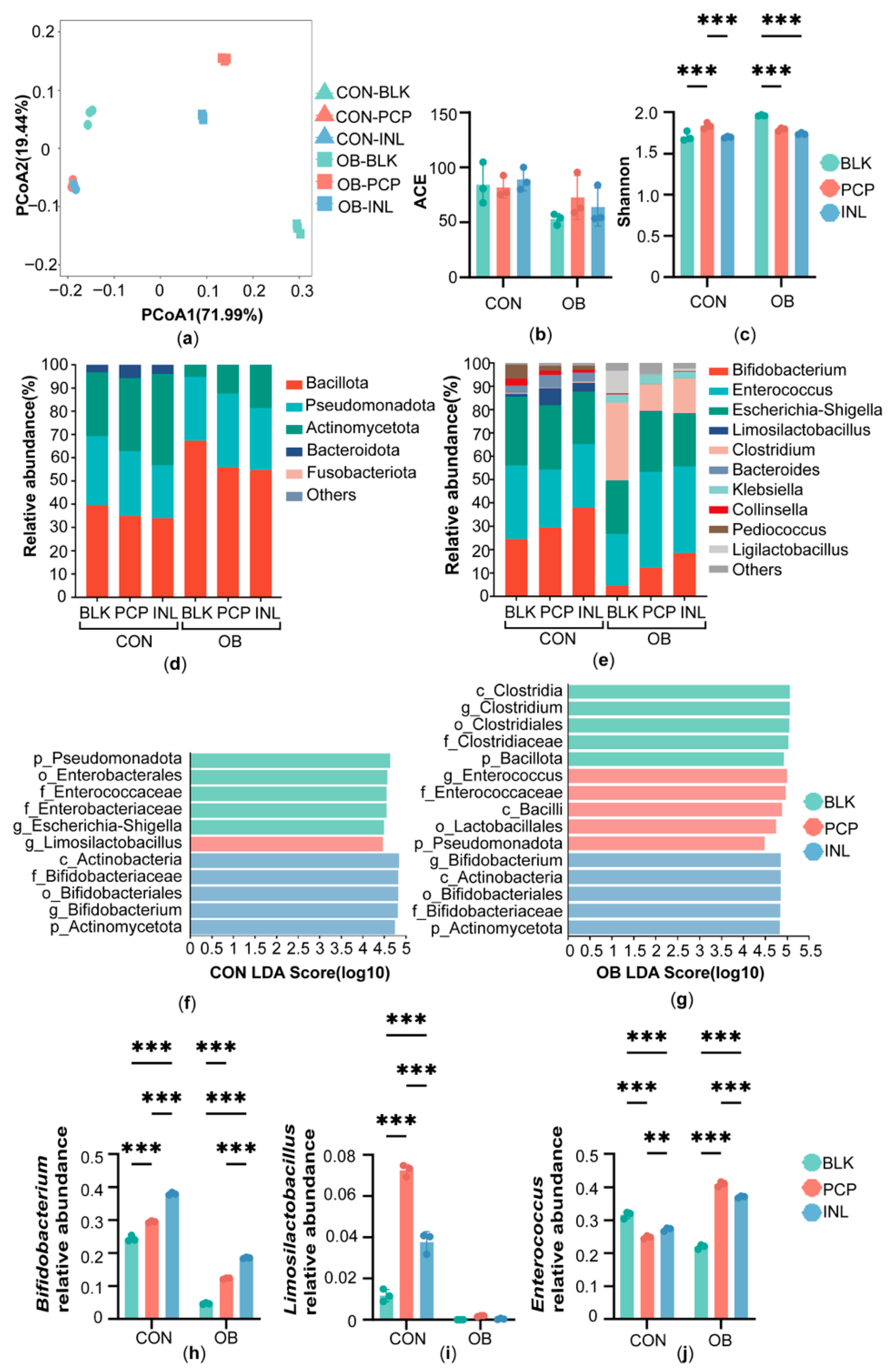
Figure 1. Gut microbiota structure after in vitro fermentation. (a) Principal coordinate analysis (PCoA) based on Bray–Curtis distances of the gut microbiota at the OTU level. Alpha diversity measured by (b) ACE (Abundance-based Coverage Estimator) index and (c) Shannon index. Gut microbiota composition at the (d) phylum and (e) genus level. Histograms illustrating the distribution of taxa identified through Linear Discriminant Analysis Effect Size (LEfSe) at two groups: (f) CON group and (g) OB group. Relative abundance of key genera: (h) Bifidobacterium, (i) Limosilactobacillus, and (j) Enterococcus. Data are expressed as the mean ± SD (n = 3), ** p < 0.01, *** p < 0.001.
Foods 14 04077 g001
Figure 2. Metabolic profiles after in vitro fermentation. (a) Partial least squares discriminant analysis (PLS-DA) of metabolites. (b) Overall metabolite composition at the class level. The levels of (c) total SCFA production (the total amount of acetic acid, ethylmethylacetic acid, propionic acid, butyric acid, isobutyric acid, valeric acid, isovaleric acid, caproic acid and isocaproic acid), (d) acetic acid, (e) propionic acid, (f) butyric acid, (g) lactic acid, (h) fumaric acid, (i) malic acid, (j) pyruvic acid, (k) tryptophan, (l) indolelactic acid. Data are expressed as the mean ± SD (n = 3), * p < 0.05, ** p < 0.01, *** p < 0.001.
Foods 14 04077 g002
Figure 3. (a) Spearman correlation analysis between the gut microbial genera and metabolites. The heatmap shows correlations between the top 10 most abundant genera and representative metabolites. (b) The correlation network highlights key genera and metabolites, with orange and green edges denoting positive and negative associations, respectively. Statistical significance is indicated by * p < 0.05, ** p < 0.01, and *** p < 0.001.
Foods 14 04077 g003
Figure 4. Effects of PCP on single-strain fermentation. OD600 of (a) Bifidobacterium pseudocatenulatum, (b) Limosilactobacillus reuteri, and (c) Enterococcus faecium after 24 h of co-culture. Data are expressed as the mean ± SD (n = 3), * p < 0.05, ** p < 0.01, *** p < 0.001.
Foods 14 04077 g004
Disclaimer/Publisher’s Note: The statements, opinions and data contained in all publications are solely those of the individual author(s) and contributor(s) and not of MDPI and/or the editor(s). MDPI and/or the editor(s) disclaim responsibility for any injury to people or property resulting from any ideas, methods, instructions or products referred to in the content.