Article

Supplementation with Probiotic Camel Milk Powder Improves Serum Glucose and Cholesterol as Well as the Related Cytokines in Patients with Type 2 Diabetes Mellitus

1
School of Food and Health, Beijing Technology and Business University, Beijing 100048, China
2
Department of Nutrition and Health, China Agricultural University, Beijing 100193, China
*
Author to whom correspondence should be addressed.
These authors contributed equally to this work.
Foods 2025, 14(19), 3318; https://doi.org/10.3390/foods14193318
Submission received: 22 August 2025 / Revised: 15 September 2025 / Accepted: 22 September 2025 / Published: 24 September 2025
(This article belongs to the Special Issue Bio-Functional Properties of Lactic Acid Bacteria in Functional Foods)

Abstract

Due to the close association between gut microbiota and diabetes, probiotic dairy products have drawn a lot of attention in the development of functional foods with anti-diabetic activity. In this study, 28 type 2 diabetic patients received 10 g of camel milk powder supplemented with Bifidobacterium animalis A6 (BBA6) twice a day, taking camel milk powder as the placebo. After 4 weeks of intervention, there was a significant decrease in fasting blood glucose, serum content of total cholesterol, and pro-inflammatory cytokines (IL-6, MCP-1). And, in the CA group, the level of irisin and osteocrin increased significantly, while the level of osteonectin also increased, but with no significance. For the adipokines, the intervention of CA decreased the adiponectin, resistin, lipocalin-2, and adipsin levels significantly. Gut microbiota analysis suggested a significant enrichment in the relative abundance of Bifidobacterium when compared with patients supplemented with camel milk powder alone. Furthermore, elevated fecal concentrations of glucose-1-phosphate, conduritol b epoxide, D-Arabitol, dehydroascorbic acid, and dl-p-Hydroxyphenyllactic acid, accompanied with a decrease in glycine, N-Acetylisatin, hydroxylamine, caprylic acid, maltotriose, and guaiacol, were found in patients of group CA. Compared with camel milk alone, the adding of BBA6 can significantly decrease fasting blood glucose in type 2 diabetic patients, while also improving dyslipidemia, chronic inflammation, and skeletal muscle functions, indicating the possibility of probiotic camel milk powder as a dietary treatment that targets metabolic syndromes such as diabetes.

1. Introduction

Diabetes is a serious, long-term condition that occurs when the body cannot effectively produce or use insulin. Type 2 diabetes mellitus (T2MD) accounts for around 90% of diabetes worldwide, which can be effectively prevented and managed through the adoption of healthy lifestyles, especially a healthy diet [1]. As a result, there are many studies focused on the evaluation of the hypoglycemic activity of functional foods or ingredients, which can be mainly divided into polyphenol-enriched plant-based foods [2,3,4,5] and proteins or peptides [6,7,8,9]. Meanwhile, since evidence has emerged about the close relationship between gut microbiota and diabetes [10,11], probiotics are used as a new effective therapeutic strategy in preventing and managing diabetes [12,13]. For example, Zhang et al. have proven that supplements of Akkermansia muciniphila can reduce the body weight and glycated hemoglobin of patients with T2DM [14]. Besides supplements with the strains themselves [15,16,17], probiotic dairy products such as probiotic soy milk [18] and probiotic fermented milk [19,20,21] were also found to improve the glycemic control of T2DM patients.
Camel milk is a rich source of vitamin C, lactic acid bacteria (LAB), beta-caseins, and milk whey proteins, including lactoferrin, lysozyme, lactoperoxidase, alpha-lactalbumin, and immunoglobulin [22]. And camel milk has a significant degree of biological activity. Compared with other milk, several studies reported the profound anti-diabetic activity of camel milk both in patients with type 1 diabetes [23,24,25,26,27] and T2MD [28,29,30]. However, camels mainly live in the desert areas of Africa/the Middle East or the cooler dry areas of Asia [31], leading to the unavailability of fresh camel milk for people living in other areas. Moreover, all the exiting clinical trials were based on fresh camel milk. It is not clear whether camel milk powder has a similar result.
Bifidobacterium animalis A6 (BBA6) was isolated from the feces of a centenarian. In our previous studies, it has been proven that BBA6 not only can promote mitochondrial biogenesis [32] but also enhanced fatty acid β-oxidation of adipose tissue [33] to alleviate obesity. Therefore, we speculated that BBA6 may have the potential to improve T2DM. Meanwhile, dairy products are important vectors for the delivery of probiotics to humans. And in order to solve the transportation and storage issues of camel milk, our lab developed a probiotic camel milk powder product. Therefore, the objective of this study was to evaluate the improving effect of camel milk power with BBA6 on T2DM patients. Except for some phenotypic indicators such as the glycemic index and serum insulin, some inflammatory factors, muscle factors, and adipokines were also detected. In addition, the changes in intestinal flora and bacterial metabolites of participants were also detected to analyze the possible mechanisms of BBA6 on improving T2DM.

2. Materials and Methods

2.1. Randomization, Blinding, and Intervention Protocol

This was a randomized, parallel, double-blind trial of T2DM patients, conducted in Beijing Chinese Medicine Hospital Pinggu Hospital, which lasted for 4 weeks. This study met the CONSORT criteria as recommended elsewhere [34]. The study was approved by the local ethics committee of China Agricultural University (CAUHR-2018026) and registered at ClinicalTrials.gov (NCT04296825).
The sample size determination and power analysis used by G* Power 3.1 program was based on a previous study [35]. The estimate sample size of 20 was calculated using the parallel clinical trial formula, assuming an alpha error of 0.05 and a power of 80%. Supposing an estimated 10% dropout rate, there were 22~23 patients for each group (45 patients in total).
A total of 45 T2DM patients were recruited from subjects attending the clinic of Beijing Chinese Medicine Hospital Pinggu Hospital. Based on previous research, the inclusion and exclusion criteria have been slightly modified and determined [14]. The inclusion criteria were as follows: (a) age of 35–68 years; (b) willingness to abstain from intake of all kinds of other milk, probiotic food, and fermented dairy products during the study, but otherwise sticking to previous eating habits. The exclusion criteria were as follows: (a) pregnancy or lactation in women; (b) patients with cancer; (c) allergy or intolerance to camel milk or cow milk. These criteria were verified during an inclusion visit that included a physical medical examination, dietary and physical activity assessments, standard anthropometrics, and an evaluation of fasting glycaemia, insulin, and lipid profile. After the verification, 40 subjects were eligible to participate in the study. Study procedure was explained to participants, and all participants provided written informed consent.
The 40 participants were randomly divided into two groups (20 individuals in each group): the camel milk+BBA6 group (CA group, camel milk powder with BBA6 at a dose of 2 × 10¹⁰ viable cells) and the control group (C group, camel milk). Both powders were packaged in the same bags (10 g each bag) and taken twice daily after breakfast and dinner, respectively, for 4 weeks.
All participants were asked to maintain their previous diet except for all kinds of other milk, probiotic food, and fermented dairy products, physical activity, and medications during the study. During the study, participants underwent interviews regarding adverse effects, symptoms, or changes in quality of life and diet every week. The allocation of groups was not revealed to the participants, or to the researchers who delivered probiotic camel milk or camel milk alone, or to those who conducted the weekly follow-ups.
The experimental design and sample collection time are shown in Table S1. The blood and feces samples were collected before consuming camel milk powder and BBA6 (W0) and after consuming for four weeks (W4). In addition, at W0 and W4, some indicators related to diabetes were tested, including fasting blood glucose, 2 h postprandial blood glucose, insulin, TG, TC, HDL-C, and LDL-C [36].

2.2. Camel Milk Powder and Probiotic Powder Preparation

The camel milk powder used in this study was provided by Xinjiang Jintuo Co., Ltd. (Urumqi, China) and packaged by Sanhe Fucheng Biological Technology Co., Ltd. (Langfang, China). The nutritional contents of camel milk powder used in this study are detailed in Supplementary Table S2.
The BBA6 was cultured in a liquid medium based on a previous study [32], which contained the following (per liter of deionized water): 10 g of beef extract, 5 g of yeast extract, 10 g of tryptone peptone, 20 g of glucose, 2 g of triamine citrate, 2 g of K2HPO4, 0.5 g of MgSO4, 5 g of sodium acetate, 0.5 g of L-cysteine, 0.25 g of MnSO4, and 1 mL of Tween-80. The chemicals and reagents used in the cultivation process were purchased from Beijing Aoboxing Biotechnology Co., Ltd. (Beijing, China). After counting and centrifugation, the bacterial cells were freeze-dried and mixed with the camel milk powder.
Patients were given sufficient supplies of the two products at the beginning of the intervention.

2.3. Blood Sample Collection and Measurements

Blood samples were collected twice at the beginning (W0) and the end (W4) of the study, respectively, and adhered to the standardized principle of fasting sample collection during collection [37]. On the day of blood sample collection, patients came to the hospital without breakfast; after the collection of the fasting blood samples, they were given the same breakfast, and the 2 h postprandial blood samples were collected after 2 h of the first bite of breakfast.
Human peripheral blood was collected in Vacutainer tubes (BD Biosciences, San Jose, CA, USA) and Vacutainer heparin tubes (BD Biosciences, San Jose, CA, USA), respectively. Blood samples were centrifuged at 1500× g for 30 min at room temperature. And within 1 h after blood collection, the fasting glycaemia, 2 h postprandial glycaemia, insulin, uric acid, and lipid were measured. The rest serum samples in Vacutainer heparin tubes were carefully removed, aliquoted, snap-frozen in liquid nitrogen, and stored in aliquots at −80 °C until further analysis.
Serum insulin was measured as in the previous study [38] by using the Architect i2000SR analyzer (Abbott Diagnostics, Abbott Park, IL, USA); blood glucose, content of total cholesterol (TC), total triglyceride (TG), high-density lipoprotein cholesterol (HDL-C), and low-density lipoprotein cholesterol (LDL-C) were measured using a Roche cobas ® e 411 analyzers (Roche, Hvidovre, Denmark) according to the manufactures’ protocol by the certified core clinical laboratory at the Beijing Chinese Medicine Hospital Pinggu Hospital.

2.4. Cytokines and Hormones Assays

The Luminex multi factor detection technology was used to detect inflammatory factors, myokines, and adipokines [39]. For determination of inflammation cytokines [tumor necrosis factor-α (TNF-α), interleukin-6 (IL-6), monocyte chemotactic protein-1 (MCP-1)], myokines [fibroblast growth factor-21 (FGF-21), irisin, osteocrin, osteonectin], and adipokines (adiponectin, resistin, lipocalin-2, adipsin), the human cytokine immunobead panels (Milliplex, Millipore Saint Charles, MO, USA) coupled with a multiplex assay (Luminex, Thermo Fisher Scientific, Waltham, MA, USA)) were used according to the manufactures’ protocol.

2.5. Fecal Sample Collection and Gut Microbiota Analysis

Fecal samples were collected in the morning of the day of blood collection by patients themselves at home based on the previous study [40]. Before defecating, waterproof paper was first put into closestool to keep feces away from liquids, then a portion of feces was put into sterile tubes containing RNAlater (Qiagen, Hilden, Germany), and the other portion was put into empty sterile tubes. Some fecal samples were collected the day before blood collection due to a higher defecation frequency. Fecal samples were brought to the hospital in ice boxes and then stored at −80 °C.
DNA was extracted from fecal samples using the phenol–chloroform extraction method [41], quantified using a NanoDrop spectrophotometer (OneC, Thermo Fisher Scientific, Waltham, MA, USA), and stored at −80 °C until further analysis. DNA was amplified using the universal primers 338F (5′-ACTCCTACGGGAGGCAGCAG-3′) and 806R (5′-GGACTACHVGGGTWTCT AAT-3′) to target the V3–V4 region of bacterial 16S rRNA. The resulting 468 bp-sized products were assessed, quantified, pooled, and sequenced on an Illumina Miseq PE300 platform (Illumina, San Diego, CA, USA) at Shanghai Majorbio Bio-pharm Technology Co., Ltd. (Shanghai, China) using a paired-end sequencing strategy. Raw data were spliced, filtered, and then used to select the operational taxonomic units (OTUs) with USEARCH software (version 7.0) and a default cutoff of 97% sequence similarity.
OTUs were further subjected to the Ribosomal Database Project classifier software (version 2.14) for taxonomic identification, with an 80% confidence threshold at the phylum, class, order, family, genus, and species levels. Further analysis such as ANOSIM/Adonis tests, principal coordinates analysis, abundance heatmap, and differences in gut microbiome composition were analyzed on the free online platform of Majorbio I-Sanger Cloud Platform (https://cloud.majorbio.com/) using weighted unifrac distance matrices.

2.6. Fecal Metabolomics Analysis

The untargeted metabolomics analysis of feces was conducted based on previous research, and some modifications have been made [42]. The 50 milligrams of feces were mixed with 40 μL internal standard (0.3 mg/mL, 2-chloro-L-phenylalanine), dissolved in methanol, and ultrasonically extracted with 360 μL methanol, then with 200 μL chloroform and 400 μL ddH2O in an ice bath for 30 min, respectively. After extraction, samples were centrifuged at 12,000 rpm at 4 °C for 10 min. Then, 400 μL supernatant was volatilized and oximated with 80 μL methoxyamine hydrochloride in pyridine (15 mg/mL), and they were shaken for 90 min at 37 °C, after which they were trimethylsilylated by adding 30 μL BSTFA (containing 1% TMCS) and 20 μL n-hexane, incubating for 1 h at 70 °C. After being left at room temperature for 30 min, samples were then subjected to GC-MS analysis. The chemicals used in this part were chromatographic grade and purchased from Thermo Fisher (Waltham, MA, USA).
Metabolic profiling of fecal samples was acquired by an Agilent 7890 A/5975C GC-MS (Agilent Technologies, Santa Clara, CA, USA) using a HP-5MS fused silica capillary column (30 m × 0.25 mm × 0.25 μm, Agilent J&W Scientific, Folsom, CA, USA). A total of 1 μL of the sample was injected in a non-split mode. The injector, ion source, and quadrupole rod temperatures were 260 °C, 230 °C, and 150 °C, respectively. High-purity helium (>99.999%) was used as the carrier gas, with a flow rate of 1.0 mL/min. The GC oven temperature program consisted of 60 °C for 2 min, after which the temperature ramped to 310 °C at 8 °C/min and held steady for 6 min. Mass spectra were acquired, and the mass scan range was set at m/z 50–600. Fecal samples were analyzed randomly.
Raw GC-MS mass spectra were converted to CDF format files by ChemStation (version E.02.02.1431, Agilent, CA, USA) and subsequently preprocessed using Chroma TOF (version 4.34, LECO, St Joseph, MI, USA), including raw signal extraction, data baseline filtering, peak identification, and integration. After alignment with the statistical comparison component, the “.CSV” file was obtained with three-dimension data sets including sample information, retention time, the mass-to-charge ratio, and peak intensity. Identification of metabolites was conducted using the Automatic Mass Spectral Deconvolution and Identification System, which was searched against commercially available databases such as the National Institute of Standards and Technology and Fiehn libraries. The signal integration area of each metabolite was normalized to the internal standard (2-chloro-L-phenylalanine) for each sample.
The normalized data were transformed using SIMCA-P 14.0 software (Umetrics AB, Umea, Sweden) for principal component analysis and partial least Squares-discriminant analysis (PLS-DA). The variable importance in projection (VIP) values of all the metabolites from the PLS-DA model were taken as criteria to find the variable importance of differential metabolites, and variables with a VIP >1.0 and a p-value  <  0.05 were considered relevant for group discrimination. The statistical significance between two groups was evaluated by a univariate Student’s t-test.

2.7. Statistical Analysis

Data entry was performed twice by two separate people. Differences between W0 and W4 of each group were evaluated by paired two-tailed Student’s t-tests using GraphPad Prism version 7.0 software (San Diego, CA, USA). Differences between the two groups at the same time point (W0 or W4) were compared by unpaired two-tailed Student’s t-tests using GraphPad Prism. Statistical significance was evaluated at an alpha level of 0.05.

3. Results and Discussion

3.1. Study Population

As shown in Figure 1, of the 40 participants that were randomized, 5 people did not come to pick up the intervention products, 2 people were lost to follow-up due to going out for travel, and 5 people were poor compliances (took other dairy products and probiotics). At the end of the 4-week intervention, 28 participants completed the experiment and were subjected to the analysis: 14 received camel milk powder supplemented with BBA6 (CA) and 14 received camel milk powder (C). None of the participants reported any adverse effects, including gastrointestinal disorders. Baseline comparison showed no significant differences in blood glucose, insulin, and lipid profiles between different groups (p > 0.05, Table 1).

3.2. Changes in Glycemic Indices and Serum Insulin

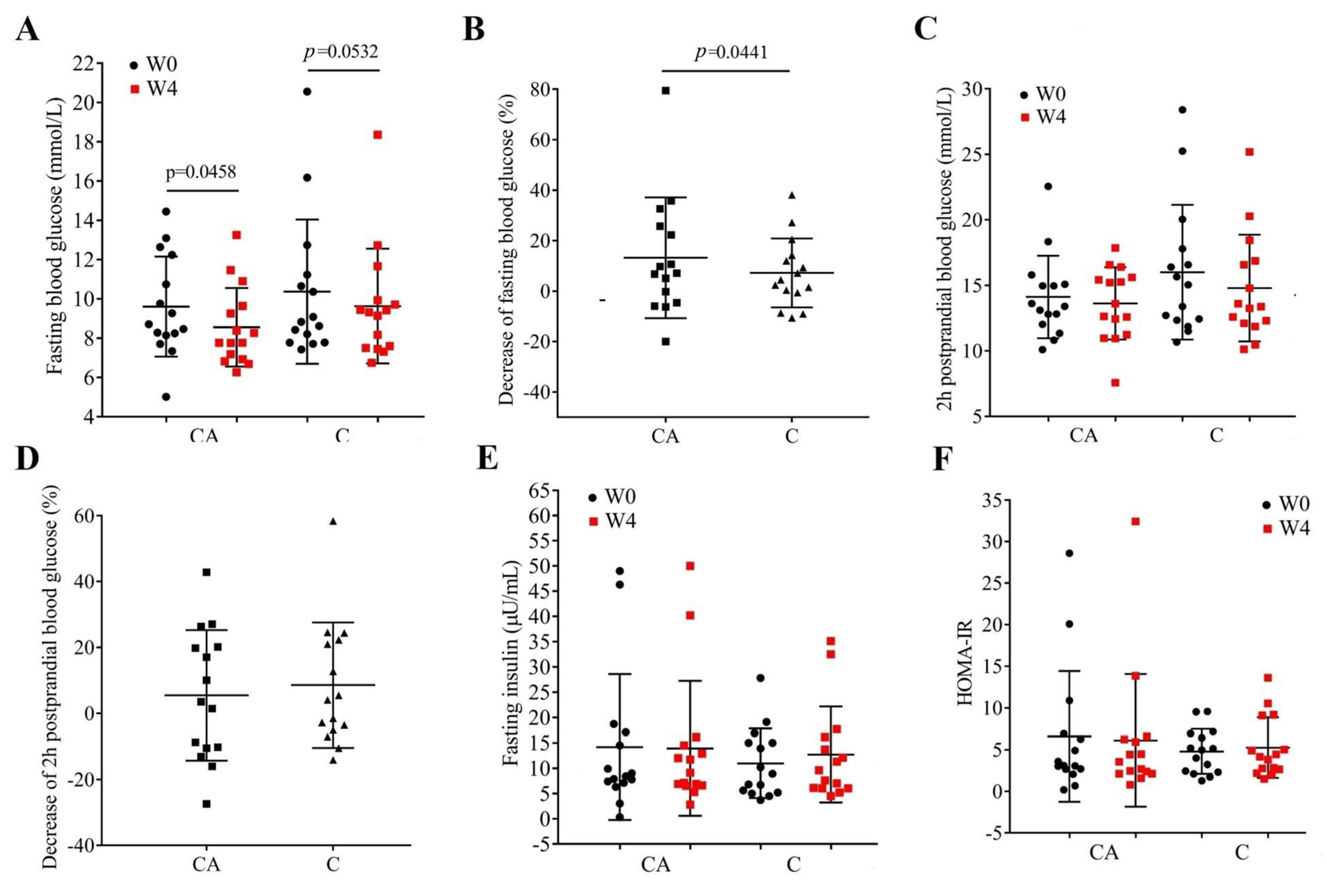
The fasting blood glucose, 2 h postprandial blood glucose, and fasting serum insulin of patients before and after the 4-week intervention are shown in Figure 2. At baseline, there were no significant differences between the two groups (p > 0.05). The hypoglycemic effect of camel milk has been proven in type 1 [24,25,26,27] and type 2 diabetic patients [28,29,30]; in this study, patients in the CA group exhibited a significant decrease in fasting blood glucose after the intervention compared with C group (p = 0.0458, Figure 2A) and a more effective hypoglycemic activity than camel milk powder alone (p = 0.0441, Figure 2B). Similarly, Alharbi et al. also found that fermented camel milk can effectively reduce the elevated blood sugar levels in Streptozotocin-induced diabetes in rats [43], which further proves our results that the combination of probiotics and camel milk exhibits better hypoglycemic effects. However, there were no significant changes in the 2 h postprandial blood glucose of patients either before and after the intervention (Figure 2C) or between the two groups (Figure 2D). This is different than Agrawal’s study: they found that, after the intervention of camel milk, the patients with T2DM from Iran showed a decrease in postprandial glucose [44]. We guess that the reason for this difference may be due to different dietary habits in the two regions.
The serum content of insulin was also not affected (Figure 2E, p > 0.05), and the intervention did not improve the insulin resistance of the patients (Figure 2F, p > 0.05). Previous studies on camel milk found a consistent unchanged insulin level in type 1 diabetic patients [24,45,46,47,48], but in T2DM patients the existing results were inconsistent [28,29], which may be due to the complicated mechanism in T2DM.

3.3. Changes in Lipid Profile and Cardiovascular Risk

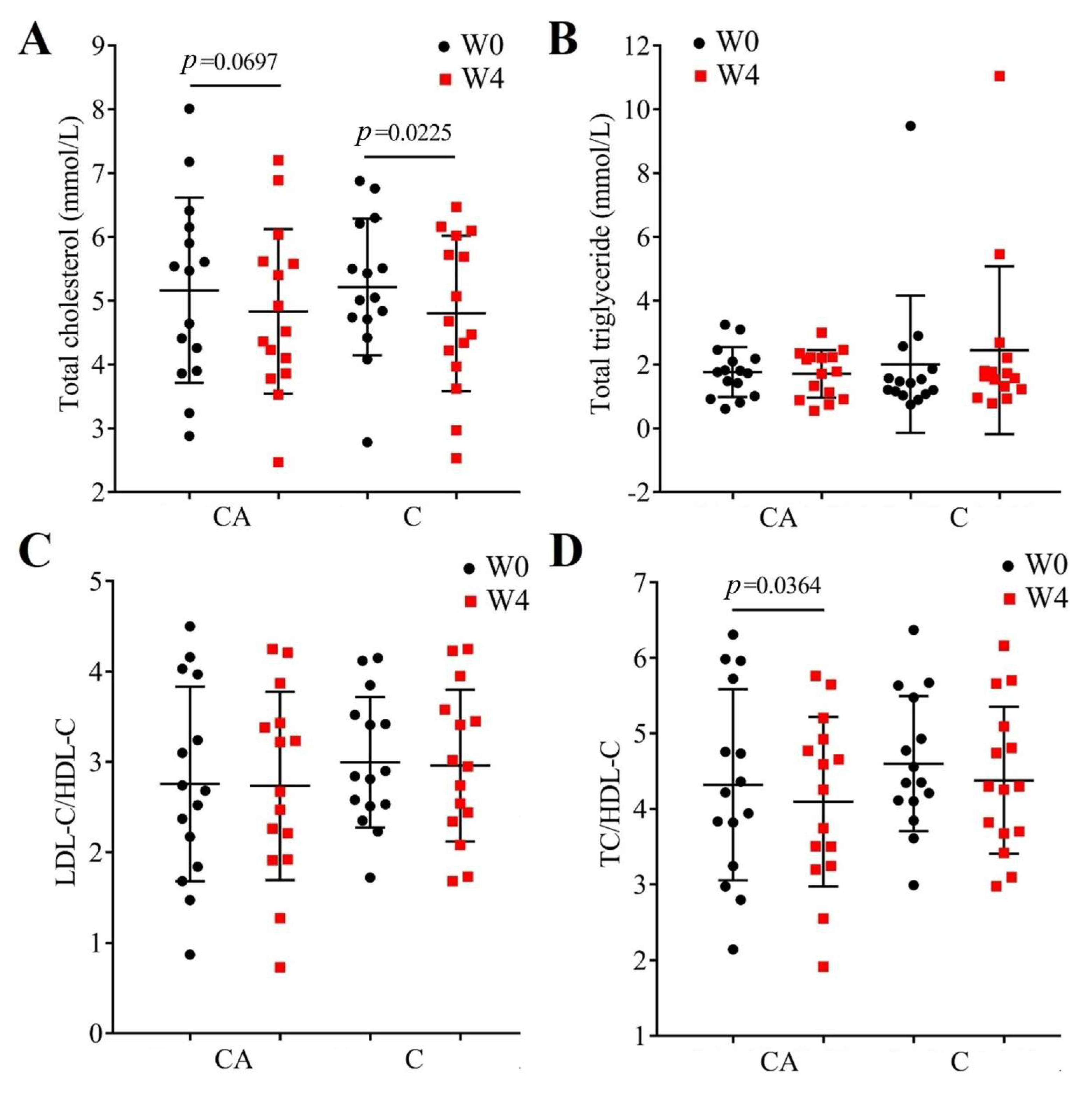
The relationship between diabetes and atherosclerotic cardiovascular disease is well established, with a significantly elevated risk for cardiovascular disease in diabetic patients [49]; therefore, we also measured the serum content of TC, TG, and the indicators of vascular risk (LDL/HDL cholesterol ratio and TC/HDL-C, Figure 3). As we can see from the results, both at baseline (W0) and post-intervention (W4), there were no significant differences between the two groups (CA-W0 vs. C-W0 and CA-W4 vs. C-W4, p > 0.05), whereas, after the intervention of CA, the TC showed a decreasing trend but was not significant (p = 0.0697, Figure 3A), but in the C group the TC significantly decreased (p = 0.0225). Furthermore, although there was no change in TG (Figure 3B) and the decreased TC in group CA was nonsignificant (p = 0.0697, Figure 3A), the intervention of CA resulted in a significant decrease in the ratio of TC and HDL-C (TC/HDL-C, p = 0.0364, Figure 3D), indicating a decreased vascular risk. However, there was no significant change in TC/HDL-C in the C group (Figure 3D). Previous clinical studies seldomly reported the effects of camel milk on the lipid profile, although animal studies reported a consistent decrease in TC [23,50,51]. In the limited studies in T2DM patients, Hanieh et al. found that patients who consumed 200 mL of camel milk for two months did not show a significant difference in blood lipids [52]. But, Sboui et al. proved that a supplement with 500 mL of camel milk can decrease the TC and TG significantly [30]. And, for the probiotic camel milk, some animal experiments have reported a decrease in TC and TG [53,54], which differs with our results. In these articles, the authors mixed the camel milk and the strains for fermentation. In this way, the probiotics may use the special nutrients in camel milk to produce unique metabolites, resulting in better probiotic effects. It is interesting that the effect of B. animal A6 on improving obesity has been reported in previous studies; we speculate whether mixed fermentation is necessary for better results.

3.4. Changes in Inflammatory Cytokines

It was reported that there was a chronic inflammation in diabetes [55] and a greater antioxidant and immunomodulatory activity of camel milk protein than bovine and other whey proteins [56,57]. So, in order to test whether probiotic camel milk has better anti-inflammatory effects, the content of TNF-α, IL-6, and MCP-1 were detected. As shown in Figure 4, there were no significant differences in the serum contents of inflammatory markers (TNF-α, IL-6, MCP-1) between groups both at baseline and post-intervention (p > 0.05). Within-group comparisons (W0 vs. W4) suggested that the decreased TNF-α (p > 0.05), IL-6 (p = 0.0103) and MCP-1 (p = 0.0814) contents in all CA groups were more obvious than those in C groups. Previous studies found that camel milk or camel milk whey proteins can reduce the pro-inflammatory IL-1β, IL-6, and TNFα in diabetic rats [29,58,59]. However, there are few or no studies proving that camel milk has a clinical effect on reducing inflammatory factors.

3.5. Changes in Adipokines and Myokines Profile

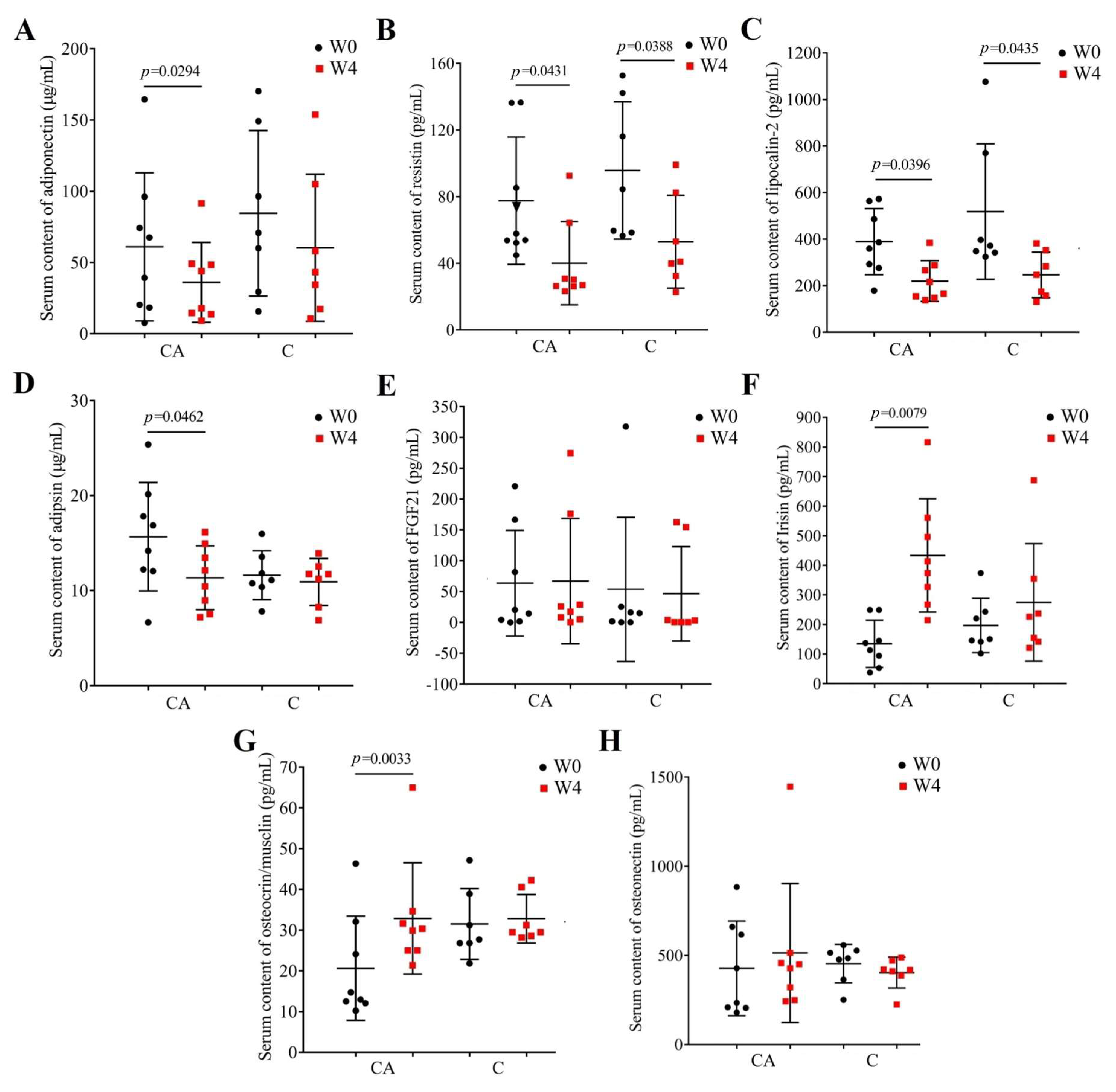
T2DM is the most common metabolic disease at present, and insulin resistance is considered to be the main reason for the occurrence and development of T2DM [60]. Lots of research has shown that the impaired function of muscles and adipose tissue is an important cause of insulin resistance. Skeletal muscle and adipocytes are secretory organs, which communicate with each other to regulate energy homeostasis and insulin sensitivity though the cytokines, called myokines and adipokines, respectively [61,62]. Therefore, we measured the serum contents of adipokines (adiponectin, resistin, lipocalin-2, adipsin) and myokines (FGF-21, irisin, osteocrin, osteonectin) in patients before and after the 4-week intervention to understand whether these interventions improved the function of patients’ skeletal muscle and adipose tissue, and the results are shown in Figure 5. There were no significant differences between different groups at baseline (W0) and after intervention (W4, p > 0.05). In the C group, results showed that it can significantly decrease the levels of resistin and lipocalin-2 (Figure 5B,C). Intervention with camel milk powder supplemented with BBA6 significantly decreased the content of adipokines (adiponectin, resistin, lipocalin-2, and adipsin) and increased myokine (irisin and osteocrin) levels. Although increased levels of adiponectin (Figure 5A) and adipsin (Figure 5D) were found to be associated with a lower risk of T2DM [63,64] in humans and improvement in pancreatic beta-cell function in mice [64,65], respectively, we found a significant decrease in patients with a significant decrease in fasting blood glucose (group CA). This further explains the relationship between adipose tissue dysfunction and diabetes. The other two adipokines, resistin (Figure 5B) and lipocalin-2 (Figure 5C), which decreased significantly in patients treated with camel milk powder alone and in combination, were reported to be good for the improvement of diabetes [66]. Similarly, Auguet et al. found that, in severely obese women, lipid carrier transporter 2 is upregulated and accompanied by an increase in pro-inflammatory cytokines [67]. Elevated serum lipocalin-2 is closely and independently associated with impaired glucose regulation and T2DM in Chinese people [68], and a lipocalin-2 deficiency attenuates the insulin resistance associated with obesity in mice [69]. Resistin promotes insulin resistance in mice, whereas whether it does so in humans is unclear [70,71], since it was synthesized in adipocytes in mice, whereas in humans it is generated by macrophages and monocytes and not adipocytes [72].
As for the myokines, no significant changes were observed in FGF21 in the two groups. And after intervention of CA, the osteonectin showed an increasing tendency, while showing a decreasing tendency in C group, although not significant (Figure 5H). Furthermore, the significant and specific decreases in irisin (p = 0.0079, Figure 5F) and osteocrin (p = 0.0033, Figure 5G) were also an indicator of the improvement of diabetes. Irisin is an adipo-myokine hormone produced during exercise. Studies have proven that irisin exhibits anti-inflammatory effects by inhibiting NF-κB signaling and countering insulin resistance [73]. Meanwhile, Irisin elevates in healthy states but reduces in diseases; this is the same as our results [74]. Osteocrin is also a regulator of bone growth as a novel vitamin D-regulated bone-specific protein [75]. Zhang et al. found that osteocrin can prevent diabetic cardiomyopathy via restoring proteasomal activity [76]. In this study, the significant increase in these two myokines in patients treated with camel milk powder supplemented with BBA6 indicated an improvement in diabetes.

3.6. Changes in Gut Microbiota

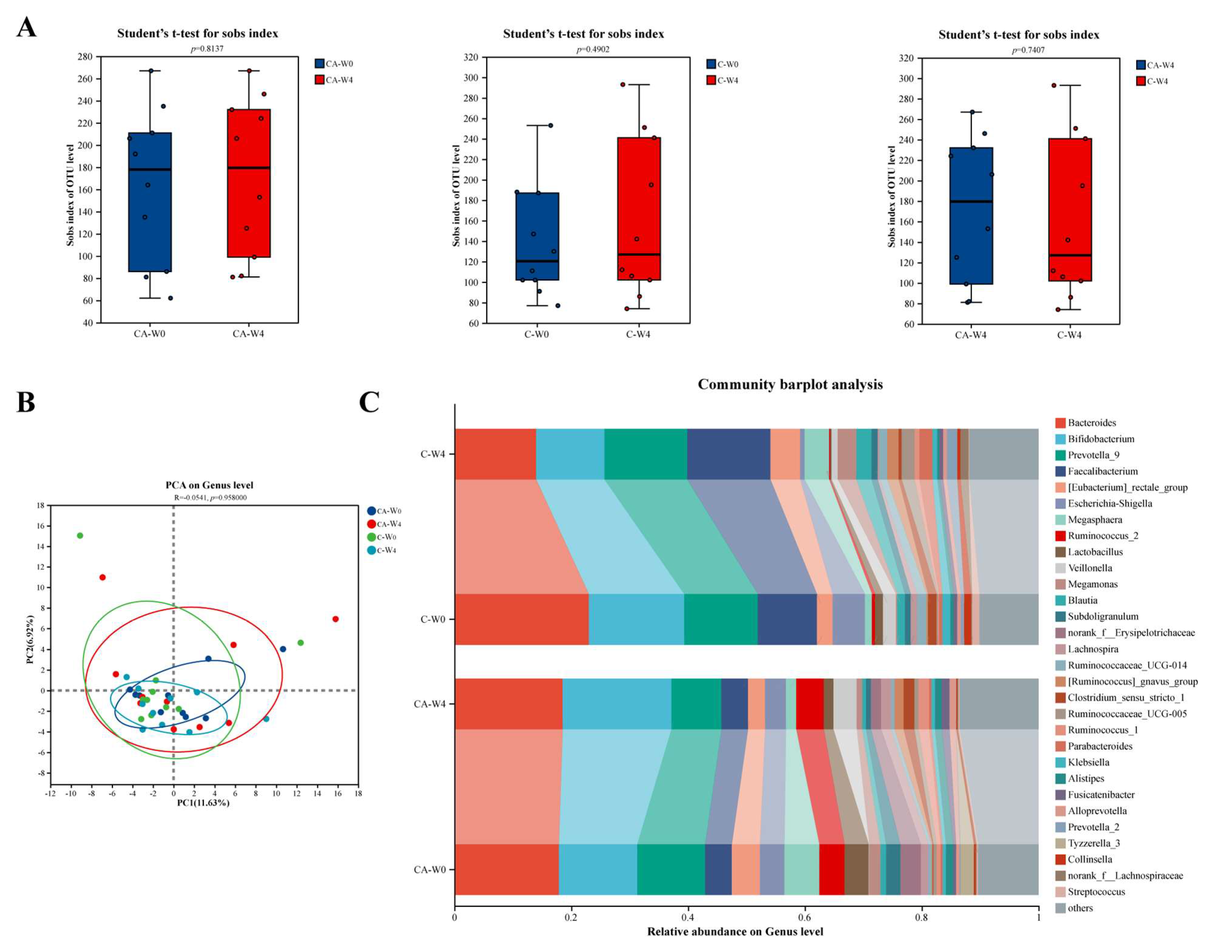
More and more evidence suggests a close relationship between gut microbiota and diabetes [77], and, since there is a component of probiotics in our study, we analyzed gut microbiota before and after the intervention (Figure 6). In Figure 6A, although there was no significant change in diversity after C and CA interventions, the species richness in the CA group was higher than that in the C group based on the post-intervention results. As shown in Figure 6B, the PCoA analysis showed that, after the interventions of C and CA, there was no significant difference in the species composition between the two groups. Such inconspicuous results have also been found by Qin et al. in the gut microbiota of healthy individuals and patients with T2DM [78]. Therefore, they believed that the gut microbiota of patients with T2DM is imbalanced, but it is a moderate imbalance, not a severe one. Thus, the reason for these results may be due to the individual differences and the short intervention time. To further observe the changes in gut microbiota before and after intervention, the community composition at the genus level is shown in Figure 6C. From the result, it was found that the interventions of C and CA have different regulatory trends on various microorganisms. The Bacteroides and Bifidobacterium are proven to have a lower abundance in the intestinal flora of patients with T2DM [79]. And their abundance increased after the intervention of CA, but decreased in group C. This may suggest that CA intervention has a potential role in improving diabetes. And for the characteristic bacteria of T2DM, the abundance of Lactobacillus decreased in both groups.
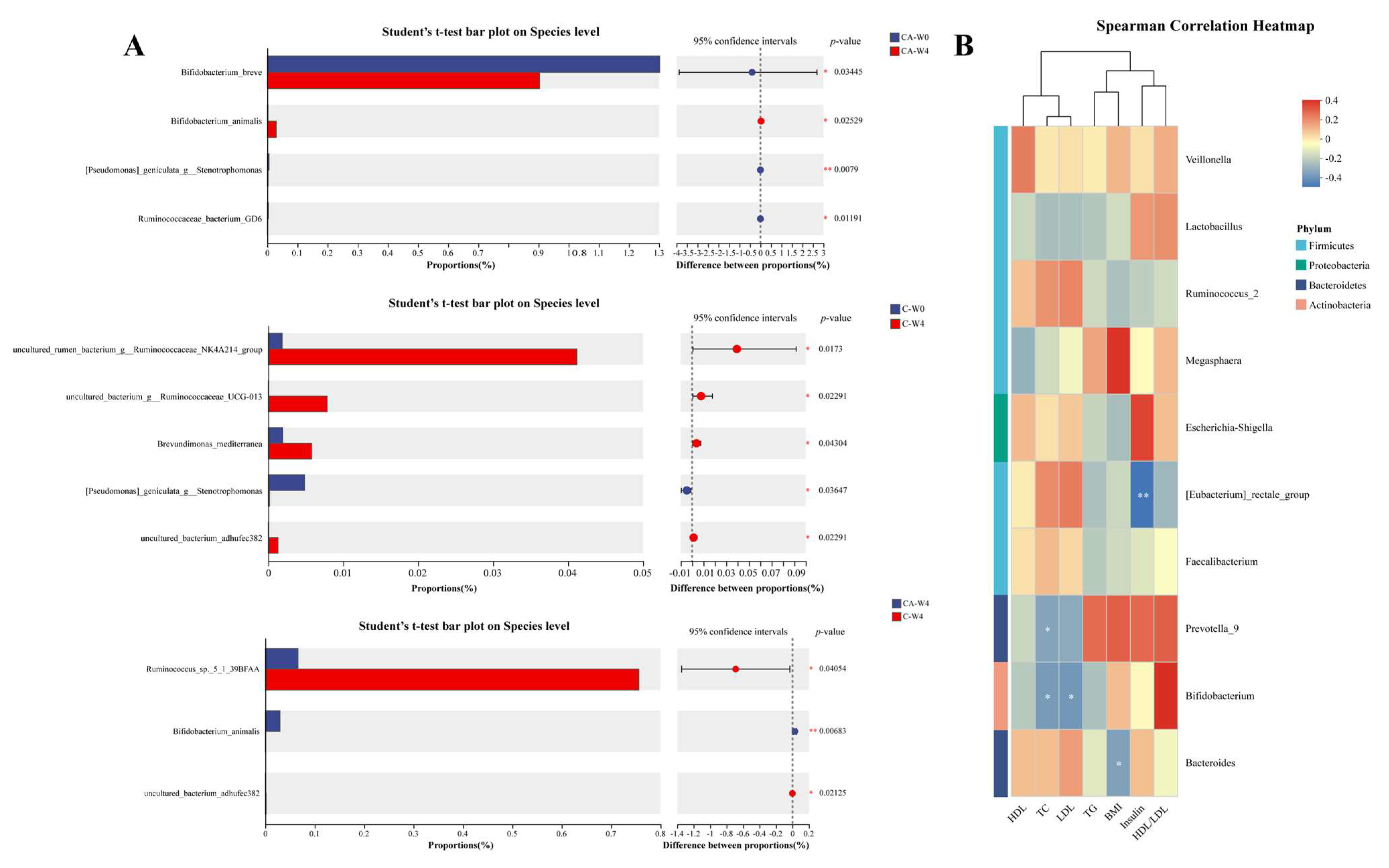
To further observe the changes in gut microbiota, we analyzed the microorganisms with significant differences before and after intervention by using Student’s t-test. As shown in Figure 7A, after the intervention of CA, the abundance of Bifidobacterium breve decreased as the Bifidobacterium animals increased. This is contrary to some research findings. For example, Chaiyavat et al. found that supplementing with B. brevis can prevent the deterioration of representative clinical parameters in T2DM subjects and change the components of the gut microbe [80]. On the contrary, a mate analysis study showed that the Lactobacillus which enrich in T2DM were positive with the B. brevis [81]. Therefore, we speculated that these results may be caused by the diversity and complexity of human gut microbiota. The increase of B. animalis effectively explains the colonization of BBA6. Meanwhile, we also observed that the C group increased the abundance of Ruminococcus, which play an important role in digesting resistant starch. Studies have proven that more consumption of resistant starch can improve T2MD, and the gut microbiota can guide the effects of it in improving T2MD [82,83]. And the Ruminococcus are the productor of butyrate, which can protect the pancreatic islet cells and regulate blood sugar [84]. Thus, we speculated that C and CA can change the digestive mode of subjects.
Then, we investigated whether gut microbes were associated with biochemical indicators of T2MD, and the results are shown in Figure 7B. Results showed that the Bacteroides were significantly negatively correlated with BMI. And the Bifidobacterium and Prevotella-9 were evidently negatively correlated with TC, while the Bifidobacterium also negatively correlated with LDL. For the content of insulin, the [Eubacterium]_rectale group showed an obviously negative association with it. Based on the difference test, we found that Ruminococcus are related to the effects of C and CA in improving T2MD. Figure 7B shows that Ruminococcus_2 is positively correlated with TC and LDL, and has a high correlation coefficient. As for the Bifidobacterium, it has the highest correlation coefficient with the HDL/LDL, which may indicate its potential mechanism for improving T2MD.

3.7. Changes in Fecal Metabolites

Figure 8 summarizes the change in metabolomics in fecal samples from participants with subjects after the intervention of CA and C. From the results, we can see that the PLS-DA analysis (Figure 8A) showed an obvious separation between the two groups, while the separation rate of two components are 19.60% and 16.50%, respectively. These indicated that the CA group drove a distinct metabolic signature. Then, the volcano analysis was used to detect the differential metabolites. A total of 220 metabolites were identified, and among them 10 metabolites were significantly upregulated and 31 downregulated in the CA group (Figure 8B). After that, 19 metabolites were regarded as key metabolites and are shown in Figure 8C. Among these key metabolites, those with a VIP value greater than 1.5 were analyzed especially. N-Acetylisatin and glycine are precursor substances of glutathione, which is regarded as an antioxidant synthesized by glycine and can protect the function of β cells [83,85].Tuell et al. found that dietary supplementation with glycine can achieve lower level of oxidative stress and oxidant damage in patients with T2MD through an increase in the synthesis of glutathione [86]. And Lei et al. proved that the N-Acetylisatin inhibits NADPH oxidase activation in diabetes and attenuates tissue oxidative damage in all organs. In this study, we observed a decrease in N-Acetylisatin and glycine, which may indicate the enhancement of antioxidant ability. For the hydroxylamine, Kimura et al. found that it can enhance the glucose uptake of C2C12 cells through the PIK3 pathway [87]. We also found a decrease in it in the CA group. Caprylic acid is a medium chain fatty acid (MCFA), which has been proven to be associated with reduced inflammation and improved insulin sensitivity in vitro [88]. Maltotriose [89] and glucose-1-phosphate [90] have been proven to be higher in T2MD patients than in healthy controls. We found a decrease in maltotriose but an increase in glucose-1-phosphate in the CA group. Studies proved that conduritol B epoxide and D-Arabitol are potential treatments for T2DM. Conduritol B epoxide is an irreversible inhibitor of β-glucosidase and can block the hydrolysis of sugar [91]. And D-Arabitol can improve systemic insulin sensitivity by reshaping the gut microbiota [92]. In this article, we observed an increase in both metabolites in the CA group. Previous studies have shown that people with T2DM have lower plasma dehydroascorbic acid concentrations than those with normal glucose control [93,94]. This research also found that, after the intervention of CA, the concentration of dehydroascorbic acid was up. In order to further explore the pathways of CA’s improvement of T2DM, we conducted a KEGG pathway enrichment analysis on differential metabolites (Figure 8D). The results showed that the CA group preferentially activated sulfur metabolism (p = 0.020), glycine/serine/threonine metabolism (p = 0.040), and thiamine metabolism (p = 0.080). These pathways converge on the generation of glutathione [86] and one-carbon units [95]—both essential for countering oxidative stress and enhancing insulin signaling.

4. Conclusions

As a traditional milk in Africa, Asia, and the Middle East, it was believed that the regular consumption of fresh camel milk may aid in the prevention and control of diabetes. In previous studies, we investigated the improvement effect of camel milk in improving T2MD. We found that, compared with cow’s milk, the supplement with camel milk can decrease fasting blood glucose, 2 hr postprandial blood glucose, serum content of total cholesterol, resistin, and lipocalin-2. And content of osteocrin, amylin, and GLP-1 showed a significant increase in the camel milk group. The gut microbiota has also changed; the camel milk powder supplement significantly enriched the relative abundance of Clostridium_sensu_stricto_1 and [Eubacterium]_eligens_group [28].
And, in this study, we evaluated the anti-diabetic activity of a probiotic camel milk product, camel milk powder supplemented with BBA6, a strain of Bifidobacterium animalis isolated by our lab from the feces of Longevity Elderly in Bama, Guangxi. Compared with the camel milk, the probiotic camel milk product showed a better improving effect on blood glucose, blood lipids, adipokines, myokines, and the gut microbiota after the 4-week intervention. These results in combination suggested that the probiotic camel milk product can be used as part of the treatment of T2MD.
However, as a clinical trial, this study inevitably has some limitations. First of all, due to the high dropout rate in this study, the sample size was relatively small; therefore, in subsequent experiments, we will expand the sample size to further validate the experimental results. In addition, the probiotics used in this experiment were from a directly freeze-dried powder added into camel milk powder instead of mixed during fermentation. Thus, it is necessary to further explore the improvement effect of mixed fermentation on T2DM patients.

Supplementary Materials

The following supporting information can be downloaded at https://www.mdpi.com/article/10.3390/foods14193318/s1. Table S1: Summary of testing indicators and methods, Table S2: Nutritional contents of camel milk powder.

Author Contributions

Conceptualization, M.Z. and B.F.; formal analysis, M.Z., Y.L. and R.W.; investigation, M.Z. and B.F.; resources, S.G.; writing—original draft preparation, M.Z., Y.L. and B.F.; writing—review and editing, B.F.; funding acquisition, M.Z. and B.F. All authors have read and agreed to the published version of the manuscript.

Funding

This work was supported by the National Key Research and Development Program of China (2024YFD2100903), the 2115 Talent Development Program of China Agricultural University, and the Cross-Innovation Open Project of Food Flavor and Health, Beijing Technology & Business University (FFHCI-2025061).

Institutional Review Board Statement

The study was conducted in accordance with the Declaration of Helsinki, and the study was approved by the local ethics committee of China Agricultural University (CAUHR-2018026, approval date 6 February 2018), and registered at ClinicalTrials.gov (NCT04296825).

Informed Consent Statement

Informed consent was obtained from all subjects involved in the study.

Data Availability Statement

The original contributions presented in the study are included in the article/Supplementary Material; further inquiries can be directed to the corresponding author.

Acknowledgments

We would like to thank all study participants and all the nurses in Beijing Chinese Medicine Hospital Pinggu Hospital for their assistance in helping to run the study visits and in the processing of blood samples and in blood biochemical items analysis.

Conflicts of Interest

The authors declare no conflicts of interest.

Abbreviations

The following abbreviations are used in this manuscript:
BBA6Bifidobacterium animalis A6
T2DMType 2 diabetes mellitus
W0The beginning of the study
W4The end of the study
CACamel milk supplemented with BBA6
CCamel milk
TCTotal cholesterol
TGTotal triglyceride
HDL-CHigh-density lipoprotein cholesterol
LDL-CLow-density lipoprotein cholesterol
TNF-αTumor necrosis factor-α
IL-6Interleukin-6
MCP-1Monocyte chemotactic protein-1
FGF-21Fibroblast growth factor-21
OTUsOperational taxonomic units
PLS-DASquares-discriminant analysis
VIPVariable importance in projection

References

  1. Ballan, R.; Saad, S.M.I. Characteristics of the gut microbiota and potential effects of probiotic supplements in individuals with type 2 diabetes mellitus. Foods 2021, 10, 2528. [Google Scholar] [CrossRef]
  2. Qin, Y.; Guo, J.; Lin, Y.; You, Y.; Huang, W.; Zhan, J. Evaluation of hypoglycemic polyphenolic compounds in blueberry extract: Functional effects and mechanisms. Antioxidants 2024, 13, 1490. [Google Scholar] [CrossRef]
  3. Esfandiar, Z.; Hosseini-Esfahani, F.; Mirmiran, P.; Yuzbashian, E.; Azizi, F. The association of dietary polyphenol intake with the risk of type 2 diabetes: Tehran lipid and glucose study. Diabetes Metab. Syndr. Obes. 2020, 13, 1643–1652. [Google Scholar] [CrossRef]
  4. Liu, Y.; Zhang, X.; Zhan, L.; Xu, C.; Sun, L.; Jiang, H.; Sun, C.; Li, X. LC-Q-TOF-MS characterization of polyphenols from white bayberry fruit and its antidiabetic effect in kk-ay mice. ACS Omega 2020, 5, 17839–17849. [Google Scholar] [CrossRef]
  5. Nwakiban Atchan, A.P.; Shivashankara, S.T.; Piazza, S.; Tchamgoue, A.D.; Beretta, G.; Dell’Agli, M.; Magni, P.; Agbor, G.A.; Kuiaté, J.-R.; Manjappara, U.V. Polyphenol-rich extracts of xylopia and aframomum species show metabolic benefits by lowering hepatic lipid accumulation in diet-induced obese mice. ACS Omega 2022, 7, 11914–11928. [Google Scholar] [CrossRef]
  6. Fernandez, M.A.; Marette, A. Novel perspectives on fermented milks and cardiometabolic health with a focus on type 2 diabetes. Nutr. Rev. 2018, 76, 16–28. [Google Scholar] [CrossRef]
  7. Mudgil, P.; Kamal, H.; Yuen, G.C.; Maqsood, S. Characterization and identification of novel antidiabetic and anti-obesity peptides from camel milk protein hydrolysates. Food Chem. 2018, 259, 46–54. [Google Scholar] [CrossRef] [PubMed]
  8. Ayoub, M.A.; Palakkott, A.R.; Ashraf, A.; Iratni, R. The molecular basis of the anti-diabetic properties of camel milk. Diabetes Res. Clin. Pract. 2018, 146, 305–312. [Google Scholar] [CrossRef] [PubMed]
  9. Valencia-Mejía, E.; Batista, K.A.; Fernández, J.J.A.; Fernandes, K.F. Antihyperglycemic and hypoglycemic activity of naturally occurring peptides and protein hydrolysates from easy-to-cook and hard-to-cook beans (Phaseolus vulgaris L.). Food Res. Int. 2019, 121, 238–246. [Google Scholar] [CrossRef]
  10. Eliasson, B.; Allansson Kjölhede, E.; Salö, S.; Fabrin Nielsen, N.; Eeg-Olofsson, K. Associations between hba1c and glucose time in range using continuous glucose monitoring in type 1 diabetes: Cross-sectional population-based study. Diabetes Ther. 2024, 15, 1301–1312. [Google Scholar] [CrossRef] [PubMed]
  11. He, L.; Yang, F.Q.; Tang, P.; Gao, T.H.; Yang, C.X.; Tan, L.; Yue, P.; Hua, Y.N.; Liu, S.J.; Guo, J.L. Regulation of the intestinal flora: A potential mechanism of natural medicines in the treatment of type 2 diabetes mellitus. Biomed. Pharmacother. 2022, 151, 113091. [Google Scholar] [CrossRef]
  12. Lee, Y.S.; Lee, D.; Park, G.S.; Ko, S.H.; Park, J.; Lee, Y.K.; Kang, J. Lactobacillus plantarum HAC01 ameliorates type 2 diabetes in high-fat diet and Streptozotocin-induced diabetic mice in association with modulating the gut microbiota. Food Funct. 2021, 12, 6363–6373. [Google Scholar] [CrossRef] [PubMed]
  13. Koay, K.-P.; Tsai, B.C.-K.; Kuo, C.-H.; Kuo, W.-W.; Luk, H.-N.; Day, C.H.; Chen, R.-J.; Chen, M.Y.-C.; Padma, V.V.; Huang, C.-Y. Hyperglycemia-induced cardiac damage is alleviated by heat-inactivated Lactobacillus reuteri gmnl-263 via activation of the IGF1r survival pathway. Probiotics Antimicrob. Proteins 2021, 13, 1044–1053. [Google Scholar] [CrossRef]
  14. Zhang, Y.; Liu, R.; Chen, Y.; Cao, Z.; Liu, C.; Bao, R.; Wang, Y.; Huang, S.; Pan, S.; Qin, L.; et al. Akkermansia muciniphila supplementation in patients with overweight/obese type 2 diabetes: Efficacy depends on its baseline levels in the gut. Cell Metab. 2025, 37, 592–605.e6. [Google Scholar] [CrossRef]
  15. Sabico, S.; Al-Mashharawi, A.; Al-Daghri, N.M.; Wani, K.; Amer, O.E.; Hussain, D.S.; Ahmed Ansari, M.G.; Masoud, M.S.; Alokail, M.S.; McTernan, P.G. Effects of a 6-month multi-strain probiotics supplementation in endotoxemic, inflammatory and cardiometabolic status of T2DM patients: A randomized, double-blind, placebo-controlled trial. Clin. Nutr. 2019, 38, 1561–1569. [Google Scholar] [CrossRef]
  16. Li, G.; Feng, H.; Mao, X.-L.; Deng, Y.-J.; Wang, X.-B.; Zhang, Q.; Guo, Y.; Xiao, S.-M. The effects of probiotics supplementation on glycaemic control among adults with type 2 diabetes mellitus: A systematic review and meta-analysis of randomised clinical trials. J. Transl. Med. 2023, 21, 442. [Google Scholar] [CrossRef]
  17. Zikou, E.; Dovrolis, N.; Dimosthenopoulos, C.; Gazouli, M.; Makrilakis, K. The effect of probiotic supplements on metabolic parameters of people with type 2 diabetes in Greece—A randomized, double-blind, placebo-controlled study. Nutrients 2023, 15, 4663. [Google Scholar] [CrossRef] [PubMed]
  18. Hasanpour, A.; Babajafari, S.; Mazloomi, S.M.; Shams, M. The effects of soymilk plus probiotics supplementation on cardiovascular risk factors in patients with type 2 diabetes mellitus: A randomized clinical trial. BMC Endocr. Disord. 2023, 23, 36. [Google Scholar] [CrossRef]
  19. Gu, Y.; Li, X.; Chen, H.; Sun, Y.; Yang, L.; Ma, Y.; Yong Chan, E.C. Antidiabetic effects of multi-species probiotic and its fermented milk in mice via restoring gut microbiota and intestinal barrier. Food Biosci. 2022, 47, 101619. [Google Scholar] [CrossRef]
  20. Deveci, G.; Çelik, E.; Ağagündüz, D.; Bartkiene, E.; Rocha, J.M.F.; Özogul, F. Certain fermented foods and their possible health effects with a focus on bioactive compounds and microorganisms. Fermentation 2023, 9, 923. [Google Scholar] [CrossRef]
  21. Ağagündüz, D.; Yilmaz, B.; Cemali, Ö.; Šimat, V.; Akkus, G.; Kulawik, P.; Ozogul, F. Impact of dairy food products on type 2 diabetes: Gut-pancreas axis for lower glucose level. Trends Food Sci. Technol. 2024, 153, 104741. [Google Scholar] [CrossRef]
  22. Khan, M.Z.; Xiao, J.; Ma, Y.; Ma, J.; Liu, S.; Khan, A.; Khan, J.M.; Cao, Z. Research Development on Anti-Microbial and Antioxidant Properties of Camel Milk and Its Role as an Anti-Cancer and Anti-Hepatitis Agent. Antioxidants 2021, 10, 788. [Google Scholar] [CrossRef] [PubMed]
  23. Hussain, H.; Wattoo, F.H.; Wattoo, M.H.S.; Gulfraz, M.; Masud, T.; Shah, I.; Ali, S.; Alavi, S.E. Camel milk as an alternative treatment regimen for diabetes therapy. Food Sci. Nutr. 2021, 9, 1347–1356. [Google Scholar] [CrossRef]
  24. Agrawal, R.P.; Jain, S.; Shah, S.; Chopra, A.; Agarwal, V. Effect of camel milk on glycemic control and insulin requirement in patients with type 1 diabetes: 2-years randomized controlled trial. Eur. J. Clin. Nutr. 2011, 65, 1048–1052. [Google Scholar] [CrossRef] [PubMed]
  25. Agrawal, R.P.; Tantia, P.; Jain, S.; Agrawal, R.; Agrawal, V. Camel milk: A possible boon for type 1 diabetic patients. Cell Mol. Biol. 2013, 59, 99–107. [Google Scholar]
  26. Agrawal, R.P.; Saran, S.; Sharma, P.; Gupta, R.P.; Kochar, D.K.; Sahani, M.S. Effect of camel milk on residual beta-cell function in recent onset type 1 diabetes. Diabetes Res. Clin. Pract. 2007, 77, 494–495. [Google Scholar] [CrossRef]
  27. Abdalla, K.; Fadlalla, A. Effects of sudanese dromedary’s camel raw milk on insulin doses and carbohydrate metabolism in type 1 diabetic patients. J. Biomol. Res. Ther. 2018, 7, 24994. [Google Scholar] [CrossRef]
  28. Zheng, Y.; Wu, F.; Zhang, M.; Fang, B.; Zhao, L.; Dong, L.; Zhou, X.; Ge, S. Hypoglycemic effect of camel milk powder in type 2 diabetic patients: A randomized, double-blind, placebo-controlled trial. Food Sci. Nutr. 2021, 9, 4461–4472. [Google Scholar] [CrossRef]
  29. Dou, Z.; Liu, C.; Feng, X.; Xie, Y.; Yue, H.; Dong, J.; Zhao, Z.; Chen, G.; Yang, J. Camel whey protein (CWP) ameliorates liver injury in type 2 diabetes mellitus rats and insulin resistance (IR) in HepG2 cells via activation of the PI3K/Akt signaling pathway. Food Funct. 2022, 13, 255–269. [Google Scholar] [CrossRef]
  30. Sboui, A.; Atig, C.; Khabir, A.; Hammadi, M.; Khorchani, T. Camel milk used as an adjuvant therapy to treat type 2 diabetic patients: Effects on blood glucose, Hba1c, cholesterol, and TG levels. J. Chem. 2022, 2022, 5860162. [Google Scholar] [CrossRef]
  31. Alhaj, O.A.; Altooq, N.J.; Alenezi, A.F.; Janahi, A.I.; Janahi, M.I.; Humood, A.M.; AlRasheed, M.M.; Bragazzi, N.L.; Jahrami, H.A.; Faye, B. Camel milk composition by breed, season, publication year, and country: A global systematic review, meta-analysis, and meta-regression. Compr. Rev. Food Sci. Food Saf. 2022, 21, 2520–2559. [Google Scholar] [CrossRef]
  32. Huo, Y.; Lu, X.; Wang, X.; Wang, X.; Chen, L.; Guo, H.; Zhang, M.; Li, Y. Bifidobacterium animalis subsp. lactis A6 Alleviates Obesity Associated with Promoting Mitochondrial Biogenesis and Function of Adipose Tissue in Mice. Molecules 2020, 25, 1490. [Google Scholar] [CrossRef]
  33. Huo, Y.; Zhao, G.; Li, J.; Wang, R.; Ren, F.; Li, Y.; Wang, X. Bifidobacterium animalis subsp. lactis A6 Enhances Fatty Acid β-Oxidation of Adipose Tissue to Ameliorate the Development of Obesity in Mice. Nutrients 2022, 14, 598. [Google Scholar] [CrossRef] [PubMed]
  34. Schulz, K.F.; Altman, D.G.; Moher, D.; CONSORT Group. Consort 2010 statement: Updated guidelines for reporting parallel group randomised trials. BMJ 2010, 340, c332. [Google Scholar] [CrossRef] [PubMed]
  35. Kang, H. Sample size determination and power analysis using the G*Power software. J. Educ. Eval. Health Prof. 2021, 18, 17. [Google Scholar] [CrossRef]
  36. Tian, L.; Lu, C.; Teng, D.; Teng, W.; Li, J. Association between blood glucose indicators and metabolic diseases in the Chinese population: A national cross-sectional study. Chin. Med. J. 2025, 138, 2159–2169. [Google Scholar] [CrossRef]
  37. Simundic, A.M.; Cornes, M.; Grankvist, K.; Lippi, G.; Nybo, M. Standardization of collection requirements for fasting samples: For the Working Group on Preanalytical Phase (WG-PA) of the European Federation of Clinical Chemistry and Laboratory Medicine (EFLM). Clin. Chim. Acta 2014, 432, 33–37. [Google Scholar] [CrossRef]
  38. Trifunovic, D.; Stankovic, S.; Sobic-Saranovic, D.; Marinkovic, J.; Petrovic, M.; Orlic, D.; Beleslin, B.; Banovic, M.; Vujisic-Tesic, B.; Petrovic, M.; et al. Acute insulin resistance in ST-segment elevation myocardial infarction in non-diabetic patients is associated with incomplete myocardial reperfusion and impaired coronary microcirculatory function. Cardiovasc. Diabetol. 2014, 13, 73. [Google Scholar] [CrossRef] [PubMed]
  39. Takanohashi, A.; Prust, M.; Wang, J.; Gordish-Dressman, H.; Bloom, M.; Rice, G.I.; Schmidt, J.L.; Crow, Y.J.; Lebon, P.; Kuijpers, T.W.; et al. Elevation of proinflammatory cytokines in patients with Aicardi-Goutières syndrome. Neurology 2013, 80, 997–1002. [Google Scholar] [CrossRef]
  40. Ahlquist, D.A.; Sargent, D.J.; Loprinzi, C.L.; Levin, T.R.; Rex, D.K.; Ahnen, D.J.; Knigge, K.; Lance, M.P.; Burgart, L.J.; Hamilton, S.R.; et al. Stool DNA and occult blood testing for screen detection of colorectal neoplasia. Ann. Intern. Med. 2008, 149, 441–450. [Google Scholar] [CrossRef]
  41. Gautam, A. Gautam, A., Ed.; Phenol-chloroform DNA isolation method. In DNA and RNA Isolation Techniques for Non-Experts; Springer International Publishing: Cham, Switzerland, 2022; pp. 33–39. [Google Scholar]
  42. Wang, H.; Ainiwaer, A.; Song, Y.; Qin, L.; Peng, A.; Bao, H.; Qin, H. Perturbed gut microbiome and fecal and serum metabolomes are associated with chronic kidney disease severity. Microbiome 2023, 11, 3. [Google Scholar] [CrossRef] [PubMed]
  43. Alharbi, Y.M.; Sakr, S.S.; Albarrak, S.M.; Almundarij, T.I.; Barakat, H.; Hassan, M.F.Y. Antioxidative, Antidiabetic, and Hypolipidemic Properties of Probiotic-Enriched Fermented Camel Milk Combined with Salvia officinalis Leaves Hydroalcoholic Extract in Streptozotocin-Induced Diabetes in Rats. Antioxidants 2022, 11, 668. [Google Scholar] [CrossRef]
  44. Agrawal, R.P.; Sharma, P.; Gafoorunissa, S.J.; Ibrahim, S.A.; Shah, B.; Shukla, D.K.; Kaur, T. Effect of camel milk on glucose metabolism in adults with normal glucose tolerance and type 2 diabetes in Raica community: A crossover study. Acta Biomed. 2011, 82, 181–186. [Google Scholar]
  45. Mohamad, R.H.; Zekry, Z.K.; Al-Mehdar, H.A.; Salama, O.; El-Shaieb, S.E.; El-Basmy, A.A.; Al-said, M.G.; Sharawy, S.M. Camel milk as an adjuvant therapy for the treatment of type 1 diabetes: Verification of a traditional ethnomedical practice. J. Med. Food 2009, 12, 461–465. [Google Scholar] [CrossRef]
  46. Agrawal, P.; Swami, S.; Beniwal, R.; Kochar, D.; Sahani, M.; Tuteja, F.; Ghouri, S. Effect of camel milk on glycemic control, risk factors and diabetes quality of life in type-1 diabetes: A randomised prospective controlled study. J. Camel Pract. Res. 2003, 10, 1048–1052. [Google Scholar]
  47. Agrawal, R.; Beniwal, R.; Sharma, S.; Kochar, D.; Tuteja, F.; Ghorui, S. Effect of raw camel milk in type 1 diabetic patients: 1 year randomised study. J. Camel Pract. Res. 2005, 12, 27–35. [Google Scholar]
  48. Agrawal, R.P.; Beniwal, R.; Kochar, D.K.; Tuteja, F.C.; Ghorui, S.K.; Sahani, M.S.; Sharma, S. Camel milk as an adjunct to insulin therapy improves long-term glycemic control and reduction in doses of insulin in patients with type-1 diabetes a 1 year randomized controlled trial. Diabetes Res. Clin. Pract. 2005, 68, 176–177. [Google Scholar] [CrossRef]
  49. Borén, J.; Öörni, K.; Catapano, A.L. The link between diabetes and cardiovascular disease. Atherosclerosis 2024, 394, 117607. [Google Scholar] [CrossRef]
  50. Magbola, S. Camel milk improvement of antioxidant and lipid profile in hypercholesterolemic rabbits. J. Cell Anim. Biol. 2018, 12, 1–4. [Google Scholar] [CrossRef]
  51. Meena, S.; Rajput, Y.S.; Sharma, R.; Singh, R. Effect of goat and camel milk vis a vis cow milk on cholesterol homeostasis in hypercholesterolemic rats. Small Rumin. Res. 2019, 171, 8–12. [Google Scholar] [CrossRef]
  52. Ejtahed, H.S.; Niasari Naslaji, A.; Mirmiran, P.; Zraif Yeganeh, M.; Hedayati, M.; Azizi, F.; Moosavi Movahedi, A. Effect of camel milk on blood sugar and lipid profile of patients with type 2 diabetes: A pilot clinical trial. Int. J. Endocrinol. Metab. 2015, 13, e21160. [Google Scholar] [CrossRef]
  53. Manaer, T.; Yu, L.; Nabi, X.-H.; Dilidaxi, D.; Liu, L.; Sailike, J. The beneficial effects of the composite probiotics from camel milk on glucose and lipid metabolism, liver and renal function and gut microbiota in db/db mice. BMC Complement. Med. Ther. 2021, 21, 127. [Google Scholar] [CrossRef]
  54. Manaer, T.; Sailike, J.; Sun, X.; Yeerjiang, B.; Nabi, X. Therapeutic effects of composite probiotics derived from fermented camel milk on metabolic dysregulation and intestinal barrier integrity in type 2 diabetes rats. Front. Pharmacol. 2025, 15, 1520158. [Google Scholar] [CrossRef] [PubMed]
  55. Zhao, L.; Hu, H.; Zhang, L.; Liu, Z.; Huang, Y.; Liu, Q.; Jin, L.; Zhu, M.; Zhang, L. Inflammation in diabetes complications: Molecular mechanisms and therapeutic interventions. MedComm 2024, 5, e516. [Google Scholar] [CrossRef]
  56. Swelum, A.A.; El-Saadony, M.T.; Abdo, M.; Ombarak, R.A.; Hussein, E.O.S.; Suliman, G.; Alhimaidi, A.R.; Ammari, A.A.; Ba-Awadh, H.; Taha, A.E.; et al. Nutritional, antimicrobial and medicinal properties of cmel’s milk: A review. Saudi J. Biol. Sci. 2021, 28, 3126–3136. [Google Scholar] [CrossRef]
  57. Badr, G.; Ramadan, N.K.; Sayed, L.H.; Badr, B.M.; Omar, H.M.; Selamoglu, Z. Why whey? Camel whey protein as a new dietary approach to the management of free radicals and for the treatment of different health disorders. Iran. J. Basic Med. Sci. 2017, 20, 338–349. [Google Scholar] [CrossRef]
  58. Badr, G.; Ramadan, N.K.; Abdel-Tawab, H.S.; Ahmed, S.F.; Mahmoud, M.H. Camel whey protein protects lymphocytes from apoptosis via the PI3K-AKT, NF-κB, ATF-3, and HSP-70 signaling pathways in heat-stressed male mice. Biochem. Cell Biol. 2018, 96, 407–416. [Google Scholar] [CrossRef]
  59. Mahmoud, M.H.; Badr, G.; El Shinnawy, N.A. Camel whey protein improves lymphocyte function and protects against diabetes in the offspring of diabetic mouse dams. Int. J. Immunopathol. Pharmacol. 2016, 29, 632–646. [Google Scholar] [CrossRef] [PubMed]
  60. Roden, M.; Shulman, G.I. The integrative biology of type 2 diabetes. Nature 2019, 576, 51–60. [Google Scholar] [CrossRef] [PubMed]
  61. de Oliveira dos Santos, A.R.; de Oliveira Zanuso, B.; Miola, V.F.B.; Barbalho, S.M.; Santos Bueno, P.C.; Flato, U.A.P.; Detregiachi, C.R.P.; Buchaim, D.V.; Buchaim, R.L.; Tofano, R.J.; et al. Adipokines, myokines, and hepatokines: Crosstalk and metabolic repercussions. Int. J. Mol. Sci. 2021, 22, 2639. [Google Scholar] [CrossRef]
  62. Chen, Z.T.; Weng, Z.X.; Lin, J.D.; Meng, Z.-X. Myokines: Metabolic regulation in obesity and type 2 diabetes. Life Metab. 2024, 3, loae006. [Google Scholar] [CrossRef] [PubMed]
  63. Wang, Y.; Meng, R.-W.; Kunutsor, S.K.; Chowdhury, R.; Yuan, J.-M.; Koh, W.-P.; Pan, A. Plasma adiponectin levels and type 2 diabetes risk: A nested case-control study in a chinese population and an updated meta-analysis. Sci. Rep. 2018, 8, 406. [Google Scholar] [CrossRef]
  64. Gómez-Banoy, N.; Guseh, J.S.; Li, G.; Rubio-Navarro, A.; Chen, T.; Poirier, B.; Putzel, G.; Rosselot, C.; Pabón, M.A.; Camporez, J.P.; et al. Adipsin preserves beta cells in diabetic mice and associates with protection from type 2 diabetes in humans. Nat. Med. 2019, 25, 1739–1747. [Google Scholar] [CrossRef]
  65. Munhoz, A.C.; Serna, J.D.C.; Vilas-Boas, E.A.; Caldeira da Silva, C.C.; Santos, T.G.; Mosele, F.C.; Felisbino, S.L.; Martins, V.R.; Kowaltowski, A.J. Adiponectin reverses β-Cell damage and impaired insulin secretion induced by obesity. Aging Cell 2023, 22, e13827. [Google Scholar] [CrossRef] [PubMed]
  66. Zhou, H.; Xiao, Y.; Li, R.; Hong, S.; Li, S.; Wang, L.; Zeng, R.; Liao, K. Quantitative analysis of secretome from adipocytes regulated by insulin. Acta Biochim. Biophys. Sin. 2009, 41, 910–921. [Google Scholar] [CrossRef]
  67. Auguet, T.; Quintero, Y.; Terra, X.; Martínez, S.; Lucas, A.; Pellitero, S.; Aguilar, C.; Hernández, M.; Del Castillo, D.; Richart, C. Upregulation of Lipocalin 2 in Adipose Tissues of Severely Obese Women: Positive Relationship with Proinflammatory Cytokines. Obesity 2011, 19, 2295–2300. [Google Scholar] [CrossRef]
  68. Wang, W.; Ye, S.; Qian, L.; Xing, Y.; Ren, A.; Chen, C.; Li, S.; Xu, J.; Liu, Q.; Dong, L.; et al. Elevated serum lipocalin 2 levels are associated with indexes of both glucose and bone metabolism in type 2 diabetes mellitus. Endokrynol. Pol. 2018, 69, 276–282. [Google Scholar] [CrossRef]
  69. Jaberi, S.A.; Cohen, A.; D’Souza, C.; Abdulrazzaq, Y.M.; Ojha, S.; Bastaki, S.; Adeghate, E.A. Lipocalin-2: Structure, function, distribution and role in metabolic disorders. Biomed. Pharmacother. 2021, 142, 112002. [Google Scholar] [CrossRef]
  70. Jiang, Y.; Lu, L.; Hu, Y.; Li, Q.; An, C.; Yu, X.; Shu, L.; Chen, A.; Niu, C.; Zhou, L.; et al. Resistin induces hypertension and insulin resistance in mice via a TLR4-dependent pathway. Sci. Rep. 2016, 6, 22193. [Google Scholar] [CrossRef] [PubMed]
  71. Yang, H.-M.; Kim, J.; Kim, B.-K.; Seo, H.J.; Kim, J.-Y.; Lee, J.-E.; Lee, J.; You, J.; Jin, S.; Kwon, Y.-W.; et al. Resistin regulates inflammation and insulin resistance in humans via the endocannabinoid system. Research 2024, 7, 0326. [Google Scholar] [CrossRef]
  72. Oh, K.-J.; Lee, D.S.; Kim, W.K.; Han, B.S.; Lee, S.C.; Bae, K.-H. Metabolic adaptation in obesity and type ii diabetes: Myokines, adipokines and hepatokines. Int. J. Mol. Sci. 2016, 18, 8. [Google Scholar] [CrossRef]
  73. Paoletti, I.; Coccurello, R. Irisin: A Multifaceted Hormone Bridging Exercise and Disease Pathophysiology. Int. J. Mol. Sci. 2024, 25, 13480. [Google Scholar] [CrossRef]
  74. Perakakis, N.; Triantafyllou, G.A.; Fernández-Real, J.M.; Huh, J.Y.; Park, K.H.; Seufert, J.; Mantzoros, C.S. Physiology and role of irisin in glucose homeostasis. Nat. Rev. Endocrinol. 2017, 13, 324–337. [Google Scholar] [CrossRef]
  75. Watanabe-Takano, H.; Ochi, H.; Chiba, A.; Matsuo, A.; Kanai, Y.; Fukuhara, S.; Ito, N.; Sako, K.; Miyazaki, T.; Tainaka, K.; et al. Mechanical load regulates bone growth via periosteal Osteocrin. Cell Rep. 2021, 36, 109380. [Google Scholar] [CrossRef]
  76. Zhang, X.; Hu, C.; Yuan, X.P.; Yuan, Y.P.; Song, P.; Kong, C.Y.; Teng, T.; Hu, M.; Xu, S.C.; Ma, Z.G.; et al. Osteocrin, a novel myokine, prevents diabetic cardiomyopathy via restoring proteasomal activity. Cell Death Dis. 2021, 12, 624. [Google Scholar] [CrossRef] [PubMed]
  77. Zhou, Z.; Sun, B.; Yu, D.; Zhu, C. Gut microbiota: An important player in type 2 diabetes mellitus. Front. Cell. Infect. Microbiol. 2022, 12, 834485. [Google Scholar] [CrossRef] [PubMed]
  78. Qin, J.; Li, Y.; Cai, Z.; Li, S.; Zhu, J.; Zhang, F.; Liang, S.; Zhang, W.; Guan, Y.; Shen, D.; et al. A metagenome-wide association study of gut microbiota in type 2 diabetes. Nature 2012, 490, 55–60. [Google Scholar] [CrossRef] [PubMed]
  79. Gurung, M.; Li, Z.; You, H.; Rodrigues, R.; Jump, D.B.; Morgun, A.; Shulzhenko, N. Role of gut microbiota in type 2 diabetes pathophysiology. EBioMedicine 2020, 51, 102590. [Google Scholar] [CrossRef]
  80. Chaiyasut, C.; Sivamaruthi, B.S.; Lailerd, N.; Sirilun, S.; Thangaleela, S.; Khongtan, S.; Bharathi, M.; Kesika, P.; Saelee, M.; Choeisoongnern, T.; et al. Influence of Bifidobacterium breve on the Glycaemic Control, Lipid Profile and Microbiome of Type 2 Diabetic Subjects: A Preliminary Randomized Clinical Trial. Pharmaceuticals 2023, 16, 695. [Google Scholar] [CrossRef]
  81. Que, Y.; Cao, M.; He, J.; Zhang, Q.; Chen, Q.; Yan, C.; Lin, A.; Yang, L.; Wu, Z.; Zhu, D.; et al. Gut Bacterial Characteristics of Patients with Type 2 Diabetes Mellitus and the Application Potential. Front. Immunol. 2021, 12, 722206. [Google Scholar] [CrossRef]
  82. Liu, H.; Zhang, M.; Ma, Q.; Tian, B.; Nie, C.; Chen, Z.; Li, J. Health beneficial effects of resistant starch on diabetes and obesity via regulation of gut microbiota: A review. Food Funct. 2020, 11, 5749–5767. [Google Scholar] [CrossRef]
  83. Pan, X.; Liu, P.; Zhang, Y.J.; Zhang, H.K.; Wei, H.; Jiang, J.Y.; Hui, Y.; Shang, E.X.; Li, W.W.; Wang, Y.; et al. Carboxymethyl chitosan-TK resistant starch complex ameliorates type 2 diabetes by regulating the gut microbiota. Int. J. Biol. Macromol. 2023, 253, 126930. [Google Scholar] [CrossRef]
  84. Sanna, S.; van Zuydam, N.R.; Mahajan, A.; Kurilshikov, A.; Vich Vila, A.; Võsa, U.; Mujagic, Z.; Masclee, A.A.M.; Jonkers, D.M.A.E.; Oosting, M.; et al. Causal relationships among the gut microbiome, short-chain fatty acids and metabolic diseases. Nat. Genet. 2019, 51, 600–605. [Google Scholar] [CrossRef] [PubMed]
  85. Lei, S.; Liu, Y.; Liu, H.; Yu, H.; Wang, H.; Xia, Z. Effects of N-acetylcysteine on nicotinamide dinucleotide phosphate oxidase activation and antioxidant status in heart, lung, liver and kidney in streptozotocin-induced diabetic rats. Yonsei Med. J. 2012, 53, 294–303. [Google Scholar] [CrossRef] [PubMed]
  86. Tuell, D.; Ford, G.; Los, E.; Stone, W. The Role of Glutathione and Its Precursors in Type 2 Diabetes. Antioxidants 2024, 13, 184. [Google Scholar] [CrossRef] [PubMed]
  87. Kimura, T.; Kato, E.; Machikawa, T.; Kimura, S.; Katayama, S.; Kawabata, J. Hydroxylamine enhances glucose uptake in C2C12 skeletal muscle cells through the activation of insulin receptor substrate 1. Biochem. Biophys. Res. Commun. 2014, 445, 6–9. [Google Scholar] [CrossRef]
  88. Thomas, D.D.; Stockman, M.C.; Yu, L.; Meshulam, T.; McCarthy, A.C.; Ionson, A.; Burritt, N.; Deeney, J.; Cabral, H.; Corkey, B.; et al. Effects of medium chain triglycerides supplementation on insulin sensitivity and beta cell function: A feasibility study. PLoS ONE 2019, 14, e0226200. [Google Scholar] [CrossRef]
  89. Oboh, G.; Isaac, A.T.; Akinyemi, A.J.; Ajani, R.A. Inhibition of key enzymes linked to type 2 diabetes and sodium nitroprusside induced lipid peroxidation in rats’ pancreas by phenolic extracts of avocado pear leaves and fruit. Int. J. Biomed. Sci. 2014, 10, 208–216. [Google Scholar] [CrossRef]
  90. Jiang, S.; Young, J.L.; Wang, K.; Qian, Y.; Cai, L. Diabetic-induced alterations in hepatic glucose and lipid metabolism: The role of type 1 and type 2 diabetes mellitus (Review). Mol. Med. Rep. 2020, 22, 603–611. [Google Scholar] [CrossRef]
  91. Grapov, D.; Fahrmann, J.; Hwang, J.; Poudel, A.; Jo, J.; Periwal, V.; Fiehn, O.; Hara, M. Diabetes Associated Metabolomic Perturbations in NOD Mice. Metabolomics 2015, 11, 425–437. [Google Scholar] [CrossRef]
  92. Li, X.; Huang, J.; Yun, J.; Zhang, G.; Zhang, Y.; Zhao, M.; Zabed, H.M.; Ravikumar, Y.; Qi, X. d-Arabitol Ameliorates Obesity and Metabolic Disorders via the Gut Microbiota–SCFAs–WAT Browning Axis. J. Agric. Food Chem. 2023, 71, 522–534. [Google Scholar] [CrossRef] [PubMed]
  93. Wilson, R.; Willis, J.; Gearry, R.; Skidmore, P.; Fleming, E.; Frampton, C.; Carr, A. Inadequate Vitamin C Status in Prediabetes and Type 2 Diabetes Mellitus: Associations with Glycaemic Control, Obesity, and Smoking. Nutrients 2017, 9, 997. [Google Scholar] [CrossRef] [PubMed]
  94. Kositsawat, J.; Freeman, V.L. Vitamin C and A1c relationship in the National Health and Nutrition Examination Survey (NHANES) 2003–2006. J. Am. Coll. Nutr. 2011, 30, 477–483. [Google Scholar] [CrossRef]
  95. Dang, S.; Jain, A.; Dhanda, G.; Bhattacharya, N.; Bhattacharya, A.; Senapati, S. One carbon metabolism and its implication in health and immune functions. Cell Biochem. Funct. 2024, 42, e3926. [Google Scholar] [CrossRef] [PubMed]
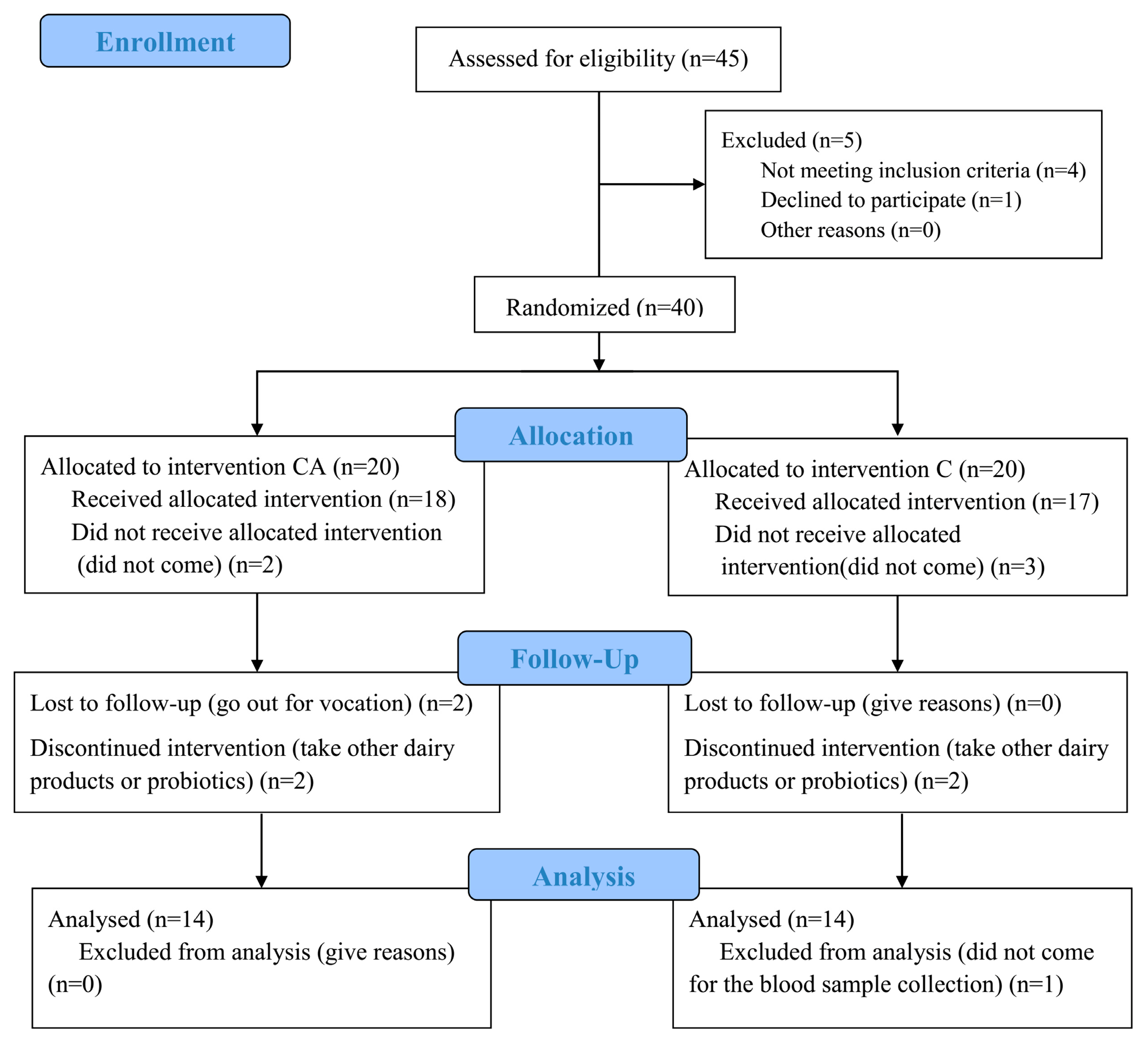
Figure 1. Flow diagram.
Foods 14 03318 g001
Figure 2. Fasting blood glucose, 2h postprandial blood glucose, fasting serum insulin, and HOMA-IR in each group before (W0) and after (W4) the intervention. (A), fasting blood glucose; (B), decrease in fasting blood glucose (squares: CA group; triangles: C group); (C), 2h postprandial blood glucose; (D), decrease of 2h postprandial blood glucose (squares: CA group; triangles: C group); (E), fasting serum insulin; and (F), HOMA-IR in patients treated with camel milk (C group) and camel milk supplemented with BBA6 (CA group).
Foods 14 03318 g002
Figure 3. Lipid profile in each group before (W0) and after (W4) the intervention. (A), total cholesterol; (B), total triglyceride; (C), the LDL cholesterol/HDL cholesterol ratio; (D), total cholesterol/HDL-cholesterol ratio in patients treated with camel milk (C) and camel milk supplemented with BBA6 (CA).
Foods 14 03318 g003
Figure 4. Serum contents of inflammatory cytokines in each group before (W0) and after (W4) the intervention. (A), TNF-α; (B), IL-6; (C), MCP-1 in patients treated with camel milk (C) and camel milk supplemented with BBA6 (CA).
Foods 14 03318 g004
Figure 5. Serum contents of adipokines and myokines in each group before (W0) and after (W4) the intervention. (A), adiponectin; (B), resistin; (C), lipocalin-2; (D), adipsin; (E), FGF-21; (F), irisin; (G), osteocrin; (H), osteonectin in patients treated with camel milk (C) and camel milk supplemented with BBA6 (CA).
Foods 14 03318 g005
Figure 6. Changes in microbial composition after intervention of camel milk (C) and camel milk supplemented with BBA6 (CA). (A) α diversity; (B) PCA analysis; (C) Species composition at the genus level.
Foods 14 03318 g006
Figure 7. Different gut bacteria at the genus level in patients intervened with camel milk (C) and camel milk supplemented with BBA6 (CA) by two-tailed Student’s t-test (A) and correlation analysis between flora and physiological indexes of diabetes (B).
Foods 14 03318 g007
Figure 8. Analysis of differential metabolites in feces in camel milk (C) and camel milk supplemented with BBA6 (CA). (A) PLS-DA analysis, (B) differential metabolite analysis, (C) VIP analysis, (D) KRGG enrichment pathway analysis of differential metabolites.
Foods 14 03318 g008
Table 1. Baseline characteristics of study participants.
ParametersCAC
Number (female/male)14 (10/4)14 (8/6)
Age (years)58.36 ± 6.2557.29 ± 7.57
BMI (kg/m2)26.56 ± 4.4026.56 ± 4.40
Fasting blood glucose (mmol/L)9.39 ± 2.5010.48 ± 3.78
2 h postprandial blood glucose (mmol/L)14.05 ± 3.2616.19 ± 5.28
Insulin (μU/mL)11.69 ± 11.1211.01 ± 7.11
TG (mmol/L)5.20 ± 1.505.10 ± 1.00
TC (mmol/L)1.77 ± 0.811.14 ± 0.14
HDL-C (mmol/L) 1.24 ± 0.261.22 ± 0.26
LDL-C (mmol/L)3.27 ± 1.253.33 ± 0.86
TC/HDL-C4.29 ± 1.314.47 ± 0.77
Notes: Data were analyzed for normal distribution by IBM SPSS Statistics 20.0 and expressed as mean ± S.D; C (camel milk), CA (camel milk with BBA6).
Disclaimer/Publisher’s Note: The statements, opinions and data contained in all publications are solely those of the individual author(s) and contributor(s) and not of MDPI and/or the editor(s). MDPI and/or the editor(s) disclaim responsibility for any injury to people or property resulting from any ideas, methods, instructions or products referred to in the content.