Article

Metabolomic Profile of Vaccinium corymbosum Leaves: Exploiting Diversity Among Ten Different Cultivars

CBQF—Centro de Biotecnologia e Química Fina—Laboratório Associado, Escola Superior de Biotecnologia, Universidade Católica Portuguesa, Rua Diogo Botelho 1327, 4169-005 Porto, Portugal
*
Author to whom correspondence should be addressed.
Foods 2025, 14(16), 2846; https://doi.org/10.3390/foods14162846
Submission received: 16 June 2025 / Revised: 11 August 2025 / Accepted: 14 August 2025 / Published: 17 August 2025
(This article belongs to the Section Foodomics)

Abstract

Blueberry (Vaccinium corymbosum) leaves are often discarded as agricultural by-products despite their potential abundance in bioactive compounds. However, comprehensive knowledge of their phytochemical profile remains limited, especially at the cultivar level. To address this gap, this study performed untargeted metabolomic profiling of blueberry leaves from ten cultivars using UHPLC-QTOF MS. Metabolites were annotated using high-resolution mass spectrometry and MS/MS fragmentation patterns. Multivariate statistical techniques were employed to investigate inter-cultivar variability and identify distinctive metabolites. A total of 76 metabolites were discovered, with 64 being confidently annotated and grouped into ten main phytochemical classes. The relative abundances of phenolic acids, flavonols, and flavan-3-ols varied significantly among cultivars. Several metabolites were annotated for the first time in V. corymbosum leaves, such as miscanthoside, glucoliquiritin, apiin, khelloside, and aromadendrin. These metabolites are known in other plants for their biological activities, demonstrating blueberry leaves’ bioactive potential. This study highlights the importance of untargeted metabolomic approaches in elucidating the biochemical diversity of plant matrices. The metabolomic data revealed significant cultivar-specific variations and novel bioactive metabolite annotation. These findings establish a complete phytochemical fingerprint for each cultivar, providing a basis for future research to validate key metabolites’ biological activities and support the valorisation of V. corymbosum leaves.

1. Introduction

Vaccinium corymbosum of the Ericaceae family is a North American species of blueberry holding significant relevance across the economic, health, and industrial sectors. This species is widely known for its rich nutritional profile and health benefits. Blueberries, like many other berries, are rich in antioxidants, particularly polyphenols, which have been linked to improved cognitive function, cardiovascular health, and reduced risk of chronic diseases such as type 2 diabetes and certain cancers [1,2]. The cosmetic industry also utilises blueberry extracts in skincare formulations due to their antioxidant properties [3]. Their antibacterial activity against specific species such as Staphylococcus aureus, Listeria monocytogenes, and Bacillus cereus has also been reported [4]. V. corymbosum boasts a remarkable diversity of cultivars, each exhibiting quite unique characteristics. These variations span crucial traits such as ripening time (early, mid, or late season), fruit size, flavour profile (sweet, tart, or aromatic), yield potential, growth habit (upright, spreading, or compact), winter hardiness, and disease resistance. This variability allows growers to choose cultivars best suited to their specific climate, soil conditions, market demands, and desired harvest window. It is easily inferred that all these differences between cultivars are mirrored in the plant metabolome, including their leaves [5]. Indeed, interest in the leaves of V. corymbosum shrubs, due to their proven potential as a source of bioactive compounds [6], has increased, and some works have been published aiming at its characterisation [7,8,9]. Routray and Orsat [7] analysed two V. corymbosum cultivars, Elliot and Nelson, and concluded that the latter possesses the highest content of monomeric anthocyanins, using a combination of ethanol and citric acid as the extraction solvent. Akšić et al. [8] characterised three cultivars (‘Duke’, Nui, and Bluecrop) and concluded that 5-O-caffeoylquinic acid was the most abundant phenolic in blueberry leaves when MeOH/H2O (70:30) plus 0.1% of HCl was used as the extraction solvent. Venskutonis et al. [9] compared seven cultivars and denoted that rutin, chlorogenic, and quinic acid concentrations for the same cultivar were highly dependent on the extraction solvent. The full characterisation of the metabolic profile of V. corymbosum cultivars’ leaves will capitalise on its use; however, most of the published works used few cultivars and diverse extraction solvents. To the best of our knowledge, the most complete study was undertaken by Wu and colleagues [10]. These authors claimed the characterisation of 73 different cultivars; however, these cultivars were grouped, and the metabolomic profile was presented for each group instead of per cultivar individually, avoiding a deep knowledge of cultivar diversity.
In this study, the metabolomic profile of V. corymbosum leaves of 14 plants belonging to 10 different cultivars was obtained by mass spectrometry in an ultra-high-performance liquid chromatography–quadrupole time-of-flight mass spectrometer (UHPLC-QTOF MS). Metabolites were annotated with their exact masses and experimentally generated fragments with the aid of online-available and licensed software. The annotated metabolites were further quantified (semi-quantitative approach), and differences between cultivars were discussed. Additionally, a principal component analysis was performed, and the corresponding variables’ importance on projection (VIP) scores were obtained allowing the annotation of the most relevant metabolites in the leaves of each cultivar.

2. Materials and Methods

2.1. The Samples

V. corymbosum leaves of 14 distinct plants belonging to 10 cultivars [‘Legacy’ (n = 3); ‘Duke’ (n = 3); ‘Liberty’ (n = 1); ‘Aurora’ (n = 1); ‘Camellia’ (n = 1); ‘Spartan’ (n = 1); ‘Susyblue’ (n = 1); ‘Star’ (n = 1); ‘Rabbiteye’ (n = 1); ‘Centrablue’ (n = 1)] were collected in spring (April) in the north of Portugal, in Arouca (40.92481384287574° N,−8.252706527709961° E). All cultivars were grown under the same geographical, soil, climatic, and cultivation conditions to ensure environmental uniformity. Leaf samples were collected in the early morning and promptly transported to the laboratory in proper bags in a refrigerated container. Immediately after arriving at the laboratory, the samples were cleaned with water, dried with paper towels, and further lyophilised. Lyophilised samples were ground and stored at −80 °C until the UHPLC-QTOF MS experiments (for no more than two weeks).

2.2. Extraction Procedure

Lyophilised leaves were taken from the refrigerator and left for 30 min in the laboratory, allowing them to reach room temperature. The samples were ground, and an average of fifty milligrams of each sample was added (in triplicate) to a 50 mL vial after adding 5 mL of pure ethanol (MS grade). Vials were sonicated for 15 min and further centrifuged. The supernatants were filtered through a 0.22 mm syringe filter, transferred to vials, and further analysed. Extracts were obtained in triplicate.

2.3. UHPLC-QTOF MS

The MS/MS experiments were conducted using ultra-high-performance liquid chromatography–quadrupole time-of-flight mass spectrometry (UHPLC-QTOF MS) equipment. The chromatographic separations were undertaken in a UHPLC UltiMate 3000 Dionex system (Thermo Fisher Scientific Inc., Waltham, MA, USA), and the mass experiments in a mass spectrometer from Bruker Daltonics Impact II, Bremen, Germany, equipped with an electrospray ionisation (ESI) source. The mass spectrometer was operated in negative ion mode, and data were recorded under the following ESI source parameters: capillary voltage, 3.5 kV; end plate offset, 500 V; nebuliser, 0.4 Bar; dry gas, 4.0 L/min; dry temperature, 180 °C; Funnel 1 RF, 400 Vpp; Funnel 2 RF, 600 Vpp; hexapole, 700 Vpp; quadrupole ion energy, 5.0 eV; collision energy, 10.0 eV; transfer time, 120.0 µs. The QTOF mass analyser scanned masses in the range of 50–1000 Da.
Extracts (5 μL) were injected on an Acclaim RSLC 120 C18 column (100 mm × 2.1 mm, 2.2 µm) (Thermo Fisher Scientific Inc., Sunnyvale, CA, USA). The mobile phase was a binary solvent system consisting of (A) water acidified with formic acid (0.1%) and (B) acetonitrile acidified with formic acid (0.1%). The gradient (0–25 min with 5–90% of B in gradual steps) was eluted at a flow rate of 0.25 mL/min at 40 °C. Each sample was run in duplicate.

2.4. Data Analysis

The data were analysed with free online-available (Compound Crawler 2.0, MetFrag v2.4.5 and MetaboAnalyst 5.0) and/or licensed software (Smart Formula 3D, DataAnalysis 4.4 and Metaboscape 2023). Raw mass spectra were calibrated and processed in Bruker Metaboscape software. Only features with MS/MS spectra were considered. The feature table was further subjected to annotation by comparison with spectral libraries (Bruker NIST 2020 MSMS Spectral Library; MassBank_NIST.msp and MassBank2RIKEN.msp). The putative compounds were annotated with a tolerance of 5.0 ppm. After the putative annotations, in silico fragmentation of all the compounds were generated, and theoretical vs. experimental fragmentation patterns were compared. The final annotation was assumed for metabolites presenting a score ≥0.99 in MetFrag software. The proposed metabolite annotation was also checked and compared with the available literature.

3. Results

3.1. Metabolic Profiling of V. corymbosum Cultivars by HPLC-QTOF MS

The metabolome of V. corymbosum cultivars was characterised by the LC-MS data, including retention time, molecular formula, experimental exact mass and m/z fragments generated, mSigma values (which are the rate for the agreement of the theoretical and measured isotopic pattern of the mass peak of interest; lower numbers correspond to better fits and higher annotation confidence), and accuracy (ppm). A total of 76 metabolites were detected in the ethanolic leaf extract, 64 of which were annotated (Table 1 and Table S1), and 12 were non-annotated (Table S2).
The annotated metabolites (64) belong to distinct compound classes: carboxylic acids (10/65); benzoic acids (14/65); caffeic acids (3/65); quinic acids (10/65); flavan-3-ols (5/65); flavonols (13/65); flavones and flavanones (4/65); chalcones (2/65); and 3/65 belonging to other classes (Figure 1).
Carboxylic acids: Compound C1 was annotated as succinic acid, presenting the m/z 73 fragment, which corresponds to the typical fragmentation ion of carboxylic acids (the loss of the CO2 unit, [M−H−44]). The unique fragment (m/z 73) observed for succinic acid agrees with previous studies [11]. Compound C2 was annotated as citramalic acid. The observed fragments m/z 87, m/z 85, and m/z 129 are due to the [C3H5O3−H]H, [C4H4O2+H]−H, and [C5H6O4−H]−H moieties, respectively. Compound C3 was annotated as hydroxypentanoic acid. Fragments of m/z 73 (typical loss of CO2) and m/z 59 ([C2H3O2−H] moiety) were observed. Compounds C4 and C5 were annotated as coumaric acids. Both present a fragment ion m/z 119 due to the loss of CO2 [M−H−44], and compound C4 presents an additional fragment ion at m/z 71 from the [C3H3O2] moiety. Mekky and colleagues [12] have annotated m- and p-coumaric acids in sesame cake and, according to the standards used, proved that when using a C18 column with acidified water and acetonitrile as mobile phases, p-coumaric acid was first eluted. This suggests that compound C4 is p-coumaric acid and compound C5 is m-coumaric acid. Compound C6 was annotated as azelaic acid, presenting m/125 and m/z 169, m/z 123, and m/z 143 (loss of CO2) fragments due to [C8H12O+H], [C9H14O3]−H, [C8H12O]−H, and [C8H14O2+H] moieties, respectively. Compound C7 was annotated as undecanoic acid with m/z 197 and m/z 153 fragments due to the [C11H18O3+H]−H and [C10H16O+H] moieties, respectively. Compounds C8 and C9 were annotated as hydroxydodecanoic acids. Both present the m/z 169 fragment due to the [C11H22O]−H moiety. The remaining fragments of both isomers are compatible with the hydroxypentanoic structure: m/z 87 ([C3H5O3−H]−H), m/z 171 ([C9H17O3−H]−H), m/z 197 ([C12H22O2]−H), and m/z 199 ([C11H21O3−H]−H). Compound C10 was annotated as a furancarboxylic acid, with fragments of m/z 209.155 ([C13H20O2+H]), m/z 210 ([C13H22O2]), m/z 59 ([C2H2O2+H]), m/z 209.120 ([C12H15O3+2H]), and m/z 89 ([C3H3O3+2H]).
Benzoic acids: Compounds B11, B12, B13, and B14 present a main fragment of m/z 93 corresponding to the typical loss of CO2 [M−H−44]. Compounds B12, B13, and B14 present nearly the same fragmentation patterns, which is suggestive of o-, m-, and p-hydroxybenzoic acids. Compound B11 presented a quite different fragmentation pattern, suggesting a di-hydroxybenzaldehyde, namely the protocatechuic aldehyde (all the fragments can be explained and correlated with this compound). Compounds B15 and B16 were annotated as di-hydroxybenzoic acids according to the fragmentation pattern: m/z 108 from the [C6H4O2], m/z 109 from the [C6H4O2+H], and m/z 123 from the [C7H5O2+2H] moieties. Compounds B17, B18, and B19 were annotated as tri-hydroxybenzoic acids, with all the fragments being explained and compatible with such a structure. Compound B17 was considered to be gallic acid (a tri-hydroxybenzoic acid) according to the exact mass and unique m/z 125 fragment. It should be noted that gallic acid was already annotated in previous studies and reported with m/z 125 as the unique fragment and an exact mass of [M−H] 162.0142 [11]. Compounds B20, B21, and B22 were annotated as hydroxybenzoic acid hexosides. The typical fragments of m/z 137 related to the loss of the glucosyl moiety [M−H−162] was observed in the three compounds. Compounds B20 and B22 additionally possess the m/z 93 fragment typical of the [C6H5O] moiety, and compound B21 possesses the fragments m/z 179, m/z 239, and m/z 151. The three isomers are compatible with a glucosyl moiety linked to both hydroxyl groups (that of the carboxylic acid or the free one in the benzene ring). Compound B23 was annotated as a di-hydroxybenzoic acid hexoside, with the main fragments being m/z 152 and m/z 108 due to the loss of the glucosyl moiety and the benzene ring with both oxygen atoms from the hydroxyl groups. Compound B24 was annotated as vanillic acid hexoside, with all fragments explained and compatible with the proposed structure, including the main fragment m/z 167, corresponding to the loss of the glucosyl moiety [M−H−162].
Caffeic acids: Compound Caf25 possesses the m/z 135 main fragment due to the loss of carbon dioxide [C8H6O2+H] and m/z 134 ([C8H6O2]), being annotated as caffeic acid (cis-/trans-3,4-dihydroxycinnamic acid) in accordance with the literature [8,13], Bruker Metaboscape software, and MassBank record MSBNK-RIKEN-PR100533. Compound Caf26 was annotated as a caffeic acid isomer due to the exact mass and the presence of m/z 135 (similar to that encountered in compound Caf25) and m/z 137 from the [C8H7O2+2H] moiety. Compound Caf27 was annotated as caffeic acid hexoside. The exact mass and fragments of m/z 179 (loss of the hexoside moiety), m/z 135 (loss of CO2 from the caffeic acid moiety), and m/z 181 (from the [C9H9O4] moiety) confirm the identification.
Quinic acid derivatives: Compounds Q28, Q29, Q30, and Q31 were annotated as caffeoyl quinic acid isomers. Typical fragments of m/z 191 and m/z 179 due to quinic acid and caffeoyl moieties, respectively, and of m/z 173 (quinic acid moiety water loss) and m/z 135 (caffeoyl moiety carbon dioxide loss) were annotated. Compound Q29 uniquely presents the m/z 191 fragment, which suggests 5-caffeoyl quinic acid. Indeed, Venskutonis et al. [9] proved that in a C18 column eluted with a gradient of water + formic acid followed by an organic solvent, 3-caffeoyl quinic acid is first eluted, then the isomer 5-caffeoyl quinic acid, which is in accordance with our observations. Compounds Q32, Q33, and Q34 were annotated as coumaroyl quinic acids. Fragments of m/z 191 and m/z 173 are associated with the quinic acid moiety (as explained above), the fragment m/z 163 corresponds to the coumaroyl moiety, while the fragment m/z 119 reflects the loss of carbon dioxide of the coumaroyl moiety. Compound Q35 was annotated as feruloylquinic acid due to its exact mass and typical fragments of m/z 191 and m/z 173 (quinic acid moiety and its loss of water), m/z 193 due to the feruloyl moiety, and m/z 93 (aromatic ring from the feruloyl moiety with the oxygen atom from the hydroxyl group). Compound Q36 was annotated as dicaffeoylquinic acid. The exact mass and typical fragments of m/z 353 (monocaffeoylquinic acid moiety), m/z 191, m/z 173 (quinic acid moiety and its loss of water), and m/z 179 (caffeoyl unit) are compatible with a dicaffeoylquinic acid structure. Compound Q37 was annotated as a feruloylcaffeoylquinic acid due to its exact mass and typical fragments of m/z 367 (loss of the caffeoyl unit) and m/z 193, m/z 191 m/z 353, m/z 173, and m/z 179 (due to the already-explained structures).
Flavan-3-ols: Five catechin derivatives were annotated in the extracts. Compounds Fla38 and Fla39 were annotated as gallocatechin/epigallocatechin and catechin/epicatechin, respectively, based on their exact masses and fragmentation patterns. Compound Fla38 presents the typical gallocatechin/epigallocatechin fragmentation pattern with the main fragment of m/z 125 ([C6H5O3]) and three additional ones (m/z 165, 167, and 137 of similar intensities from [C8H6O4]−H, [C8H8O4]−H, and [C7H6O3]−H moieties, respectively) reported previously [13] and compatible with MassBank records BS003906 (gallocatechin) and BS003952 (epigallocatechin). Regarding catechin/epicatechin, compound Fla39, fragments of m/z 245 and m/z 203 from [C14H11O4+2H] and [C12H9O3+2H], respectively, were observed herein and reported in other studies [13], as is typical of these compounds. Three trimeric catechin isomers (compounds Fla40, Fla41, and Fla42) were annotated through their exact masses and fragmentation patterns as type A/B (one of the units bonded to the central one with an A-type bonding, and the other bonded to the central one with a B-type bonding). Hokkanen et al. [13] reported the same fragmentation pattern obtained herein for compounds Fla41 and Fla42 in their studies: m/z 711 due to RDA fragmentation; m/z 451 loss of the extension A-type unit plus pholoroglucinol from the central unit; m/z 411 extension unit and pholoroglucinol from the central unit; m/z 289 the terminal unit; and m/z 573 loss of the B-type terminal unit. A different study [14] has also annotated two procyanin type A/B isomers through their exact masses and the m/z 298 fragment.
Flavonols: Compound Flo43 was annotated as aromadendrin. The observed fragments of m/z 259, m/z 143, m/z 125, and m/z 152 from the [C14H12O5]−H, [C14H10O4+H], [C6H4O3+H], and [C7H4O4] moieties, respectively, are compatible with the proposed compound. Compound Flo44 was annotated as isorhamnetin. Fragments of m/z 300 ([C15H9O7]−H) and m/z 301 ([C15H9O7]), due to the loss of the CH4 and CH3· moieties, respectively, and m/z 151 from ring C cleavage between positions 1 and 3 ([C7H4O4]−H) were observed. Compound Flo45 was annotated as kaempferol-7-O-rhamnoside. The main fragments of m/z 285 ([C15H9O6]) and m/z 284 ([C15H9O6]−H), due to the loss of the rhamnose moiety and m/z 191 ([C7H9O6+2H]), were observed. Compound Flo46 was annotated as myricetin-3-hexoside. The typical loss of the hexosyl moiety (−162) was observed through the presence of the fragment m/z 317 ([C15H9O8]). Additional fragments of m/z 191 and m/z 301 from the [C7H11O6] and [C15H9O7] moieties were also observed. Compound Flo47 was annotated as syringetin-3-hexoside due to the presence of the typical fragment of m/z 344 ([C17H13O8]−H) due to the loss of the hexosyl moiety (−162). Compound Flo48 was annotated as quercetin-3-O-pentosylpentosilpentoside. The presence of the typical fragment of m/z 300 ([C15H9O7]−H), due to the loss of the sugar moieties, and the additional fragments of m/z 463 and m/z 191 from [C21H18O12+H] and [C7H12O6]−H moieties, respectively, confirms the identification. Compounds Flo49 and Flo50 were annotated as possessing a flavonol base with a 3-O-hexoside-hexoside moiety. The loss of the sugar moieties was the main fragment observed (m/z 285 from [C15H9O6] and m/z 284 from [C15H9O6]−H). Compound Flo49 was tentatively annotated as kaempferol 3-O-rutinoside with a score of 1.0 in Bruker MetaboScape software. Additional m/z 463 and m/z 327 fragments from [C15H19O12] and [C21H12O7]−H moieties were also observed. Compound Flo51 was annotated as rutin through the exact mass and the typical unique fragment of m/z 300 from the [C15H9O7]−H moiety. Compound Flo52 was annotated as quercetin 3-gentiobioside, with the five most intense fragments perfectly explained, namely, m/z 445 ([C21H19O11−H]−H), m/z 463 ([C21H19O12]H), m/z 301 ([C15H9O7]), m/z 464 ([C21H19O12+H]), and m/z 300 ([C15H9O7]−H). Compound Flo53 was annotated as possessing a flavonol base plus three sugar moieties. A higher identification score (1.0) was obtained for helieianeoside B in Bruker MetaboScape software, with the five most intense fragments compatible with the proposed identification: m/z 300 ([C15H9O7]−H), m/z 191 ([C7H12O6]−H), m/z 353 ([C16H15O9+2H]), m/z 417 ([C21H19O9+2H]), and m/z 178 ([C8H4O5]−H). Compound Flo54 was annotated as kaempferol-3-O-rutinoside-7-O-β-D-glucopyranoside with a score of 1.0 in Bruker MetaboScape software (both fragments explained): m/z 593 ([C27H29O15]) and m/z 285 ([C15H9O6]). The fragments and exact mass were also in accordance with the literature [10]. Compound Flo55 was annotated as variabiloside with a score of 1.0 in Bruker MetaboScape software (both fragments explained): m/z 593 ([C30H25O13]) and m/z 285 ([C15H9O6]).
Flavones and flavanones: Flavones and flavanones differ in the saturation degree of the carbon–carbon bond of ring C. Four compounds were annotated: three flavanones (Flav56, Flav 57, and Flav59) and one flavone (Flav58). Compound Flav56 was annotated as 3,9-dihydroeucomin through the presence of its typical fragments at m/z 179 ([C9H7O4]), m/z 151 ([C7H4O4]−H), m/z 135 ([C8H8O2]−H), m/z 165 ([C8H6O4]−H), m/z 121 ([C7H4O2+H]), m/z 229 ([C13H11O4−H]−H), and m/z 149 ([C9H9O2]). Compound Flav57 was annotated as miscanthoside, with fragments at m/z 287 (C15H11O6]), m/z 191 ([C7H11O6]), m/z 151 ([C7H3O4]), m/z 257 ([C15H11O4+2H]), m/z 301 [C15H10O7−H], and m/z 135 ([C8H8O2]−H). Compound Flav58 was annotated as apiin though its fragments at m/z 191 ([C7H12O16]−H) and at m/z 353 ([C16H15O9+2H]). Compound Flav59 was annotated as glucoquiritin through its fragments at m/z 417 ([C21H21O9]), m/z 418 ([C21H21O9+H]), m/z 307 ([C15H15O7]), m/z 335 ([C16H17O8−H]−H), and m/z 191 ([C7H11O6]).
Chalcones: Two chalcones were annotated (compounds Cha60 and Cha61). Compound Cha60 was annotated as naringenin chalcone through the fragments m/z 151 ([C7H5O4−H]−H), m/z 165 ([C9H7O3+2H]), m/z 119 ([C8H7O]), m/z 177 ([C9H7O4−H]−H), and m/z 228 ([C14H11O3+H]). The naringenin isomer was discarded due to its exact mass (272.25601). Compound Cha61 was annotated as cardamonin, with fragments at m/z 134 ([C8H6O2]), m/z 178 ([C9H6O4]), m/z 137 ([C7H7O3−H]−H), m/z 133 ([C8H5O2]), and m/z 139 ([C7H7O3]), justifying the identification.
Miscellaneous compounds: Four additional compounds belonging to diverse chemical classes were annotated. Compound M62 was annotated as uridine, possessing m/z 200, m/z 42, m/z 153, m/z 140, and m/z 71 fragments due to the [C8H11NO5]−H, [CHNO]−H, [C6H6N2O3]−H, [C6H8NO3−H]−H, and [C3H3O2] moieties, respectively. Compound M63 was annotated as guanosine, presenting the typical fragments of m/z 150, m/z 133, m/z 117, m/z 113, and m/z 191 from [C5H4N5O], [C5H3N4O−H]−H, [C5H2N4]−H, [C5H7O3−H]−H, and [C7H7N5O2−H]−H moieties, respectively. Compound M64 was annotated as khelloside, possessing the typical fragment of m/z 246 ([C13H9O5]), corresponding to the hexoside moiety loss [M−H−162].
Non-annotated compounds: Twelve metabolites detected in the V. corymbosum extracts were not annotated. Nid1 and Nid2 are two isomers possessing the structural formula C5H10O4 and exact mass [M−H] of 133.05058(86), respectively, but were not annotated. Compound Nid3 has a structural formula (C6H8O2) and an exact mass compatible with a hexadienoic acid, including the well-known sorbic acid. However, its unique fragment of m/z 69 (despite being present in the mass spectra of hexadienoic acids) prevents its unequivocal identification. Compounds Nid4 and Nid5 appear to be two isomers of structural formula C6H10O3 with an exact mass [M−H] of 129.05580(69), which were not possible to identify. Compound Nid6 appears to be an acidic compound due to the fragment of m/z 121, which corresponds to the typical loss of −CO2 in carboxylic acids [M−44]. Compounds Nid7 and Nid8, Nid9 and Nid10, and Nid11 and Nid12 appear to correspond to three pairs of isomers due to their structural formulas and exact masses; however, it was not possible to identify them. Isomers Nid9 and Nid10 have a strong probability of having a glucosyl moiety in their structure due to their typical fragment of m/z 161.

3.2. Metabolome Semi-Quantitative Evaluation

3.2.1. Global Metabolome Cultivars Comparison

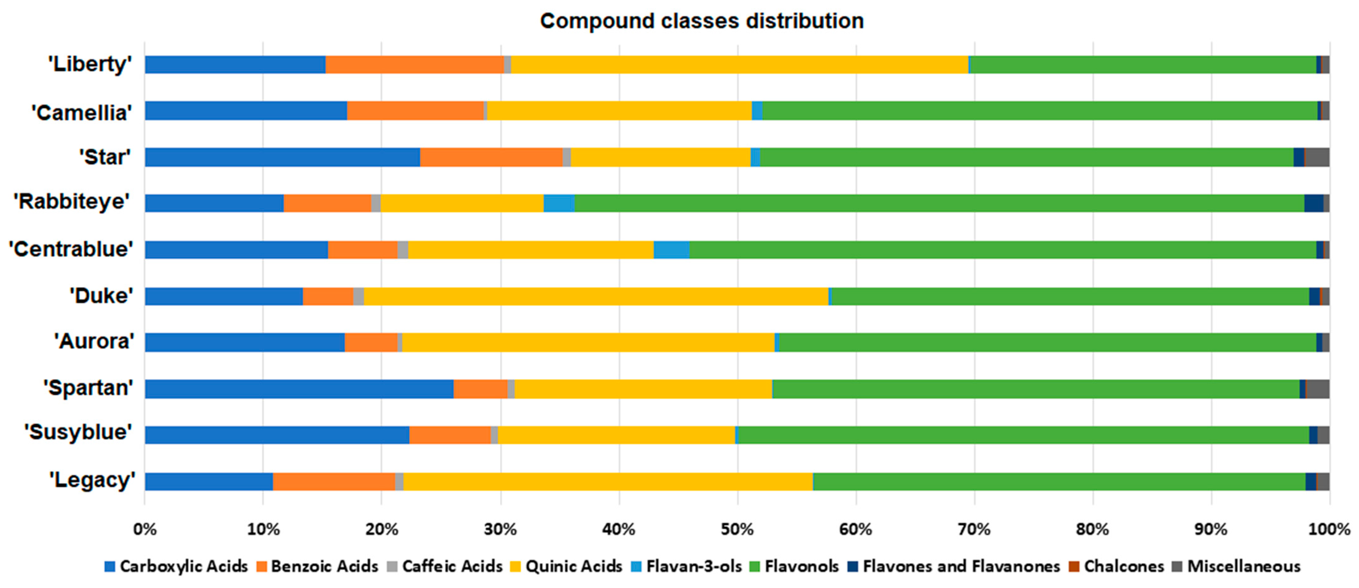
The annotated V. corymbosum metabolites were grouped into 10 distinct compound classes, and their relative amounts per cultivar are presented in Figure 2. Carboxylic, benzoic, and quinic acids and flavonols represent the great majority of the annotated metabolites in all cultivars. Carboxylic acids represent nearly 11–26% of the annotated metabolites, with the lowest and the highest quantities observed for the ‘Legacy’ and ‘Spartan’ cultivars, respectively. Benzoic acids represent around 4–15%, with ‘Aurora’ and ‘Liberty’ possessing the lowest and the highest amount of this compound class. Quinic acids vary from just under 13% in ‘Star’ and ‘Rabbiteye’ to nearly 38% in the ‘Liberty’ and ‘Duke’ cultivars. Flavonols range from around 30% of the annotated metabolites in ‘Liberty’ to 60% in ‘Rabbiteye’.
‘Rabbiteye’ and ‘Centrablue’ were the cultivars possessing the highest amount of flavan-3-ols, nearly 3%, while ‘Star’ and ‘Spartan’ were the ones possessing the highest amount of the miscellaneous compounds (uridine, guanosine, and khelloside). Overall, caffeic acids, flavones, flavanones, and chalcones each represent less than 2% of the annotated V. corymbosum metabolites.
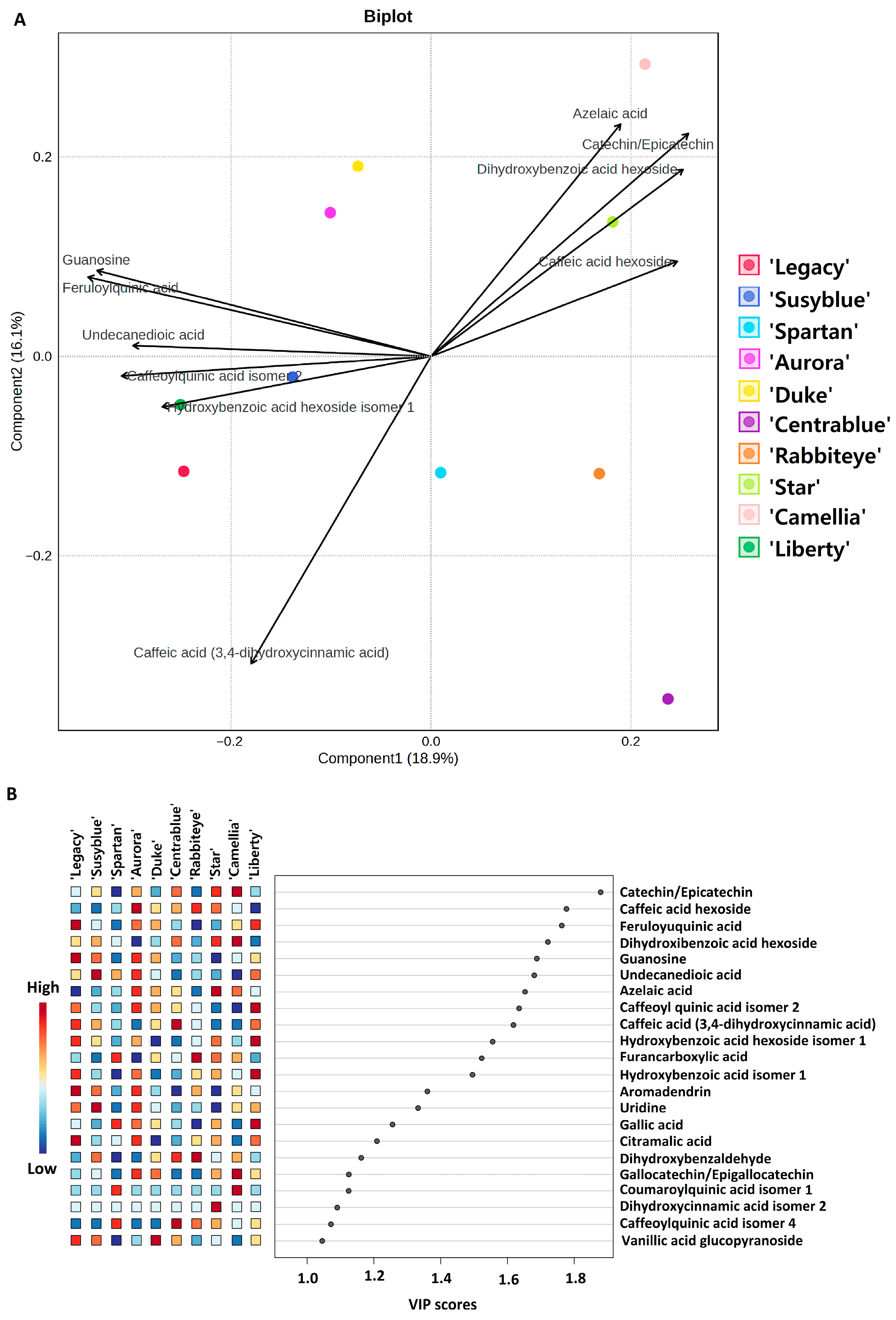
A PLSDA model and the corresponding VIP scores were obtained from the metabolites’ relative amount in all cultivars (Figure 3A,B) with two main goals: (I) to infer about the similarities/differences among cultivars’ metabolome, and (II) to identify the metabolites that strongly correlate with each cultivar. Figure 3A presents the biplot of the PLSDA models, including the scores of the models and the 10 most relevant metabolites (for simplification proposes) contributing to the cultivars’ distribution across the score map. The PLSDA was cross-validated (CV) through a fivefold CV method (R2—0.99618; Q2—0.98151). In Figure 3B, the 25 most relevant metabolites are presented, including the degree of correlation with each cultivar. The 10 metabolites that differ the most among the 10 analysed V. corymbosum cultivars are as follows: azelaic acid; catechin/epicatechin; dihydroxybenzoic acid hexoside; caffeic acid hexoside; caffeic acid; guanosine, feruloylquinic acid; undecanedioic acid; caffeoylquinic acid isomer 2; and hydroxybenzoic acid hexoside isomer 1. The ‘Star’ and ‘Camellia’ cultivars appear in the positive part of both components, suggesting a similar metabolome profile. These two cultivars positively correlate with the amount of azelaic acid, catechin/epicatechin, dihydroxybenzoic acid hexoside, and caffeic acid hexoside and negatively correlate with caffeic acid. The ‘Spartan’, ‘Rabbiteye’, and ‘Centrablue’ cultivars all appear in the positive part of component 1 and the negative part of component 2, pointing to a similar metabolome profile. These three cultivars positively correlate mainly with the relative amount of caffeoylquinic acid isomer 4, dihydroxybenzaldehyde, furancarboxylic acid, and coumaroylquinic acid (only the ‘Spartan’ cultivar), as can be seen in the VIP scores (Figure 3B). Also, they negatively correlate with the amount of guanosine and feruloylquinic acid. The ‘Duke’ and ‘Aurora’ cultivars cluster in the negative part of the first component and the positive part of the second and positively correlate mainly with caffeic acid hexoside, feruloyl quinic acid, guanosine, azelaic acid (‘Aurora’), and with vanillic acid glucopyranoside (‘Duke’). ‘Legacy’, ‘Liberty’, and ‘Susyblue’ appear in the negative part of both components and strongly correlate with the relative amount of caffeoylquinic acid isomer 2, hydroxybenzoic acid hexoside isomer 1, and caffeic acid. These three cultivars negatively correlate with azelaic acid, catechin/epicatechin, dihydroxybenzoic acid hexoside, and caffeic acid hexoside.
Additionally, a non-parametric ANOVA was performed for the 10 most discriminating metabolites (according to the VIP scores). The comparative analysis of the metabolites revealed statistically significant differences for all compounds according the Kruskal–Wallis test. Subsequent pairwise comparisons using the Mann–Whitney U test with Holm correction identified specific differences among cultivars, particularly involving ‘Camellia’, ‘Aurora’, and ‘Star’, which consistently exhibited negligible or null concentrations for several metabolites, in contrast to cultivars such as ‘Liberty’ and ‘Susyblue’, which showed markedly elevated levels (Table 2).
Table 2 Pairwise comparisons of cultivars for each compound showing statistically significant differences in metabolite concentrations. Results are based on Mann–Whitney U tests with Holm correction for multiple comparisons. Differences were considered statistically significant at a corrected p-value threshold of p < 0.05

3.2.2. Specific Metabolites’ Variation Between Cultivars

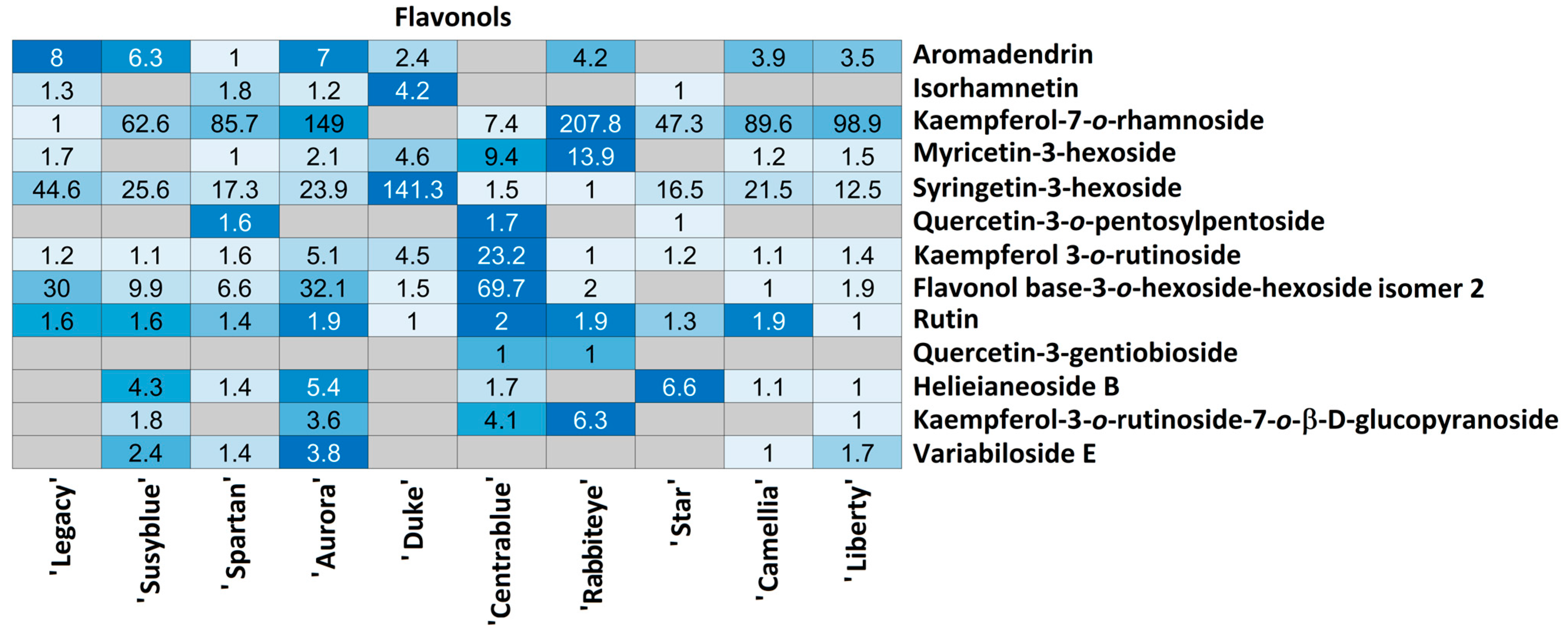
For each compound class, a heatmap was generated (Figure 4, Figure 5, Figure 6, Figure 7, Figure 8, Figure 9, Figure 10, Figure 11 and Figure 12) according to the method described in Section 2.4. Data Analysis. The values inside each cell correspond to the ratio between the corresponding cultivar area and the lowest cultivar area for each metabolite. Grey cells correspond to an undetected metabolite in the corresponding cultivar when the extract ion chromatograms were obtained for each exact mass with a tolerance of ±0.01. It should be stressed that these heatmaps allow metabolite semi-quantitative comparisons between cultivars but not quantitative comparisons among metabolites of a single cultivar.
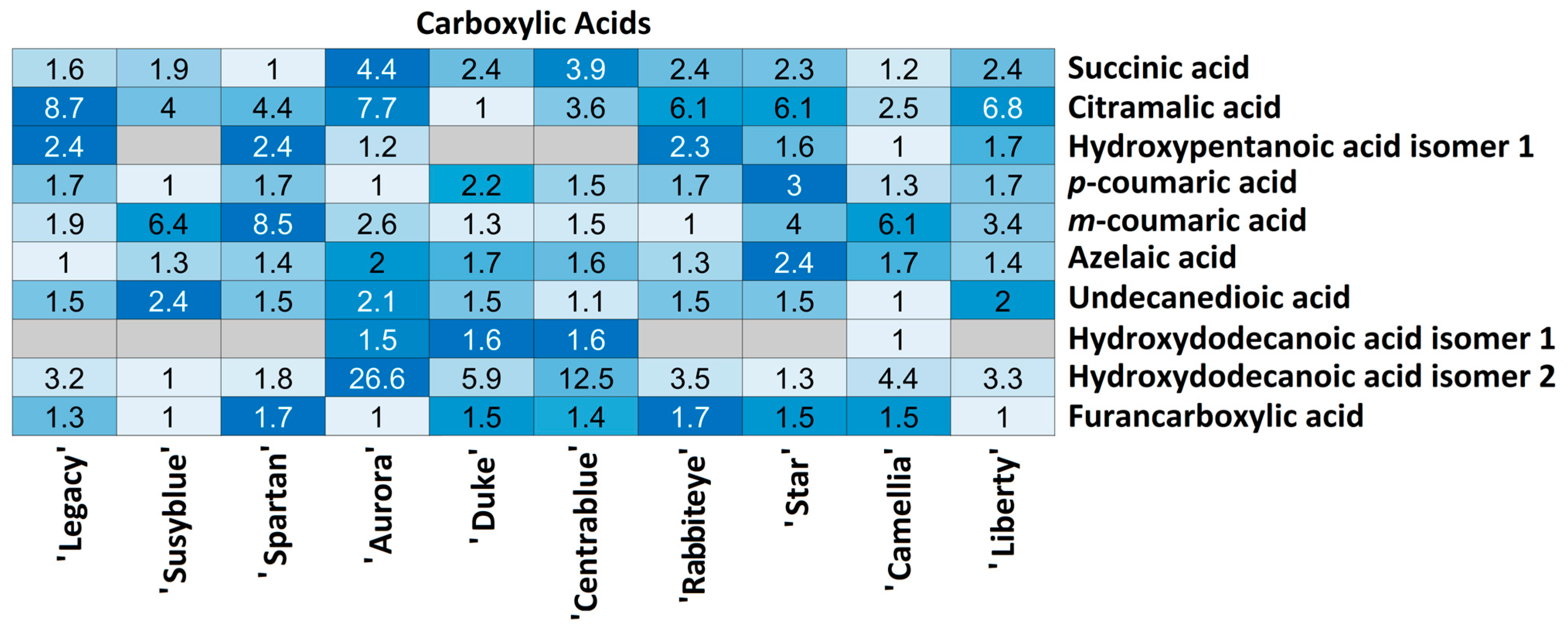
In total, 10 carboxylic acids were annotated, of which 8 were detected in all cultivars, while 2 of them (hydroxypentanoic isomer 1 and hydroxydodecanoic isomer 1) were undetected in 3/10 and 6/10 cultivars (Figure 4), respectively.
In the ‘Aurora’ and ‘Camellia’ cultivars, all the carboxylic acids were detected. Regarding the relative quantity of each metabolite among the cultivars, quite different variations can be observed. Hydroxypentanoic acid isomer 1, p-coumaric acid, azelaic acid, undecanedioic acid, hydroxydodecanoic acid isomer 1, and furancarboxylic acid (6/10) relative quantities do not vary more than 2.5 times between cultivars. However, metabolites such as succinic acid, citramalic acid, and m-coumaric acid present inter-cultivar variations of 8.7 times. Hydroxydodecanoic acid isomer 2 is the metabolite presenting the highest variation among cultivars, with the lowest quantity present in the ‘Susyblue’ cultivar and the highest in the ‘Aurora’ cultivar (26.6 times).
It should be emphasised that all the carboxylic acids annotated were detected only in the ‘Camellia’ and ‘Aurora’ cultivars.
Regarding benzoic acids, 14 metabolites were annotated, 7 of which were present in all cultivars, while the remaining 7 were randomly annotated among cultivars (Figure 5).
‘Rabbiteye’ emerges as the cultivar presenting the fewest benzoic acids (9/14), while ‘Susyblue’ and ‘Camellia’ present all the annotated ones. Most of the annotated benzoic acids strongly varied among cultivars. A few exceptions occur for hydroxybenzoic acid isomer 2 and trihydroxybenzoic acid isomers 1 and 2, for which the variations between cultivars are less than or equal to 2.2. Metabolites such as dihydroxybenzaldehyde, hydroxybenzoic acid hexoside isomer 3, and vanillic acid glucopyranoside present inter-cultivar variations of up to 10 times. Higher metabolite variations among cultivars were observed for hydroxybenzoic acid isomer 1 (up to 15.5 times) and isomer 3 (up to 19.3 times), dihydroxybenzoic acid isomer 1 (up to 28.4 times) and isomer 2 (up to 16.9 times), gallic acid (up to 11.5 times), dihydroxybenzoic acid hexoside (up to 21.7 times), and with a considerable variation between cultivars (up to 50.6 times), hydroxybenzoic acid hexoside isomer 1. This benzoic acid strongly fluctuates among cultivars, with the lowest quantity annotated in ‘Duke’ and the highest in ‘Liberty’ (50.6 times higher). ‘Legacy’ and ‘Star’ cultivars also present high amounts of this compound, 26.7 and 12.2 times higher than ‘Duke’. It should be stressed that all the annotated benzoic acids were detected in the ‘Camellia’ and ‘Susyblue’ cultivars.
Three caffeic acids were annotated in V. corymbosum cultivars (Figure 6). Caffeic acid was annotated in 7/10 cultivars with quite a similar amount in all cultivars (variations were no higher than 1.9 times). This metabolite was undetected in the ‘Aurora’, ‘Star’, and ‘Camellia’ cultivars. Dihydroxycinnamic acid was only annotated in the ‘Star’ cultivar, preventing additional comparison. Caffeic acid hexoside was annotated in all cultivars, with inter-cultivar variation no higher than 4.8 times. The lowest amounts was measured in ‘Liberty’ and ‘Susyblue’ (1.2 times higher than ‘Liberty’), and the highest amounts were annotated in ‘Rabbiteye’ (4.2 times more than ‘Liberty’) and ‘Aurora’ (4.8 times more than ‘Liberty’).
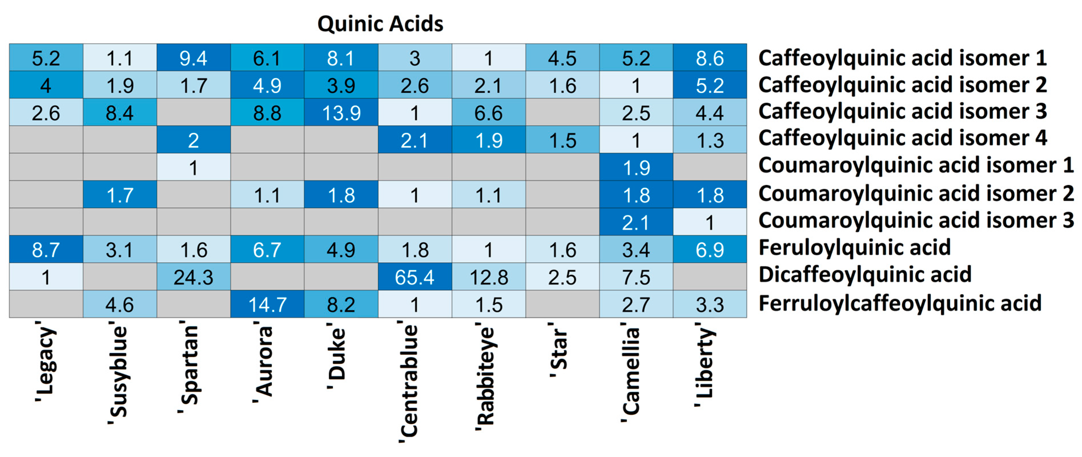
Quinic acids were randomly detected between cultivars (Figure 7). Ten metabolites were annotated, of which only (3/10) were annotated in all cultivars: caffeoylquinic acid isomers 1 and 2 and feruloylquinic acid. Four isomers of caffeoylquinic acid were annotated: isomers 1 and 2 in all cultivars, with relative amounts between cultivars no higher than 9.4 and 5.2, respectively, and isomers 3 and 4 randomly annotated among cultivars. The lowest relative amount of isomer 3 was detected in ‘Rabbiteye’ and the highest in ‘Duke’ (13.9 times higher), while isomer 4 was detected in the lowest relative amount in ‘Camellia’ and the highest in ‘Centrablue’ (2.1 times higher). Three coumaroylquinic acid isomers were randomly annotated between cultivars in very similar amounts (no more than 2.1 times). ‘Camellia’ was the unique cultivar in which all the isomers were annotated and also presented the highest quantity of all of them, while none of the three were annotated in the ‘Star’ cultivar. Feruloylquinic acid was annotated in all cultivars, with the lowest relative amount observed in ‘Rabbiteye’ and the highest in ‘Legacy’ (8.7 times higher). Dicaffeoylquinic acid was annotated in 5/10 cultivars with a huge variation among them; the lowest relative amount was observed in ‘Legacy’, while the highest was observed in ‘Centrablue’ (65.4 times higher). Feruloylquinic acid was detected in 7/10 cultivars; the lowest amount was observed in ‘Centrablue’, and the highest was observed in ‘Aurora’ (14.7 times higher). It should be stressed that all the annotated quinic acids were detected only in the ‘Camellia’ cultivar.
Five metabolites from the flavan-3-ol class were annotated in the V. corymbosum extracts (Figure 8). Gallocatechin/epigallocatechin was observed in 7/10 cultivars, catechin/epicatechin in all cultivars, and three procyanidin trimer type A/B isomers only in the ‘Centrablue’ and ‘Rabbiteye’ cultivars. Isomer 3 was also annotated in the ‘Spartan’ cultivar. ‘Legacy’ possesses the lowest gallocatechin/epigallocatechin relative amount, while ‘Camellia’ has the highest (7.4 times more). This metabolite was undetected in ‘Spartan’, ‘Centrablue’, and ‘Rabbiteye’. A lower amount of catechin/epigallocatechin was detected in ‘Spartan’ and the highest in ‘Camellia’ (10 times higher). ‘Rabbiteye’ possesses the lowest relative quantity of procyanidin isomer 1 and ‘Centrablue’ the highest (4.1 times). Regarding isomer 2, the lowest relative amount was observed in ‘Centrablue’, while the highest was observed in ‘Rabbiteye’ (1.7 times higher). Isomer 3 was observed in the lowest amount in ‘Spartan’, while ‘Rabbiteye’ possesses 16.8 times more and ‘Centrablue’ 22.8 times more.
Flavonols were randomly annotated among the ten cultivars (Figure 9). Rutin was annotated in all cultivars, presenting very little variation among them (no more than 1.9 times). Kaempferol-3-O-rutinoside was also annotated in all cultivars; however, it exhibited higher variation: the lowest amount was measured in ‘Rabbiteye’ and the highest in ‘Centrablue’ (23.2 times higher). Quercetin-3-gentibioside was only annotated in ‘Centrablue’ and ‘Rabbiteye’ in the same relative amount. Aromadendrin, isorhamnetin, myricetin-3-hexoside, and quercetin-3-O-pentosylpentoside were randomly detected among cultivars, presenting variations no higher than 13.9 times. Among the metabolites that present higher variation among cultivars are kaempferol-7-O-rhamnoside (‘Legacy’—lowest; ‘Rabbiteye’—207.8 times more), syrigetin-3-hexoside (‘Rabbiteye’—lowest; ‘Duke’—141.3 times more), and flavonol base-3-O-hexoside-hexoside isomer 2 (‘Camellia’—lowest; ‘Centrablue’—69.7 times more). Helieianeoside B, kaempferol-3-O-rutinoside-7-O-B-D-glucopyranoside, and variabiloside E were not detected in the ‘Legacy’ and ‘Duke’ cultivars. These metabolites were randomly detected in the remaining cultivars, presenting variation among them no higher than 6.6 times.
Three flavanones (3,9-dihydroeucomin, miscanthoside, and glucoliquiritin) and one flavone (apiin) were annotated among V. corymbosum cultivars (Figure 10). 3,9-dihydroeucomin was detected only in ‘Legacy’, ‘Aurora’, and ‘Duke’ in the same relative amount and glucoliquiritin only in ‘Susyblue’. Miscanthoside was annotated in all the cultivars, with ‘Camellia’ being the one with the lowest amount and ‘Rabbiteye’ the one with the highest (5.5 times higher). The unique flavone annotated in V. corymbosum extracts was only detected in ‘Aurora’ (lowest relative amount) and ‘Centrablue’ (1.5 times higher).
Two chalcones were annotated among V. corymbosum cultivars (Figure 11), with naringenin chalcone detected uniquely in ‘Centrablue’. Cardamonin was not detected in ‘Susyblue’, ‘Centrablue’, or ‘Rabbiteye’ and was detected in nearly the same amount in the remaining cultivars. The lowest relative amount was measured in ‘Camellia’, while the highest was in ‘Duke’ (1.9 times higher).
Three additional metabolites were annotated in V. corymbosum extracts (Figure 12). Uridine and guanosine were annotated in all cultivars, presenting a slight variation among them. ‘Star’ was the cultivar presenting the lowest quantity of these two metabolites, while the highest quantity was measured in ‘Susyblue’ (4.1 times higher) and ‘Legacy’ (2.3 times higher), respectively. Khelloside was detected in the lowest amount in both the ‘Camellia’ and ‘Susyblue’ cultivars, while also being detected in ‘Legacy’ (4.8 times more) and in ‘Spartan’ and ‘Star’ (nearly 22 times more for both).

4. Discussion

In this work, LC-MS/MS was used to decipher the metabolic profile of V. corymbosum ethanolic leaf extracts of 10 distinct cultivars (‘Legacy’, ‘Susyblue’, ‘Spartan’, ‘Aurora’, ‘Duke’, ‘Centrablue’, ‘Rabbiteye’, ‘Star’, ‘Camellia’, and ‘Liberty’). Three plants of the ‘Duke’ and ‘Legacy’ cultivars and a single plant of each of the remaining eight cultivars (the only ones provided) were included. Despite the well-known intrinsic plant variability, we have observed for the ‘Duke’ and ‘Legacy’ cultivars a strong metabolite (m/z values) consistency among the replicates. Replicate variability was only observed in a few m/z values presenting intensities bellow the defined threshold (1000) for analysis/identification. Considering the above, we believe that the results presented herein are not compromised. A total of 76 metabolites were detected in the extracts, of which 64 were annotated and 12 were non-annotated. The major compound classes were carboxylic, benzoic, caffeic, and quinic acids, flavan-3-ols, flavonols, flavones, flavanones, and chalcones. These compound classes had already been found in the berries and leaves of some Vaccinium species, including in bilberries (Vaccinium vitis-idaea) and blueberries (V. corymbosum) [7,9,10,15]. Among the already-published studies, a wide variety of extraction solvents, geographic origins, cultivars, and harvest seasons were found, preventing direct comparisons with the results herein obtained. It is well known that all these factors highly affect plant metabolites. Venskutonis and colleagues [9] obtained V. corymbosum leaf extracts with different polarity solvents to obtain diverse polarity fractions and to discuss the impact of the solvent in the extraction and further plant metabolome identification. These authors compared diverse cultivars (not included in this work) and observed that rutin, chlorogenic, and quinic acid concentrations for the same cultivar were highly dependent on the extraction solvent. This solvent-dependent metabolome extraction was also reported in berries [16]. Other authors have used different solvents to extract different compound classes [17]. The harvest season has also been linked to different metabolome several times [7,18]. Despite recognised metabolomic variation between plant cultivars, to the best of our knowledge, no studies have been conducted to compare the metabolome of V. corymbosum cultivars’ leaves. Of the more complete exhaustive studies, all but one (Wu et al.) include no more than three cultivars. Wu et al. [10] claimed the analysis of 73 blueberry cultivars from V. corymbosum and V. ashei species (including ‘Duke’, ‘Legacy’, and ‘Spartan’ included in this work), identifying 23 phenolic compounds. However, these authors grouped all cultivars (in five categories) and presented the overall results per category, preventing specific cultivar characterisation. Eight of the most predominant phenolics were identified in Wu et al.’s study and further quantified, namely five caffeoylquinic acids (3-, 4-, and 5-O-caffeoylquinic acid; 3,5- and 4,5-dicaffeoylquinic acid), two quercetin-glycosides (quercetin-3-O-glucoside and quercetin-3-O-galactoside), and one kaempferol glycoside (kaempferol-3-O-glucoside). 3-O-caffeoylquinic acid was identified as the predominant compound among the eight quantified in the five categories, followed by quercetin-3-O-galactoside. The lowest content overall was obtained for 4-O-caffeoylquinic acid. This study annotated four isomers of caffeoylquinic acid (instead of three), but a single dicaffeoylquic acid (instead of two) was annotated. Also, two quercetin- and two kaempferol-based metabolites were annotated, but not the ones reported in Wu et al.’s study. The identification of a higher number of compounds and their correlation with each cultivar is a step forward from the previous knowledge about V. corymbosum leaves’ metabolome. In the context of more sustainable agricultural practices, this specific metabolite/cultivar information could be used to guide cultivar selection for cultivation. Akšić et al. [8] also characterised three blueberry cultivar leaves (including ‘Duke’, characterised in this work) and recognised 5-O-caffeoylquinic acid and quercetin-3-O-galactoside as the major compounds in V. corymbosum berries and leaves. These authors also have identified catechin and epigallocatechin, one caffeoylquinic acid (isomer 5-), caffeic acid, one hydroxybenzoic acid (p-hydroxybenzoic acid), and p-coumaric acid in the ‘Duke’ cultivar. Despite not being able to discriminate among the caffeoylquinic acids isomers detected in the 10 cultivars analysed in our work, isomers 1 and 2 were annotated in all cultivars, suggesting that one of them is 5-O-caffeoylquinic acid and the other one is 3-O-caffeoylquinic acid. Notably, isomer 2 appeared as one of the ten most relevant metabolites in the PLSDA analysis performed (Figure 4). Also, isomer 5- was identified as the dominant metabolite in Vaccinium myrtilus leaves [19], proving its high occurrence in Vaccinium species.
Ten carboxylic acids were annotated among the 10 cultivars herein studied; some of them had already been reported in V. corymbosum, while others had not. Of note, succinic acid, C1, is a compound belonging to the tricarboxylic acid cycle, which is a key metabolic pathway in plants (linked with energy metabolism) and has already been identified in blueberry buds [20] and in Cicer arietinum seeds [11]. Citramalic acid, C2, is naturally present as an acidic taste component in fruits and has already been identified in apple juice but not in blueberries [21] and Sesamum indicum L. cake [12]. Hydroxypentanoic acids, C3, are common organic acids already identified in Solanum lycopersicum fruits [22], namely the 2-hydroxypentanoic isomer. Coumaric acids (C4C5) have been widely identified in plant extracts, including in blueberries [8], Sesamum indicum L. cake [12], and Cicer arietinum seeds [11]. Azelaic acid has been identified in an Egyptian cultivar of Sesame indicum L. cake [11]. Undecanoic acid (C7) has already been reported in Eugenia winzerlinggii leaves [23] and identified as being involved in plant defence mechanisms. Despite not mentioning hydroxydodecanoic acids (C8C9), this study also reported a mixture of other fatty acids involved in the same plant defence mechanism, including tridecanoic and dodecanoic acids. To the best of our knowledge, furancarboxylic acid derivatives had never been annotated in plant materials.
Regarding benzoic acids, compound B11 was annotated as protocatechuic aldehyde, which is a naturally occurring phenolic compound that can be found in many plants such as grapevine leaves [24] or in the roots of Taraxacum ohwianum [25]. Hydroxybenzoic acids (B12B14), namely the para- isomer, have been widely identified in raspberry and blackberry [26] and in blueberry leaf [8] extracts. However, the ortho- and the meta- isomers have never been mentioned in blueberries. Di- and trihydroxybenzoic acids (B15B19) have also been widely identified in plants, namely gallic acid [12,27]. Hydroxybenzoic acid hexosides (B20B23) have been reported in Helichrysum italicum extracts [28]. Also, three isomers of vanillic acid hexoside (B24) have been identified in sesame cake [12].
Caffeic acids and caffeic acid hexosides (Caf25Caf27) are widely identified and well known as plant metabolites, including being present in blueberry leaves [13,28].
Quinic acids are a class of compounds widely distributed among plant material. Several caffeoyl quinic acid isomers (Q28Q31) had already been identified in V. corymbosum leaves [10], sesame cake [12], and in the bilberry and bog bilberry [27], among others, as reported above. Coumaroyl-, feruloyl-, dicaffeoyl-, and ferruloylcaffeoylquinic acids (Q32Q37) have also been identified as plant metabolites being broadly present across species.
With regard to flavan-3-ols, five metabolites were annotated. Catechins (epi) and gallocatechins (epi) (Fla38Fla39) have been broadly identified as plant metabolites being present in blueberry leaves and fruits [8], bilberries and bog bilberries [27], and in lingonberries (Vaccinium vitis-idaea L.) [13]. Procyanidin trimers (type A/B and type B) (Fla40Fla42) have been identified in Vaccinium species in several studies [13,29,30].
Flavonols are widely recognised as one of the most abundant subclasses of flavonoids in plants, known for their antioxidant, anti-inflammatory, antimicrobial, cardioprotective, and neuroprotective properties. Their occurrence in plant tissues contributes not only to pigmentation but also to stress response and defence mechanisms, such as protection from UV radiation, oxidative stress, and pathogen attack [31]. In this study, thirteen distinct flavonols were annotated. Aromadendrin (Flo43) had already been identified in the aerial parts of Plume armeniaca [32] but never in Vaccinium species. This compound has been associated with a broad spectrum of biological activities, including antioxidant, anti-inflammatory, antimicrobial, and hepatoprotective effects, making its presence in blueberry leaves particularly relevant [33]. Isorhamnetin (Flo44) has been identified in Helichrysum italicum extracts [28]. Kaempferol-3-O-rhamnoside (Flo45) has previously been identified in bilberries [27]. Myricetin hexosides (Flo46) have been widely identified in several plant species and/or by-products [10,34]. Syringetin-3-hexoside (Flo47) has been identified in bilberry and blueberry liqueurs [34] and in bog bilberries (Vaccinum uliginosum) but not in bilberries [27]. Quercetin and kaempferol sugar derivatives (Flo48Flo54) have been reported as being present in several plant leaves species, including in V. corymbosum species, V. mirtylus (bilberry and bog bilberry) [27], lingonberry, and hybrid bilberry leaves [13], among others. Variabiloside (Flo55) has already been identified in Strychnos variabilis leaves [35].
To the best of our knowledge, the flavones and flavanones herein annotated had never been reported in Vaccinium species. However, 3,9-dihydroeucomin (Flav56) had already been identified in Agave sisalana [36], miscanthoside (Flav57) in Acer truncatum [37], apiin (Flav58) in Apium graveolens [38], and glucoliquiritin (Flav59) in Glycyrrhiza uralensis [39]. Among these, apiin has demonstrated significant anti-inflammatory effects through inhibition of nitric oxide production in activated macrophages [38]. Glucoliquiritin has shown promising antioxidant, anti-inflammatory, and antimicrobial potential in silico, though further in vitro validation is necessary [40]. Khelloside, while not directly tested in isolation, is found in Ammi visnaga extracts known for antimicrobial and antioxidant activity [41]. Moreover, khelloside and its aglycone khellin have been described as potent coronary vasodilators and bronchodilators and have also shown hypocholesterolemic effects.
Two chalcones were identified, namely naringenin chalcone (Cha60), also present in blueberry fruits [42], and cardamonin (Cha61), a chalconoid that has been identified in Alpina katsumadai and Alpina conchigera [43]. Cardamonin has been extensively studied for its pharmacological properties, including strong anti-inflammatory effects through inhibition of NF-κB and MAPK signalling pathways, as well as notable antioxidant and antimicrobial activities [44]. These bioactivities further support the potential relevance of chalcone-type compounds in V. corymbosum leaves for functional and therapeutic applications.
Regarding the three compounds not included in the above-mentioned compound classes, uridine (M62) and guanosine (M63) have been identified in the seeds of seven distinct Egyptian cultivars of Cicer arietinum L. [11], while kelloside (M64) has been identified in Eranthis longistipitata leaves [45].
Overall, some of the identified metabolites play a key role in disease resistance, environmental adaptability, defence, and stress response in plants. Of note, gallic acid has been reported to be involved in defence response and allelopathic interactions inhibiting pathogen growth and modulating plant–microbial interactions [46]. Rutin and quercetin are linked to enhancing resistance to oxidative stress, UV radiation, and pathogen attack [46]. Azelaic acid is known to prime systemic acquired resistance (SAR) in plants [46]. Catechin/epicatechin and gallocatechin/epigallocatechin are strongly linked to resistance to fungal pathogens (biotic stress) and herbivory insects as well as to abiotic stress [46]. Quinic acids, such as ferruloy-, caffeoyl-, and coumaroyl-, and caffeic acid are also associated with high tolerance to biotic and abiotic stress, with their accumulation often induced under stress conditions [46,47]. p-coumaric acid, besides its involvement in plant defence mechanisms, also plays a key role in response to oxidative stress [46,47].
V. corymbosum leaves have traditionally been consumed as functional herbal teas, particularly in East Asia and parts of Europe, where they are valued for their antidiabetic, anti-inflammatory, and antioxidant properties [48]. These effects are largely attributed to their rich content of phenolic acids (e.g., caffeic, gallic, and chlorogenic acids) and flavonoids (e.g., quercetin, rutin, and catechins), which are known to scavenge free radicals and modulate oxidative stress pathways [49]. Beyond health benefits, these metabolites also exhibit antimicrobial activity, making them promising candidates as natural food preservatives to enhance shelf life and safety [49]. Recent studies have shown that the total flavonoid content in blueberry leaves can vary significantly among cultivars, influencing their suitability for processing [48]. Cultivars with higher concentrations of flavonols and hydroxycinnamic acids are particularly attractive for extract-based applications in functional beverages, nutraceuticals, and clean-label food products. Industrial pathways may include standardised leaf extract production, encapsulation for controlled release, or integration into active packaging. As consumer demand for plant-based antioxidants grows, blueberry leaves represent a sustainable and underutilised resource with strong potential for value-added food innovation.
Due to its recognised bioactive properties, the identified metabolites in the analysed blueberry ethanolic extracts position this by-product as a good candidate to be used as a functional food in the form of a plant infusion. Despite the fact that most studies have focused on the fruit itself, the available data suggest that the leaves are non-toxic at typical consumption levels, especially when used in infusions or extracts. Indeed, blueberry leaf infusions have traditionally been used in folk medicine to manage blood sugar levels and inflammation. This long-standing use provides some support for their general safety. However, comprehensive safety assessments (e.g., chronic toxicity and genotoxicity) are still lacking and are mandatory for regulatory approval in food applications [50].

5. Conclusions

This study presents the most detailed cultivar-specific metabolomic profiling of V. corymbosum leaves reported to date. Using UHPLC-QTOF MS, 76 metabolites were detected, of which 64 were confidently annotated and classified into ten major chemical groups. A marked inter-cultivar variability was observed, particularly in the relative abundance of phenolic acids, flavonols, and flavan-3-ols. Multivariate analysis enabled the annotation of discriminant compounds, highlighting the potential of untargeted metabolomics to differentiate cultivars based on their biochemical fingerprints. Several compounds were reported for the first time in V. corymbosum leaves, including miscanthoside, glucoliquiritin, apiin, khelloside, aromadendrin, and cardamonin. These metabolites have been associated in other plant sources with relevant bioactivities, such as antioxidant, anti-inflammatory, antimicrobial, vasodilator, and hypocholesterolemic effects. Their presence reinforces the potential of blueberry leaves as a source of bioactive compounds with possible applications in functional foods, nutraceuticals, or cosmetic formulations. These results contribute to the sustainable revalorisation of blueberry leaves, currently considered an agro-industrial residue, and support their integration into bio-based and circular economy strategies. Future work should focus on targeted validation of these bioactivities through in vitro or in vivo assays, as well as the integration of transcriptomic or agronomic data to better understand the regulation of these metabolites. Moreover, extending the comparative metabolomic analysis to other Vaccinium species or to different harvest periods would provide further insights into the genetic and environmental factors underlying the observed variability. Altogether, these findings consolidate the role of V. corymbosum leaves as a promising matrix for the development of value-added products.

Supplementary Materials

The following supporting information can be downloaded at: https://www.mdpi.com/article/10.3390/foods14162846/s1. Table S1. Structural formula of the fragments (m/z) experimentally obtained for the annotated V. corymbosum metabolites. Table S2. Non-identified metabolites detected in V. corymbosum ethanolic leaf extracts.

Author Contributions

Conceptualisation, T.R. and C.S.; methodology, T.R. and C.S.; validation, C.S. and M.E.P.; writing—original draft preparation, T.R. and C.S.; writing—review and editing, M.E.P. and C.S. All authors have read and agreed to the published version of the manuscript.

Funding

This work was supported by National Funds from FCT (Fundação para a Ciência e a Tecnologia) through the project UIDB/50016/2020.

Data Availability Statement

The datasets presented in this article are not readily available because the data are part of an ongoing study. Requests to access the datasets should be directed to the corresponding author.

Acknowledgments

The authors would like to acknowledge Eng. Paulo Lúcio Gomes for providing Vaccinium spp. leaves through the co-op Bagas de Portugal (https://www.bagasdeportugal.pt/, (accessed on 12 June 2025)).

Conflicts of Interest

The authors declare no conflicts of interest.

References

  1. Hong, S.M.; Soe, K.H.; Lee, T.H.; Kim, I.S.; Lee, Y.M.; Lim, B.O. Cognitive improving effects by highbush blueberry (Vaccinium corymbosum L.) vinegar on scopolamine-induced amnesia mice model. J. Agric. Food Chem. 2018, 66, 99–107. [Google Scholar] [CrossRef]
  2. Pervin, M.; Hasnat, M.A.; Lim, J.H.; Lee, Y.M.; Kim, E.O.; Um, B.H.; Lim, B.O. Preventive and therapeutic effects of blueberry (Vaccinium corymbosum) extract against DSS-induced ulcerative colitis by regulation of antioxidant and inflammatory mediators. J. Nutr. Biochem. 2016, 28, 103–113. [Google Scholar] [CrossRef] [PubMed]
  3. Heberlé, G.; Dos Santos, M.A.; Magri, S. Cosmetic formulations containing blueberry extracts (Vaccinium myrtillus L.). TOJSAT 2012, 2, 1–6. [Google Scholar]
  4. Tian, Y.; Puganen, A.; Alakomi, H.-L.; Uusitupa, A.; Saarela, M.; Yang, B. Antioxidative and antibacterial activities of aqueous ethanol extracts of berries, leaves and branches of berry plants. Food Res. Int. 2018, 106, 291–303. [Google Scholar] [CrossRef] [PubMed]
  5. Connor, A.M.; Luby, J.J.; Tong, C.B.S.; Finn, C.E.; Hancock, J.F. Genotypic and environmental variation in antioxidant activity, total phenolic content, and anthocyanin content among blueberry cultivars. J. Am. Soc. Hortic. Sci. 2002, 127, 89–97. [Google Scholar] [CrossRef]
  6. Ehlenfeldt, M.K.; Prior, R.L. Oxygen radical absorbance capacity (ORAC) and phenolic and anthocyanin concentrations in fruit and leaf tissues of highbush blueberry. J. Agric. Food Chem. 2001, 49, 2222–2227. [Google Scholar] [CrossRef]
  7. Routray, W.; Orsat, V. Variation of phenolic profile and antioxidant activity of North American highbush blueberry leaves with variation of time and cultivar. Ind. Crops Prod. 2014, 62, 147–155. [Google Scholar] [CrossRef]
  8. Akšić, M.; Zagorac, D.; Sredojević, M.; Milivojević, J.; Gašić, U.; Meland, M.; Natić, M. Chemometric characterization of strawberries and blueberries according to their phenolic profile: Combined effect of cultivar and cultivation system. Molecules 2019, 24, 4310. [Google Scholar] [CrossRef]
  9. Venskutonis, P.R.; Barnackas, S.; Kazernavic, R.; Mazdzieriene, R.; Pukalskas, A.; Sipailiene, A.; Labokas, J.; Loziene, K.; Abrutiene, G. Variations in antioxidant capacity and phenolics in leaf extracts isolated by different polarity solvents from seven blueberry (Vaccinium L.) genotypes at three phenological stages. Acta Physiol. Plant. 2016, 38, 33. [Google Scholar] [CrossRef]
  10. Wu, H.; Chaia, Z.; Tathagat, R.P.; Zeng, Q.; Niu, L.; Li, D.; Yu, H.; Huang, W. Blueberry leaves from 73 different cultivars in southeastern China as nutraceutical supplements rich in antioxidants. Food Res. Int. 2019, 122, 548–560. [Google Scholar] [CrossRef]
  11. Mekky, R.H.; Contreras, M.M.; El-Gindi, M.R.; Abdel-Monem, A.R.; Abdel-Sattar, E.; Segura-Carretero, A. Profiling of phenolic and other compounds from Egyptian cultivars of chickpea (Cicer arietinum L.) and antioxidant activity: A comparative study. RSC Adv. 2015, 5, 17751–17767. [Google Scholar] [CrossRef]
  12. Mekky, R.H.; Abdel-Sattar, E.; Segura-Carretero, A.; Contreras, M.M. Phenolic compounds from Sesame cake and antioxidant activity: A new insight for Agri-food residues’ significance for sustainable development. Foods 2019, 8, 432. [Google Scholar] [CrossRef] [PubMed]
  13. Hokkanen, J.; Mattila, S.; Jakkola, L.; Pirttilä, A.M.; Tolonen, A. Identification of phenolic compounds from lingonberry (Vaccinium vitis-idaea L.), bilberry (Vaccinium myrtillus L.) and hybrid bilberry (Vaccinium x intermedium Ruthe L.) leaves. J. Agric. Food Chem. 2009, 57, 9437–9447. [Google Scholar] [CrossRef]
  14. Liu, P.; Lindstedt, A.; Markkinen, N.; Sinkkonen, J.; Suomela, J.; Yang, B. Characterization of metabolite profiles of leaves of bilberry (Vaccinium myrtillus L.) and lingonberry (Vaccinium vitis-idaea L.). J. Agric. Food Chem. 2014, 62, 12015–12026. [Google Scholar] [CrossRef] [PubMed]
  15. Wu, Y.; Yang, H.; Huang, Z.; Zhang, C.; Lyu, L.; Li, W.; Li, W.; Wu, W. Metabolite profiling and classification of highbush blueberry leaves under different shade treatments. Metabolites 2022, 12, 79. [Google Scholar] [CrossRef]
  16. Tabart, J.; Kevers, C.; Pincemail, J.; Defraigne, J.-O.; Dommes, J. Antioxidant capacity of black currant varies with organ, season and cultivar. J. Agric. Food Chem. 2006, 54, 6271–6276. [Google Scholar] [CrossRef] [PubMed]
  17. Vilkickyte, G.; Motiekaityte, V.; Vainoriene, R.; Raudone, L. Promising cultivars and intraspecific taxa of lingonberries (Vaccinium vitis-idaea L.): Profiling of phenolics and triterpenoids. J. Food Compos. Anal. 2022, 114, 104796. [Google Scholar] [CrossRef]
  18. Fan, M.; Lian, W.; Li, T.; Fan, Y.; Rao, Z.; Li, Y.; Qian, H.; Zhang, H.; Wu, G.; Qi, X.; et al. Metabolomics approach reveals discriminatory metabolites associating with the blue pigments from Vaccinium bracteatum thunb. leaves at different growth stages. Ind. Crops Prod. 2020, 147, 112252. [Google Scholar] [CrossRef]
  19. Shamilov, A.A.; Olennikov, D.N.; Pozdnyakov, D.I.; Bubenchikova, V.N.; Garsiya, E.R.; Larskii, M.V. Caucasian blueberry: Comparative study of phenolic compounds and neuroprotective and antioxidant potential of Vaccinium myrtilus and Vaccinium arctostaphylos leaves. Life 2022, 12, 2079. [Google Scholar] [CrossRef]
  20. Wang, H.; Xia, X.; An, L. Metabolomics analysis reveals the mechanism of hydrogen cyanamide in promoting flower bud break in blueberry. Agronomy 2021, 11, 102. [Google Scholar] [CrossRef]
  21. Umino, M.; Onozato, M.; Sakamoto, T.; Koishi, M.; Fukushima, T. Analyzing citramalic acid enantiomers in apples and commercial fruit juice by liquid chromatography-tandem mass spectrometry with pre-column derivatization. Molecules 2023, 28, 1556. [Google Scholar] [CrossRef]
  22. Luengwilai, K.; Saltveit, M.; Beckles, D.M. Metabolite content of harvested Micro-Tom tomato (Solanum lycopersicum L.) fruit is altered by chilling and protective heat-shock treatments as shown by GC–MS metabolic profiling. Postharvest Biol. Technol. 2012, 63, 116–122. [Google Scholar] [CrossRef]
  23. Cruz-Estrada, A.; Ruiz-Sánchez, E.; Cristobal-Alejo, J.; González-Coloma, A.; Andrés, M.F.; Gamboa-Angulo, M. Medium-chain fatty acids from Eugenia winzerlingii leaves causing insect settling deterrent, nematicidal, and phytotoxic effects. Molecules 2019, 24, 1724. [Google Scholar] [CrossRef]
  24. Weber, B.; Hoesch, L.; Rast, D.M. Protocatechualdehyde and other phenols as cell wall components of grapevine leaves. Phytochemistry 1995, 40, 433–437. [Google Scholar] [CrossRef]
  25. Kim, T.W.; Kim, T.H. Pancreatic lipase inhibitors in the roots of Taraxacum ohwianum, a herb used in Korean traditional medicine. Korean J. Food Preserv. 2011, 18, 53–58. [Google Scholar] [CrossRef]
  26. Pavlović, A.V.; Papetti, A.; Zagorac, D.; Gašić, U.; Mišić, D.; Tešić, Ž.; Natić, M.M. Phenolics composition of leaf extracts of raspberry and blackberry cultivars grown in Serbia. Ind. Crops Prod. 2016, 87, 304–314. [Google Scholar] [CrossRef]
  27. Stanoeva, J.; Stefova, M.; Andonovska, K.; Vankova, A.; Stafilov, T. Phenolics and mineral content in bilberry and bog bilberry from Macedonia. Int. J. Food Prop. 2017, 20, S863–S883. [Google Scholar] [CrossRef]
  28. Kramberger, K.; Barlic-Maganja, D.; Bandelj, D.; Arbeiter, A.B.; Peeters, K.; Višnjevec, A.M.; Pražnikar, Z.J. HPLC-DAD-ESI-QTOF-MS determination of bioactive compounds and antioxidant activity comparison of the hydroalcoholic and water extracts from two Helichrysum italicum species. Metabolites 2020, 10, 403. [Google Scholar] [CrossRef]
  29. Tian, Y.; Liimatainen, J.; Alanne, A.-L.; Lindstedt, A.; Liu, P.; Sinkkonen, J.; Kallio, H.; Yang, B. Phenolic compounds extracted by acidic aqueous ethanol from berries and leaves of different berry plants. Food Chem. 2017, 220, 266–281. [Google Scholar] [CrossRef]
  30. Vilkickyte, G.; Petrikaite, V.; Pukalskas, A.; Sipailiene, A.; Raudone, L. Exploring Vaccinium vitis-idaea L. as a potential source of therapeutic agents: Antimicrobial, and anti-inflammatory activities of extracts and fractions. J. Ethnopharmacol. 2022, 292, 115207. [Google Scholar] [CrossRef]
  31. Chagas, M.S.S.; Behrens, M.D.; Moragas-Tellis, C.J.; Penedo, G.X.M.; Silva, A.R.; Gonçalves-de-Albuquerque, C.F.; Angeloni, C. Flavonols and flavones as potential ani-inflammatory, antioxidant, and antibacterial compounds. Oxidative Med. Cell. Longev. 2022, 2022, 9966750. [Google Scholar] [CrossRef]
  32. Rawat, M.S.M.; Geeta Pant, G.; Devi Prasad, D.; Joshi, R.K.; Pande, C.B. Plant growth inhibitors (Proanthocyanidins) from Prunus armeniaca. Biochem. Syst. Ecol. 1998, 26, 13–23. [Google Scholar] [CrossRef]
  33. Patel, K.; Patel, D.K. Biological potential of aromadendrin against human disorders: Recent development in pharmacological activities and analytical aspects. Pharmacol. Res. Mod. Chin. Med. 2024, 11, 100424. [Google Scholar] [CrossRef]
  34. Medic, A.; Smrke, T.; Hudina, M.; Veberic, R.; Zamljen, T. HPLC-Mass spectrometry analysis of phenolics comparing traditional bilberry and blueberry liqueurs. Food Res. Int. 2023, 173, 113373. [Google Scholar] [CrossRef]
  35. Brasseur, T.; Angenot, L. Six flavonol glycosides from leaves of Strychnos variabilis. Phytochemistry 1988, 27, 1487–1490. [Google Scholar] [CrossRef]
  36. Chen, P.Y.; Kuo, Y.C.; Chen, C.H.; Kuo, Y.H.; Lee, C.K. Isolation and immunomodulatory effect of homoisoflavones and flavones from Agave sisalana Perrine ex Engelm. Molecules 2009, 14, 1789–1795. [Google Scholar] [CrossRef]
  37. Gu, R.-H.; Morcol, T.; Liu, B.; Shi, M.-J.; Kennelly, E.J.; Long, C.-L. GC-MS, UPLC-QTOF-MS, and bioactivity characterization of Acer truncatum seeds. Ind. Crops Prod. 2019, 138, 111480. [Google Scholar] [CrossRef]
  38. Mencherini, T.; Cau, A.; Bianco, G.; Della, L.R.; Aquino, R.P.; Autore, G. An extract of Apium graveolens var. dulce leaves: Structure of the major constituent, apiin, and its anti-inflammatory properties. J. Pharm. Pharmacol. 2007, 59, 891–897. [Google Scholar] [CrossRef]
  39. Zhou, Y.; Wang, M.K.; Liao, X.; Zhu, X.M.; Peng, S.L.; Ding, L.S. Rapid identification of compounds in Glycyrrhiza uralensis by liquid chromatography/tandem mass spectrometry. Chin. J. Anal. Chem. 2004, 32, 174–178. [Google Scholar]
  40. Fatoki, T.H.; Ajiboye, B.O.; Aremu, A.O. In silico evaluation of the antioxidant, antii-inflammatory, and dermatocosmetic activities of phytoconstituents in licorice (Glycyrrhiza glabra L.). Cosmetics 2023, 10, 69. [Google Scholar] [CrossRef]
  41. Aziz, I.M.; Alshalan, R.M.; Rizwana, H.; Alkhelaiwi, F.; Almuqrin, A.M.; Aljowaie, R.M.; Alkubaisi, N.A. Chemical composition, antioxidant, anticancer, and anibacterial activities of roots and seeds of Ammi visnaga L. methanol extracts. Pharmaceuticals 2024, 17, 121. [Google Scholar] [CrossRef] [PubMed]
  42. Yang, H.; Duan, Y.; Wei, Z.; Wu, Y.; Zhang, C.; Wu, W.; Lyu, L.; Li, W. Integrated physiological and metabolomic analyses reveal the differences in the fruit quality of the blueberry cultivated in three soilless substrates. Foods 2023, 11, 3965. [Google Scholar] [CrossRef] [PubMed]
  43. Lee, J.-H.; Jung, H.S.; Giang, P.M.; Jin, X.; Lee, S.; Son, P.T.; Lee, D.; Hong, Y.S.; Lee, K.; Lee, J.J. Blockade of nuclear factor-B signaling pathway and anti-Inflammatory activity of cardamomin, a chalcone analog from Alpinia conchigera. J. Pharmacol. Exp. Ther. 2005, 316, 271–278. [Google Scholar] [CrossRef] [PubMed]
  44. Nawaz, J.; Rasul, A.; Shah, M.A.; Hussain, G.; Riaz, A.; Sarfraz, I.; Zafar, S.; Adnan, M.; Khan, A.H.; Selamoglu, Z. Cardamini: A new player to fight cancer via multiple signaling pathways. Life Sci. 2020, 250, 117591. [Google Scholar] [CrossRef]
  45. Erst, A.S.; Chernonosov, A.A.; Petrova, N.V.; Kulikovskiy, M.S.; Maltseva, S.Y.; Wang, W.; Kostikova, V.A. Investigation of chemical constituents of Eranthis longistipitata (Ranunculaceae): Coumarins and furochromones. Int. J. Mol. Sci. 2022, 23, 406. [Google Scholar] [CrossRef]
  46. da Silva, A.P.G.; Sganzerla, W.G.; John, O.D.; Marchiosi, R. A comprehensive review of the classification, sources, biosynthesis, and biological properties of hydroxybenzoic and hydroxycinnamic acids. Phytochem. Rev. 2025, 24, 1061–1090. [Google Scholar] [CrossRef]
  47. Hădărugă, N.G.; Hădărugă, D.I. Hydroxycinnamic Acids. In Handbook of Food Bioactive Ingredients; Jafari, S.M., Rashidinejad, A., Simal-Gandara, J., Eds.; Springer: Cham, Switzerland, 2022. [Google Scholar]
  48. Li, Y.C.; Li, B.X.; Geng, L.J. Hypolipidemic and antioxidant effects of total flavonoids from blueberry leaves. Eur. Food Res. Technol. 2011, 233, 897–903. [Google Scholar] [CrossRef]
  49. Mathivha, P.L.; Msagati, T.A.M.; Thibane, V.S.; Mudau, F.N. Phytochemical Analysis of Herbal Teas and Their Potential Health, and Food Safety Benefits: A Review. In Herbal Medicine in India; Sen, S., Chakraborty, R., Eds.; Springer: Singapore, 2020. [Google Scholar]
  50. Song, G.Q. Blueberry (Vaccinium corymbosum L.). In Agrobacterium Protocols. Methods in Molecular Biology; Wang, K., Ed.; Springer: New York, NY, USA, 2015; Volume 1224. [Google Scholar]
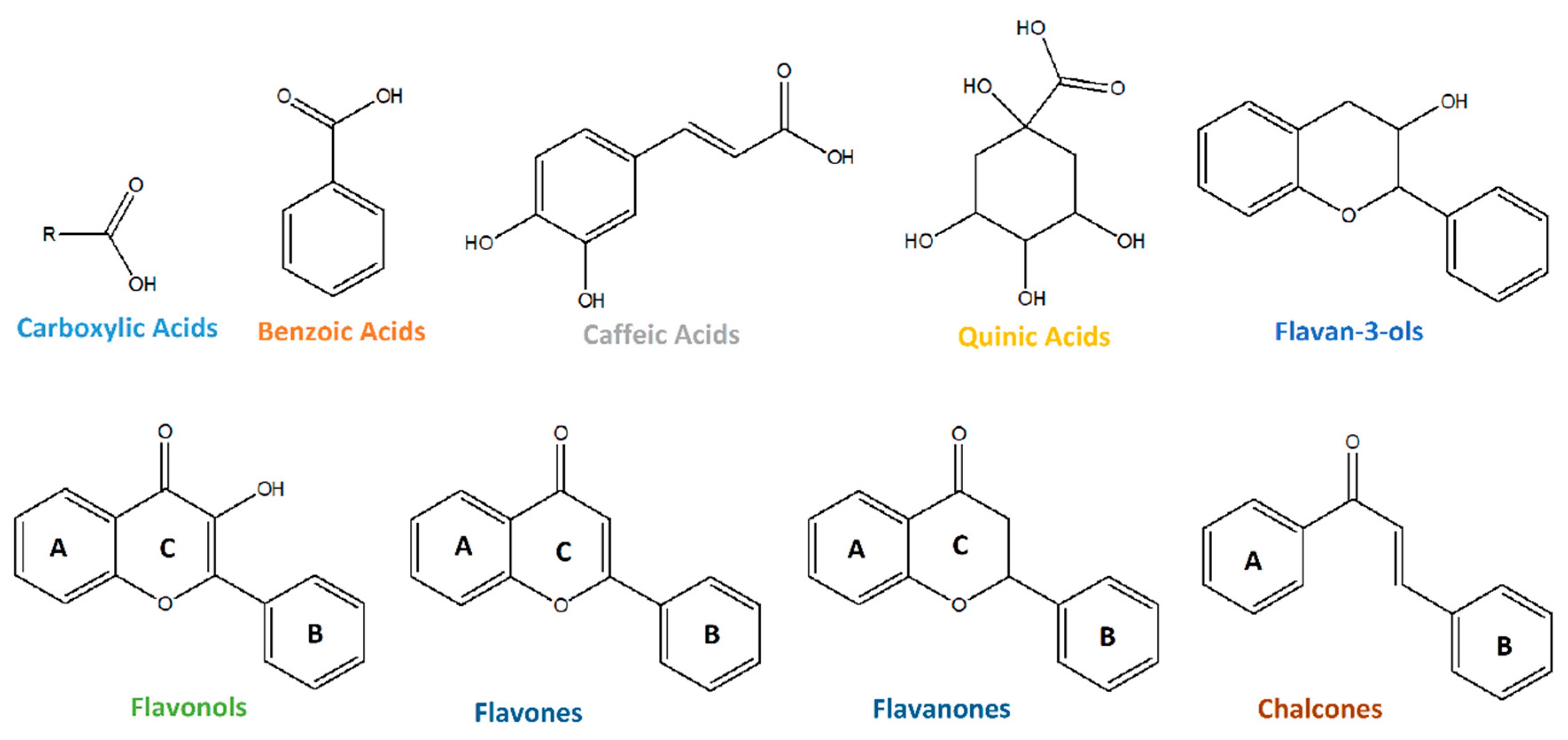
Figure 1. Compounds classes annotated in the ethanolic leaf extracts of V. corymbosum cultivars.
Foods 14 02846 g001
Figure 2. Compounds class distribution in V. corymbosum cultivars.
Foods 14 02846 g002
Figure 3. (A) PLSDA biplot (scores and loadings—10 most relevant) obtained with the metabolite profile (semi-quantitative data) of V. corymbosum, and (B) variables’ importance on projection (VIP scores) of the PLSDA model—23 most relevant (VIP > 1).
Foods 14 02846 g003
Figure 4. Carboxylic acids’ relative abundance in V. corymbosum cultivars.
Foods 14 02846 g004
Figure 5. Benzoic acids’ relative abundance in V. corymbosum cultivars.
Foods 14 02846 g005
Figure 6. Caffeic acids’ relative abundance in V. corymbosum cultivars.
Foods 14 02846 g006
Figure 7. Quinic acids’ relative abundance in V. corymbosum cultivars.
Foods 14 02846 g007
Figure 8. Flavan-3-ols’ relative abundance in V. corymbosum cultivars.
Foods 14 02846 g008
Figure 9. Flavonols’ relative abundance in V. corymbosum cultivars.
Foods 14 02846 g009
Figure 10. Flavones’ and flavanones’ relative abundance in V. corymbosum cultivars.
Foods 14 02846 g010
Figure 11. Chalcones’ relative abundance in V. corymbosum cultivars.
Foods 14 02846 g011
Figure 12. Miscellaneous compounds’ relative abundance in V. corymbosum cultivars.
Foods 14 02846 g012
Table 1. Metabolite profiling of V. corymbosum ethanolic leaf extracts.
#Formula[M−H]−TrFragmentsmSigma|Accuracy|(ppm)Annotation
Carboxylic AcidsC1C4H6O4117.019041.9873.0294.72.48Succinic acid
C2C5H8O5147.030241.9787.009; 85.029; 129.0197.12.38Citramalic acid
C3C5H10O3117.055362.9573.029; 59.01421.03.08Hydroxypentanoic acid isomer 1
C4C9H8O3163.040524.95119.050; 71.0146.93.01p-coumaric acid
C5163.0404810.28119.05011.82.45m-coumaric acid
C6C9H16O4187.0979113.31125.097; 169.089; 123.082; 143.1089.41.92Azelaic acid
C7C11H20O4215.1288818.03197.119; 153.12917.20.01Undecanedioic acid
C8C12H24O3215.1651917.40169.157; 87.008; 171.1038.3−0.93Hydroxydodecanoic acid isomer 1
C9215.1652619.21169.160; 197.055; 199.1338.40.88Hydroxydodecanoic acid isomer 2
C10C14H22O4253.1443519.69209.155; 210.159; 59.015; 209.120; 89.02713.21.34Furancarboxylic acid
Benzoic AcidsB11C7H6O3137.024841.7493.038; 111.008; 81.0344.983.07Dihydoxybenzaldehyde
B12137.024752.8693.035; 94.0387.862.26Hydroxybenzoic acid isomer 1
B13137.0247911.9993.035; 108.020>302.70Hydroxybenzoic acid isomer 2
B14137.0247913.0493.035; 108.022; 94.038>302.70Hydroxybenzoic acid isomer 3
B15C7H6O4153.019597.39109.030; 108.022; 123.04610.992.03Dihydroxybenzoic acid isomer 1
B16153.019529.82109.030>301.24Dihydroxybenzoic acid isomer 2
B17C7H6O5169.014532.51125.025>301.66Gallic acid
B18169.014413.44125.025; 124.017>300.95Trihydroxybenzoic acid isomer 1
B19169.014504.20151.004; 125.024; 83.012>301.48Tri-hydroxybenzoic acid isomer 2
B20C13H16O8299.077162.86137.025; 93.034; 139.04015.170.50Hydroxybenzoic acid hexoside isomer 1
B21299.076814.05137.025; 179.036; 239.057; 151.04116.621.17Hydroxybenzoic acid hexoside isomer 2
B22299.076665.64137.024; 93.033>301.94Hydroxybenzoic acid hexoside isomer 3
B23C13H16O9315.071963.58152.012; 108.02216.920.63Dihydroxybenzoic acid hexoside
B24C14H18O9329.087078.45167.036; 191.035; 209.047; 123.0453.370.36Vanillic acid glucopyranoside
Caffeic AcidsCaf25C9H8O4179.035278.63135.046; 134.038>301.62Caffeic acid (3,4-dihydroxycinnamic acid)
Caf26179.0352913.63135.047; 137.060>301.56Dihydroxycinnamic acid isomer 2
Caf27C15H18O9341.087776.20179.035; 135.035; 181.05018.71.88Caffeic acid hexoside
Quinic AcidsQ28C16H18O9353.087671.39191.057; 179.036; 135.045; 173.04615.70.20Caffeoylquinic acid isomer 1
Q29353.086959.09191.05727.62.44Caffeoylquinic acid isomer 2
Q30353.0868912.65191.057; 179.035; 135.046; 192.059; 165.018; 173.0466.02.66Caffeoylquinic acid isomer 3
Q31353.0866213.61191.057; 173.046; 179.036; 135.04526.23.37Caffeoylquinic acid isomer 4
Q32C16H18O8337.0923710.59191.057; 173.04618.11.57Coumaroylquinic acid isomer 1
Q33337.0922514.27163.041; 191.057; 119.0514.01.51Coumaroylquinic acid isomer 2
Q34337.0924916.12163.041; 191.057; 173.047; 164.044; 119.05021.51.54Coumaroylquinic acid isomer 3
Q35C17H20O9367.103049.14191.057; 173.046; 193.053; 93.03520.30.57Feruloylquinic acid 
Q36C25H24O12515.1183715.48353.089; 173.046; 179.036; 191.0571.22.19Dicaffeoylquinic acid 
Q37C26H26O12529.1335814.90367.104; 193.051; 191.056; 353.088; 173.046; 179.03710.73.27Feruloylcaffeoylquinic acid 
Flavan-3-olsFla38C15H14O7305.066393.88125.025; 165.020; 167.036; 137.02516.70.43Gallocatechin/Epigallocatechin
Fla39C15H14O6289.071285.81245.082; 203.071; 205.051; 137.025; 179.03621.81.45Catechin/Epicatechin
Fla40C45H36O18863.183135.04411.073; 289.072; 285.041;712.137; 451.104>300.28Procyanidin trimer type A/B isomer 1
Fla41863.184247.95289.072; 411.072; 451.104; 711.136; 573.10425.81.15Procyanidin trimer type A/B isomer 2
Fla42863.182858.58411.073; 289.073; 451.104; 711.134; 573.103>300.30Procyanidin trimer type A/B isomer 3
FlavonolsFlo43C15H12O6287.0555313.52259.060; 243.069; 125.024; 152.01024.21.43Aromadendrin
Flo44C16H12O7315.0505818.35300.028; 301.032; 151.00416.91.46Isorhamnetin
Flo45C21H20O10431.0976014.60285.040; 284.033; 191.05622.11.79Kaempferol-7-O-rhamnoside
Flo46C21H20O13479.0816911.82317.029; 191.056; 301.033>301.42Myricetin-3-hexoside
Flo47C23H24O13507.1126913.11344.05418.51.77Syringetin-3-hexoside
Flo48C25H26O15565.1183411.34300.027; 463.087; 191.056>303.11Quercetin-3-O-pentosylpentoside
Flo49C27H30O15593.1498911.81284.033; 463.088; 327.05013.92.24kaempferol 3-O-rutinoside
Flo50593.1510512.38285.042; 284.03424.80.42Flavonol base-3-O-hexoside-hexoside isomer 2
Flo51C27H30O16609.1466711.00300.029>300.72Rutin
Flo52C27H30O17625.1392313.20445.077; 463.088; 301.035; 464.090; 300.029>302.86Quercetin-3-gentiobioside
Flo53C32H38O20741.188379.60300.028; 191.057; 353.087; 417.123; 178.998>300.05Helieianeoside B
Flo54C33H40O20755.2050212.40593.152; 285.042>301.40Kaempferol-3-O-rutinoside-7-O-β-D-glucopyranoside
Flo55C36H36O18755.1824016.96593.130; 285.04077.90.04Variabiloside E
Flavones and FlavanonesFlav56C17H16O5299.0934418.24179.035; 151.004; 135.046; 165.020; 121.030; 229.053; 149.06315.73.813,9-dihydroeucomin
Flav57C21H22O11449.1070113.53287.056; 191.057; 151.004; 257.082; 301.033; 135.04529.94.14Miscanthoside
Flav58C26H28O14563.139897.74191.057; 353.088>301.31Apiin
Flav59C27H32O14579.170458.66417.118; 418.123; 307.083; 335.077; 191.057>302.56Glucoliquiritin
ChalconesCha60C15H12O5271.0606913.03151.003; 165.055; 119.050; 177.019; 228.076>301.88Naringenin chalcone
Cha61C16H14O4269.0823119.83134.037; 178.028; 137.025; 133.029; 139.04213.91.26Cardamonin
MiscellaneousM62C9H12N2O6243.062111.80200.055; 42.000; 153.029; 140.035; 71.01310.10.45Uridine
M63C10H13N5O5282.084302.00150.043; 133.018; 117.019; 113.025; 191.05510.80.21Guanosine
M64C19H20O10407.097455.77245.04512.0−2.65Khelloside
Table 2. ANOVA of the ten most discriminating metabolites.
CompoundCultivar 1Cultivar 2Corrected p-Value (Holm)
Azelaic acid‘Aurora’‘Susyblue’0.0042
Azelaic acid‘Aurora’‘Liberty’0.0042
Azelaic acid‘Camellia’‘Susyblue’0.0042
Azelaic acid‘Camellia’‘Liberty’0.0042
Azelaic acid‘Star’‘Susyblue’0.0042
Azelaic acid‘Star’‘Liberty’0.0042
Undecanedioic acid‘Camellia’‘Susyblue’0.0042
Undecanedioic acid‘Camellia’‘Liberty’0.0042
Hydroxybenzoic acid hexoside isomer 1‘Camellia’‘Liberty’0.0042
Dihydroxybenzoic acid hexoside‘Camellia’‘Liberty’0.0042
Caffeic acid (3,4-dihydroxycinnamic acid)‘Aurora’‘Susyblue’0.0042
Caffeic acid (3,4-dihydroxycinnamic acid)‘Aurora’‘Liberty’0.0042
Caffeic acid (3,4-dihydroxycinnamic acid)‘Camellia’‘Susyblue’0.0042
Caffeic acid (3,4-dihydroxycinnamic acid)‘Camellia’‘Liberty’0.0042
Caffeic acid (3,4-dihydroxycinnamic acid)‘Star’‘Susyblue’0.0042
Caffeic acid (3,4-dihydroxycinnamic acid)‘Star’‘Liberty’0.0042
Caffeic acid hexoside‘Camellia’‘Liberty’0.0042
Caffeoylquinic acid isomer 2‘Camellia’‘Liberty’0.0042
Feruloylquinic acid‘Camellia’‘Liberty’0.0042
Catechin/Epicatechin‘Camellia’‘Liberty’0.0042
Guanosine‘Camellia’‘Liberty’0.0042
Disclaimer/Publisher’s Note: The statements, opinions and data contained in all publications are solely those of the individual author(s) and contributor(s) and not of MDPI and/or the editor(s). MDPI and/or the editor(s) disclaim responsibility for any injury to people or property resulting from any ideas, methods, instructions or products referred to in the content.