Article

Metabolic Master Switch: Pyruvate Carboxylase Fuels Antimicrobial Resistance and Virulence in Foodborne Staphylococcus aureus

State Key Laboratory of Microbial Metabolism, Department of Food Science & Technology, School of Agriculture & Biology, Shanghai Jiao Tong University, Shanghai 200240, China
*
Author to whom correspondence should be addressed.
Foods 2025, 14(15), 2566; https://doi.org/10.3390/foods14152566
Submission received: 22 June 2025 / Revised: 11 July 2025 / Accepted: 19 July 2025 / Published: 22 July 2025
(This article belongs to the Section Food Microbiology)

Abstract

Staphylococcus aureus, a major cause of foodborne illness globally, presents significant challenges due to its multidrug resistance and biofilm-forming capabilities. Pyruvate carboxylase (PycA), a metabolic master switch linking glycolysis and the tricarboxylic acid (TCA) cycle, is a potential target for controlling S. aureus. In this study, a pycA mutant was constructed and analyzed using phenotypic assays and proteomics to investigate its role in virulence and antimicrobial resistance. The results showed that deletion of pycA in the foodborne methicillin-resistant strain ATCC BAA1717 resulted in a 4- to 1024-fold reduction in resistance to β-lactams, aminoglycosides, and macrolides; a 23.24% impairment in biofilm formation; and a 22.32% decrease in staphyloxanthin production, a key antioxidant essential for survival in oxidative food environments. Proteomic analysis revealed downregulation of the TCA cycle, purine biosynthesis, surface adhesins (FnbA/B, SasG), and β-lactamase (BlaZ), linking PycA-mediated metabolism to phenotypes relevant to food safety. These findings underscore the importance of PycA as a metabolic regulator crucial for S. aureus resilience in food systems, suggesting novel strategies to combat foodborne staphylococcal infections through metabolic interference.

1. Introduction

Staphylococcus aureus is a prevalent foodborne pathogen responsible for staphylococcal food poisoning through the production of heat-stable enterotoxins [1]. In addition to its toxigenic potential, the pathogenicity of S. aureus is mediated by an arsenal of virulence factors, including surface adhesins, the antioxidant pigment staphyloxanthin (STX), and various immune evasion molecules, which facilitate effective colonization, biofilm formation, and persistence in hostile environments [2]. Worsening the situation is the widespread emergence of methicillin-resistant S. aureus (MRSA) in food environments [3,4], which exhibits multidrug resistance and severely limits therapeutic options [5]. Given its dual threat of toxicity and resistance, S. aureus presents a significant challenge to food safety and public health, highlighting the urgent need for novel antimicrobial strategies targeting both resistance mechanisms and virulence regulation.
Recent studies have highlighted the critical role of central metabolic pathways in regulating both antimicrobial resistance and virulence. For example, glycolysis mitigates energy depletion during antimicrobial stress, contributing to resistance against cyclic lipopeptide antibiotics such as polymyxin B and daptomycin [6]. Moreover, glycolysis enables S. aureus to sustain energy metabolism and redox balance under nitric oxide-induced hypoxic conditions, thereby facilitating immune evasion and promoting virulence during infection [7]. Similarly, disruption of the TCA cycle leads to ATP depletion and reduced membrane potential, promoting the formation of persister cells and enhancing tolerance to multiple antimicrobial classes [8,9,10].
Pyruvate carboxylase (PycA) is a key enzyme in central carbon metabolism, catalyzing the carboxylation of pyruvate to oxaloacetate, thereby linking glycolysis to the TCA cycle. Previous genome-wide screening studies have identified pycA as a critical gene involved in virulence attenuation in S. aureus [11]. In a murine systemic infection model, disruption of pycA markedly reduced bacterial burden in vivo and mitigated infection-induced tissue necrosis. Moreover, the pycA mutant displayed increased susceptibility to macrophage-mediated clearance [12]. During a preliminary screen for virulence-associated genes using a transposon mutant library in S. aureus, we unexpectedly found that mutation of the pycA gene led to a noticeable decrease in both virulence and antimicrobial resistance. However, its role in regulating antimicrobial resistance remains unclear. Here, we investigated the impact of pycA deletion on antimicrobial resistance, virulence, and metabolic pathways in S. aureus, providing insights into its potential as a control target for food safety management.

2. Materials and Methods

2.1. Bacterial Strains and Plasmids

The strains and plasmids used in this study are listed in Table 1. All strains were stored at –80 °C in tryptic soy broth (TSB) supplemented with 25% glycerol. Unless otherwise specified, strains were cultured in TSB at 37 °C prior to use. For the pycA-complemented (cpycA) and pycA-overexpression strains (WT-ppycA), pycA expression was induced with 0.15 μg/mL anhydrotetracycline.

2.2. Mutated Strain Construction

In this study, deletion of the pycA in S. aureus BAA1717 (wild-type, WT) was performed using a modified λ-Red homologous recombination strategy, as previously reported with minor modifications [17]. Briefly, upstream and downstream homologous arms of the pycA were amplified by PCR and ligated into the Sal I and Kpn I sites of the temperature-sensitive plasmid pKZ2. The resulting recombinant plasmid was first introduced into E. coli JTU006 for restriction-modification and subsequently electroporated into S. aureus BAA1717. Homologous recombination facilitated the deletion of the pycA locus, and the ΔpycA mutant was verified by PCR. For gene complementation and overexpression assays, the full-length pycA gene along with its native promoter was cloned into the shuttle vector pCL55 to construct the recombinant plasmid pCL55-pycA. This plasmid was subsequently introduced into both the ΔpycA mutant and the WT for functional complementation and overexpression, respectively.

2.3. Minimum Inhibitory Concentration Determination

According to the Clinical and Laboratory Standards Institute (CLSI) guidelines [18], the minimum inhibitory concentrations (MICs) of antimicrobials against S. aureus strains were determined using the broth microdilution method. In brief, antimicrobials were two-fold serially diluted in TSB across 96-well microtiter plates. Bacterial suspensions were prepared from overnight cultures and diluted to a final concentration of 1 × 10⁶ CFU/mL. Equal volumes of the antimicrobial dilutions and bacterial suspensions were added to each well. The plates were incubated at 37 °C for 24 h. The MIC was defined as the lowest antimicrobial concentration that visibly inhibited bacterial growth.

2.4. Time-Kill Curve Assay

Time-kill assays were performed on WT and ΔpycA strains following previously described protocols [19]. Antimicrobials were used at 1/4 MIC, based on the MIC values determined for the WT BAA1717 strain.

2.5. Biofilm Quantification

Biofilm formation was assessed using crystal violet staining in a 96-well microtiter plate [20]. Briefly, bacterial suspensions were diluted in TSB to achieve a final concentration of 1 × 10⁶ CFU/mL, and 200 μL of this mixture was added to each well, followed by incubation at 37 °C for 24 h. After incubation, the wells were gently washed three times with phosphate-buffered saline (PBS, pH 7.4) to remove planktonic cells, then fixed at 60 °C for 15 min. The biofilms were stained with 0.1% crystal violet for 15 min, washed with PBS, and decolorized with 200 μL of absolute ethanol for 5 min. Absorbance was measured at 595 nm using a microplate reader. TSB without bacterial inoculation served as the blank control. All experiments were performed in triplicate, with five technical replicates per group.

2.6. Staphyloxanthin Quantification

Staphyloxanthin (STX) quantification was performed using a previously described method with minor modifications [21]. S. aureus strains were inoculated into brain heart infusion (BHI) medium and incubated at 37 °C with shaking at 200 rpm for 30 h. After incubation, 2 mL of the culture was centrifuged to collect the bacterial pellet, which was subsequently washed three times with PBS (pH 7.4). The pellet was resuspended in PBS, and the bacterial suspension was adjusted to a final concentration of 109 CFU/mL. The normalized bacterial pellet was resuspended in 800 μL of 99% methanol and mixed thoroughly. The mixture was incubated at 55 °C in the dark for 2 h, followed by centrifugation. A 200 μL aliquot of the supernatant was collected, and the absorbance at 462 nm was measured using a microplate reader to quantify STX, as this wavelength corresponds to its characteristic absorbance peak. All experiments were conducted in triplicate.

2.7. 2,2-Diphenyl-1-Picrylhydrazyl Free Radical Scavenging Activity Assay

The 2,2-diphenyl-1-picrylhydrazyl (DPPH) radical scavenging activity was evaluated using a modified version of a previously described method [22]. S. aureus was cultured overnight in TSB, and the bacterial cells were subsequently harvested and washed three times with PBS (pH 7.4). The cell suspension was then standardized to ensure equal bacterial counts across all experimental groups. Cell disruption was performed by sonication on ice (200 W, 3 s on/10 s off, 60 cycles). The lysates were then centrifuged, and the supernatants were collected for antioxidant analysis. For the DPPH assay, 30 μL of each sample was mixed with 170 μL of 0.1 mM DPPH in ethanol in a 96-well plate. PBS served as the blank control. The reaction mixtures were incubated in the dark at room temperature for 30 min, and absorbance was measured at 490 nm using a microplate reader. All experiments were conducted in triplicate.

2.8. L-Aspartate Auxotrophy Assay

The minimum essential medium (MEM) for S. aureus growth was prepared following previously described protocols [23]. S. aureus strains were cultured in 96-well plates containing either MEM alone or MEM supplemented with 100 μg/mL L-aspartate. The plates were incubated at 37 °C under static conditions for 24 h. Bacterial growth was measured by optical density (OD) at 600 nm using a microplate reader. The experiment was conducted with three independent biological replicates. The composition of MEM is provided in Table S1.

2.9. Growth Curve Measurement

An overnight culture of S. aureus was harvested and diluted in fresh TSB to a final bacterial concentration of 1 × 10⁶ CFU/mL. Bacterial growth was monitored at 37 °C for 26 h using a growth curve analyzer, with OD600 readings recorded every 30 min. Each condition was measured in five technical replicates, and the experiment was repeated in three independent biological replicates.

2.10. Real-Time PCR Analysis

Total RNA was extracted from 12 h S. aureus cultures using the SPARKeasy Bacteria RNA Kit (Sparkjade Biotechnology, Jinan, China). Complementary DNA (cDNA) was synthesized with the HiScript® III RT SuperMix for qPCR (+gDNA wiper) kit (Vazyme Biotech, Nanjing, China), according to the manufacturer’s instructions. Quantitative PCR (qPCR) and reverse transcription qPCR (RT-qPCR) were performed to evaluate the relative expression of genes using 2 × SYBR Green qPCR Mix (with ROX) (Sparkjade Biotechnology, Jinan, China). Gene expression levels were normalized to 16S rRNA as the internal control, and foldchanges were calculated using the 2−ΔΔCt method. All experiments were conducted with three independent biological replicates. Primer sequences are listed in Table S2.

2.11. Scanning Electron Microscopy Analysis

Scanning electron microscopy (SEM) was conducted according to a previous protocol with slight modifications [24]. S. aureus was cultured in TSB at 37 °C for 12 h. Bacterial cells were washed with PBS (pH 7.4) and fixed in 2.5% (v/v) glutaraldehyde at 4 °C for 8–12 h. The samples were then dehydrated through a graded ethanol series (30%, 50%, 70%, 90%, and 100%, v/v), with each step lasting 10 min; the 30% and 100% steps were repeated twice. The dehydrated samples were stored in a desiccator and sputter-coated with gold for SEM analysis.

2.12. Transmission Electron Microscopy Analysis

Transmission electron microscopy (TEM) analysis was conducted according to a previous protocol [25]. S. aureus cultures were fixed in 2.5% (v/v) glutaraldehyde at 4 °C for 8–12 h, followed by three washes with PBS (pH 7.4). Post-fixation was carried out using 1% osmium tetroxide for 1 h at room temperature. The samples were then dehydrated through a graded ethanol series (30%, 50%, 70%, 90%, and 100%, v/v), with each step lasting 10 min. Dehydrated cells were embedded in epoxy resin, and ultrathin sections (70–90 nm) were prepared using an ultramicrotome. These sections were stained with uranyl acetate and lead citrate before being examined under a transmission electron microscope.

2.13. Intracellular ATP Analysis

Intracellular ATP levels were measured using an ATP assay kit (Beyotime, Beijing, China) with minor modifications. S. aureus strains were standardized to a final concentration of 1 × 10⁹ CFU/mL and washed three times with PBS (pH 7.4). Cells from 2 mL of culture were collected by centrifugation, resuspended in 400 μL lysis buffer with several 2 mm steel beads, and vortexed on ice for 15 min. After centrifugation, 60 μL of the supernatant was used for ATP detection following the kit instructions. ATP concentration was calculated using a standard curve.

2.14. Proteomic Analysis

Total proteins from the WT and ΔpycA strains were extracted, quantified using the bicinchoninic acid (BCA) assay, and digested with trypsin. The resulting peptides were desalted, dried, and reconstituted in a solvent containing indexed retention time (iRT) standard peptides for LC-MS analysis. Approximately 500 ng of peptides per sample were separated using a Vanquish Neo Nano-scale Ultra Performance Liquid Chromatography (nano-UPLC) system coupled with an Astral mass spectrometer (Thermo Fisher Scientific, Waltham, MA, USA) and analyzed in the data-independent acquisition (DIA) mode. The MS data were processed using the DIA-NN software against the S. aureus UniProt database, with quantification based on a 2 m/z isolation window and a 1% false discovery rate (FDR) threshold. Differentially expressed proteins (|log2FC| ≥ 0.5, p < 0.05) were identified and further analyzed using Gene Ontology (GO), the Kyoto Encyclopedia of Genes and Genomes (KEGG), principal component analysis (PCA), and statistical methods.

2.15. A549 Cells Infection Assay

A549 is a human lung adenocarcinoma epithelial cell line that is widely used in cytotoxicity testing and drug screening assays. The A549 cell infection assay was conducted following a previously established protocol, with slight modifications [26]. Briefly, A549 cells were seeded into 96-well plates at a density of 2 × 10⁴ cells per well and incubated for 16 h. The culture medium was then replaced with fresh Dulbecco’s Modified Eagle Medium (DMEM) supplemented with 10% fetal bovine serum (FBS). Subsequently, S. aureus strains were added at a multiplicity of infection (MOI) of 100, and the cells were co-incubated for 6 h. Cell viability was assessed using calcein AM/PI dual staining (Beyotime, Beijing, China), which allowed for the differentiation of live and dead cells via fluorescence microscopy. Untreated cells served as the negative control. Cytotoxicity was evaluated by measuring lactate dehydrogenase (LDH) release after 6 h of incubation using a commercial LDH assay kit (Beyotime, Beijing, China). Supernatants were collected for analysis. The A549 cell line was purchased from Wuhan Procell Life Science & Technology Co., Ltd. (Wuhan, China).

2.16. Statistical Analyses

Statistical analyses were conducted using GraphPad Prism 10.1.2 (GraphPad Software, San Diego, CA, USA). Group differences were evaluated using unpaired t-tests or one-way ANOVA, as appropriate. Results with p-values less than 0.05 were deemed statistically significant.

3. Results

3.1. Deletion of pycA Increases Antimicrobial Susceptibility in S. aureus

Deletion of pycA significantly increased the susceptibility of S. aureus to a broad range of antimicrobials (Table 2). Notably, the MICs of ΔpycA for β-lactams, including oxacillin, ampicillin, and amoxicillin, were reduced by 64- to over 1024-fold compared to the WT. Enhanced sensitivity was also observed to aminoglycosides, including kanamycin sulfate and amikacin (4- to 64-fold reductions), as well as for macrolides, with erythromycin and azithromycin MICs decreased by 128-fold. Complementation with pycA (cpycA) partially restored antimicrobial resistance to WT levels, while overexpression of pycA (WT-ppycA) further elevated MICs, indicating enhanced resistance. These results indicate that pycA may indirectly contribute to S. aureus resistance to β-lactams, aminoglycosides, and macrolides. This observation is further corroborated by time-kill assays (Figure S1), which reveal that all tested antimicrobials exert enhanced bactericidal activity against the ΔpycA relative to the WT.
Antimicrobial resistance in S. aureus is typically multifactorial. Previous studies have shown that downregulation of central carbon metabolism in S. aureus leads to ATP depletion, promoting the formation of persister cells and enhancing antimicrobial resistance [10,27]. However, pycA, encoding a key enzyme bridging glycolysis and the TCA cycle, appears to play an inverse role. Deletion of pycA reversed resistance to multiple antimicrobial classes in the WT, suggesting that PycA may act upstream to modulate the expression of established resistance determinants. Notably, the partial restoration of resistance upon cpycA indicates that pycA deletion may involve potentially irreversible metabolic reprogramming, thereby hindering full restoration of the original metabolic state and resistance phenotype. Furthermore, the increased sensitivity to β-lactams observed in the ΔpycA mutant may be attributed to reduced blaZ expression rather than mecA, as no difference in cefoxitin resistance was detected between ΔpycA and WT. In the case of aminoglycosides, the heightened susceptibility observed in ΔpycA may be linked to decreased activity of 3’,5’’-aminoglycoside phosphotransferase (type III), an enzyme that inactivates aminoglycosides via phosphorylation [28]. Similarly, the increased susceptibility to macrolide antimicrobials may be explained by impaired expression of macrolide phosphotransferases or reduced activity of ATP-dependent efflux pumps [29]. Given the observed ATP reduction in the ΔpycA mutant, it is plausible that energy deficiency compromises efflux efficiency, leading to intracellular accumulation of macrolides and enhanced bactericidal effects. Collectively, these findings indicate that PycA acts as a key regulator of antimicrobial resistance in S. aureus, potentially via metabolic alterations.

3.2. PycA Is Dispensable for Growth Under Nutrient-Rich Conditions but Essential in the Absence of L-Aspartate

To rule out the possibility that the reduced antimicrobial resistance in the ΔpycA strain was due to a growth defect, growth curves were evaluated in TSB. As shown in Figure 1a, deletion of pycA does not significantly impair bacterial growth under standard culture conditions, as further supported by the absence of significant morphological changes observed by SEM and TEM (Figure 1c,d). PycA catalyzes the carboxylation of pyruvate to form oxaloacetate, a precursor for aspartic acid (ASP) synthesis via aspartate aminotransferase [30]. To determine whether pycA deletion disrupts ASP biosynthesis, a nutritional limitation assay was conducted. As shown in Figure 1b, ΔpycA exhibited significantly reduced growth without exogenous L-ASP, while supplementation restored growth to levels comparable to the cpycA strain. No growth defects were observed in the WT or WT-ppycA strains, suggesting that pycA is essential for survival under ASP-limited conditions.
These findings indicate that pycA deletion does not affect S. aureus growth under nutrient-rich conditions but impairs the biosynthesis of ASP. Under normal physiological conditions, the concentration of free ASP is approximately 2.8 μg/mL in human serum [31] and around 2 μg/mL in mouse serum [32], concentrations that are insufficient to support the growth of the ΔpycA. ASP is an essential metabolic intermediate, and the pathogen must rely on endogenous ASP synthesis to sustain purine biosynthesis and survival [33]. Therefore, PycA is essential for the survival and virulence of S. aureus during infection and represents a potential target for novel antimicrobial strategies.

3.3. Impaired Virulence isConfirmed in the ΔpycA Mutant

3.3.1. Deletion of pycA Inhibits Biofilm Formation by S. aureus In Vitro

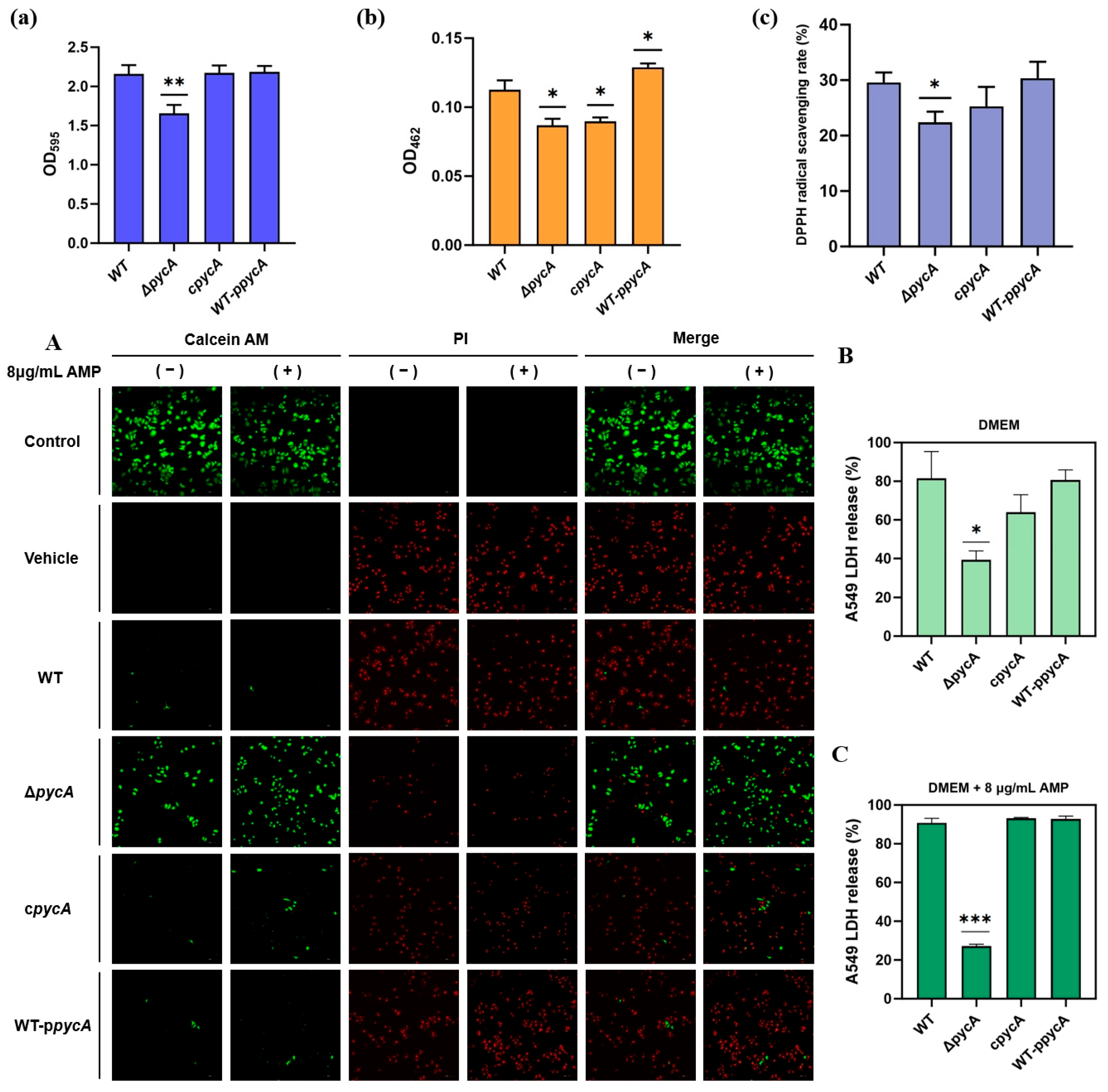
In addition to mediating resistance phenotypes, PycA may also influence key virulence-associated factors that facilitate surface adhesion and persistence, which are critical for S. aureus survival and contamination in food processing environments. Among these, biofilm formation is a well-recognized virulence strategy in S. aureus [34,35]. Quantitative analysis revealed that pycA deletion significantly reduced biofilm biomass by 23.24% compared to the WT (Figure 2a). This defect was fully restored in the cpycA strain, indicating that PycA plays a critical role in maintaining biofilm-forming capacity. Given that the biofilm matrix impedes antimicrobial penetration and supports persister cell survival [36], the diminished biofilm formation in the ΔpycA strain likely contributes to its increased antimicrobial susceptibility and attenuated pathogenicity.

3.3.2. Deletion of pycA Inhibits STX Production and Reduces Total Antioxidant Capacity in S. aureus

During cultivation, visual inspection indicated a reduction in STX production in the ΔpycA compared to the WT. Quantitative analysis further confirmed a 22.32% decrease in STX levels in the ΔpycA strain, while overexpression of pycA increased STX production by 15.18% (Figure 2b). However, complementation of pycA did not fully restore STX levels to those observed in the WT. STX, a golden carotenoid pigment unique to S. aureus, enhances resistance to oxidative stress through its antioxidant properties by scavenging reactive oxygen species [37]. Additionally, STX intercalates into the bacterial membrane, reducing fluidity and impairing the insertion of antimicrobial peptides, thus enhancing bacterial resistance [38,39]. To further investigate oxidative stress resistance, the total antioxidant capacity of the strains was measured using the DPPH radical scavenging assay. Consistent with the reduction in STX levels, the ΔpycA strain exhibited a 24.26% decrease in total antioxidant activity (Figure 2c), which was partially restored in the cpycA strain. These findings further confirm that reduced STX levels in the ΔpycA strain impair its antioxidant capacity, which may compromise its resistance to host innate immune defenses and oxidative disinfectants, potentially contributing to its attenuated virulence.

3.3.3. Deletion of pycA Reduces the Cytotoxicity of S. aureus and Its Survival Under Antimicrobial Pressure

Confocal microscopy and LDH assays demonstrated that the ΔpycA exhibited significantly reduced cytotoxicity toward A549 cells compared to the WT, as evidenced by increased live-cell staining and lower LDH release (Figure 2A,B). The complementation of pycA restored its cytotoxicity to WT levels. Under ampicillin treatment (8 μg/mL), only the ΔpycA failed to induce significant cell death (Figure 2A,C). These findings suggest that pycA is required for full virulence in host cells and enhances bacterial survival under antimicrobial stress. The reduced cytotoxicity and antimicrobial resistance observed in the ΔpycA strain indicate that PycA plays a critical role as a metabolic node linking central metabolism to both virulence and drug resistance. Previous studies have demonstrated that PycA is essential for the intracellular replication of Listeria monocytogenes, as its deletion impairs bacterial growth within mammalian host cells [40]. Similarly, in S. aureus, PycA replenishes the TCA cycle and supports the biosynthesis of essential precursors such as amino acids, nucleotides, and cell wall components. Disruption of this metabolic flux likely limits the availability of key intermediates required for the synthesis of virulence factors and other biomolecules necessary for survival within mammalian host cells.

3.4. Key Proteins Involved in the Regulation of Virulence and Antimicrobial Resistance via Proteomic Analysis

3.4.1. Deletion of pycA Affects the Overall Metabolism of S. aureus

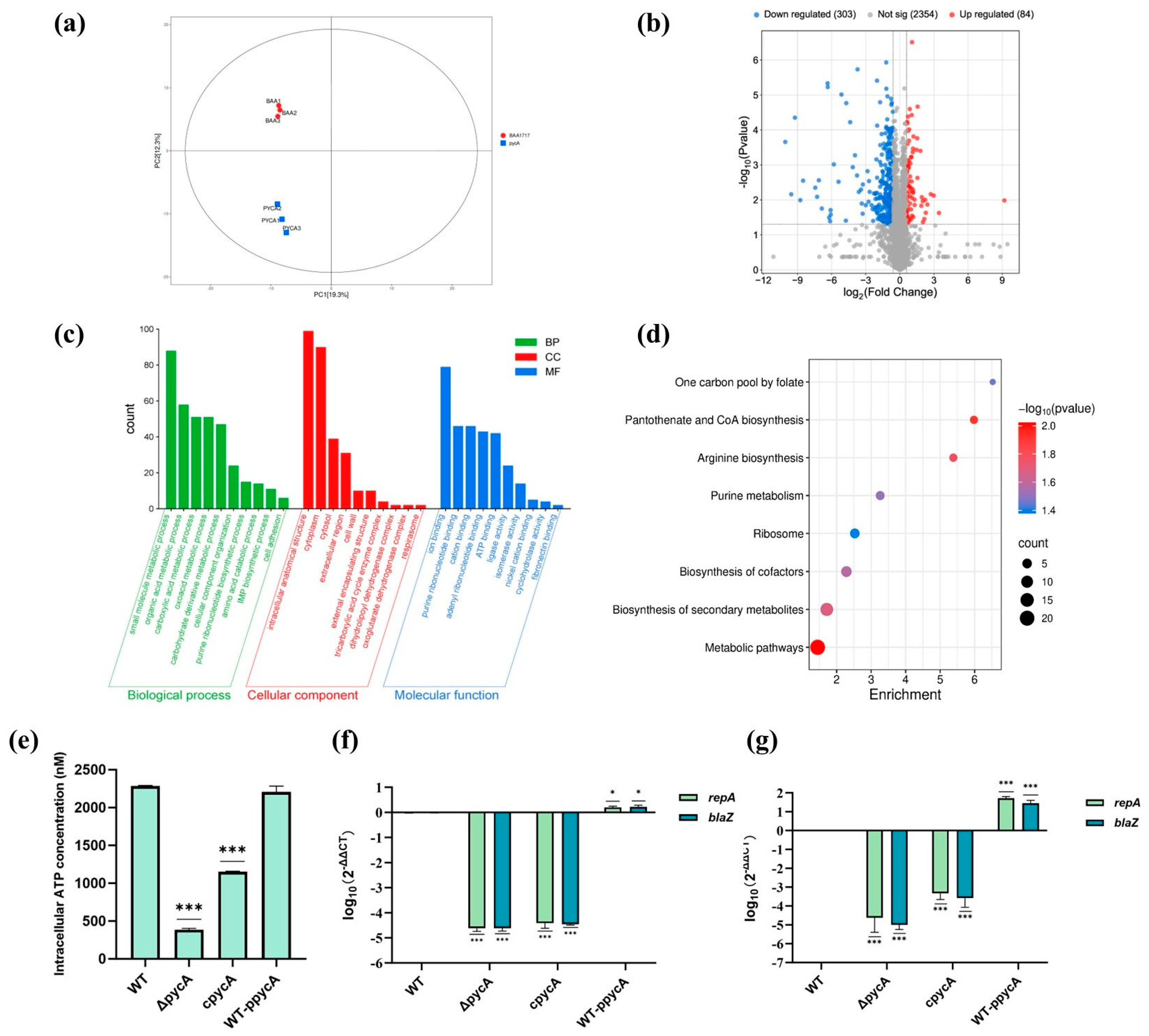
PCA analysis revealed distinct proteomic profiles between WT and ΔpycA (Figure 3a). Comparative proteomic analysis identified 2741 unique peptides across the proteomes. Among these, 84 proteins were significantly upregulated and 303 were significantly downregulated in the ΔpycA (Figure 3b). GO analysis revealed significant changes in biological processes such as carboxylic acid metabolism, purine ribonucleotide metabolism, amino acid metabolism, and cell adhesion (Figure 3c). Notably, the enrichment of ATP-binding proteins in the Molecular Function category further suggests impaired energy metabolism in the ΔpycA strain.
KEGG pathway enrichment analysis revealed that the differentially expressed proteins were predominantly involved in central metabolic pathways, particularly those related to pantothenate and CoA biosynthesis, purine metabolism, and arginine biosynthesis (Figure 3d). These pathways are tightly linked to central carbon metabolism and precursor synthesis, supporting the hypothesis that pycA deletion disrupts oxaloacetate production and its downstream metabolic network. Additionally, the enrichment of the ribosomal pathway and folate-mediated one-carbon metabolism indicates potential impairments in protein and nucleotide biosynthesis.
Further analysis of functional modules of differentially expressed proteins (Table 3) revealed that key TCA cycle enzymes (FumC, SucC, SucD, and Mqo) were significantly downregulated in the ΔpycA. This suggests that reduced oxaloacetate production suppresses TCA activity. Consistently, intracellular ATP quantification showed a dramatic reduction in the ΔpycA compared to the WT, confirming the energy-deficient phenotype associated with pycA deletion (Figure 3e). Notably, despite this significant decline in ATP levels, the growth of S. aureus remained unaffected. A plausible explanation is that under energy-limiting conditions, the bacterium may prioritize survival by downregulating non-essential ATP-consuming processes, such as virulence factor production, thereby conserving energy for essential cellular functions [41]. Similar findings have been reported in other studies, where inactivation of the TCA cycle in S. aureus does not impair its growth under nutrient-rich conditions, supporting that pycA deletion-induced ATP reduction does not affect growth rate [10]. While TCA cycle inactivation has been linked to persister formation via ATP depletion [9], the ΔpycA strain exhibited increased antimicrobial susceptibility, indicating that the metabolic changes induced by pycA deletion may be insufficient to induce a classical persister-like state.
Enzymes involved in de novo purine biosynthesis were also significantly downregulated in ΔpycA. This may be attributed to reduced ASP synthesis, as ASP is an essential amino donor in the purine nucleotide biosynthetic pathway [42]. Furthermore, purine biosynthesis is a highly energy-consuming process, requiring six ATP molecules per IMP produced [43]. ATP limitation may further restrict this pathway in ΔpycA. Purine metabolism is central to both the virulence and antimicrobial resistance in S. aureus. It is upregulated during biofilm formation to support energy generation and extracellular DNA synthesis, both critical for biofilm stability [44]. Moreover, purF and purN contribute to ATP and ppGpp production, which regulate the formation of antimicrobial-tolerant persister cells and activate virulence-related regulators such as the SaeRS [45,46]. These findings suggest that PycA may promote S. aureus virulence by sustaining purine biosynthesis, thereby linking central carbon metabolism to both energy metabolism and virulence gene regulation.

3.4.2. Deletion of pycA Downregulates Plasmid-Associated Antimicrobial Resistance Proteins

In the ΔpycA, the expression levels of β-lactam resistance-associated proteins (BlaZ, BlaI, and BlaR1) were significantly decreased (Table 3). In S. aureus BAA1717 (the WT strain), the blaZ is encoded on the plasmid pUSA300HOUMR [47], which also carries resistance genes against macrolides and aminoglycosides (Figure S2). To examine whether pycA influences plasmid maintenance, qPCR and RT-qPCR were used to assess the expression of the plasmid replication initiator gene repA and the resistance gene blaZ (Figure 3f,g). Both repA and blaZ expression were significantly reduced in the ΔpycA strain and markedly upregulated upon pycA overexpression. However, complementation of pycA did not fully restore their expression to WT levels, suggesting that PycA exerts a threshold-dependent or partially irreversible effect on plasmid stability. The coordinated downregulation of repA and blaZ following pycA deletion indicates that metabolic impairment, likely due to reduced oxaloacetate availability, may disrupt nucleotide pool balance and cellular energy status. As resistance plasmids impose a metabolic burden on S. aureus by diverting resources from essential cellular functions [48], the metabolic stress caused by pycA deletion may reduce plasmid replication. These findings suggest that PycA indirectly regulates plasmid-mediated antimicrobial resistance by maintaining metabolic homeostasis, highlighting the link between core metabolism and extrachromosomal resistance mechanisms.

3.4.3. Deletion of pycA Downregulates the Expression of Cell Adhesion-Associated Proteins

In the ΔpycA strain, the expression of multiple cell surface-associated virulence proteins was significantly reduced, including FnbA, FnbB, SasG, Fib, SpA, and IsaAB (Table 3). FnbA and FnbB mediate adhesion by interacting with host fibronectin through their C-terminal domains [49] and also promote interbacterial aggregation and biofilm formation [50]. SasG facilitates adherence via homophilic interactions and binding to glycosylated receptors on keratinocytes, promoting skin colonization [51,52]. Fib binds host fibrinogen, mediating bacterial attachment to wound surfaces and plasma proteins, and contributes to biofilm structural stability [53]. SpA binds the Fc region of IgG with high affinity, effectively blocking immune recognition and phagocytosis to facilitate immune evasion [54]. Additionally, SpA can bind to host cell surface proteins, assisting bacterial adherence and invasion [55]. The downregulation of these proteins in the ΔpycA strain likely impairs epithelial adhesion, biofilm formation, and immune evasion, collectively contributing to reduced virulence.
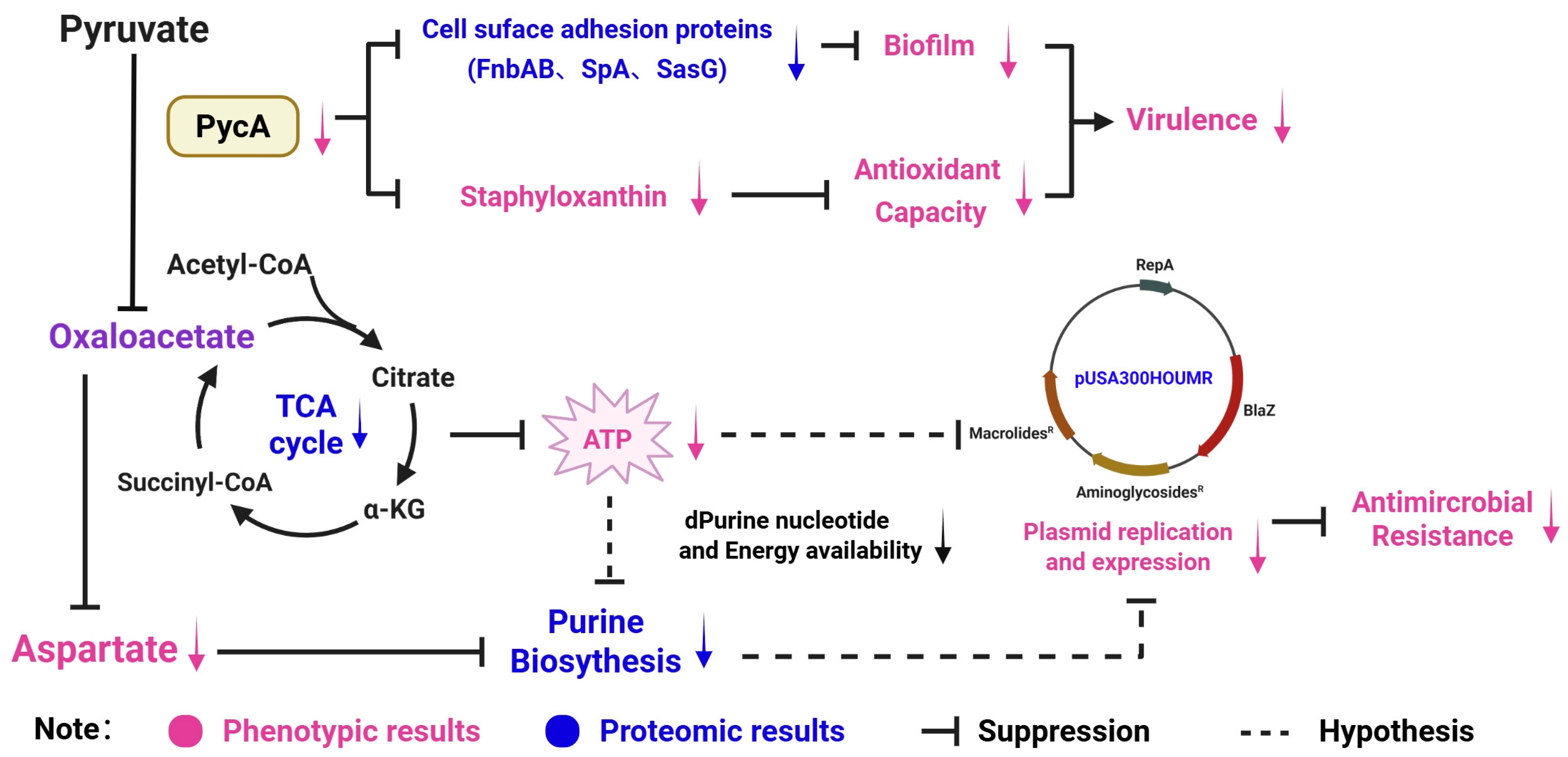
Based on integrated phenotypic observations and proteomic data, we propose a hypothetical model wherein PycA functions as a central metabolic regulator, promoting both virulence and antimicrobial resistance in S. aureus (Figure 4). Reduced PycA activity leads to the downregulation of cell surface adhesion proteins (e.g., FnbAB, SpA, SasG) and STX biosynthesis, thereby impairing biofilm formation and antioxidant capacity—key determinants of bacterial virulence. Additionally, diminished PycA function decreases oxaloacetate production, reducing TCA cycle activity and disrupting aspartate biosynthesis. These metabolic impairments likely constrain ATP generation and purine nucleotide synthesis, leading to compromised DNA replication capacity. In response to this energy-deprived state, S. aureus may downregulate the replication and expression of resistance-related plasmids such as pUSA300HOUMR. This cascade ultimately results in increased susceptibility to multiple classes of antimicrobials. Collectively, these findings suggest that PycA-mediated metabolic flux is a critical upstream driver of both virulence expression and plasmid-based antimicrobial resistance in foodborne S. aureus.

3.5. Outlook: PycA as a Target for Food Safety Interventions

S. aureus remains a major concern in the food processing industry due to its ability to form biofilms, tolerate environmental stresses, and produce enterotoxins associated with foodborne illnesses. Disruption of pycA has been shown to impair stress tolerance and suppress virulence-associated traits, such as biofilm formation and STX production. These changes may increase the bacterial susceptibility to environmental stressors, including food-grade preservatives. Natural compounds, such as phenolic acids and flavonoids, have demonstrated potential to interfere with bacterial metabolism and virulence regulation [56,57,58]. High-throughput virtual screening [59] and fluorescence-based enzyme assays [60] offer a promising approach for identifying PycA inhibitors from natural product libraries. Recent studies have identified potent small-molecule inhibitors of PycA, such as 2-hydroxy-3-(quinoline-2-yl)propenoic acid, which selectively target the carboxyltransferase domain with micromolar potency [61]. In addition, biotinyl-acylsulfamide adenosine, a biotin protein ligase inhibitor, has been shown to indirectly suppress PycA activity by blocking the biotinylation of biotin-dependent enzymes [62]. While the regulatory impact of these inhibitors on S. aureus has not been fully defined, their potential to diminish virulence and compromise preservative tolerance suggests a novel approach to mitigating contamination in food environments. When used alongside reduced concentrations of oxidative preservatives, PycA inhibitors could enhance antimicrobial efficacy through synergistic interactions, serving as effective adjuncts to conventional preservation approaches. Furthermore, incorporating PycA-targeting agents into antimicrobial coatings, surface rinses, or packaging materials may help reduce S. aureus biofilm formation and persistence within food processing environments. Similar effects have been documented in previous studies on plant essential oils [63,64].
While this study provides comprehensive in vitro phenotypic evidence for the regulatory role of pycA in coordinating antimicrobial resistance and virulence in S. aureus, several limitations remain. First, our findings are based primarily on in vitro assays and a single clinical strain (BAA1717). This study may not fully capture the strain-to-strain variability or the complexity of host–pathogen interactions in vivo. Second, although consistent phenotypic and proteomic changes were observed following pycA deletion, the precise regulatory mechanisms by which PycA influences specific virulence pathways remain unclear. Likewise, the mechanism underlying the observed reduction in plasmid copy number of resistance gene-bearing plasmids upon pycA deletion lacks sufficient experimental validation. From an application perspective, translating PycA inhibitions into industrial settings poses challenges, including ensuring compound stability during food processing and achieving regulatory compliance.
Future studies should focus on identifying food-grade inhibitors and evaluating their efficacy in real food systems. In addition, in vivo studies using appropriate infection models will be essential to better understand the physiological role of PycA. Furthermore, clarifying the downstream regulatory pathways linked to PycA, particularly its interaction with resistance plasmids and virulence factors, is essential for a comprehensive understanding of its functional role.

4. Conclusions

This study identifies PycA as a central metabolic hub coordinating S. aureus virulence and antimicrobial resistance—traits critical for its persistence in food systems. By disrupting PycA, we observed attenuated biofilm formation, reduced antioxidant capacity, diminished cytotoxicity toward mammalian cells, and heightened susceptibility to antimicrobials. These findings provide a foundation for developing PycA-targeted strategies, such as metabolic inhibitors or nutrient-limiting packaging, to mitigate S. aureus contamination in food production and storage.

Supplementary Materials

The following supporting information can be downloaded at https://www.mdpi.com/article/10.3390/foods14152566/s1, Figure S1: Time-kill curves of antimicrobials against WT BAA1717 and ΔpycA; Figure S2: Detailed information of the WT BAA1717 strains pUSA300HOUMR plasmid; Table S1: Minimal nutritional medium (MEM) for S. aureus; Table S2: Primer information used in this study.

Author Contributions

Z.M.: methodology, investigation, and writing—original draft preparation. J.L., Z.Z., M.H., X.T. and W.H.: review and editing. C.S.: conceptualization, supervision, funding acquisition, and review and editing. All authors have read and agreed to the published version of the manuscript.

Funding

This study was supported by the National Key R&D Program of China (No. 2024YFE0199000), the National Natural Science Foundation of China (No. 32472458), and the Natural Science Foundation of Shanghai (24ZR1436200).

Institutional Review Board Statement

Not applicable. This study was conducted using the A549 cell line, which was purchased from Wuhan Procell Life Science & Technology Co., Ltd. (Wuhan, China). No human participants or animals were involved in the study.

Informed Consent Statement

Not applicable.

Data Availability Statement

The data in the study are available from the corresponding author on reasonable request.

Conflicts of Interest

The authors declare no financial, personal, or professional interests that could influence the content or evaluation of this manuscript.

Abbreviations

The following abbreviations are used in this manuscript:
PycAPyruvate Carboxylase
MRSAMethicillin-resistant Staphylococcus aureus
TCATricarboxylic Acid
TSBTryptic Soy Broth
BHIBrain Heart Infusion
PBSPhosphate-Buffered Saline
STXStaphyloxanthin
MICMinimum Inhibitory Concentration
CLSIClinical and Laboratory Standards Institute
DPPH2,2-Diphenyl-1-Picrylhydrazyl
MEMMinimum Essential Medium
ODOptical Density
DMEMDulbecco’s Modified Eagle Medium
FBSFetal Bovine Serum
LDHLactate Dehydrogenase
SEMScanning Electron Microscopy
TEMTransmission Electron Microscopy
MOIMultiplicity of Infection
PCAPrincipal Component Analysis
BCABicinchoninic Acid
iRTIndexed Retention Time
nano-UPLCNano-scale Ultra Performance Liquid Chromatography
DIAData-Independent Acquisition
FDRFalse Discovery Rate
GOGene Ontology
KEGGKyoto Encyclopedia of Genes and Genomes
ppGppGuanosine Tetraphosphate

References

  1. Wakabayashi, Y.; Umeda, K.; Yonogi, S.; Nakamura, H.; Yamamoto, K.; Kumeda, Y.; Kawatsu, K. Staphylococcal food poisoning caused by Staphylococcus argenteus harboring staphylococcal enterotoxin genes. Int. J. Food Microbiol. 2018, 265, 23–29. [Google Scholar] [CrossRef] [PubMed]
  2. Ford, C.A.; Hurford, I.M.; Cassat, J.E. Antivirulence Strategies for the Treatment of Staphylococcus aureus Infections: A Mini Review. Front. Microbiol. 2021, 11, 632706. [Google Scholar] [CrossRef] [PubMed]
  3. Mama, O.M.; Gómez-Sanz, E.; Ruiz-Ripa, L.; Gómez, P.; Torres, C. Diversity of staphylococcal species in food producing animals in Spain, with detection of PVL-positive MRSA ST8 (USA300). Vet. Microbiol. 2019, 233, 5–10. [Google Scholar] [CrossRef] [PubMed]
  4. Sivakumar, M.; Dubal, Z.B.; Kumar, A.; Bhilegaonkar, K.; Vinodh Kumar, O.R.; Kumar, S.; Kadwalia, A.; Shagufta, B.; Grace, M.R.; Ramees, T.P.; et al. Virulent methicillin resistant Staphylococcus aureus (MRSA) in street vended foods. J. Food Sci. Technol. 2019, 56, 1116–1126. [Google Scholar] [CrossRef] [PubMed]
  5. Noskin, G.A.; Rubin, R.J.; Schentag, J.J.; Kluytmans, J.; Hedblom, E.C.; Smulders, M.; Lapetina, E.; Gemmen, E. The Burden of Staphylococcus aureus Infections on Hospitals in the United States: An Analysis of the 2000 and 2001 Nationwide Inpatient Sample Database. Arch. Intern. Med. 2005, 165, 1756–1761. [Google Scholar] [CrossRef] [PubMed]
  6. Yu, W.-B.; Pan, Q.; Ye, B.-C. Glucose-Induced Cyclic Lipopeptides Resistance in Bacteria via ATP Maintenance through Enhanced Glycolysis. iScience 2019, 21, 135–144. [Google Scholar] [CrossRef] [PubMed]
  7. Vitko, N.P.; Spahich, N.A.; Richardson, A.R. Glycolytic Dependency of High-Level Nitric Oxide Resistance and Virulence in Staphylococcus aureus. mBio 2015, 6, e00045-15. [Google Scholar] [CrossRef] [PubMed]
  8. Matsumoto, Y.; Nakashima, T.; Cho, O.; Ohkubo, T.; Kato, J.; Sugita, T. Pyruvate-triggered TCA cycle regulation in Staphylococcus aureus promotes tolerance to betamethasone valerate. Biochem. Biophys. Res. Commun. 2020, 528, 318–321. [Google Scholar] [CrossRef] [PubMed]
  9. Nabb, D.L.; Song, S.; Kluthe, K.E.; Daubert, T.A.; Luedtke, B.E.; Nuxoll, A.S. Polymicrobial Interactions Induce Multidrug Tolerance in Staphylococcus aureus Through Energy Depletion. Front. Microbiol. 2019, 10, 2803. [Google Scholar] [CrossRef] [PubMed]
  10. Wang, Y.; Bojer, M.S.; George, S.E.; Wang, Z.; Jensen, P.R.; Wolz, C.; Ingmer, H. Inactivation of TCA cycle enhances Staphylococcus aureus persister cell formation in stationary phase. Sci. Rep. 2018, 8, 10849. [Google Scholar] [CrossRef] [PubMed]
  11. Benton, B.M.; Zhang, J.P.; Bond, S.; Pope, C.; Christian, T.; Lee, L.; Winterberg, K.M.; Schmid, M.B.; Buysse, J.M. Large-Scale Identification of Genes Required for Full Virulence of Staphylococcus aureus. J. Bacteriol. 2004, 186, 8478–8489. [Google Scholar] [CrossRef] [PubMed]
  12. Kim, G.-L.; Hooven, T.A.; Norambuena, J.; Li, B.; Boyd, J.M.; Yang, J.H.; Parker, D. Growth and Stress Tolerance Comprise Independent Metabolic Strategies Critical for Staphylococcus aureus Infection. mBio 2021, 12, e0081421. [Google Scholar] [CrossRef] [PubMed]
  13. Han, T.; Yamada-Mabuchi, M.; Zhao, G.; Li, L.; Liu, G.; Ou, H.Y.; Deng, Z.; Zheng, Y.; He, X. Recognition and cleavage of 5-methylcytosine DNA by bacterial SRA-HNH proteins. Nucleic Acids Res. 2015, 43, 1147–1159. [Google Scholar] [CrossRef] [PubMed]
  14. Teng, Z.; Guo, Y.; Liu, X.; Zhang, J.; Niu, X.; Yu, Q.; Deng, X.; Wang, J. Theaflavin-3,3′-digallate increases the antibacterial activity of β-lactam antibiotics by inhibiting metallo-β-lactamase activity. J. Cell. Mol. Med. 2019, 23, 6955–6964. [Google Scholar] [CrossRef] [PubMed]
  15. Li, F.; Zhai, D.; Wu, Z.; Zhao, Y.; Qiao, D.; Zhao, X. Impairment of the Cell Wall Ligase, LytR-CpsA-Psr Protein (LcpC), in Methicillin Resistant Staphylococcus aureus Reduces Its Resistance to Antibiotics and Infection in a Mouse Model of Sepsis. Front. Microbiol. 2020, 11, 557. [Google Scholar] [CrossRef] [PubMed]
  16. Karinou, E.; Schuster, C.F.; Pazos, M.; Vollmer, W.; Gründling, A. Inactivation of the Monofunctional Peptidoglycan Glycosyltransferase SgtB Allows Staphylococcus aureus To Survive in the Absence of Lipoteichoic Acid. J. Bacteriol. 2019, 201, e00574-18. [Google Scholar] [CrossRef] [PubMed]
  17. Wang, B.; Duan, J.; Jin, Y.; Zhan, Q.; Xu, Y.; Zhao, H.; Wang, X.; Rao, L.; Guo, Y.; Yu, F. Functional Insights of MraZ on the Pathogenicity of Staphylococcus aureus. Infect. Drug Resist. 2021, 14, 4539–4551. [Google Scholar] [CrossRef] [PubMed]
  18. Delgado-Valverde, M.; Valiente-Mendez, A.; Torres, E.; Almirante, B.; Gómez-Zorrilla, S.; Borrell, N.; Aller-García, A.I.; Gurgui, M.; Almela, M.; Sanz, M.; et al. MIC of amoxicillin/clavulanate according to CLSI and EUCAST: Discrepancies and clinical impact in patients with bloodstream infections due to Enterobacteriaceae. J. Antimicrob. Chemother. 2017, 72, 1478–1487. [Google Scholar] [CrossRef] [PubMed]
  19. Gan, C.; Langa, E.; Valenzuela, A.; Ballestero, D.; Pino-Otín, M.R. Synergistic Activity of Thymol with Commercial Antibiotics against Critical and High WHO Priority Pathogenic Bacteria. Plants 2023, 12, 1868. [Google Scholar] [CrossRef] [PubMed]
  20. Stepanović, S.; Vuković, D.; Hola, V.; Di Bonaventura, G.; Djukić, S.; Cirković, I.; Ruzicka, F. Quantification of biofilm in microtiter plates: Overview of testing conditions and practical recommendations for assessment of biofilm production by staphylococci. APMIS Acta Pathol. Microbiol. Immunol. Scand. 2007, 115, 891–899. [Google Scholar] [CrossRef] [PubMed]
  21. Liu, G.Y.; Essex, A.; Buchanan, J.T.; Datta, V.; Hoffman, H.M.; Bastian, J.F.; Fierer, J.; Nizet, V. Staphylococcus aureus golden pigment impairs neutrophil killing and promotes virulence through its antioxidant activity. J. Exp. Med. 2005, 202, 209–215. [Google Scholar] [CrossRef] [PubMed]
  22. Brand-Williams, W.; Cuvelier, M.E.; Berset, C. Use of a free radical method to evaluate antioxidant activity. LWT-Food Sci. Technol. 1995, 28, 25–30. [Google Scholar] [CrossRef]
  23. Machado, H.; Weng, L.L.; Dillon, N.; Seif, Y.; Holland, M.; Pekar, J.E.; Monk, J.M.; Nizet, V.; Palsson, B.O.; Feist, A.M. Strain-Specific Metabolic Requirements Revealed by a Defined Minimal Medium for Systems Analyses of Staphylococcus aureus. Appl. Environ. Microbiol. 2019, 85, e01773-19. [Google Scholar] [CrossRef] [PubMed]
  24. Zhang, J.J.; Qu, L.B.; Bi, Y.F.; Pan, C.X.; Yang, R.; Zeng, H.J. Antibacterial activity and mechanism of chloroform fraction from aqueous extract of mugwort leaves (Artemisia argyi L.) against Staphylococcus aureus. Lett. Appl. Microbiol. 2022, 74, 893–900. [Google Scholar] [CrossRef] [PubMed]
  25. Li, J.; Ahn, J.; Liu, D.; Chen, S.; Ye, X.; Ding, T. Evaluation of Ultrasound-Induced Damage to Escherichia coli and Staphylococcus aureus by Flow Cytometry and Transmission Electron Microscopy. Appl. Environ. Microbiol. 2016, 82, 1828–1837. [Google Scholar] [CrossRef] [PubMed]
  26. Liu, L.; Wang, L.; Liu, X.; Wang, B.; Guo, X.; Wang, Y.; Xu, Y.; Guan, J.; Zhao, Y. Elucidating the potential of isorhapontigenin in targeting the MgrA regulatory network: A paradigm shift for attenuating MRSA virulence. Antimicrob. Agents Chemother. 2024, 68, e00611–e00624. [Google Scholar] [CrossRef] [PubMed]
  27. Pan, Z.; Fan, L.; Zhong, Y.; Guo, J.; Dong, X.; Xu, X.; Wang, C.; Su, Y. Quantitative proteomics reveals reduction in central carbon and energy metabolisms contributes to gentamicin resistance in Staphylococcus aureus. J. Proteom. 2023, 277, 104849. [Google Scholar] [CrossRef] [PubMed]
  28. Collatz, E.; Carlier, C.; Courvalin, P. The chromosomal 3′,5″-aminoglycoside phosphotransferase in Streptococcus pneumoniae is closely related to its plasmid-coded homologs in Streptococcus faecalis and Staphylococcus aureus. J. Bacteriol. 1983, 156, 1373–1377. [Google Scholar] [CrossRef] [PubMed]
  29. Miklasińska-Majdanik, M. Mechanisms of Resistance to Macrolide Antibiotics Among Staphylococcus aureus. Antibiotics 2021, 10, 1406. [Google Scholar] [CrossRef] [PubMed]
  30. Zhao, G.; Zhang, D.; Zhou, B.; Li, Z.; Liu, G.; Li, H.; Hu, X.; Wang, X. Fine-Regulating the Carbon Flux of l-Isoleucine Producing Corynebacterium glutamicum WM001 for Efficient l-Threonine Production. ACS Synth. Biol. 2024, 13, 3446–3460. [Google Scholar] [CrossRef] [PubMed]
  31. Algawwam, G.; Mnaather, A.A.; Al-Mahdawi, A.M.; Fawzi, H.A.; Shareef, L.G. Serum Aspartic Acid as a Marker of Epilepsy. Glob. J. Health Sci. 2019, 11, 142. [Google Scholar] [CrossRef]
  32. Nuzzo, T.; Sekine, M.; Punzo, D.; Miroballo, M.; Katane, M.; Saitoh, Y.; Galbusera, A.; Pasqualetti, M.; Errico, F.; Gozzi, A.; et al. Dysfunctional d-aspartate metabolism in BTBR mouse model of idiopathic autism. Biochim. Biophys. Acta (BBA)-Proteins Proteom. 2020, 1868, 140531. [Google Scholar] [CrossRef] [PubMed]
  33. Potter, A.D.; Butrico, C.E.; Ford, C.A.; Curry, J.M.; Trenary, I.A.; Tummarakota, S.S.; Hendrix, A.S.; Young, J.D.; Cassat, J.E. Host nutrient milieu drives an essential role for aspartate biosynthesis during invasive Staphylococcus aureus infection. Proc. Natl. Acad. Sci. USA 2020, 117, 12394–12401. [Google Scholar] [CrossRef] [PubMed]
  34. Shin, H.-J.; Yang, S.; Lim, Y. Antibiotic susceptibility of Staphylococcus aureus with different degrees of biofilm formation. J. Anal. Sci. Technol. 2021, 12, 41. [Google Scholar] [CrossRef]
  35. Tran, N.N.; Morrisette, T.; Jorgensen, S.C.J.; Orench-Benvenutti, J.M.; Kebriaei, R. Current therapies and challenges for the treatment of Staphylococcus aureus biofilm-related infections. Pharmacotherapy 2023, 43, 816–832. [Google Scholar] [CrossRef] [PubMed]
  36. Guo, H.; Tong, Y.; Cheng, J.; Abbas, Z.; Li, Z.; Wang, J.; Zhou, Y.; Si, D.; Zhang, R. Biofilm and Small Colony Variants—An Update on Staphylococcus aureus Strategies toward Drug Resistance. Int. J. Mol. Sci. 2022, 23, 1241. [Google Scholar] [CrossRef] [PubMed]
  37. Hall, J.W.; Yang, J.; Guo, H.; Ji, Y. The Staphylococcus aureus AirSR Two-Component System Mediates Reactive Oxygen Species Resistance via Transcriptional Regulation of Staphyloxanthin Production. Infect. Immun. 2017, 85, 10-1128. [Google Scholar] [CrossRef] [PubMed]
  38. Braungardt, H.; Singh, V.K. Impact of Deficiencies in Branched-Chain Fatty Acids and Staphyloxanthin in Staphylococcus aureus. BioMed Res. Int. 2019, 2019, 2603435. [Google Scholar] [CrossRef] [PubMed]
  39. Mishra, N.N.; Liu, G.Y.; Yeaman, M.R.; Nast, C.C.; Proctor, R.A.; McKinnell, J.; Bayer, A.S. Carotenoid-related alteration of cell membrane fluidity impacts Staphylococcus aureus susceptibility to host defense peptides. Antimicrob. Agents Chemother. 2011, 55, 526–531. [Google Scholar] [CrossRef] [PubMed]
  40. Schär, J.; Stoll, R.; Schauer, K.; Loeffler, D.I.; Eylert, E.; Joseph, B.; Eisenreich, W.; Fuchs, T.M.; Goebel, W. Pyruvate carboxylase plays a crucial role in carbon metabolism of extra- and intracellularly replicating Listeria monocytogenes. J. Bacteriol. 2010, 192, 1774–1784. [Google Scholar] [CrossRef] [PubMed]
  41. Joo, H.-S.; Chatterjee, S.S.; Villaruz, A.E.; Dickey, S.W.; Tan, V.Y.; Chen, Y.; Sturdevant, D.E.; Ricklefs, S.M.; Otto, M. Mechanism of Gene Regulation by a Staphylococcus aureus Toxin. mBio 2016, 7, e01579-16. [Google Scholar] [CrossRef] [PubMed]
  42. Ayoub, N.; Gedeon, A.; Munier-Lehmann, H. A journey into the regulatory secrets of the de novo purine nucleotide biosynthesis. Front. Pharmacol. 2024, 15, 1329011. [Google Scholar] [CrossRef] [PubMed]
  43. Zalkin, H.; Dixon, J.E. De Novo Purine Nucleotide Biosynthesis. Prog. Nucleic Acid Res. Mol. Biol. 1992, 42, 259–287. [Google Scholar] [CrossRef] [PubMed]
  44. Gélinas, M.; Museau, L.; Milot, A.; Beauregard, P.B. The de novo Purine Biosynthesis Pathway Is the Only Commonly Regulated Cellular Pathway during Biofilm Formation in TSB-Based Medium in Staphylococcus aureus and Enterococcus faecalis. Microbiol. Spectr. 2021, 9, e0080421. [Google Scholar] [CrossRef] [PubMed]
  45. Peng, Q.; Guo, L.; Dong, Y.; Bao, T.; Wang, H.; Xu, T.; Zhang, Y.; Han, J. PurN Is Involved in Antibiotic Tolerance and Virulence in Staphylococcus aureus. Antibiotics 2022, 11, 1702. [Google Scholar] [CrossRef] [PubMed]
  46. Li, L.; Abdelhady, W.; Donegan, N.P.; Seidl, K.; Cheung, A.; Zhou, Y.-F.; Yeaman, M.R.; Bayer, A.S.; Xiong, Y.Q. Role of Purine Biosynthesis in Persistent Methicillin-Resistant Staphylococcus aureus Infection. J. Infect. Dis. 2018, 218, 1367–1377. [Google Scholar] [CrossRef] [PubMed]
  47. Kennedy, A.D.; Porcella, S.F.; Martens, C.; Whitney, A.R.; Braughton, K.R.; Chen, L.; Craig, C.T.; Tenover, F.C.; Kreiswirth, B.N.; Musser, J.M.; et al. Complete Nucleotide Sequence Analysis of Plasmids in Strains of Staphylococcus aureus Clone USA300 Reveals a High Level of Identity among Isolates with Closely Related Core Genome Sequences. J. Clin. Microbiol. 2010, 48, 4504–4511. [Google Scholar] [CrossRef] [PubMed]
  48. San Millan, A.; MacLean, R.C. Fitness Costs of Plasmids: A Limit to Plasmid Transmission. Microbiol. Spectr. 2017, 5, 10-1128. [Google Scholar] [CrossRef] [PubMed]
  49. Foster, T.J. The remarkably multifunctional fibronectin binding proteins of Staphylococcus aureus. Eur. J. Clin. Microbiol. Infect. Dis. 2016, 35, 1923–1931. [Google Scholar] [CrossRef] [PubMed]
  50. Gries, C.M.; Biddle, T.; Bose, J.L.; Kielian, T.; Lo, D.D. Staphylococcus aureus Fibronectin Binding Protein A Mediates Biofilm Development and Infection. Infect. Immun. 2020, 88, e00859-19. [Google Scholar] [CrossRef] [PubMed]
  51. Kuroda, M.; Ito, R.; Tanaka, Y.; Yao, M.; Matoba, K.; Saito, S.; Tanaka, I.; Ohta, T. Staphylococcus aureus surface protein SasG contributes to intercellular autoaggregation of Staphylococcus aureus. Biochem. Biophys. Res. Commun. 2008, 377, 1102–1106. [Google Scholar] [CrossRef] [PubMed]
  52. Mills, K.B.; Maciag, J.J.; Wang, C.; Crawford, J.A.; Enroth, T.J.; Keim, K.C.; Dufrêne, Y.F.; Robinson, D.A.; Fey, P.D.; Herr, A.B.; et al. Staphylococcus aureus skin colonization is mediated by SasG lectin variation. Cell Rep. 2023, 43, 114022. [Google Scholar] [CrossRef] [PubMed]
  53. Speziale, P.; Pietrocola, G.; Rindi, S.; Provenzano, M.; Provenza, G.; Di Poto, A.; Visai, L.; Arciola, C.R. Structural and functional role of Staphylococcus aureus surface components recognizing adhesive matrix molecules of the host. Future Microbiol. 2009, 4, 1337–1352. [Google Scholar] [CrossRef] [PubMed]
  54. Varshney, A.K.; Kuzmicheva, G.A.; Lin, J.; Sunley, K.M.; Bowling, R.A., Jr.; Kwan, T.-Y.; Mays, H.R.; Rambhadran, A.; Zhang, Y.; Martin, R.L.; et al. A natural human monoclonal antibody targeting Staphylococcus Protein A protects against Staphylococcus aureus bacteremia. PLoS ONE 2018, 13, e0190537. [Google Scholar] [CrossRef] [PubMed]
  55. Sun, Y.; Emolo, C.; Holtfreter, S.; Wiles, S.; Kreiswirth, B.; Missiakas, D.; Schneewind, O. Staphylococcal Protein A Contributes to Persistent Colonization of Mice with Staphylococcus aureus. J. Bacteriol. 2018, 200, 10-1128. [Google Scholar] [CrossRef] [PubMed]
  56. Cui, W.; Wang, Y.; Zhang, L.; Liu, F.; Duan, G.; Chen, S.; Long, J.; Jin, Y.; Yang, H. Recent advances in the use of resveratrol against Staphylococcus aureus infections (Review). Med. Int. 2024, 4, 67. [Google Scholar] [CrossRef] [PubMed]
  57. Elmasri, W.A.; Zhu, R.; Peng, W.; Al-Hariri, M.; Kobeissy, F.; Tran, P.; Hamood, A.N.; Hegazy, M.F.; Paré, P.W.; Mechref, Y. Multitargeted Flavonoid Inhibition of the Pathogenic Bacterium Staphylococcus aureus: A Proteomic Characterization. J. Proteome Res. 2017, 16, 2579–2586. [Google Scholar] [CrossRef]
  58. Wu, Y.; Bai, J.; Liu, X.; Liu, L.; Zhong, K.; Huang, Y.; Gao, H. Antibacterial effect of 3-p-trans-coumaroyl-2-hydroxyquinic acid, a phenolic compound from needles of Cedrus deodara, on cellular functions of Staphylococcus aureus. RSC Adv. 2018, 8, 4969–4975. [Google Scholar] [CrossRef] [PubMed]
  59. Azam, M.A.; Jupudi, S. Structure-based virtual screening to identify inhibitors against Staphylococcus aureus MurD enzyme. Struct. Chem. 2019, 30, 2123–2133. [Google Scholar] [CrossRef]
  60. Guo, X.; Wang, L.; Zhang, J.; Liu, Q.; Wang, B.; Liu, D.; Gao, F.; Lanzi, G.; Zhao, Y.; Shi, Y. Thwarting resistance: MgrA inhibition with methylophiopogonanone a unveils a new battlefront against S. aureus. npj Biofilms Microbiomes 2024, 10, 15. [Google Scholar] [CrossRef] [PubMed]
  61. Burkett, D.J.; Wyatt, B.N.; Mews, M.; Bautista, A.; Engel, R.; Dockendorff, C.; Donaldson, W.A.; Maurice, M.S. Evaluation of α-hydroxycinnamic acids as pyruvate carboxylase inhibitors. Bioorganic Med. Chem. 2019, 27, 4041–4047. [Google Scholar] [CrossRef] [PubMed]
  62. Hayes, A.J.; Satiaputra, J.; Sternicki, L.M.; Paparella, A.S.; Feng, Z.; Lee, K.J.; Blanco-Rodriguez, B.; Tieu, W.; Eijkelkamp, B.A.; Shearwin, K.E.; et al. Advanced Resistance Studies Identify Two Discrete Mechanisms in Staphylococcus aureus to Overcome Antibacterial Compounds that Target Biotin Protein Ligase. Antibiotics 2020, 9, 165. [Google Scholar] [CrossRef] [PubMed]
  63. Zheng, H.; Mei, J.; Liu, F.; Chen, L.; Li, F.; Zeng, Q.; Wang, J.J. Preparation and characterization of carvacrol essential oil-loaded halloysite nanotubes and their application in antibacterial packaging. Food Packag. Shelf Life 2022, 34, 100972. [Google Scholar] [CrossRef]
  64. Cui, H.; Zhang, C.; Li, C.; Lin, L. Preparation and antibacterial activity of Litsea cubeba essential oil/dandelion polysaccharide nanofiber. Ind. Crops Prod. 2019, 140, 111739. [Google Scholar] [CrossRef]
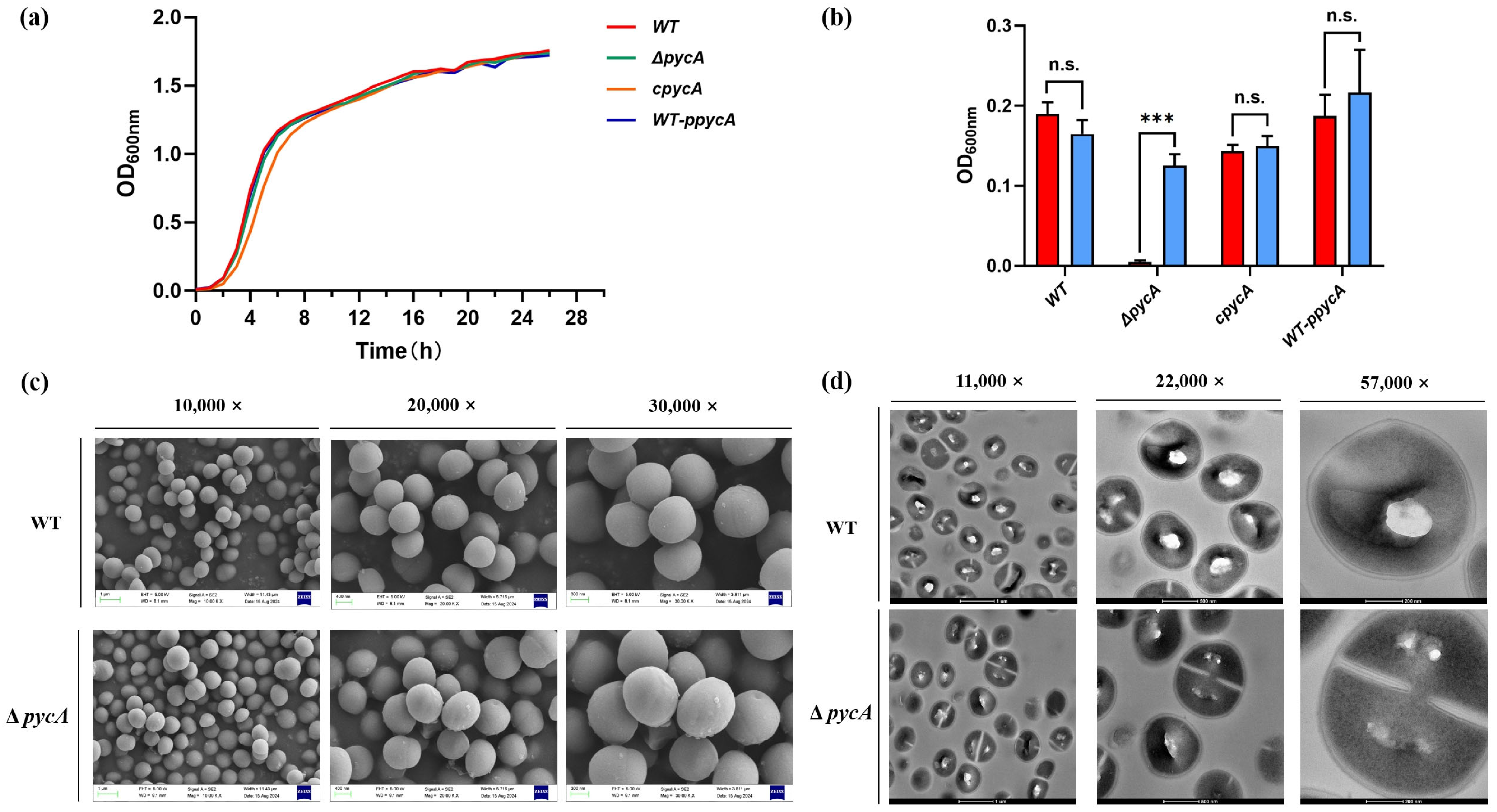
Figure 1. The effect of PycA on the growth of S. aureus. (a) Growth curves of S. aureus strains in TSB medium; (b) L-ASP auxotrophy assay in MEM with or without 100 μg/mL L-ASP supplementation; (c) SEM images showing surface morphology at 10,000×, 20,000×, and 30,000× magnification; (d) TEM images of cellular ultrastructure at 11,000×, 22,000×, and 57,000× magnification. *** p < 0.001; n.s., not significant.
Foods 14 02566 g001
Figure 2. In vitro assessment of virulence-associated phenotypes in S. aureus strains. (a) Quantification of biofilm formation; (b) quantification of STX production; (c) total antioxidant capacity assessed using DPPH radical scavenging assay; (A) fluorescence microscopy of A549 cells after infection with S. aureus strains, in the absence (−) or presence (+) of 8 μg/mL ampicillin; and (B,C) LDH release assays measuring cytotoxicity of different S. aureus strains to A549 cells under (B) normal and (C) ampicillin-treated conditions. * p < 0.05; ** p < 0.01; *** p < 0.001 compared to the WT group.
Foods 14 02566 g002
Figure 3. Proteomic and functional analysis of the ΔpycA mutant in S. aureus. (a) PCA showing distinct clustering of WT and ΔpycA; (b) volcano plot showing differentially expressed proteins between WT and ΔpycA; (c) GO enrichment analysis of differentially expressed proteins; (d) KEGG pathway enrichment analysis of differentially expressed proteins; (e) measurement of intracellular ATP levels in S. aureus strains; and (f,g) qPCR (f) and RT-qPCR (g) analysis of repA and blaZ genes associated with resistance plasmids. * p < 0.05; *** p < 0.001 compared to the WT group.
Foods 14 02566 g003
Figure 4. Proposed regulatory role of PycA in coordinating metabolism, virulence, and antimicrobial resistance in S. aureus. Purple words represent phenotypic results; blue words represent proteomic results; black solid bars (⊥) indicate suppression; and black dashed lines indicate hypothesized relationships based on indirect evidence.
Foods 14 02566 g004
Table 1. Strains and plasmids used in this study.
NameDescriptionSource or Reference
Strains
E. coli JTU006Based on the E. coli DH10B lineage and engineered to be restriction deficient.Han et al. [13]
S. aureus ATCC BAA1717A standard MRSA strain originating from the USA300 lineage.Teng et al. [14]
ΔpycABAA1717 mutant strain with pycA deletion.This study
cpycAΔpycA complemented with pCL55-pycA, cmR.This study
WT-ppycABAA1717 overexpressing pycA with pCL55-pycA, cmR.This study
Plasmids
pKZ2Thermosensitive E. coliS. aureus shuttle plasmid for gene deletion; AmpR/E. coli, CmR/S. aureus.Li et al. [15]
pKZ2-pycApKZ2-pycA homologous arm sequence.This study
pCL55Anhydrotetracycline-inducible E. coliS. aureus shuttle plasmid; AmpR/E. coli, CmR/S. aureus.Karinou et al. [16]
pCL55-pycApCL55-pycA CDSThis study
Note: AmpR indicates ampicillin resistance, and CmR indicates chloramphenicol resistance.
Table 2. MICs of antimicrobials against S. aureus strains.
Class of AntimicrobialsAntimicrobialMIC (μg/mL)Foldchange
WTΔpycAcpycAWT-ppycAWT/ΔpycAWT-ppycA/WT
β-lactamsOxacillin160.253264644
Amoxicillin40960.532819281922
Ampicillin1024132204810242
Carbenicillin disodium10241128409610244
Penicillin potassium204841640965122
Imipenem10.254848
Meropenem414842
Cefoxitin6464646411
CephalosporinsCefotaxime1643212848
Cephalothin sodium320.51632641
AminoglycosidesKanamycin sulfate409628819220482
Amikacin160.251664644
MacrolidesErythromycin320.251641282
Azithromycin640.511281282
Table 3. Significantly expressed differential proteins of WT vs. ΔpycA.
FunctionProteinDescriptionAccession No.Foldchangep-Value
TCA cycleFumCFumarate hydratase class IIQ5HES40.6050.0225
SucCSuccinyl-CoA synthetase subunit beta A5ISD00.5870.0003
SucDSuccinyl-CoA synthetase subunit alphaP668660.5650.0001
MqoMalate quinone oxidoreductase Q5HDJ00.6230.0251
Purine metabolismPurCPhosphoribosylaminoimidazole-succinocarboxamide synthase A5IRV30.1780.0140
PurMPhosphoribosylformylglycinamidine cyclo-ligase A5IRV80.3000.0040
PurLPhosphoribosylformylglycinamidine synthase A6QFS70.2230.0160
PurQPhosphoribosylaminoimidazole carboxylase catalytic subunit P659040.2250.0060
PurHIMP cyclohydrolase Q2FI050.2990.0080
PurDPhosphoribosylamine-glycine ligase Q5HH100.3050.0020
PurNPhosphoribosylglycinamide formyltransferase Q5HH120.2270.0060
PurFAmidophosphoribosyltransferase Q5HH140.2400.0006
PurKPhosphoribosylaminoimidazole carboxylase non-catalytic subunit Q5HH190.2520.0090
PurSPhosphoribosylformylglycinamidine synthase subunit A0A0E1VKY10.2480.0060
PurEPhosphoribosylaminoimidazole carboxylase A0A5F0HIJ80.4720.0820
Antimicrobial
resistance
BlaZBeta-lactamase D2J6840.0289.67 × 10−6
BlaIBeta-lactamase repressor P0A0420.0650.0005
BlaR1Beta-lactamase sensor–transducer protein P183570.0010.0069
Cell adhesionFnbAFibronectin-binding protein A P147380.4910.0105
FnbBFibronectin-binding protein B A0A0H2XKG30.3970.0125
FibFibrinogen-binding protein A6QG590.4710.0006
SasGSurface-anchored protein G Q2G2B20.4180.0049
SpAImmunoglobulin G-binding protein A Q70AB90.1530.0029
IsaAImmunodominant staphylococcal antigen A A6QK590.5960.0001
IsaBImmunodominant staphylococcal antigen BQ2FDM10.4770.0019
Disclaimer/Publisher’s Note: The statements, opinions and data contained in all publications are solely those of the individual author(s) and contributor(s) and not of MDPI and/or the editor(s). MDPI and/or the editor(s) disclaim responsibility for any injury to people or property resulting from any ideas, methods, instructions or products referred to in the content.