Article

Proteomic Analysis of Listeria monocytogenes Subjected to Pulsed Magnetic Field

1
School of Food and Biological Engineering, Jiangsu University, 301 Xuefu Road, Zhenjiang 212013, China
2
Institute of Food Physical Processing Engineering, Jiangsu University, 301 Xuefu Road, Zhenjiang 212013, China
3
College of Engineering, China Agricultural University, Beijing 100083, China
*
Author to whom correspondence should be addressed.
Foods 2025, 14(11), 1871; https://doi.org/10.3390/foods14111871
Submission received: 3 April 2025 / Revised: 21 May 2025 / Accepted: 22 May 2025 / Published: 24 May 2025

Abstract

:
As one of the non-thermal technologies, the pulsed magnetic field (PMF) has increasingly attracted attention for its application in food microbial inactivation. In this study, a proteomic analysis was conducted to elucidate the molecular mechanism underlying the inactivation of Listeria monocytogenes (L. monocytogenes) by a PMF. A total of 79 proteins, comprising 65 upregulated and 14 downregulated proteins, were successfully identified as differentially expressed proteins (DEPs, >1.2-fold or <0.83-fold, p-value < 0.05) in Listeria monocytogenes exposed to a PMF at 8 T with 20 pulses. Gene Ontology (GO) and Kyoto Encyclopedia of Genes and Genomes (KEGG) pathway analyses revealed that PMF exposure significantly impacted nutrient transport, the composition of cytoplasmic and intracellular substances, and various metabolic processes in L. monocytogenes, such as carbohydrate metabolism, amino acid metabolism, and nicotinate and nicotinamide metabolism. The disruption of cellular functions and metabolic pathways may contribute to the death of L. monocytogenes under PMF treatment. These findings provide valuable insights and serve as a reference for further investigations into the inactivation mechanisms induced by PMFs.

1. Introduction

The growing demand for high-quality or minimally processed food is driving the development of innovative technologies in food processing. Non-thermal technologies do not generate high temperatures during food processing and have relatively short processing times. Non-thermal processing results in better retention of nutrients in foods than that in traditional thermal processing [1]. Non-thermal technologies, such as high pressure [2], ultrasound [3], and pulsed electric field [4], are capable of inactivating microorganisms while inducing minimal changes to the sensory and nutritional properties of food. The PMF has emerged as one of the non-thermal technologies and can effectively reduce microbial loads in liquid foods, such as fruit juices, under ambient pressure and temperature conditions [5]. Several studies have highlighted its efficacy in eliminating pathogens like Escherichia coli O157:H7 [6] and Listeria grayi [7] in liquid matrices, including juice and buffer solutions. The study by Ragupathi et al. showed that a PMF effectively inhibited the growth of tomato pathogenic bacteria [8]. Compared with conventional heat treatment and chemical processing methods, PMF technology shows advantages such as reduced process duration, environmentally friendly operation without additional damage, and no chemical residues [7].
However, the inactivation mechanisms of the PMF toward the microorganisms have not been fully elucidated. Cell membranes and walls are the main targets of the PMF. The PMF induced the formation of pores, changes in permeability, and structural disruption of the cell membrane, resulting in the leakage of cellular components [5]. Furthermore, changes in mobility, carbohydrate metabolism, energy metabolism, amino acid metabolism, phosphorylation and dephosphorylation, membrane properties, quorum sensing, two-component regulatory systems, and ATP-binding cassette (ABC) transporters were related to cell death after PMF treatment [9].
L. monocytogenes is one of the major foodborne pathogens that can cause a rare and severe disease called listeriosis [10]. In recent years, there has been a notable rise in L. monocytogenes outbreaks across Europe and the United States. L. monocytogenes was identified as the primary cause of zoonosis-related deaths in 37 European countries, accounting for 52% of deaths associated with outbreaks in the United States. Dairy products, such as soft cheese [11] and raw fluid milk [12]; ready-to-eat foods, such as smoked salmon [13] and cooked meats [14]; and fresh produce, such as apple [15] and lettuce [16], have been closely associated with L. monocytogenes contamination. The pathogenic microorganism L. monocytogenes, a significant concern in the food industry, can be transmitted to humans, posing a substantial risk to immunocompromised individuals, pregnant women, newborns, and the elderly [17]. This pathogen not only poses a significant threat to public health but also presents substantial challenges to food safety management, necessitating urgent attention from public health authorities globally [18]. Therefore, the inactivation of L. monocytogenes in both processed and fresh foods is essential for ensuring food safety.
The efficacy of non-thermal approaches in reducing L. monocytogenes has been documented in several studies. High-Pressure Processing (HPP) treatments, particularly at 600 MPa and to a lesser extent at 500 MPa, are effective to eliminate L. monocytogenes in fresh cheeses [19], and a reduction of 6.2 log CFU/g was achieved when simulated meat was treated with HPP at 600 MPa for 5 min. Pulsed electric field (PEF) treatment resulted in L. monocytogenes population reductions ranging from 18.98% to 43.64% as the field strength was increased from 15 to 30 kV/cm in milk [20]. Power was the predominant factor influencing L. monocytogenes inactivation in almond milk using ultrasound, and a 1 log CFU/mL reduction in viable count was achieved when the power was set to 80% for 8 min [21].
Proteomic approaches have been employed to investigate bacterial cellular responses under specific conditions [22,23]. By detecting and identifying proteins and analyzing protein–protein interactions, biological events can be elucidated. Moreover, insights into bacterial metabolic pathways can be gained, and this knowledge can facilitate a deeper understanding of the mechanisms underlying cellular responses to stimuli. Proteomics analysis has been effectively utilized to investigate global changes in protein expression in biological organisms under diverse environmental conditions [24]. Therefore, proteomic approaches have been widely utilized as a primary tool to investigate microbial responses to antibacterial agents [25,26]. In this study, a tandem mass tag (TMT)-based quantitative proteomic analysis was performed to investigate the proteomic changes of L. monocytogenes exposed to a PMF, thereby elucidating the molecular mechanisms underlying PMF-induced inactivation.

2. Materials and Methods

2.1. L. monocytogenes Sample Preparation

L. monocytogenes (ATCC 19111, American Tissue Culture Collection, ATCC) was stored at −20 °C in a 10% glycerol solution. Cells were subcultured in brain heart infusion broth (BHI, Shanghai Sinopharm Group Chemical Reagent Co., Ltd., Shanghai, China) at 37 °C for 24 h. A single colony was selected and inoculated into 50 mL of BHI broth, incubated at 37 °C for 24 h. Subsequently, 1 mL of the culture was transferred to 50 mL of fresh BHI broth and incubated in a shaking incubator at 37 °C with a shaking speed of 180 rpm for 4 h to reach the exponential growth phase. The cells were harvested by centrifugation at 6000× g for 5 min at 4 °C and subsequently resuspended in 4 mL of phosphate-buffered saline (PBS, pH 7.2).

2.2. PMF Treatment

The pulsed magnetic field device (TSK-H15300, Tingjin Magnetic Components, Nanjing, China) consisted of a magnetic field generator and a treatment chamber. A magnetic field generator was employed to produce a pulsed magnetic field by charging and discharging current within the treatment chamber. The treatment chamber consisted of a coil measuring 0.5 m in length and 5 cm in diameter. The intensity of PMF was quantified using a teslameter (LZ-610H, Hunan Linkjoin Technology, Hunan, China). The L. monocytogenes samples were exposed to PMF intensities ranging from 2 to 8 Tesla (T) and pulse numbers varying between 10 and 50. The duration of a single pulse was maintained at 0.3 ms throughout the experimental treatments. Circulating cooling water was employed to maintain the treatment temperature at room temperature throughout the process. The samples that were not exposed to PMF treatment served as the control group. All the experiments were conducted in triplicate.

2.3. Determination of L. monocytogenes Inactivation

An aliquot of 0.1 mL of the sample was subjected to a 10-fold serial dilution with 0.9 mL PBS, and 0.1 mL of appropriately diluted sample an appropriate dilution was evenly spread onto the BHI agar plates. Each dilution was performed in duplicate, and the plates were incubated at 37 °C for 24 h before the colonies were counted. The detection limit of colony count is defined as the lowest sample concentration at which microorganisms can be reliably detected, corresponding to a colony count of 20 CFU per plate. The survival fraction (N/N0) was calculated to represent the inactivation efficiency, where N0 and N were colony-forming units (CFUs) of L. monocytogenes before and after PMF treatment, respectively.

2.4. Protein Extraction and Quantitation

L. monocytogenes samples before and after PMF treatment were lysed with 300 µL lysis buffer supplemented with 1 mM PMSF. The samples were sonicated at 80 W with a pulse cycle of 1 s on/1 s off applied to the samples for 3 min. After the sonication, the samples were centrifuged at 15,000× g for 15 min to remove insoluble particles. To ensure consistent protein loading amounts across all the samples prior to electrophoresis and mass spectrometry (MS) analysis, the protein concentrations were normalized using the Bicinchoninic Acid Assay (BCA) method. The protein aliquots were stored at −80 °C.

2.5. SDS-PAGE Electrophoresis

Protein was separated by a 12% SDS-PAGE gel as described by Candiano et al. [27]. First, the gel was fixed for 2 h and stained with Coomassie Brilliant Blue R-250 for 12 h. After staining, the gel was washed with water until the protein bands became clearly visible. Finally, the stained gel was scanned by an Image Scanner (GE Healthcare, IL, USA) at a resolution of 300 dpi.

2.6. Protein Digestion and TMT Labeling

The Filter-Aided Sample Preparation (FASP) method [28] was used to perform protein digestion and labeling. About 100 μg of protein extract was mixed with 120 μL reduction buffer (10 mM DTT, 8 M urea, 100 mM TEAB, pH 8.0). The solution was incubated at 60 °C for 1 h. Iodoacetamide (IAA) was added to the solution to achieve a final concentration of 50 mM and reacted in the dark at room temperature for 40 min. Then, the solution was centrifuged at 12,000 rpm for 20 min at 4 °C. The pellet was resuspended in 100 μL of TEAB (100 mM) and centrifuged at 12,000 rpm for 20 min; this step was repeated twice. After washing, 100 μL of TEAB (100 mM) was added, followed by 2 μL of sequencing-grade trypsin (1 μg/μL), and then, the solution was incubated for digestion at 37 °C for 12 h. The digestate was centrifuged at 12,000 rpm for 20 min. Finally, 50 μL of TEAB (100 mM) was added and centrifuged again. The solution was collected and lyophilized using a vacuum refrigerated centrifuge.
For TMT labeling, the lyophilized samples were resuspended in 100 μL of 50 mM triethylammonium bicarbonate (TEAB), and 40 μL aliquots from each sample were taken for labeling. The TMT reagent was equilibrated to room temperature, and 41 μL of anhydrous acetonitrile was added to the TMT reagent. The TMT reagent was vortexed for 5 min to ensure complete dissolution. Then, 41 μL of the TMT labeling reagent was added to each 100 μL sample for mixing, and the mixture was incubated at room temperature for 1 h. Finally, 8 µL of 5% hydroxylamine was added to each sample and incubated for 15 min to quench the labeling reaction. The labeled peptide solutions were lyophilized and stored at −80 °C.

2.7. Reverse-Phase Liquid Chromatography (RPLC) Analysis

Reversed-phase separation was performed on an Agilent 1100 Series HPLC System (Agilent Technologies, Santa Clara, CA, USA) with an Agilent Zorbax Extend RP column (5 μm, 150 mm × 2.1 mm). Mobile phase A (2% acetonitrile in ultrapure water) and mobile phase B (98% acetonitrile in ultrapure water) were used. The solvent gradient was as follows: 0–8 min, 98% A; 8.00–8.01 min, 98–95% A; 8.01–38 min, 95–75% A; 38–50 min, 75–60% A; 50–50.01 min, 60–10% A; 50.01–60 min, 10% A; 60–60.01 min, 10–98% A; 60.01–65 min, 98% A. Peptides were separated at a flow rate of 300 μL/min and detected at wavelengths of 210 nm and 280 nm. The eluted peptides were collected from 8 min to 50 min, and the elution fractions were collected at 1 min intervals. The separated peptides were lyophilized and stored at −80 °C for subsequent MS analysis.

2.8. MS Analysis

Analyses were performed using a Q Exactive™ Hybrid Quadrupole-Orbitrap mass spectrometer (Thermo Fisher Scientific, Waltham, MA, USA) equipped with a Nanospray Flex source (Thermo, USA). Samples were loaded by a capillary C18 trap column (3 cm × 100 µm) and then separated by a C18 column (15 cm × 75 µm i.d., 2 μm particle size, 100 Å pore size, Acclaim PepMap RSLC) on an EASY-nLCTM 1200 system (Thermo, USA). The flow rate was 300 nL/min, and a 90 min linear gradient from 5% to 100% mobile phase B (0.1% FA in 80% ACN) was applied. Eluent A was 0.1% (v/v) formic acid (FA) in ultrapure water. The gradient program was as follows: 8% B, 0–55 min; 30% B, 55–79 min; 50% B, 79–80 min; 100% B, 80–90 min; 90–100% B over 5 min.
Full MS scans were acquired in the mass range of 300–1600 m/z with a mass resolution of 70,000, and the automatic gain control (AGC) target value was 1 × 10⁶. The top 10 most intense precursor ions in full MS were fragmented with higher-energy collisional dissociation (HCD) with a collision energy of 30. MS/MS spectra were obtained with a resolution of 17,500, an AGC target of 200,000, and a maximum injection time of 80 ms. The Q-E dynamic exclusion was set for 15.0 s and operated in positive ion mode. Three biological replicates were established for both the PMF treatment group and the control group. Each sample underwent three technical replicates of TMT labeling and MS analysis to assess reproducibility.

2.9. Database Search

Proteome Discoverer (version 2.2) was employed to search all of the Q Exactive MS/MS raw data thoroughly against the L. monocytogenes protein database. The database searches were conducted with trypsin digestion specificity, and the cysteine alkylation was specified as a fixed modification during the database searching. For protein quantification, TMT 6-plex labeling was selected. The normalization method applied to the TMT data was median normalization. Specifically, this approach standardizes the ratios of all the peptides by referencing the median of the protein ratios and adjusts the median protein ratio to 1 after standardization. A global false discovery rate (FDR) of <0.01 was applied, and peptide groups considered for quantification required at least one peptide per protein group.

2.10. Bioinformatic Analysis

Proteins with a fold change >1.2-fold or <0.83-fold and p-value < 0.05 were defined as differentially expressed proteins (DEPs). Gene Ontology (GO) analysis was performed using the Blast2GO (Version 2.8.0) tool. Kyoto Encyclopedia of Genes and Genomes (KEGG) pathway enrichment analysis was performed to analyze the biological pathways. GO and KEGG pathway enrichment analysis was performed using a background dataset constructed from the total proteome of L. monocytogenes obtained from UniProt. The screening criteria were set as p-value < 0.05 and FDR < 0.01.

3. Results

3.1. Inactivation of L. monocytogenes by PMF

The inactivation efficiency of L. monocytogenes treated by PMF intensities of 2–8 T with pulse numbers of 10–50 is shown in Figure 1. The survival rate decreased after PMF treatment, and the lowest survival fraction was 9.6% when a PMF at the intensity of 8 T with a pulse number of 20 was applied to L. monocytogenes. However, the survival rate did not continue to decrease with increasing intensity and pulse number, which may be attributed to the “window effect”, one of the characteristics of electromagnetic fields. Specific microorganisms exhibit biological effects under electromagnetic fields with specific intensity and frequency ranges; thus, the “window effect” includes both the “Frequency (or Time) Window” and the “Intensity (or Power Density) Window” [29]. Due to the influence of the window effect, the sterilization efficacy exhibits a systematic fluctuation trend in response to variations in key parameters such as magnetic field intensity and pulse [30]. Furthermore, series of valley values appear due to “window effect”. The survival rate reached its lowest point at 35 pulses, which was significantly lower than the survival rates at the adjacent pulse numbers of 30 and 40 (p-value < 0.05). This finding aligns with the characteristic of “window effect”. Pulses of 35 might be one of the “frequency windows” in the inactivation of L. monocytogenes by a PMF at the intensity of 2–8 T. It has been found that different intensities also induce different biological effects. Guo et al. [31] proposed that, owing to the “window effect”, the application of a high-power PMF might not necessarily inhibit microbial growth under specific conditions. Naskar et al. [32] demonstrated that short-duration, low-power PMFs were as effective in inactivating Enterococcus faecalis as a prolonged exposure to high-power static fields.

3.2. Identification of Differentially Expressed Proteins

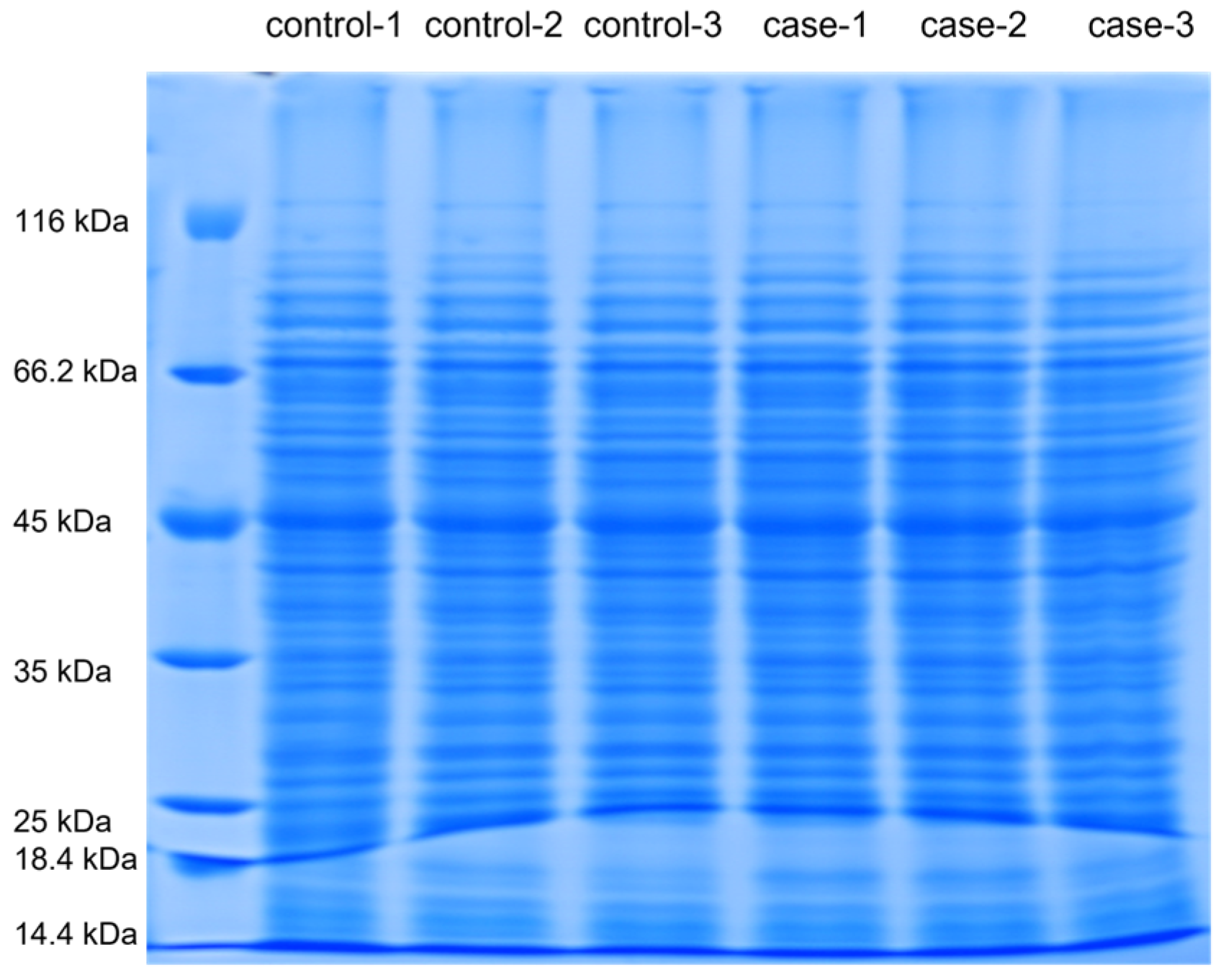
The SDS-PAGE gel electropherogram of proteins from L. monocytogenes that were untreated and treated with a PMF (8 T, 20 pulses) is shown in Figure 2. In the SDS-PAGE gel experiments of the control group and the treatment group, 15 μg of protein was precisely loaded into each independent sample well. Overloaded samples can lead to high background noise, bulky bands, streaks, and smears [33]. Therefore, during the experimental procedures, the volume of the sample added should be carefully controlled to maintain the accuracy and reliability of the results. The protein bands were clear and were distributed between 20 and 80 kDa. Moreover, there was a high-abundance band at 50 kDa in each sample. The band was excised from the gel for identification. The complete raw data of the proteomics experiment are provided in the Supplementary Materials. Based on fold change (FC) > 1.2 and p-value < 0.05, 101 differentially expressed proteins (DEPs) were screened. Meanwhile, 79 DEPs (65 upregulated and 14 downregulated) were successfully identified, and the protein names and other information are listed in Table 1.
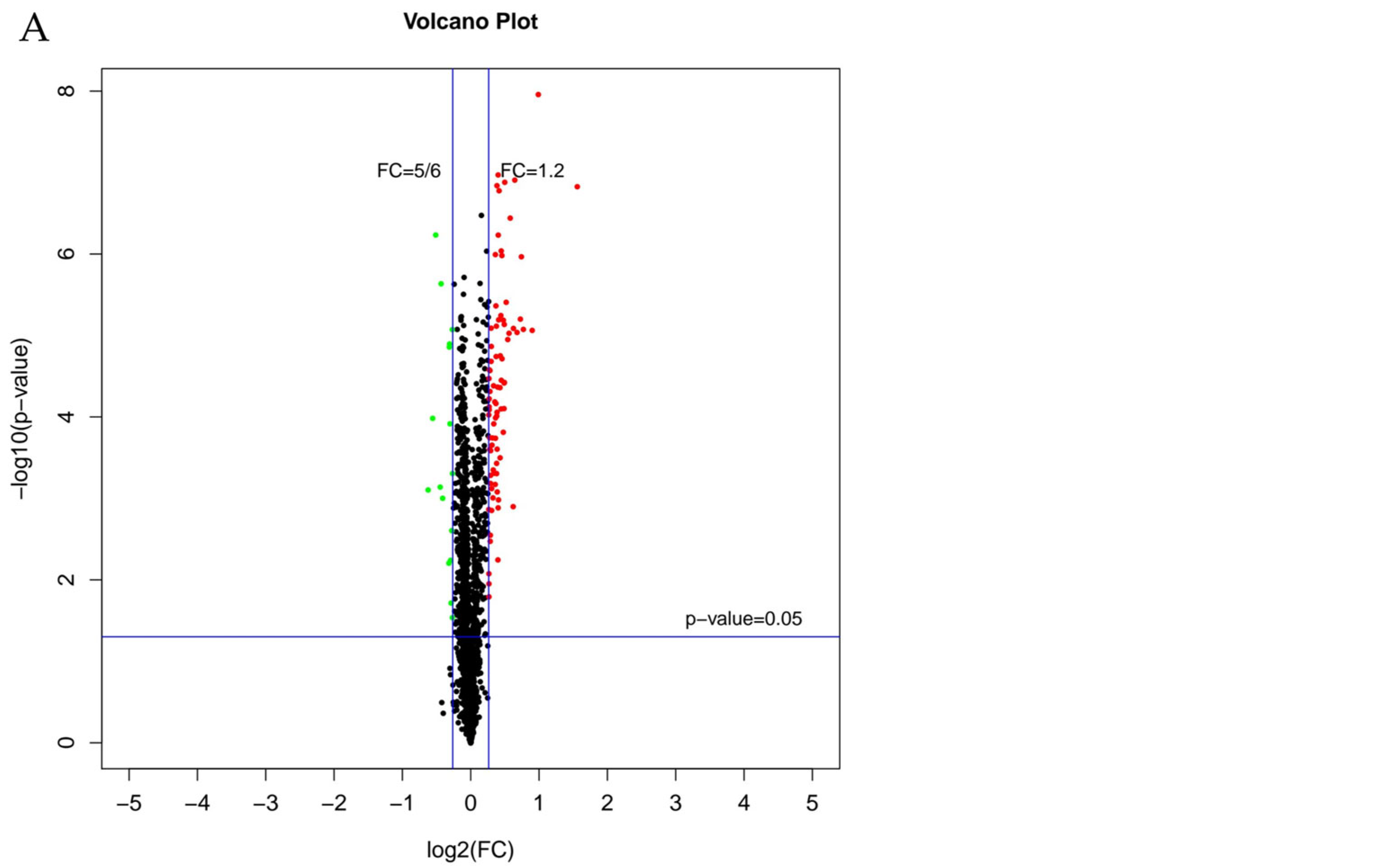
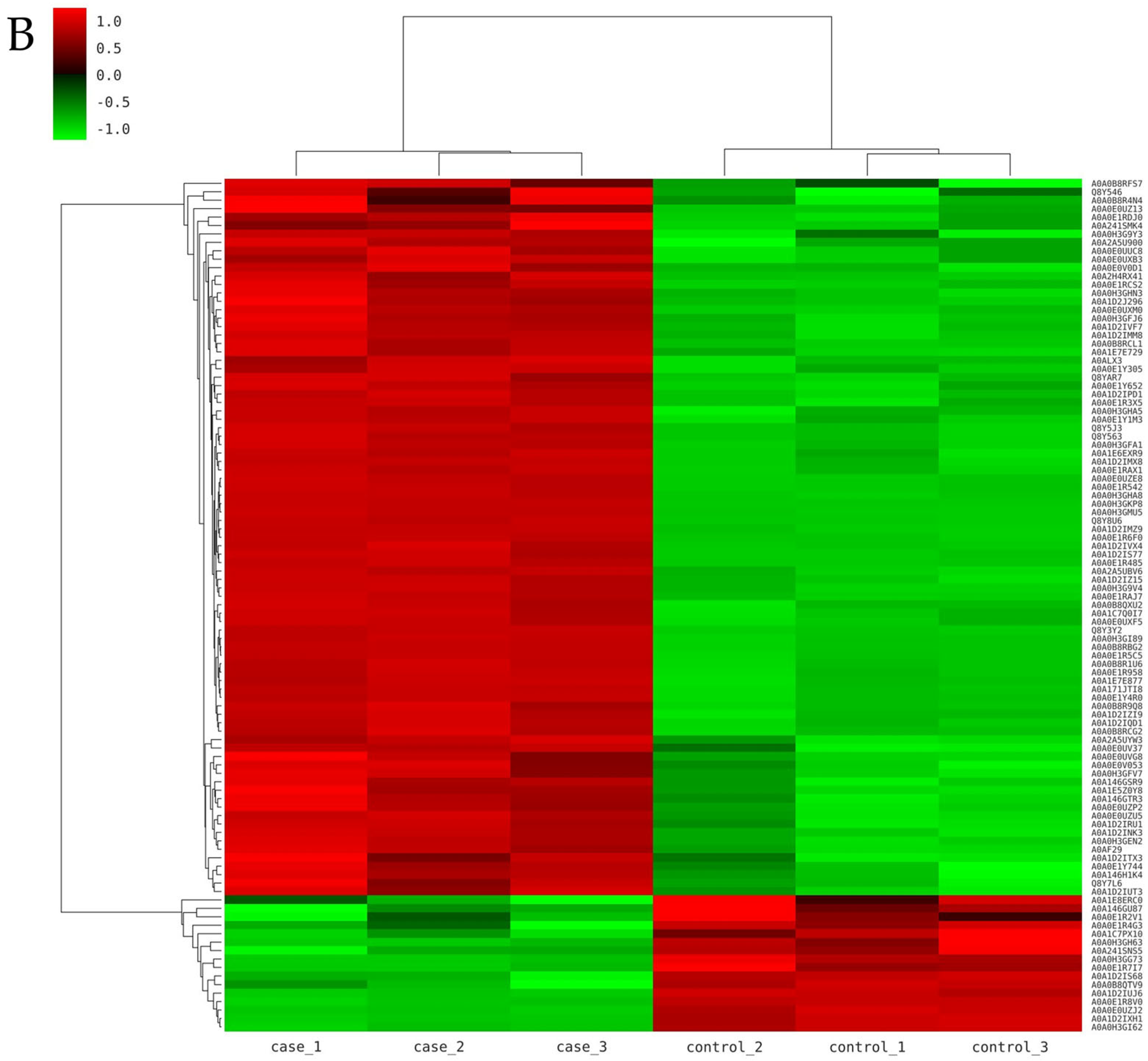
The volcano plot (Figure 3A) showed all DEPs between untreated and treated samples. Black dots in Figure 3A represented proteins that had no significant difference; green dots represented downregulated proteins and red dots represented upregulated proteins. Figure 3B was the clustered heatmap of protein expression patterns. The green lines represented the downregulated proteins, while red lines represented the upregulated proteins. After PMF treatment, most proteins in L. monocytogenes were upregulated.

3.3. Analysis of Differentially Expressed Proteins

3.3.1. GO Analysis of DEPs

Bioinformatics analyses were performed to better elucidate the response of L. monocytogenes to PMF treatment [34]. GO functional annotation was performed to identify the biological process, cellular components, and molecular functions related to DEPs. The top ten terms sorted by significance in GO analysis are shown in Figure 4. In biological processes, DEPs after PMF treatment were mainly classified into terms such as glucose import into cell, carbohydrate import into cell, and mannose transport. In cellular components, the DEPs were involved in cytoplasm, intracellular regions, pyruvate dehydrogenase complex, and so on. In molecular function, terms in DEPs were dominated by mannose transmembrane transporter activity, protein-N(PI)-phosphohistidine-mannose phosphotransferase system transporter activity, oxidoreductase activity, and so on.

3.3.2. KEGG Analysis of DEPs

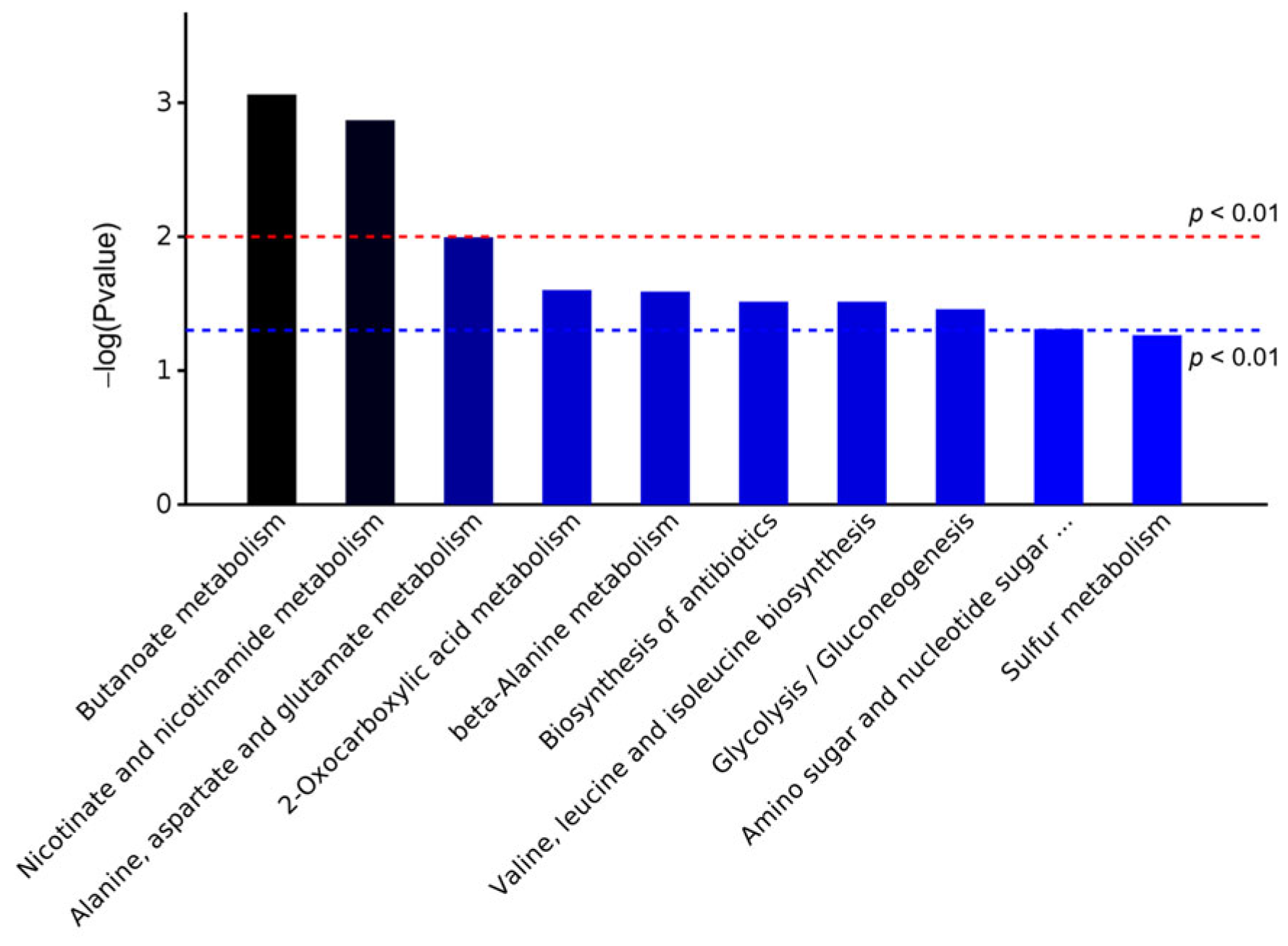
The KEGG database was used to analyze the pathway enrichment of DEPs. The KEGG database system systematically integrates and stores the functional annotation information of genes and genomes, encompassing a wide range of biological processes [35]. Specifically, it includes core modules such as metabolic pathways, membrane transport mechanisms, signal transduction networks, cell cycle regulation, and conserved sub-pathways across species [36]. This database enables the visualization and functional analysis of cellular biochemical processes, thereby offering substantial support for bioinformatics research. The top 10 pathways of KEGG enrichment for DEPs based on significance are shown in Figure 5. The metabolic pathways involved in DEPs were mainly butanoate metabolism, nicotinate and nicotinamide metabolism, alanine, aspartate and glutamate metabolism, and so on.
Similar to the results of the GO analysis, DEPs participated in metabolic pathways related to glycerol metabolism, sulfur metabolism, and so on. In addition, DEPs participated in metabolic pathways involved in amino acid metabolism.

3.3.3. DEPs Involved in Transportation

PTS mannose/fructose/sorbose transporter subunit IIB (CDR86_01030), mannose permease IID component (manZ_3), PTS system mannose-specific EIIAB component (manX_2), and Lmo2697 protein (Lmo2697) were upregulated after PMF treatment. These proteins belong to the phosphoenolpyruvate-dependent sugar phosphotransferase system (PTS), and the function of the phosphotransferase system is to transport sugars from the environment into bacterial cells [37]. The PTS is a carbohydrate transport and phosphorylation system found in all the different phyla of bacteria and archaea. The PTS not only functions as a carbohydrate transport protein but also regulates many cellular processes by phosphorylating its target proteins or interacting with them in a phosphorylation-dependent manner [38]. Therefore, those proteins participate in biological processes in L. monocytogenes, such as glucose intracellular transport, carbohydrate import into the cell, and mannose transport [39]. At the same time, these proteins have certain molecular functions, including mannose transmembrane transporter activity and protein-N(PI)-phosphohistidine-mannose phosphotransferase system transporter activity. The upregulation of these proteins may be attributed to carbon catabolite repression (CCR). The bacteria primarily utilize preferred carbon sources (such as sucrose and fructose) to produce energy. If the preferred carbon source is exhausted, the bacteria will synthesize the enzymes needed to transport those less preferred carbon sources, such as mannose and sorbitol [40,41]. Related studies have shown that bacteria can be susceptible to adaptive evolution depending on substrate availability [42]. After PMF treatment, L. monocytogenes might increase the uptake of nutrients (including less preferred carbon sources) from the environment to repair the damage caused by the PMF.
In addition, the cadmium-translocating P-type ATPase (Lmrg_00327), which is involved in cation transmembrane transport, was significantly upregulated under the PMF, suggesting enhanced ion transmembrane transport in L. monocytogenes. Concurrently, several transporters potentially involved in ion transport were identified, including a putative Zn/Cd/Fe cation exporter (Fief), a putative efflux ABC transporter/ATP-binding permease protein (Lmm7_0961), and a sodium-dependent phosphate transporter (LmNIHS28_00558). The ion transmembrane transport in L. monocytogenes might be increased by the PMF. Qian et al. [43] reported that a PMF promoted Ca2+ transmembrane transport in L. monocytogenes. The acceleration of ion transmembrane transport may lead to the disruption of cell membrane integrity, resulting in the loss of cell viability. However, the SecE protein translocase subunit was downregulated, indicating that intracellular protein transmembrane transport and amino acid transport decreased in L. monocytogenes.

3.3.4. DEPs Involved in Transcription and Translation

Concerning transcription, the heat-inducible transcription repressor HrcA and the transcriptional regulator CtsR were upregulated. CtsR participates in the positive regulation of DNA-templated transcription, while HrcA participates in the negative regulation of DNA-templated transcription. The transcription of some heat-shock proteins, such as molecular chaperones (e.g., DnaK and GroEL) and proteases (e.g., Lon, Clp, FtsH, and DegP), can be regulated by HrcA [44]. Therefore, the upregulation of HrcA indicates that the heat shock response was triggered to enable cells to resist the PMF to a certain extent.
The DEPs involved in the translation process were 30S ribosomal protein S14 type Z (rpsN) and 50S ribosomal protein L35 (rpmI). The downregulation of these two proteins might not only indicate a decreased tolerance of L. monocytogenes to the PMF but also suggest that PMF potentially affects ribosomal function, thereby disrupting the translation processes. Zheng et al. [45] reported that the abundance of 30S ribosomal proteins S14 and S18, as well as 50S ribosomal proteins L13, L18, and L20, was reduced in the ΔclpP mutant strain, and the reduction might contribute to the decreased tolerance of the ΔclpP mutant strain to linezolid or minocycline. Liu et al. [46] also demonstrated that high hydrostatic pressure (HHP) could upregulate ribosome-related pathways, including the expression of 30S and 50S ribosomal proteins, and HHP might disrupt ribosomal function, consequently impairing transcription and translation processes and ultimately contributing to the mortality of L. monocytogenes. Tian et al. [47] found that both high-voltage short-time ohmic (HVST) and low-voltage long-time ohmic (LVLT) caused greater damage to ribosomal proteins, stalling the protein translation process.

3.3.5. DEPs Involved in Carbohydrate Metabolism

Carbohydrates play a pivotal role in bacterial physiology not only supplying the essential energy required for cellular life processes but also acting as a critical carbon source for the synthesis of various biomolecules [48]. The DEPs involved in carbohydrate metabolic processes include PTS-dependent dihydroxyacetone kinase, the dihydroxyacetone-binding subunit dhaK (dhaK_2), fructosamine deglycase (Lmm7_2086), a putative dihydroxyacetone kinase, a C-terminal domain protein (dhaL), 2,3-bisphosphoglycerate-dependent phosphoglycerate mutase (gpmA), Lmo2697 protein (Lmo2697), and tagatose 1,6-diphosphate aldolase (lacD). Notably, all these proteins were significantly upregulated following PMF treatment.
Among them, dhaK_2 and dhaL participate in glycerol metabolism; gpmA is related to glycolysis; and lacD is associated with lactose catabolism via the tagatose-6-phosphate pathway. Dihydroxyacetone (dha) kinases are homologous proteins that use different phosphoryl donors, and they are also multiphosphorylation proteins of the phosphoenolpyruvate-dependent carbohydrate-phosphotransferase system in bacteria [49]. Dha kinase consists of dhaL, dhaK, and the multiphosphorylation protein DhaM (dhaM) [50]. The upregulation of dhaK may provide substrates for the production of different types of carbohydrates [51]. Upregulation of dhaL and dhaM was also observed in the presence of methanol. GpmA is a key enzyme involved in glycolysis [52], converting 2- phosphoglycerate to 3-phosphoglycerate in glycolysis. The upregulation of gpmA indicates the increase in glycolysis of L. monocytogenes treated with a PMF. GpmA is also upregulated when Bacillus anthracis is exposed to hydrogen peroxide [53]. LacD transforms tagatose-1, 6-diphosphate to glyceraldehyde-3-phosphate (G3P) and dihydroxyacetone phosphate (DHAP) [54], which are metabolized via the glycolysis pathway. The upregulation of lacD might result in the acceleration of glycolysis pathway. L. monocytogenes might accelerate carbohydrate metabolism to respond to a PMF.

3.3.6. DEPs Involved in Amino Acid Metabolism

Amino acids serve as the fundamental building blocks for the synthesis of enzymes and proteins in bacteria. Their concentrations not only influence bacterial growth but also play an essential role in cell wall and membrane biosynthesis, material transport, and biofilm formation [55]. Environmental disturbances cause changes in the amino acid metabolism of microorganisms [56]. The DEPs involved in amino acid metabolism were glutamate decarboxylase (LMRG_01479), glutamate decarboxylase (gadG), ketol-acid reductoisomerase (NADP(+)) (ilvC), and cystathionine beta-lyase (metC). They were all upregulated as were DEPs involved in carbohydrate metabolism.
LMRG_01479 and gadG are associated with glutamate metabolic processes, while ilvC is related to leucine, isoleucine, and valine biosynthetic processes. Glutamate decarboxylase catalyzes the irreversible conversion of L-glutamate to γ-aminobutyric acid (GABA) [57]. An incremental expression of the glutamate decarboxylase system explains one of the mechanisms underlying acid resistance in microorganisms [58]. The metabolic processes in L. monocytogenes that alleviate acid stress primarily involve proton-consuming reactions, including glutamate decarboxylation, arginine/agmatine deimination, and fermentative acetoin production [59]. Under PMF conditions, the gadG gene, which is associated with acid tolerance, was found to be upregulated by 1.43-fold compared with the control group. This gene maintains intracellular pH homeostasis through the GABA metabolic pathway, thereby enhancing the cell’s adaptability and survival in acidic environments. Boura et al. [60] indicated that glutamate decarboxylase plays a crucial role in the acid tolerance and oxidative stress resistance of L. monocytogenes. Branched-chain amino acid (BCAA) biosynthesis starts with pyruvate and threonine, and ilvC participates in the second or third step of the biosynthesis of Val, Leu, and Ile, respectively. Moreover, in the Ehrlich pathway, ilvC utilizes nicotinamide adenine dinucleotide phosphate (NADPH) as a cofactor. IlvC plays some roles in managing stress, such as pH and starvation [61]. It is reported that the downregulation of ilvC affected the ability of Mycobacterium tuberculosis to persist and its survival in macrophages and in mice [62]. In bacteria, cystathionine beta-lyase (metC) is responsible for the hydrolysis of L-cystathionine (L-Cth) to L-homocysteine (L-Hcys), pyruvate, and ammonia [63]. MetC not only participates in transsulfuration in amino acid metabolic process but also participates in transsulfuration in sulfur metabolic process. The upregulation of metC may also enhance the sulfur metabolism. The enhancement of the amino acid metabolism might improve the survival ability of L. monocytogenes exposed to a PMF.

3.3.7. DEPs Involved in Nicotinate and Nicotinamide Metabolism

As a core component in the metabolic pathways of nicotinic acid and nicotinamide, niacinamide exerts anti-oxidative stress and anti-inflammatory effects through the regulation of cellular energy metabolism [64]. The DEPs including fumarate reductase (LmNIHS28_02228), cystathionine beta-lyase (metC), Lmo0047, Lmo2213, and putative nico-tinamidase (pncA) were related to nicotinate and nicotinamide metabolism. Nicotinamidase is capable of catalyzing the conversion of nicotinamide into nicotinic acid. Feng et al. [45] demonstrated that pncA plays a critical role in the salvage synthesis pathway of nicotinamide adenine dinucleotide (NAD+). The PMF affected the homeostasis of L. monocytogenes, and pncA was upregulated to help L. monocytogenes to maintain metabolic homeostasis and cope with PMF-induced damage. Shats et al. [65] demonstrated that the pncA gene in Escherichia coli plays a critical role in responding to adverse environmental stimuli by modulating the nicotinamide metabolic pathway, thereby maintaining intracellular metabolic homeostasis effectively. In general, the nicotinate and nicotinamide metabolism was increased to help L. monocytogenes survive in a PMF.

3.3.8. DEPs Involved in Other Metabolism

Aldehyde–alcohol dehydrogenase (LMRG_01332) was upregulated after PMF treatment and participates in the butanoate metabolism, biosynthesis of antibiotics, glycolysis/gluconeogenesis, and biosynthesis of secondary metabolites in L. monocytogenes. Aldehyde–alcohol dehydrogenases (ADHEs) convert acyl-CoAs and aldehydes to their corresponding alcohols and play an important role in butanol biosynthesis. Martin et al. [66] identified the aldehyde–alcohol dehydrogenase as the primary bottleneck in butanol production, and increasing the bi-functional aldehyde–alcohol dehydrogenase resulted in a much larger improvement in the butanol titer than increasing any other butanol pathway enzymes. Furthermore, Chlamydomonas reinhardtii with the upregulation of aldehyde–alcohol dehydrogenase increased its survival under dark anoxia [67]. Marina Uroz et al. [68] assert that cell cycle progression and communication between cellular components are mediated by two-component signal transduction systems and signaling pathways involving the activation of transcription factors. In addition, bacterial cells can adapt their transcriptional cues to changing environments and respond correctly to stimulation by transcriptional regulators [69]. The upregulation of aldehyde–alcohol dehydrogenase may be a self-protective stress response of L. monocytogenes, which increases its survival ability under PMF treatment.

4. Conclusions

A proteomic analysis of L. monocytogenes exposed to a PMF was performed to investigate the relationship between changes in protein expression and loss of cell viability. Under the PMF treatment at 8 T with 20 pulses, 101 differentially expressed proteins (DEPs) were annotated, and 79 proteins were successfully identified. Meanwhile, 65 proteins were upregulated, and 14 proteins were downregulated. These DEPs were related to transport, cytoplasmic processes, metabolism, and other functions of L. monocytogenes. The PMF affected the transport capacity and ribosomal structure in L. monocytogenes. L. monocytogenes transiently enhanced nutrient uptake; accelerated the metabolism of carbohydrates, amino acids, nicotinate, and nicotinamide; and activated the heat-shock response as compensatory adaptations to the PMF. These findings indicate that, although a PMF disrupts essential biological functions and metabolic pathways, eventually resulting in cell death, L. monocytogenes can temporarily alleviate PMF-induced stress through these adaptive responses. However, the adaptive mechanisms were insufficient to fully counteract the cumulative damage, ultimately leading to impaired cellular viability. Future research should employ integrated multi-omics approaches, combining proteomics, transcriptomics and metabolomics, to achieve a more in-depth and comprehensive elucidation of the molecular mechanisms underlying the response of L. monocytogenes to a PMF.

Supplementary Materials

The following supporting information can be downloaded at: https://www.mdpi.com/article/10.3390/foods14111871/s1, Table S1: Raw data of proteomic experiments.

Author Contributions

Writing—original draft preparation, D.C.; conceptualization, writing—review and editing, J.Q.; funding acquisition, S.H.; resources and validation, F.W.; supervision, H.M., reviewing and editing, S.L. All authors have read and agreed to the published version of the manuscript.

Funding

This work was supported by the National Natural Science Foundation of China (21978120) and the Key R&D Projects in Jiangsu Province (BE2020405).

Institutional Review Board Statement

Not applicable.

Informed Consent Statement

Not applicable.

Data Availability Statement

The original contributions presented in this study are included in the article/Supplementary Materials. Further inquiries can be directed to the corresponding author.

Conflicts of Interest

The authors declare no conflicts of interest.

Abbreviations

AbbreviationTerm
PMFPulsed magnetic field
L. monocytogenesListeria monocytogenes
GOGene Ontology
KEGGKyoto Encyclopedia of Genes and Genomes
HPPHigh-Pressure Processing
PEFPulsed electric field
TMTTandem Mass Tag
BHIBrain heart infusion broth
CFUColony-forming unit
IAAIodoacetamide
TEABTriethylammonium bicarbonate
MSMass spectrometry
FAFormic acid
AGCAutomatic gain control
HCDHigher-energy collisional dissociation
FDRFalse discovery rate
DEPsDifferentially expressed proteins
FCFold change
CCRCarbon catabolite repression
HVSTHigh-voltage short-time ohmic
LVLTLow-voltage long-time ohmic
ADHEAldehyde–alcohol dehydrogenases
TTesla
HHPHigh hydrostatic pressure

References

  1. Zhang, Z.H.; Wang, L.H.; Zeng, X.A.; Han, Z.; Brennan, C.S. Non-thermal technologies and its current and future application in the food industry: A review. Int. J. Food Sci. Tech. 2018, 54, 1–13. [Google Scholar] [CrossRef]
  2. Sevenich, R.; Mathys, A. Continuous Versus Discontinuous Ultra-High-Pressure Systems for Food Sterilization with Focus on Ultra-High-Pressure Homogenization and High-Pressure Thermal Sterilization: A Review. Compr. Rev. Food Sci. Food Saf. 2018, 17, 646–662. [Google Scholar] [CrossRef] [PubMed]
  3. Shen, Q.-J.; Sun, J.; Pan, J.-N.; Zheng, X.; Zhong, J.-J.; Zhou, W.-W. Ultrasound-synergized gas in ensuring the sterilization and physicochemical quality of fruit and vegetables: A review. Postharvest Biol. Technol. 2024, 209, 112705. [Google Scholar] [CrossRef]
  4. Li, L.; Yang, R.; Zhao, W. The Effect of Pulsed Electric Fields (PEF) Combined with Temperature and Natural Preservatives on the Quality and Microbiological Shelf-Life of Cantaloupe Juice. Foods 2021, 10, 2606. [Google Scholar] [CrossRef]
  5. Qian, J.; Yan, G.; Huo, S.; Dai, C.; Ma, H.; Kan, J. Effects of pulsed magnetic field on microbial and enzymic inactivation and quality attributes of orange juice. J. Food Process. Preserv. 2021, 45, 1553. [Google Scholar] [CrossRef]
  6. Lin, L.; Wang, X.; He, R.; Cui, H. Action mechanism of pulsed magnetic field against E. coli O157:H7 and its application in vegetable juice. Food Control 2019, 95, 150–156. [Google Scholar] [CrossRef]
  7. Wu, P.; Qu, W.; Abdualrahman, M.A.Y.; Guo, Y.; Xu, K.; Ma, H. Study on inactivation mechanisms of Listeria grayi affected by pulse magnetic field via morphological structure, Ca2+ transmembrane transport and proteomic analysis. Int. J. Food Sci. Technol. 2017, 52, 2049–2057. [Google Scholar] [CrossRef]
  8. Kp, R.; Renganayaki, P.R.; Sundareswaran, S.; Kumar, S.M.; Kamalakannan, A. Effect of pulsed magnetic field on seed borne pathogen and germination of tomato. Int. J. Chem. Stud. 2021, 9, 1141–1144. [Google Scholar] [CrossRef]
  9. Qian, J.; Zhang, M.; Dai, C.; Huo, S.; Ma, H. Transcriptomic analysis of Listeria monocytogenes under pulsed magnetic field treatment. Food Res. Int. 2020, 133, 109195. [Google Scholar] [CrossRef]
  10. Li, M.M.H.; MacDonald, M.R.; Rice, C.M. To translate, or not to translate: Viral and host mRNA regulation by interferon-stimulated genes. Trends Cell Biol. 2015, 25, 320–329. [Google Scholar] [CrossRef]
  11. Jackson, K.A.; Gould, L.H.; Hunter, J.C.; Kucerova, Z.; Jackson, B. Listeriosis Outbreaks Associated with Soft Cheeses, United States, 1998–2014. Emerg. Infect. Dis. 2018, 24, 1116–1118. [Google Scholar] [CrossRef] [PubMed]
  12. Kabir, M.N.; Aras, S.; George, J.; Wadood, S.; Chowdhury, S.; Fouladkhah, A.C. High-pressure and thermal-assisted pasteurization of habituated, wild-type, and pressure-stressed Listeria monocytogenes, Listeria innocua, and Staphylococcus aureus. LWT 2021, 137, 110445. [Google Scholar] [CrossRef]
  13. Huang, J.; Chen, B.; Zeng, Q.-H.; Liu, Y.; Liu, H.; Zhao, Y.; Wang, J.J. Application of the curcumin-mediated photodynamic inactivation for preserving the storage quality of salmon contaminated with L. monocytogenes. Food Chem. 2021, 359, 129974. [Google Scholar] [CrossRef] [PubMed]
  14. Serra-Castelló, C.; Jofré, A.; Belletti, N.; Garriga, M.; Bover-Cid, S. Modelling the piezo-protection effect exerted by lactate on the high pressure resistance of Listeria monocytogenes in cooked ham. Food Res. Int. 2021, 140, 110003. [Google Scholar] [CrossRef]
  15. Nangul, A.; Bozkurt, H.; Gupta, S.; Woolf, A.; Phan-thien, K.-y.; McConchie, R.; Fletcher, G.C. Decline of Listeria monocytogenes on fresh apples during long-term, low-temperature simulated international sea-freight transport. Int. J. Food Microbiol. 2021, 341, 109069. [Google Scholar] [CrossRef]
  16. Kyere, E.O.; Foong, G.; Palmer, J.; Wargent, J.J.; Fletcher, G.C.; Flint, S. Rapid attachment of Listeria monocytogenes to hydroponic and soil grown lettuce leaves. Food Control 2019, 101, 77–80. [Google Scholar] [CrossRef]
  17. Lin, L.; Gu, Y.; Cui, H. Moringa oil/chitosan nanoparticles embedded gelatin nanofibers for food packaging against Listeria monocytogenes and Staphylococcus aureus on cheese. Food Packag. Shelf Life 2019, 19, 86–93. [Google Scholar] [CrossRef]
  18. Liu, R.; Zhang, Y.; Ali, S.; Haruna, S.A.; He, P.; Li, H.; Ouyang, Q.; Chen, Q. Development of a fluorescence aptasensor for rapid and sensitive detection of Listeria monocytogenes in food. Food Control 2021, 122, 107808. [Google Scholar] [CrossRef]
  19. Evert-Arriagada, K.; Trujillo, A.J.; Amador-Espejo, G.G.; Hernández-Herrero, M.M. High pressure processing effect on different Listeria spp. in a commercial starter-free fresh cheese. Food Microbiol. 2018, 76, 481–486. [Google Scholar] [CrossRef]
  20. Zhao, W.; Yang, R.; Shen, X.; Zhang, S.; Chen, X. Lethal and sublethal injury and kinetics of Escherichia coli, Listeria monocytogenes and Staphylococcus aureus in milk by pulsed electric fields. Food Control 2013, 32, 6–12. [Google Scholar] [CrossRef]
  21. Iorio, M.C.; Bevilacqua, A.; Corbo, M.R.; Campaniello, D.; Sinigaglia, M.; Altieri, C. A case study on the use of ultrasound for the inhibition of Escherichia coli O157:H7 and Listeria monocytogenes in almond milk. Ultrason. Sonochem. 2019, 52, 477–483. [Google Scholar] [CrossRef] [PubMed]
  22. Chen, T.-Y.; Kuo, S.-H.; Chen, S.-T.; Hwang, D.-F. Differential proteomics to explore the inhibitory effects of acidic, slightly acidic electrolysed water and sodium hypochlorite solution on Vibrio parahaemolyticus. Food Chem. 2016, 194, 529–537. [Google Scholar] [CrossRef] [PubMed]
  23. Zhao, F.; Wang, Y.; An, H.; Hao, Y.; Hu, X.; Liao, X.; Doores, S. New Insights into the Formation of Viable but Nonculturable Escherichia coli O157:H7 Induced by High-Pressure CO2. mBio 2016, 7, e00961-16. [Google Scholar] [CrossRef] [PubMed]
  24. Li, M.; Chen, C.; Xia, X.; Garba, B.; Shang, L.; Wang, Y. Proteomic analysis of the inhibitory effect of chitosan on Penicillium expansum. Food Sci. Technol. 2020, 40, 250–257. [Google Scholar] [CrossRef]
  25. Han, J.; Gao, P.; Zhao, S.; Bie, X.; Lu, Z.; Zhang, C.; Lv, F. iTRAQ-based proteomic analysis of LI-F type peptides produced by Paenibacillus polymyxa JSa-9 mode of action against Bacillus cereus. J. Proteom. 2017, 150, 130–140. [Google Scholar] [CrossRef]
  26. Ning, Y.; Fu, Y.; Hou, L.; Ma, M.; Wang, Z.; Li, X.; Jia, Y. iTRAQ-based quantitative proteomic analysis of synergistic antibacterial mechanism of phenyllactic acid and lactic acid against Bacillus cereus. Food Res. Int. 2021, 139, 109562. [Google Scholar] [CrossRef]
  27. Candiano, G.; Bruschi, M.; Musante, L.; Santucci, L.; Ghiggeri, G.M.; Carnemolla, B.; Orecchia, P.; Zardi, L.; Righetti, P.G. Blue silver: A very sensitive colloidal Coomassie G-250 staining for proteome analysis. Electrophoresis 2004, 25, 1327–1333. [Google Scholar] [CrossRef]
  28. Wiśniewski, J.R.; Zougman, A.; Nagaraj, N.; Mann, M. Universal sample preparation method for proteome analysis. Nat. Methods 2009, 6, 359–362. [Google Scholar] [CrossRef] [PubMed]
  29. Zhu, Y.-M.; Xu, D.; Ren, H.; Geng, J.; Xu, K. Metagenomic insights into the “window” effect of static magnetic field on nitrous oxide emission from biological nitrogen removal process at low temperature. J. Environ. Manag. 2021, 298, 113377. [Google Scholar] [CrossRef]
  30. Su, D.B.; Zhao, Z.X.; Yin, D.C.; Ye, Y.J. Promising application of pulsed electromagnetic fields on tissue repair and regeneration. Prog. Biophys. Mol. Bio. 2024, 187, 36–50. [Google Scholar] [CrossRef]
  31. Guo, L.; Azam, S.M.R.; Guo, Y.; Liu, D.; Ma, H. Germicidal efficacy of the pulsed magnetic field against pathogens and spoilage microorganisms in food processing: An overview. Food Control 2022, 136, 108496. [Google Scholar] [CrossRef]
  32. Naskar, S.; Chandan; Baskaran, D.; Roy Choudhury, A.N.; Chatterjee, S.; Karunakaran, S.; Murthy, B.V.S.; Basu, B. Dosimetry of pulsed magnetic field towards attaining bacteriostatic effect on Enterococcus faecalis: Implications for endodontic therapy. Int. Endod. J. 2021, 54, 1878–1891. [Google Scholar] [CrossRef]
  33. Dao, T.T.M.; Truong, D.D.; Duong, L.N.H.; Nguyen, N.N.Y.; Nguyen, H.D. Preparation of Bacillus subtilis Cell Samples and Generation of an SDS-PAGE. BioTechniques 2023, 74, 123–129. [Google Scholar] [CrossRef]
  34. Bassey, A.P.; Zhang, Y.; Zhu, Y.; Cui, X.; Zhang, X.; Corradini, M.G.; Singh, M.; Liu, X.; Zhang, H. Tandem mass tag-based quantitative proteomics elucidates the inactivation mechanisms of high-power pulsed microwave treatment on Pseudomonas aeruginosa PAO1. Innov. Food Sci. Emerg. 2024, 91, 103532. [Google Scholar] [CrossRef]
  35. Jia, Q.; Zhang, J.; Wang, S.; Xu, F. Proteomic Analysis Reveals Differentially Expressed Proteins in Cordyceps militaris Cultured with Different Media. Curr. Microbiol. 2024, 82, 29. [Google Scholar] [CrossRef]
  36. Liu, X.; Suo, R.; Wang, H.; Wang, W.; Sun, J.; Wang, J. TMT proteomics establishes correlations between solar drying and quality modifications in Penaeus vannamei. Food Chem. 2024, 441, 138330. [Google Scholar] [CrossRef]
  37. Hudek, L.; Premachandra, D.; Webster, W.A.J.; Bräu, L.; Liu, S.J. Role of Phosphate Transport System Component PstB1 in Phosphate Internalization by Nostoc punctiforme. Appl. Environ. Microbiol. 2016, 82, 6344–6356. [Google Scholar] [CrossRef]
  38. Galinier, A.; Deutscher, J. Sophisticated Regulation of Transcriptional Factors by the Bacterial Phosphoenolpyruvate: Sugar Phosphotransferase System. J. Mol. Biol. 2017, 429, 773–789. [Google Scholar] [CrossRef]
  39. Li, H.; Li, C.; Shi, C.; Alharbi, M.; Cui, H.; Lin, L. Phosphoproteomics analysis reveals the anti-bacterial and anti-virulence mechanism of eugenol against Staphylococcus aureus and its application in meat products. Int. J. Food Microbiol. 2024, 414, 110621. [Google Scholar] [CrossRef] [PubMed]
  40. Deutscher, J. The mechanisms of carbon catabolite repression in bacteria. Curr. Opin. Microbiol. 2008, 11, 87–93. [Google Scholar] [CrossRef] [PubMed]
  41. Woo, H.; Kim, Y.; Kim, D.; Yoon, S.H. Machine learning identifies key metabolic reactions in bacterial growth on different carbon sources. Mol. Syst. Biol. 2024, 20, 170–186. [Google Scholar] [CrossRef] [PubMed]
  42. Nascimento, V.M.; Fonseca, G.G. Effects of the carbon source and the interaction between carbon sources on the physiology of the industrial Saccharomyces cerevisiae CAT-1. Prep. Biochem. Biotechnol. 2019, 50, 349–356. [Google Scholar] [CrossRef]
  43. Qian, J.Y.; Fall, A.N.; Zhang, M.; Huo, S.H.; Ma, H.L. Increase of intracellular Ca2+ concentration in Listeria monocytogenes under pulsed magnetic field. J. Magn. Magn. Mater. 2022, 553, 7. [Google Scholar] [CrossRef]
  44. Liu, J.; Huang, C.; Shin, D.-H.; Yokota, H.; Jancarik, J.; Kim, J.-S.; Adams, P.D.; Kim, R.; Kim, S.-H. Crystal Structure of a Heat-inducible Transcriptional Repressor HrcA from Thermotoga maritima: Structural Insight into DNA Binding and Dimerization. J. Mol. Biol. 2005, 350, 987–996. [Google Scholar] [CrossRef]
  45. Zheng, J.; Wu, Y.; Lin, Z.; Wang, G.; Jiang, S.; Sun, X.; Tu, H.; Yu, Z.; Qu, D. ClpP participates in stress tolerance, biofilm formation, antimicrobial tolerance, and virulence of Enterococcus faecalis. BMC Microbiol. 2020, 20, 30. [Google Scholar] [CrossRef]
  46. Liu, X.; Zhang, L.; Pang, X.; Wu, Y.; Wu, Y.; Shu, Q.; Chen, Q.; Zhang, X. Synergistic antibacterial effect and mechanism of high hydrostatic pressure and mannosylerythritol Lipid-A on Listeria monocytogenes. Food Control 2022, 135, 108797. [Google Scholar] [CrossRef]
  47. Tian, X.; Yu, Q.; Wu, W.; Li, X.; Dai, R. Comparative proteomic analysis of Escherichia coli O157:H7 following ohmic and water bath heating by capillary-HPLC-MS/MS. Int. J. Food Microbiol. 2018, 285, 42–49. [Google Scholar] [CrossRef]
  48. Roth, P.; Jeckelmann, J.-M.; Fender, I.; Ucurum, Z.; Lemmin, T.; Fotiadis, D. Structure and mechanism of a phosphotransferase system glucose transporter. Nat. Commun. 2024, 15, 1878–1891. [Google Scholar] [CrossRef]
  49. Wei, D.; Wang, M.; Jiang, B.; Shi, J.; Hao, J. Role of dihydroxyacetone kinases I and II in the dha regulon of Klebsiella pneumoniae. J. Biotechnol. 2014, 177, 13–19. [Google Scholar] [CrossRef] [PubMed]
  50. Kataoka, N.; Hirata, K.; Matsutani, M.; Ano, Y.; Nguyen, T.M.; Adachi, O.; Matsushita, K.; Yakushi, T. Three ATP-dependent phosphorylating enzymes in the first committed step of dihydroxyacetone metabolism in Gluconobacter thailandicus NBRC3255. Appl. Microbiol. Biotechnol. 2021, 105, 1227–1236. [Google Scholar] [CrossRef]
  51. Ethayathulla, A.S.; Yousef, M.S.; Amin, A.; Leblanc, G.; Kaback, H.R.; Guan, L. Structure-based mechanism for Na+/melibiose symport by MelB. Nat. Commun. 2014, 5, 7992. [Google Scholar] [CrossRef]
  52. Fraser, H.I.; Kvaratskhelia, M.; White, M.F. The two analogous phosphoglycerate mutases of Escherichia coli. FEBS Lett. 1999, 455, 344–348. [Google Scholar] [CrossRef] [PubMed]
  53. Pohl, S.; Tu, W.Y.; Aldridge, P.D.; Gillespie, C.; Hahne, H.; Mäder, U.; Read, T.D.; Harwood, C.R. Combined proteomic and transcriptomic analysis of the response of Bacillus anthracis to oxidative stress. Proteomics 2011, 11, 3036–3055. [Google Scholar] [CrossRef]
  54. Cui, Y.; Qu, X. Genetic mechanisms of prebiotic carbohydrate metabolism in Lactic acid bacteria: Emphasis on Lacticaseibacillus casei and Lacticaseibacillus paracasei as flexible, diverse and outstanding prebiotic carbohydrate starters. Trends Food Sci. Technol. 2021, 115, 486–499. [Google Scholar] [CrossRef]
  55. Liu, Y.; Liu, Y.; Hao, L.; Cao, J.; Jiang, L.; Yi, H. Metabolomic Approaches to Study the Potential Inhibitory Effects of Plantaricin Q7 against Listeria monocytogenes Biofilm. Foods 2024, 13, 2573. [Google Scholar] [CrossRef]
  56. Mishra, Y.K.; Wu, N.; Yu, Y.; Li, T.; Ji, X.; Jiang, L.; Zong, J.; Huang, H. Investigating the Influence of MoS2 Nanosheets on E. coli from Metabolomics Level. PLoS ONE 2016, 11, e0167245. [Google Scholar] [CrossRef]
  57. Yarabbi, H.; Mortazavi, S.A.; Yavarmanesh, M.; Javadmanesh, A. Molecular cloning, gene overexpression and characterization of glutamate decarboxylase from Enterococcus faecium DO. LWT 2021, 148, 111699. [Google Scholar] [CrossRef]
  58. Azcarate-Peril, M.A.; Altermann, E.; Hoover-Fitzula, R.L.; Cano, R.J.; Klaenhammer, T.R. Identification and Inactivation of Genetic Loci Involved with Lactobacillus acidophilus Acid Tolerance. Appl. Environ. Microbiol. 2004, 70, 5315–5322. [Google Scholar] [CrossRef]
  59. Wu, J.; Wang, C.; O’Byrne, C. Metabolic reprogramming in the food-borne pathogen Listeria monocytogenes as a critical defence against acid stress. FEMS Microbiol. Lett. 2024, 371, fnae060. [Google Scholar] [CrossRef]
  60. Boura, M.; Brensone, D.; Karatzas, K.A.G. A novel role for the glutamate decarboxylase system in Listeria monocytogenes; protection against oxidative stress. Food Microbiol. 2020, 85, 103284. [Google Scholar] [CrossRef]
  61. Singh, N.; Chauhan, A.; Kumar, R.; Singh, S.K. Biochemical and functional characterization of Mycobacterium tuberculosis ketol-acid reductoisomerase. Microbiology 2021, 167, 1087. [Google Scholar] [CrossRef] [PubMed]
  62. Singh, N.; Chauhan, A.; Kumar, R.; Singh, S.K. Mycobacterium tuberculosis ketol-acid reductoisomerase down-regulation affects its ability to persist, and its survival in macrophages and in mice. Microb. Infect. 2022, 24, 105000. [Google Scholar] [CrossRef]
  63. Jaworski, A.F.; Aitken, S.M. Exploration of the six tryptophan residues of Escherichia coli cystathionine β-lyase as probes of enzyme conformational change. Arch. Biochem. Biophys. 2013, 538, 138–144. [Google Scholar] [CrossRef]
  64. Ji, L.; Chen, C.; Zhu, J.; Hong, X.; Liu, X.; Wei, C.; Zhu, X.; Li, W. Integrated time-series biochemical, transcriptomic, and metabolomic analyses reveal key metabolites and signaling pathways in the liver of the Chinese soft-shelled turtle (Pelodiscus sinensis) against Aeromonas hydrophila infection. Front. Immunol. 2024, 15, 1376860. [Google Scholar] [CrossRef] [PubMed]
  65. Shats, I.; Williams, J.G.; Liu, J.; Makarov, M.V.; Wu, X.; Lih, F.B.; Deterding, L.J.; Lim, C.; Xu, X.; Randall, T.A.; et al. Bacteria Boost Mammalian Host NAD Metabolism by Engaging the Deamidated Biosynthesis Pathway. Cell Metab. 2020, 31, 564–579. [Google Scholar] [CrossRef]
  66. Martin, J.P.; Rasor, B.J.; DeBonis, J.; Karim, A.S.; Jewett, M.C.; Tyo, K.E.J.; Broadbelt, L.J. A dynamic kinetic model captures cell-free metabolism for improved butanol production. Metab. Eng. 2023, 76, 133–145. [Google Scholar] [CrossRef]
  67. van Lis, R.; Popek, M.; Couté, Y.; Kosta, A.; Drapier, D.; Nitschke, W.; Atteia, A. Concerted Up-regulation of Aldehyde/Alcohol Dehydrogenase (ADHE) and Starch in Chlamydomonas reinhardtii Increases Survival under Dark Anoxia. J. Biol. Chem. 2017, 292, 2395–2410. [Google Scholar] [CrossRef]
  68. Uroz, M.; Wistorf, S.; Serra-Picamal, X.; Conte, V.; Sales-Pardo, M.; Roca-Cusachs, P.; Guimerà, R.; Trepat, X. Regulation of cell cycle progression by cell–cell and cell–matrix forces. Nat. Cell Biol. 2018, 20, 646–654. [Google Scholar] [CrossRef] [PubMed]
  69. Pis Diez, C.M.; Juncos, M.J.; Villarruel Dujovne, M.; Capdevila, D.A. Bacterial Transcriptional Regulators: A Road Map for Functional, Structural, and Biophysical Characterization. Int. J. Mol. Sci. 2022, 23, 2179. [Google Scholar] [CrossRef]
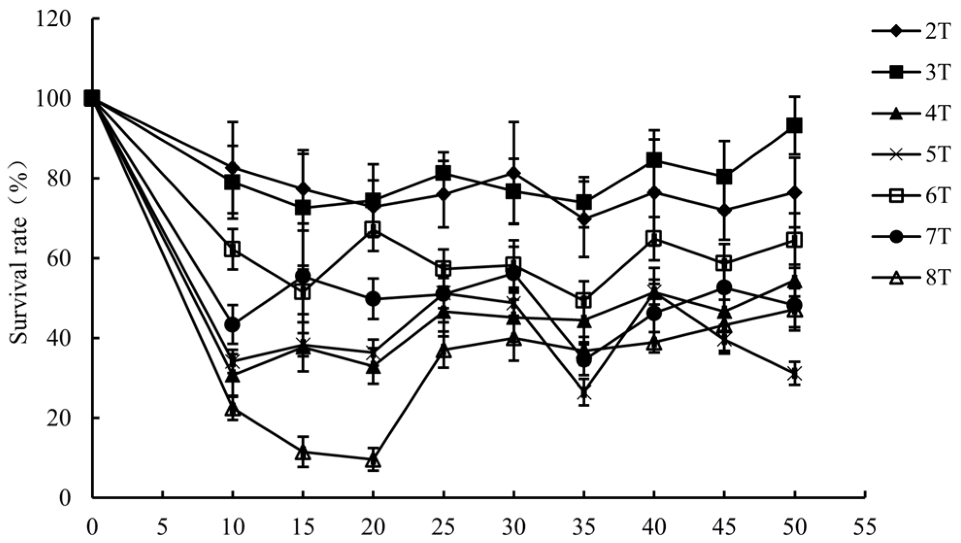
Figure 1. Survival rate of L. monocytogenes under PMF treatment.
Foods 14 01871 g001
Figure 2. SDS-PAGE gel electrophoresis of proteins extracted from L. monocytogenes before and after PMF treatment.
Foods 14 01871 g002
Figure 3. Protein volcano plot (A) and clustering heatmap of expression profiles (B).
Foods 14 01871 g003aFoods 14 01871 g003b
Figure 4. Gene Ontology (GO) functional enrichment analysis.
Foods 14 01871 g004
Figure 5. KEGG pathway enrichment analysis.
Foods 14 01871 g005
Table 1. DEPs identified by MALDI-TOF MS.
Spot No.Gene/ORF NameDescriptionAccession No.pI/MW (kDa)ScoresSeq Cov (%)Fold Change
1ylbF *Regulatory protein ylbFA0A0E1R7I75.68/18.75.816−1.54
2LMRG_00836UPF0176 protein LMRG_00836A0A0H3GG734.98/36.3219.7559−1.47
3rpmI50S ribosomal protein L35A0A0E1R8V012.56/7.7116.3135−1.43
4hemA *Glutamyl-tRNA reductaseA0A0H3GH635.33/49.221.345−1.37
5CDR86_15435tRNA-dihydrouridine synthaseA0A1D2IUJ66.44/36.8326.2864−1.35
6A410_1127 *UPF0358 protein A410_1127A0A241SNS58.63/12.62.510−1.33
7SAMD00023519_01241Dipicolinate synthaseA0A146GU878.66/20.817.6613−1.25
8lysP *Lysine-specific permeaseA0A0E1R4G39.41/53.14.522−1.23
9CDR86_13105Carbonic anhydraseA0A1D2IXH14.97/27.2405.449−1.23
10BB718_05970DNA-binding response regulatorA0A1D2IS686.13/28.561.2222−1.23
11secE *Protein translocase subunit SecEA0A0E1R2V19.54/6.92.2414−1.20
12LmNIHS28_00558 *Sodium-dependent phosphate transporterA0A0B8QTV95.45/59.75.771−1.20
13rpsN30S ribosomal protein S14 type ZA0A0E0UZJ210.49/7.121.4134−1.20
14CDR86_09705Rhodanese-like domain-containing proteinA0A1C7PX104.84/10.852.8858−1.20
15LMRG_01479Glutamate decarboxylaseA0A0H3GMU55.22/53.5305.63382.95
16LMRG_01332Aldehyde–alcohol dehydrogenaseA0A0H3GKP86.93/94.62625.08571.99
17LMRG_01480 *Glutamate/gamma-aminobutyrate antiporterA0A0H3GFA19.09/55.121.7541.87
18lmo2067Lmo2067 proteinQ8Y5J35.15/36.838.58191.71
19inlB *Internalin BA0A0E1R4859.41/71.220.5551.67
20CDR86_05685GNAT family N-acetyltransferaseA0A1D2IS774.94/10.259.52231.65
21LMRG_00327Cadmium-translocating P-type ATPaseA0A0H3G9V45.72/67.6238.61231.60
22spsBSignal peptidase IA0A0E1R6F05.19/21.150.79281.56
23LmNIHS28_02228Fumarate reductaseA0A0B8RBG25.94/54.5754.17571.49
24dhaK_2PTS-dependent dihydroxyacetone kinase, dihydroxyacetone-binding subunit dhaKA0A0E1R9584.93/34.9123.73191.48
25gadGGlutamate decarboxylaseA0A0E1RAJ75.22/54.876.2151.43
26lmo0796Lmo0796 proteinQ8Y8U64.84/19.3404.6881.41
27LMM7_2086 *Fructosamine deglycaseA0A0E0UXM05.54/3821.2591.41
28NT04LM_1576Lipoprotein, putative (Fragment)A0A0E1Y6524.77/8.423.34191.40
29CDR86_01030PTS mannose/fructose/sorbose transporter subunit IIBA0A1C7Q0I76.34/17.221.69171.40
30CXL08_12865NAD-dependent dehydrataseA0A1D2IMX86.38/22.776.14351.39
31LMM7_2092 *Putative transcription regulator, GntR familyA0A0E0UXB35.45/28.02.0931.39
32dhaLDihydroxyacetone kinase, C-terminal domain proteinA0A0E0UZE85.33/21.5228.46521.37
33CDR86_02755Carbohydrate kinaseA0A1D2IMM84.96/40.635.57101.37
34lwe2587lipoproteinA0ALX35.91/32.91369.32531.36
35gpmA2,3-Bisphosphoglycerate-dependent phosphoglycerate mutaseA0A0H3GI895.69/26.4285.71391.36
36manZ_3 *Mannose permease IID componentA0A0E1RAX18.34/31.835.571.36
37CDR86_08010Anaerobic ribonucleoside-triphosphate reductaseA0A1D2IZ155.94/82.1131.03241.35
38BN389_17220Epimerase family protein SE_0553A0A0E1RDJ07.68/34.620.27171.35
39NT04LM_1565DNA protection during starvation protein 2A0A0E1Y4R04.93/18.0915.53791.33
40LMRG_00977Short chain dehydrogenaseA0A0H3GHA86.19/20.991.47381.33
41LMRG_00977Lmo1261 proteinQ8Y7L69.41/42.3122.24181.33
42manX_2PTS system mannose-specific EIIAB componentA0A0E1R5429.33/19.7262.23461.32
43lmo2697Lmo2697 proteinQ8Y3Y24.7/13.4115.1541.32
44Fief *Putative Zn/Cd/Fe cation exporterA0A0E0UZ135.78/31.810.5561.32
45CLN77_13670FMN-binding proteinA0A2H4RX416.65/32.71370.82501.31
46psuGPseudouridine-5’-phosphate glycosidaseA0A1D2IMZ94.84/32.5385.23611.31
47SAMD00023519_01958 *Putative activator of (R)-hydroxyglutaryl-CoAA0A146GTR36.34/164.16171.31
48A4P56_12940 *Cell surface proteinA0A2A5UYW35.15/34.614.81161.31
49AF973_12935 *Amino acid ABC transporter permeaseA0A1E6EXR99.66/23.99.9661.31
50CDR86_13550Pyruvate oxidaseA0A1D2INK35.02/62.8266.31351.30
51lmo2213Lmo2213 proteinQ8Y5637.21/19.5101.16351.30
52SAMD00023519_01961 *PTS mannose transporter subunit IICA0A146GSR95.54/32.224.9841.30
53AJL15_04430FMN-binding proteinA0A1E7E8776.65/32.71328.39501.29
54pdxHPutative general stress protein 26 putative pyridoxamine/pyridoxine 5’-phosphate oxidaseA0A0E0UZU54.68/15.759.03351.29
55lacDTagatose 1,6-diphosphate aldolaseA0A0E1R5C55.1/37.9599.72681.28
56hrcAHeat-inducible transcription repressor HrcAA0A0E1Y3055.58/40.467.28181.28
57AJZ74_10515 *Serine/threonine protein phosphataseA0A1E5Z0Y85.08/26.716141.28
58SAMD00023520_02065 *Cyclic nucleotide-binding proteinA0A146H1K49.22/32.83.9461.27
59deoCDeoxyribose-phosphate aldolaseA0A1D2IZI95.39/23.5454.21701.26
60mgtA *Magnesium-translocating P-type ATPaseA0A1D2IRU17.99/94.852.5691.26
61ilvCKetol-acid reductoisomerase (NADP(+))A0A0H3GHN35.36/36.433.9161.26
62LMRG_02097ATP-dependent Clp protease ATP-binding subunit ClpEA0A0H3GFJ65.21/80.2226.18311.25
63LMM7_0960Putative efflux ABC transporter, ATP-binding protein (N-terminal part)A0A0E0UVG86.11/25.941.68301.25
64LMM7_0961 *Putative efflux ABC transporter, ATP binding and permease proteinA0A0E0UUC87.18/41.315.7151.24
65pncA *Putative nicotinamidaseA0A0E0V0534.77/23.55.2731.24
66CDR86_04330Carnitine transport ATP-binding protein OpuCAA0A1D2IQD15.16/45.2289.6521.23
67gabDNAD-dependent succinate-semialdehyde dehydrogenaseA0A2A5UBV66.09/53.1335.88381.23
68metC *Cystathionine beta-lyaseA0A0E0UXF55.41/41.74.8521.23
69mntAManganese-binding lipoprotein MntAA0A2A5U9005.52/34.4717.31461.22
70ctsRTranscriptional regulator CtsRA0AF296.23/17.590.67371.22
71LmNIHS28_01175UPF0473 protein LmNIHS28_01175A0A0B8QXU23.89/12.159.49291.22
72phoU *Phosphate-specific transport system accessory protein PhoUA0A1D2ITX35.05/25.07.64131.22
73lmo0047Lmo0047 proteinQ8YAR74.56/22.7126.33351.21
74BB664_03185LipaseA0A1D2J2964.61/40.440.15141.21
75CDR86_03830DUF1049 domain-containing proteinA0A1D2IVF79.33/12.729.94141.21
76uspF *Putative universal stress protein UspA and related nucleotide-binding proteinA0A0E0V0D18.88/17.211.5181.21
77opuCAGlycine betaine/carnitine/choline transport ATP-binding protein OpuCAA0A0E1RCS25.29/36.766.89341.20
78lmo2230 *Lmo2230 proteinQ8Y5464.87/15.97.32191.20
79Ung *Uracil-DNA glycosylaseA0A0B8RFS77.91/26.44.4141.20
* Proteins with a score below 20 or coverage lower than 10% are categorized as low confidence.
Disclaimer/Publisher’s Note: The statements, opinions and data contained in all publications are solely those of the individual author(s) and contributor(s) and not of MDPI and/or the editor(s). MDPI and/or the editor(s) disclaim responsibility for any injury to people or property resulting from any ideas, methods, instructions or products referred to in the content.