Article

Evaluation of Simultaneous Growth of Escherichia coli O157:H7, Salmonella spp., and Listeria monocytogenes in Ground Beef Samples in Different Growth Media

1
CEB—Centre of Biological Engineering, LIBRO—Laboratory of Research in Biofilms Rosário Oliveira, Campus de Gualtar, University of Minho, 4710-057 Braga, Portugal
2
LEPABE—Laboratory for Process Engineering, Environment, Biotechnology and Energy, Faculty of Engineering, University of Porto, Rua Dr. Roberto Frias, 4200-465 Porto, Portugal
3
ALiCE—Associate Laboratory in Chemical Engineering, Faculty of Engineering, University of Porto, Rua Dr. Roberto Frias, 4200-465 Porto, Portugal
4
INIAV, IP—National Institute for Agrarian and Veterinary Research, Rua dos Lagidos, Lugar da Madalena, Vairão, 4485-655 Vila do Conde, Portugal
5
LABBELS—Associate Laboratory, 4710-057 Braga, Portugal
*
Author to whom correspondence should be addressed.
These authors contributed equally to this work.
Foods 2024, 13(13), 2095; https://doi.org/10.3390/foods13132095
Submission received: 28 May 2024 / Revised: 20 June 2024 / Accepted: 24 June 2024 / Published: 1 July 2024
(This article belongs to the Special Issue Advances and Future Challenges to Microbial Food Safety—Volume II)

Abstract

:
Several multiplex approaches for the simultaneous detection of pathogens in food have been developed in recent years, but the use of a single enrichment medium remains a problem. In this study, six enrichment broths (five non-selective media, tryptic soy broth (TSB), brain heart infusion broth (BHI), buffered peptone water (BPW), universal pre-enrichment broth (UPB), no. 17 broth, and a selective, Salmonella Escherichia Listeria broth (SEL)), were studied for the simultaneous detection of E. coli O157:H7, Salmonella spp., and L. monocytogenes, to validate the suitable enrichment broth to be used for the detection methods. Different ratios of E. coli O157:H7, Salmonella spp., and L. monocytogenes were used. Almost all non-selective broths evaluated in this study showed similar growth parameters and profiles among each other. The only selective enrichment broth under analysis (SEL) showed distinct growth features compared to the non-selective media, allowing for a slower but balanced growth of the three pathogens, which could be beneficial in preventing the overgrowth of fast-growing bacteria. In addition, when tested in ground beef samples, SEL broth seems to be the most distinctive medium with a balanced growth pattern observed for the three pathogens. Overall, this study is intended to provide the basis for the selection of suitable enrichment broths according to the technology detection to be used, the desired time of enrichment, and the expected balanced concentration of pathogens.

1. Introduction

Foodborne diseases represent an important cause of morbidity and mortality that have a negative impact on public health, the economy, and society worldwide [1]. Foodborne pathogens are responsible, in the US, for nearly 48 million cases of foodborne illness each year, which result in 128,000 hospitalizations and 3000 deaths [2,3,4]. Pathogens such as Salmonella spp., Listeria monocytogenes, Shiga-toxin-producing Escherichia coli (STEC) O157:H7, Campylobacter spp., Yersinia spp., and Shigella spp. are considered to be the major bacterial foodborne pathogens [4,5]. From these, E. coli O157:H7, Salmonella spp., and L. monocytogenes are probably the most studied and surveyed due to their high infectious potential; the severity of the illness caused; the relatively high prevalence of the population; and their capacity to contaminate a wide range of food samples [6]. Given the complexity of global food supply chains, the application of systems analysis methods to food security is of paramount importance. The standardization of the food analysis protocols can increase the simplicity of analysis, which would drive the implementation of food security measures in the food supply chain.
Technology is evolving towards the more rapid and simple detection of foodborne pathogens, allying simplicity, accuracy, and time-to-result to surpass the socio-economic problems associated with food contamination. Immunoassays, polymerase chain reaction (PCR), biosensors, and fluorescence in situ hybridization (FISH)-based techniques are some examples of new approaches to pathogen detection [7,8,9,10]. However, all the food sampling protocols need to be well defined to optimize the detection method performance.
Traditionally, the detection methods require the use of an enrichment step to increase the target pathogen concentration to detectable levels in a short period of time, usually not exceeding 24 h [11,12], to avoid the procedure being extended to an additional working day. For PCR-based applications and immunological and biosensor assays, the target pathogen concentration should reach a detection limit of around 103–104 colony-forming units per millilitre (CFUs/mL) [9], and for the FISH technology, the limit is about 103–106 CFUs/mL [13,14]. The enrichment protocols are also important to recover injured or stressed cells that result from food processing and storage [15] and to decrease the interference of inhibitors [8,16,17], improving the efficiency of the target pathogen detection [18,19].
Allied with this, the simultaneous detection of foodborne agents is advantageous because it reduces the associated costs and labour, allowing for the testing of the same food sample for contamination with different pathogens at the same time [8,15]. The most often used media for the simultaneous enrichment of foodborne pathogens (especially E. coli O157:H7, Salmonella spp., and L. monocytogenes) include tryptic soy broth (TSB) [20,21], brain heart infusion broth [22], buffered peptone water (BPW) [23], universal pre-enrichment broth (UPB) [24], no. 17 broth [25], and the selective Salmonella Escherichia Listeria broth (SEL) [16]. However, their performances have never been compared extensively and some media compositions may not be able to provide a balanced growth of different pathogens, which might hide some of the populations in the analysis [26]. This is especially important for culture-based detection methods, where the dominant population forms a lawn that masks the results [16].
In this study, the six enrichment broths mentioned were studied for the simultaneous detection of E. coli O157:H7, Salmonella spp., and L. monocytogenes, in order to validate the suitable enrichment broths to be used. The media were then evaluated in terms of growth profile and kinetic parameters in single and co-cultures. Further assays with raw ground beef samples were carried out to evaluate the performance of enrichment media in the presence of a natural competing microbiota.

2. Materials and Methods

2.1. Bacterial Strains and Culture Conditions

To study the kinetic parameters in different enrichment media, three different food-borne bacterial species were included and distributed into group I and group II. Each group contains one strain of each pathogen in analysis. The strains belong to the Spanish Type Culture Collection, Spain (CECT); South Georgia State College Collection, Georgia, USA (SGSC); or the National Collection of Type Cultures, UK (NCTC). Group I comprised Escherichia coli O157:H7 CECT 4783, Salmonella enterica serovar Enteritidis SGSC 2476, and Listeria monocytogenes CECT 4031T; group II included E. coli O157:H7 CECT 4267, Salmonella serovar Typhimurium NCTC 12416, and L. monocytogenes 747. These two groups were used for the single-culture assays. For co-culture experiments, only group II of strains was used. All strains were maintained on tryptic soy agar (Liofilchem, Roseto degli Abruzzi, Italy) at 37 °C.

2.2. Enrichment Media

Six media were selected, namely, tryptic soy broth (TSB) (Liofilchem), buffered peptone water (BPW) (Liofilchem), brain heart infusion broth (BHI) (Liofilchem), universal pre-enrichment broth (UPB) (Dehydrated Culture Media: Universal Preenrichment Broth) (Becton Dickinson, Franklin Lakes, NJ, USA), no. 17 broth, and Salmonella Escherichia Listeria broth (SEL) (Table 1). All media were already purchased in a ready-to-use form, except SEL and no. 17 broth. SEL was prepared with a base medium (buffered listeria enrichment broth, BLEB) (Liofilchem) to which the selective agents were added, 0.01 g/L Acriflavine (Sigma-Aldrich, St. Louis, MO, USA), 0.05 g/L Cycloheximide (Fluka, Buchs Switzerland), 0.002 g/L Nalidixic acid (Applichem, Darmstadt, Germany), and 0.05 g/L Fosfomycin (Sigma-Aldrich, St. Louis, MO, USA), as described in Kim and Bhunia [26]. No. 17 broth was prepared according to the formulation described by Kawasaki and colleagues [25] with some modifications suggested by Garrido and colleagues [18] and by Omiccioli and coworkers [27], and it included 10 g/L tryptose (Liofilchem), 5 g/L beef extract (Merck, Darmstadt, Germany), 5g/L yeast extract (Liofilchem), 5 g/L sodium chloride (Sigma-Aldrich), 19.3 g/L disodium phosphate (Sigma-Aldrich), and 3.4 g/L monopotassium phosphate (Sigma-Aldrich).

2.3. Evaluation of Growth Kinetics in Single Cultures

For the determination of growth kinetic parameters (exponential growth rate and doubling time) in a single culture, a protocol similar to that described by Garrido and colleagues [18] was followed. A loopful of each bacterium was inoculated into 20 mL of TSB and the cultures were allowed to grow at 37 °C, for approximately 18 h, with an agitation of 120 rpm (Stuart Scientific, Stone, UK). An amount of 25 µL of the stationary phase culture was then inoculated in 20 mL of each enrichment medium under analysis and incubated until the stationary growth phase, at 37 °C, in a shaking incubator at 120 rpm. In the case of no. 17 broth, the temperature used was 35 °C as recommended by Kawasaki and colleagues [25]. The growth was monitored through measurements of optical density at 600 nm (OD600nm) overtime, with a microplate reader device (Tecan, Männedorf, Switzerland). All the experiments were repeated at least twice.

2.4. Evaluation of Growth Kinetics in Co-Cultures

Growth kinetic parameters in co-cultures were determined as described before [26], with minor modifications. The experiments were performed using group II of strains described in Section 2.1. Each experiment involved the co-inoculation of the pathogens in different proportions. In addition, herein, only four growth media were tested here: TSB, UPB, no. 17, and SEL.
For this evaluation, different concentration ratios of the three pathogens were used as inoculum. In experiment I, equal concentrations of the three pathogens (E. coli O157:H7, Salmonella spp., and L. monocytogenes) were inoculated in a 1:1:1 proportion, with ~1 CFU/mL of each pathogen. In the other experiments, E. coli, Salmonella spp., and L. monocytogenes ratios were as follows: experiment II—1:1000:10; experiment III—10:1:1000; and experiment IV—1000:10:1.
Briefly, pre-inocula of E. coli, Salmonella spp., and L. monocytogenes were prepared by inoculating a loopful of each bacterium into 20 mL of TSB. Cultures were allowed to grow at 37 °C, overnight (~18 h), with an agitation of 120 rpm (Stuart Scientific). Afterward, the OD600nm of each pre-inoculum was measured and adjusted to attain a concentration of 108 colony-forming units per millilitre (CFUs/mL). Serial ten-fold dilutions were made in phosphate-buffered saline (PBS, 8 g/L NaCl, 0.2 g/L KCl, 1.44 g/L Na2HPO4, 0.2 g/L KH2PO4, pH 7.2) and then used to inoculate 20 mL of the enrichment media under analysis, in the proportions described above (experiment I, II, III, and IV). Inoculum concentrations were confirmed by plating the appropriate dilutions on tryptic soy agar (TSA) plates. Next, each enrichment medium was incubated at 37 °C (35 °C for no. 17 broth), for 24 h, in a shaking incubator at 120 rpm. Samples were taken at different incubation times for 24 h, and cell counts for each pathogen were determined by plating the cells (and the adequate ten-fold dilution in PBS), in triplicate, onto plates with the appropriate selective agar. MacConkey agar (Liofilchem) was used for E. coli O157:H7 and Salmonella spp., and Oxford Listeria agar (Liofilchem) was used for L. monocytogenes. Lastly, the morphology of bacteria was accessed by the Stereo-Microscope SZ61TR (Olympus, Hamburg, Germany). These experiments were repeated twice for each group of strains.

2.5. Kinetic Parameters Determination

Calculations for the determination of specific growth rates and doubling times were determined through the equation of exponential growth using GraphPad Prism version 8® software (GraphPad Software, San Diego, CA, USA) both in pure and co-culture assays. The equations for co-culture assays are presented in Table S1. The exponential specific growth rate and doubling time were determined through the linear regression determination. For pure culture assays, growth parameters were calculated through OD600nm versus time graphical data; while for co-culture assays, graphics of log10 CFUs/mL versus time were used.

2.6. Performance on Ground Beef Samples

In order to evaluate real conditions of enrichment, 25 g of ground beef samples acquired on the previous day from a local retailer (Pingo Doce, Braga) were artificially contaminated with low levels of the three pathogens of group II described in Section 2.1, as previously described [31], in a proportion of 1:1:1, with 1 CFU/mL. Of note, a non-inoculated batch was also kept and analysed simultaneously as an experimental control.
For the artificial contamination of ground beef samples, pre-inocula of E. coli O157:H7 CECT 4267, Salmonella serovar Typhimurium NCTC 12416, and L. monocytogenes 747 (group II of strains) were prepared as described above. The inoculums were then adjusted to the desired concentration, confirmed by plating the bacterial suspensions on TSA. Twenty-five grams of artificially contaminated ground beef was mixed in aseptic conditions with 225 mL of pre-warmed enrichment medium, in 1000 mL flasks, and incubated at 37 °C for 24 h in a shaking incubator at 120 rpm. Assays involving no. 17 broth were performed at 35 °C. Samples were taken at different incubation times for 24 h. Cell counts for each pathogen were determined by plating the cells in appropriate selective agar: CT-SMAC agar (Thermo Scientific, Waltham, MA, USA) for E. coli O157:H7 (Thermo Scientific, Waltham, MA, USA) for Salmonella spp. and Oxford Listeria agar for L. monocytogenes. The total viable cell counts were assessed using TSA media through the following process: samples were first serially diluted in sterile saline to obtain a range of dilutions. Aliquots of each dilution were then spread onto TSA plates. The plates were incubated at 37 °C for 24 to 48 h to allow for colony formation. After incubation, the colonies were counted manually. The number of colonies was then multiplied by the dilution factor to calculate the total viable cell counts, expressed as colony-forming units per millilitre (CFUs/mL). This method ensures the accurate and reproducible quantification of viable cells in the sample.

2.7. Statistical Analysis

The data were analysed using the statistical package GraphPad Prism version 8 by one-way ANOVA, with multiple comparisons by Tukey’s multiple comparisons test or Bartlett’s test. For non-linear curves, a non-parametric analysis was performed by using the Kruskal–Wallis test. Values with p < 0.05 were considered statistically significant.

3. Results and Discussion

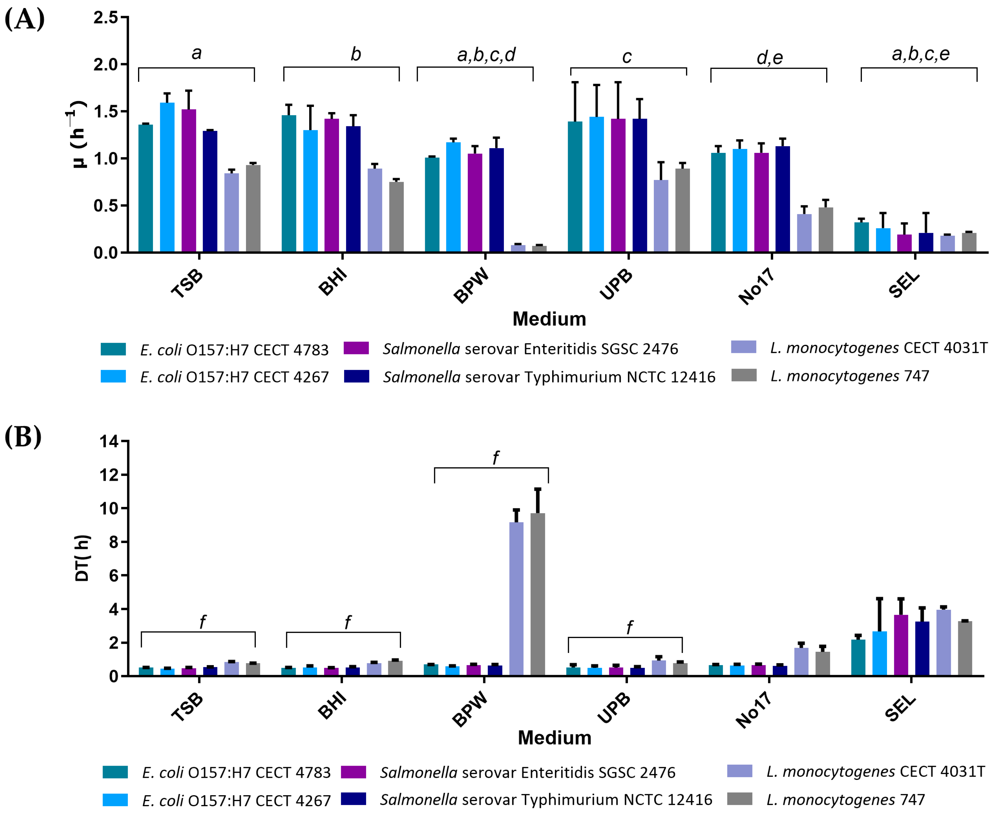
3.1. Evaluation of Growth Kinetics in Single Cultures

The growth profile of E. coli O157:H7, Salmonella spp., and L. monocytogenes was determined individually in each of the six selected enrichment broths through OD600nm measurements over time as shown in Figure 1. The assays in single cultures intended to identify the media with the best features regarding the exponential growth rate (µ) and doubling time (DT).
As it is possible to observe in Figure 1A, TSB, BHI, and UPB generally presented the highest values of µ and the lowest values of DT in all strains analysed. The results for these three media are very similar (p > 0.05), which might be explained by their highly nutritive nature [27]. In addition, the results are very similar between the two strains of each species, and no statistical difference was found (p > 0.05). Among the tested conditions, overall, the highest µ values were observed in E. coli O157:H7 CECT 4783, E. coli O157:H7 CECT 4267, Salmonella serovar Enteritidis SGSC 2476, and Salmonella serovar Typhimurium NCTC 12416, while the lowest µ value was observed for L. monocytogenes CECT 4031T and L. monocytogenes 747. It is also important to highlight that in the case of UPB, the high content of salts (buffering ability) helps the recovery of sub-lethally injured cells [30]. Although the study here was carried out with single cells in the exponential phase, this may be advantageous in food matrices where pathogens are often present in low numbers and under sub-lethal conditions.
The results obtained with BPW and no. 17 broth show that these media perform well for E. coli and Salmonella spp. but slightly worse than TSB, BHI, or UPB. Concerning the L. monocytogenes values in BPW, the results are considerably different. The µ values of L. monocytogenes are extremely low and, consequently, the DT values are very high (Figure 1B). These results are probably related to the fastidious behaviour of L. monocytogenes [32] and the lower content of nutrients of BPW compared to the other rich broths. It is also possible to observe that even in highly nutritive broths (TSB, BHI, UPB, and no. 17), L. monocytogenes always show lower µ and higher DT due to the fastidious behaviour referred to above.
SEL is the only selective enrichment broth in the analysis. This medium was developed [26] as a new selective enrichment broth for simultaneous growth of E. coli O157:H7, Salmonella spp., and L. monocytogenes, with promising results for enriching the mixed cultures of these three pathogens. The results show that µ values obtained with SEL assays are much lower compared to other broths (Figure 1A). This feature is probably related to the selective nature of the medium [33]. The presence of antibiotics and limitative agents appears to interfere with the growth, making it slower and less pronounced. An interesting trait is that SEL appears to balance the growth of the three pathogens once they reveal identical values of µ and DT.

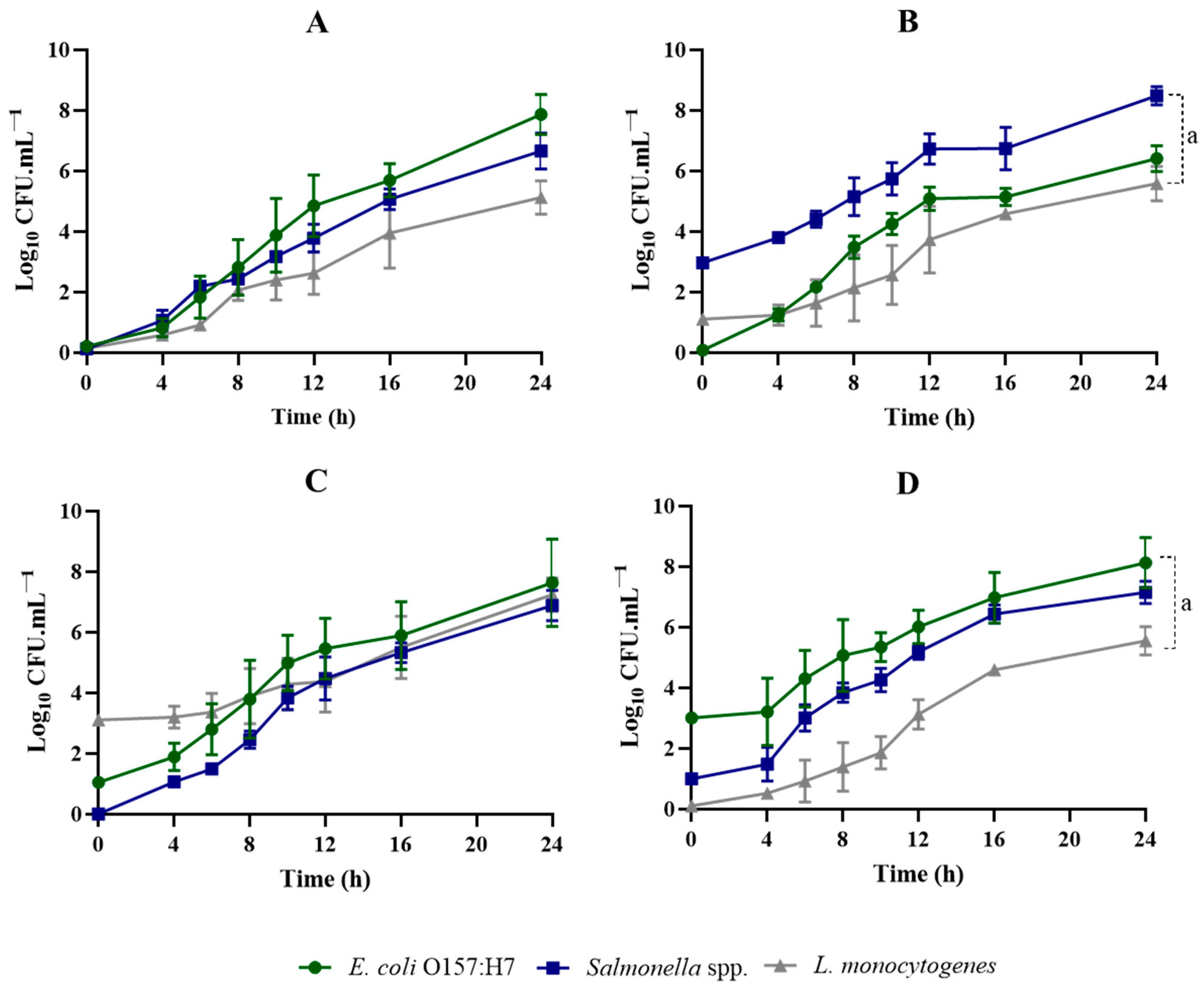
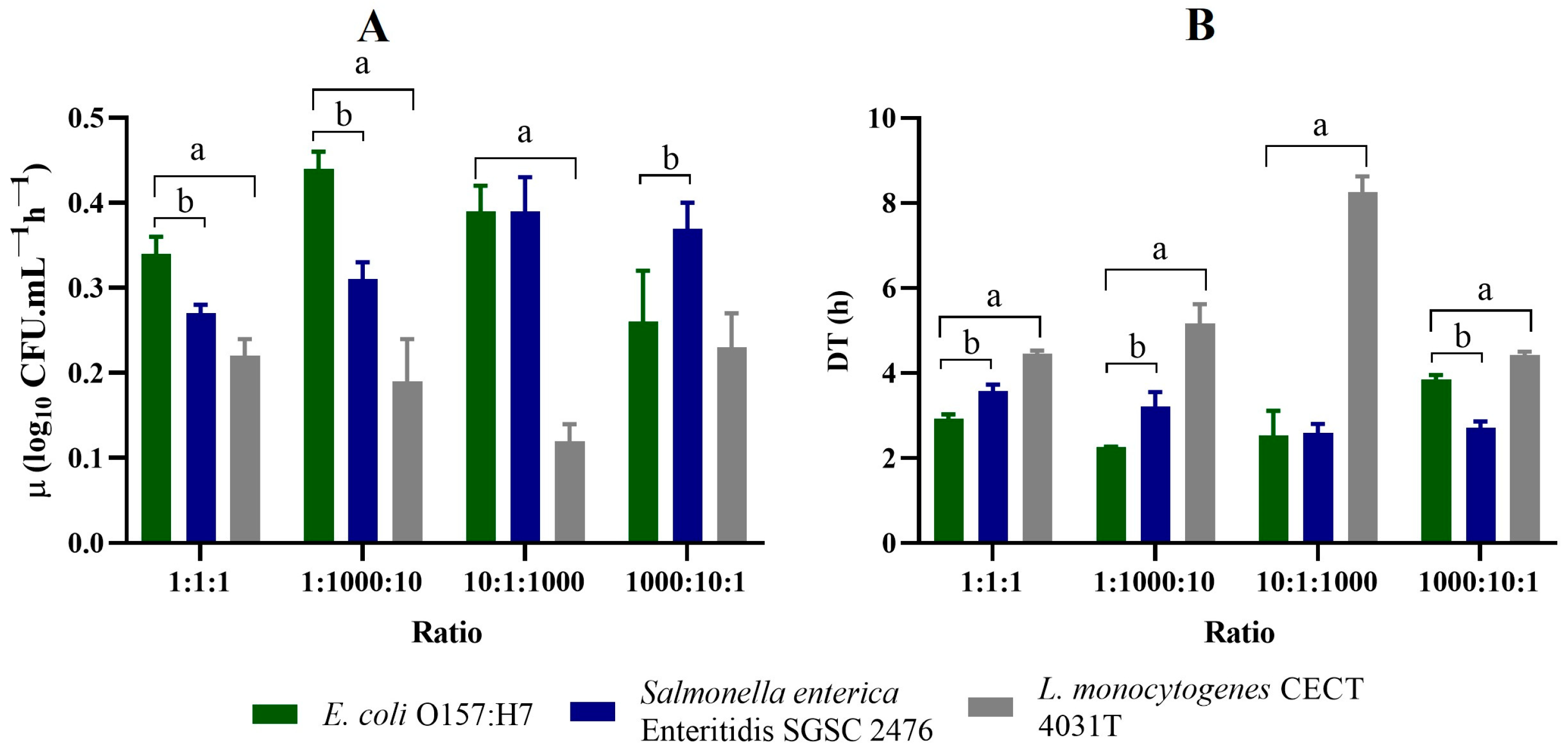
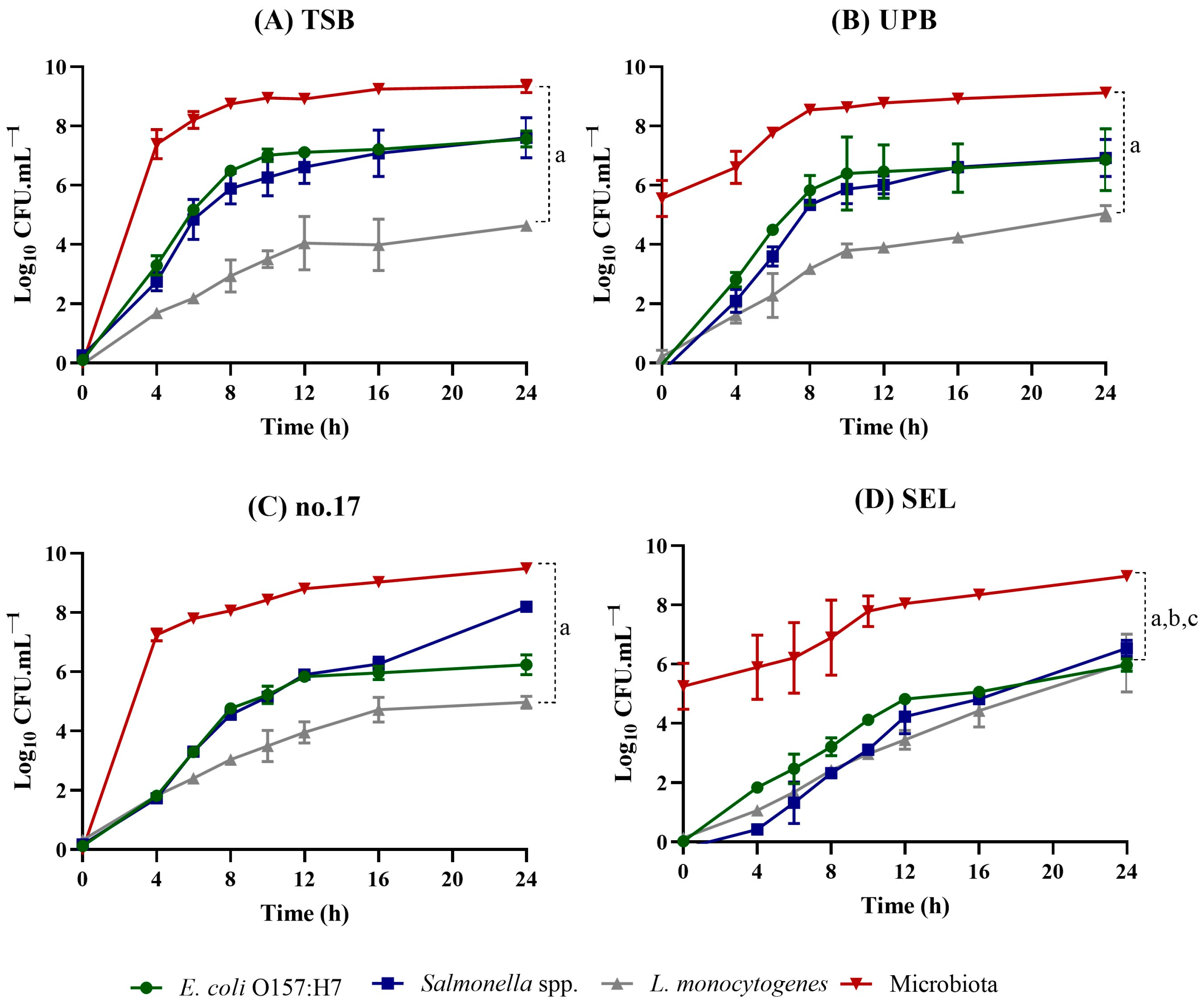
3.2. Evaluation of Growth Kinetics in Co-Cultures

After selecting the media best fitted to single pathogen cultures, their performance in co-cultures was evaluated. Since no statistical differences were found between the bacterial strains belonging to the same bacterial species, as mentioned above, only group II strains were selected here.
For further evaluation of the performance of enrichment broths in co-cultures, four media were selected: TSB, UPB, no. 17, and SEL. BHI was excluded due to its high similarities with TSB and UPB in terms of kinetic parameters, while BPW was discarded due to the poor results obtained for L. monocytogenes.
TSB, UPB, no. 17, and SEL were inoculated at different ratios to better evaluate the influence of a dominant species on the performance of the others. In experiment I, equal concentrations of the three pathogens (E. coli, Salmonella spp., and L. monocytogenes) were inoculated in a 1:1:1 proportion (with ~1CFU/mL of each pathogen). In the other experiments, E. coli, Salmonella spp., and L. monocytogenes ratios were as follows: experiment II—1:1000:10; experiment III—10:1:1000; and experiment IV—1000:10:1.
Regarding the growth patterns of the three pathogens in SEL broth (Figure 2 and Figure 3), there were considerable differences when compared with those of TSB, UPB, and no. 17 (Figure S1, Figure S2, and Figure S3, respectively).
The selective enrichment broth under examination (SEL) exhibited unique growth characteristics (Figure 2) compared to the non-selective media (Table S2).
It facilitated slower yet well-balanced growth of all three pathogens, which could prove beneficial in preventing the dominance of fast-growing bacteria.
Looking at the growth patterns for all the proportions accessed (Figure 2), it is possible to observe that SEL does not have the same ability to recover low numbers of cells, compared to TSB, UPB, or no. 17 broth (Table S2). As expected, SEL has demonstrated to be much slower in recovering both lower and higher levels of the three pathogens. At the end of the 24 h assay, the E. coli and Salmonella spp. concentrations hardly reached values around 108 CFUs/mL, even for higher inoculum concentrations. In the case of L. monocytogenes, at the end of the 24 h assay, it reached values around 105 CFUs/mL and up to 106 when inoculated with higher concentrations. The presence of selective agents appears to retard and diminish the growth of all the target bacteria (Figure 3 and Table S2). Nonetheless, selective agents can constitute simultaneously the main limitation factor and the main advantage of SEL, when using food samples. This effect will be further evaluated in the next step.
Another interesting feature of the growth patterns in the SEL medium is the presence of some kind of adapting period (until ~4 h), where cell numbers remain at the initial inoculation level or even lower (Figure 2). In the CFU counts, it was possible to observe that some colonies of E. coli and Salmonella spp. showed irregular morphology within this period (Figure 4).
This was only observed for cells grown in SEL. With further streaking of colonies into fresh plates, the irregular colonies showed regular shape. This behaviour might be related to the induction of some level of stress in bacteria, affecting its morphology and growth in the early stages.
The growth patterns of the pathogens in TSB, at similar initial inoculation levels (Table S2), showed a similar growth behaviour to E. coli and Salmonella spp., while L. monocytogenes growth was slightly slower. E. coli and Salmonella spp. µ values were 0.65 ± 0.02 and 0.63 ± 0.12 log10 CFUs mL−1 h−1, respectively, while L. monocytogenes reached an µ value of 0.35 ± 0.03 log10 CFUs mL−1 h−1 (Table S2). These data express that E. coli and Salmonella spp. were able to grow better in TSB when compared to L. monocytogenes, even with similar initial concentrations. L. monocytogenes might be affected by some level of nutrient depletion once E. coli and Salmonella spp. are fast-growing bacteria. But, in fact, the µ and DT values observed in co-cultures are very similar to those observed in pure cultures. This means that these three species do not seem to greatly affect the growth of each other.
In experiment II (Figure S1B) the initial pathogen ratio was 1:1000:10 for E. coli, Salmonella spp., and L. monocytogenes, respectively. The growth profiles of Salmonella spp. and E. coli seem to be proportional to the initial inoculums, reaching a differential final concentration (at 24 h) of about 2 log10 CFUs/mL (Figure S1B), with Salmonella spp. reaching higher cell concentrations than E. coli, as would be expected. The growth pattern of L. monocytogenes is very similar to the one observed in experiment I (Figure S1A).
The observed µ and DT values do not differ significantly from the ones at experiment I (Table S2), regardless of the initial concentration of L. monocytogenes, which was 10 times higher than E. coli. L. monocytogenes was rapidly surpassed by the other species in terms of cell number. This behaviour has already been reported by Mellefont and co-workers [34]. However, it is important to observe that L. monocytogenes shows interesting behaviour when both Salmonella spp. and E. coli reach the stationary phase. It managed to maintain the cellular growth, which may indicate that the conditions that are limiting the growth of Salmonella spp. and E. coli (such as the nutrient depletion, accumulation of toxic metabolites, and higher number of cells) do not limit the growth of L. monocytogenes, at least until this bacterium reaches 106–107 CFUs/mL.
Regarding experiments III and IV, in TSB media (Figure S1C,D), the exponential growth phase parameters do not seem to be greatly affected by the initial inoculums (Table S2). Actually, when inoculated at a high concentration, L. monocytogenes reaches its maximum concentration (and, consequently, the stationary phase) early in the assay; but still the maximum concentration is fixed at 107 CFUs/mL. It always reached lower concentrations no matter the initial concentration used.
The other non-selective media in analysis (UPB and no. 17 broth) present high similarities with the results observed for TSB (Figures S1–S3), both in terms of the growth profile and kinetic parameters (Table S2).

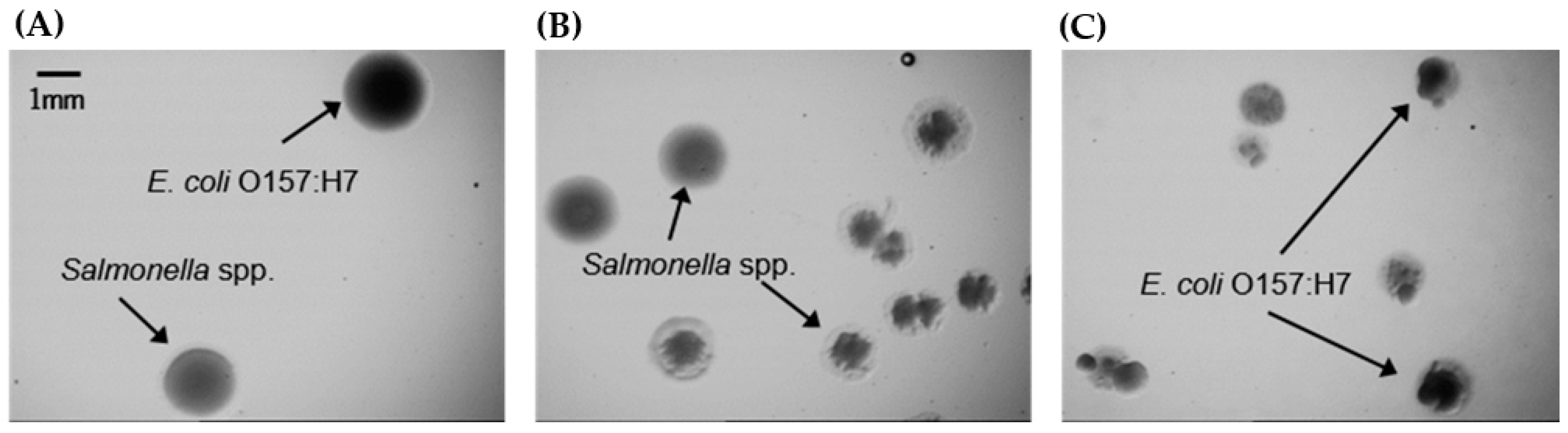
3.3. Performance on Ground Beef Samples

In order to simulate the real conditions of pre-enrichment, with the presence of a competing microbiota, a food matrix was included. Ground beef was chosen as it is colonized by a variety of microorganisms [35,36] and it is often associated with foodborne infections caused by the three pathogens under study. Samples of 25 g of ground beef were inoculated with 1 CFU/mL of each pathogen, homogenized with 225 mL of enrichment broth, and incubated for 24 h (35 or 37 °C). CFU counts were performed using several selective media and also TSA to assess the total culturable microbiota. Results are shown in Figure 5.
It is clear that TSB, UPB, and no. 17 broth provide similar growth conditions (Figure 5A–C). The growth patterns on no. 17 broth show that the growth is slightly slower than that observed in TSB and UPB assays, probably due to the use of a lower incubation temperature (35 °C). At the end of the 24 h of enrichment, the concentration of E. coli and Salmonella spp. ranged between 106 CFUs/mL and 108 CFUs/mL and L. monocytogenes ranged from 104 CFUs/mL to 105 CFUs/mL in these three media. For the screening approach to be performed subsequently, the concentrations obtained following the enrichment step should provide pathogen concentrations to detectable levels. The results obtained for the three pathogens enable the application of PCR techniques or immunology assays [9]; however, to apply FISH technology, the levels of L. monocytogenes achieved may not be sufficient to obtain a favourable result [13]. The slow and less pronounced growth profile of L. monocytogenes in the presence of natural microbiota may be due to both the growth-limiting effect of a larger competing microbiota (when compared to the assay in co-cultures) and the saturation effect of the medium [37]. This behaviour of L. monocytogenes has been reported previously [28] for the enrichment of ground pork samples. During the enrichment, the vacant media transforms into a saturated culture, usually corresponding to a cell count of ~109 CFUs/mL. When the culture reaches this value, the physical space available becomes limiting.
Regarding the enrichment in SEL broth, once again, the most distinctive feature is the balanced growth pattern observed for the three pathogens (Figure 4). The selective properties of SEL seem to only affect the overall microorganism concentration. Even though the concentration of the pathogens was balanced (between 105 and 106 CFUs/mL), a slower and less pronounced growth was observed. This could be a disadvantage for those detection methods with high detection levels. This might be the case with immunological techniques such as some ELISA assays, known for their low limit of detection [9,38]; several types of biosensors [38]; and some fluorescence in situ hybridization (FISH) procedures, where the detection limit is usually around 105 CFUs/mL [13]. In those particular cases, the use of a rich medium might be advisable, instead of a selective one. Nonetheless, the choice of the preferred medium will depend on the properties of the detection technique to be applied.

4. Conclusions

Multiplex detection possesses a number of advantages, but the need for a universal enrichment step is a limitative factor. The choice of an enrichment broth depends on several factors such as the detection limit of the technique to be used, the growth capacity of the target microorganism, and, ultimately, the characteristics of the medium itself.
Regarding this, the selective medium SEL provides a slower and less pronounced growth for all three pathogens tested, which are clearly affected by the medium’s composition. This medium in fact has been demonstrated to be much slower in recovering low levels of E. coli O157 and Salmonella spp., and it also failed to recover L. monocytogenes up to 106 CFUs/mL; however, it seems to present an advantage in preventing the overgrowth of fast-growing bacteria.
In conclusion, the present study shows that the choice of an enrichment broth is clearly a limiting factor for obtaining detection and balanced levels of the pathogens, which has implications for the detection technology to be applied in the future. Furthermore, it is important to understand the behaviour of the selective medium in other food matrixes.

Supplementary Materials

The following supporting information can be downloaded at https://www.mdpi.com/article/10.3390/foods13132095/s1, Figure S1: Growth curves for E. coli O157:H7, Salmonella spp., and L. monocytogenes co-cultured in TSB, at different pathogen ratios. Figure S2: Growth curves for E. coli O157:H7, Salmonella spp., and L. monocytogenes co-cultured in UPB, at different pathogen ratios. Figure S3: Growth curves for E. coli O157:H7, Salmonella spp., and L. monocytogenes co-cultured in no. 17 broth, at different pathogen ratios. Table S1: Equations for measuring the exponential growth and doubling time for TSB, UPB, no. 17 broth, and SEL for the co-culture assays. Table S2: Values of DT and µ for TSB, UPB, no. 17 broth, and SEL for the co-culture assays.

Author Contributions

Conceptualization, L.C., C.A. and N.F.A.; methodology: J.M.S. and A.B.; software, A.B., D.A. and J.C.; investigation, J.M.S., A.B., D.A. and J.C.; writing—original draft preparation, J.M.S.; writing—review and editing, A.B., D.A., J.C., L.C., C.A. and N.F.A.; funding acquisition, L.C., C.A. and N.F.A. All authors have read and agreed to the published version of the manuscript.

Funding

This work is financially supported by national funds through the FCT/MCTES (PIDDAC), under the project 2022.07654.PTDC—NAMSal—Nucleic acid mimics as a new tool against salmonellosis in the poultry industry with DOI 10.54499/2022.07654.PTDC (https://doi.org/10.54499/2022.07654.PTDC). In addition, this work was financially supported by LA/P/0045/2020 (ALiCE), UIDB/00511/2020, and UIDP/00511/2020 (LEPABE), funded by national funds through the FCT/MCTES (PIDDAC) and the Portuguese Foundation for Science and Technology (FCT) under the scope of the strategic funding of the UIDB/04469/2020 unit, LABBELS—Associate Laboratory in Biotechnology, Bioengineering and Microelectromechanical Systems—LA/P/0029/2020. A.B. thanks FCT for the PhD fellowship 2022.11840.BD. J.C. also thanks FCT for the CEEC Individual (https://doi.org/10.54499/2022.06886.CEECIND/CP1737/CT0001).

Institutional Review Board Statement

Not applicable.

Informed Consent Statement

Not applicable.

Data Availability Statement

The original contributions presented in the study are included in the article, further inquiries can be directed to the corresponding author.

Conflicts of Interest

The authors declare no conflict of interest.

References

  1. Fung, F.; Wang, H.S.; Menon, S. Food Safety in the 21st Century. Biomed. J. 2018, 41, 88–95. [Google Scholar] [CrossRef]
  2. Bintsis, T. Foodborne Pathogens. AIMS Microbiol. 2017, 3, 529–563. [Google Scholar] [CrossRef]
  3. Vieira, K.C.d.O.; Silva, H.R.A.d.; Rocha, I.P.M.; Barboza, E.; Eller, L.K.W. Foodborne Pathogens in the Omics Era. Crit. Rev. Food Sci. Nutr. 2022, 62, 6726–6741. [Google Scholar] [CrossRef]
  4. Jeon, B.; Luangtongkum, T.; Shen, Z.; Logue, C.M.; Lin, J. Editorial: Resistance and Tolerance in Food-Borne Pathogens: Mechanisms, Public Health Impact, and Control Measures. Front. Microbiol. 2021, 12, 769931. [Google Scholar] [CrossRef]
  5. Yeni, F.; Yavaş, S.; Alpas, H.; Soyer, Y. Most Common Foodborne Pathogens and Mycotoxins on Fresh Produce: A Review of Recent Outbreaks. Crit. Rev. Food Sci. Nutr. 2016, 56, 1532–1544. [Google Scholar] [CrossRef] [PubMed]
  6. Abdelhaseib, M.U.; Singh, A.K.; Bhunia, A.K. Simultaneous Detection of Salmonella enterica, Escherichia coli and Listeria monocytogenes in Food Using a Light Scattering Sensor. J. Appl. Microbiol. 2019, 126, 1496–1507. [Google Scholar] [CrossRef] [PubMed]
  7. Ali, A.A.; Altemimi, A.B.; Alhelfi, N.; Ibrahim, S.A. Application of Biosensors for Detection of Pathogenic Food Bacteria: A Review. Biosensors 2020, 10, 58. [Google Scholar] [CrossRef]
  8. Bundidamorn, D.; Supawasit, W.; Trevanich, S. A New Single-Tube Platform of Melting Temperature Curve Analysis Based on Multiplex Real-Time PCR Using EvaGreen for Simultaneous Screening Detection of Shiga Toxin-Producing Escherichia coli, Salmonella spp. and Listeria monocytogenes in Food. Food Control 2018, 94, 195–204. [Google Scholar] [CrossRef]
  9. Jasson, V.; Jacxsens, L.; Luning, P.; Rajkovic, A.; Uyttendaele, M. Alternative Microbial Methods: An Overview and Selection Criteria. Food Microbiol. 2010, 27, 710–730. [Google Scholar] [CrossRef]
  10. Azinheiro, S.; Carvalho, J.; Prado, M.; Garrido-Maestu, A. Multiplex Detection of Salmonella spp., E. coli O157 and L. monocytogenes by qPCR Melt Curve Analysis in Spiked Infant Formula. Microorganisms 2020, 8, 1359. [Google Scholar] [CrossRef]
  11. Bhunia, A.K. One Day to One Hour: How Quickly Can Foodborne Pathogens Be Detected? Future Microbiol. 2014, 9, 935–946. [Google Scholar] [CrossRef] [PubMed]
  12. McMeekin, T.A.; Hill, C.; Wagner, M.; Dahl, A.; Ross, T. Ecophysiology of Food-Borne Pathogens: Essential Knowledge to Improve Food Safety. Int. J. Food Microbiol. 2010, 139, S64–S78. [Google Scholar] [CrossRef] [PubMed]
  13. Almeida, C.; Azevedo, N.F.; Iversen, C.; Fanning, S.; Keevil, C.W.; Vieira, M.J. Development and Application of a Novel Peptide Nucleic Acid Probe for the Specific Detection of Cronobacter Genomospecies (Enterobacter sakazakii) in Powdered Infant Formula. Appl. Environ. Microbiol. 2009, 75, 2925–2930. [Google Scholar] [CrossRef] [PubMed]
  14. Rohde, A.; Hammerl, J.A.; Appel, B.; Dieckmann, R.; Al Dahouk, S. FISHing for Bacteria in Food—A Promising Tool for the Reliable Detection of Pathogenic Bacteria? Food Microbiol. 2015, 46, 395–407. [Google Scholar] [CrossRef] [PubMed]
  15. Ding, T.; Suo, Y.; Zhang, Z.; Liu, D.; Ye, X.; Chen, S.; Zhao, Y. A Multiplex RT-PCR Assay for S. aureus, L. monocytogenes, and Salmonella spp. Detection in Raw Milk with Pre-Enrichment. Front. Microbiol. 2017, 8, 989. [Google Scholar] [CrossRef] [PubMed]
  16. Suo, B.; Wang, Y. Evaluation of a Multiplex Selective Enrichment Broth SEL for Simultaneous Detection of Injured Salmonella, Escherichia coli O157:H7 and Listeria monocytogenes. Braz. J. Microbiol. 2014, 44, 737–742. [Google Scholar] [CrossRef] [PubMed]
  17. Wu, V.C.H. A Review of Microbial Injury and Recovery Methods in Food. Food Microbiol. 2008, 25, 735–744. [Google Scholar] [CrossRef] [PubMed]
  18. Garrido, A.; Chapela, M.J.; Román, B.; Fajardo, P.; Vieites, J.M.; Cabado, A.G. In-House Validation of a Multiplex Real-Time PCR Method for Simultaneous Detection of Salmonella spp., Escherichia coli O157 and Listeria monocytogenes. Int. J. Food Microbiol. 2013, 164, 92–98. [Google Scholar] [CrossRef]
  19. Hahm, B.K.; Kim, H.; Singh, A.K.; Bhunia, A.K. Pathogen Enrichment Device (PED) Enables One-Step Growth, Enrichment and Separation of Pathogen from Food Matrices for Detection Using Bioanalytical Platforms. J. Microbiol. Methods 2015, 117, 64–73. [Google Scholar] [CrossRef]
  20. Germini, A.; Masola, A.; Carnevali, P.; Marchelli, R. Simultaneous Detection of Escherichia coli O175:H7, Salmonella spp., and Listeria monocytogenes by Multiplex PCR. Food Control 2009, 20, 733–738. [Google Scholar] [CrossRef]
  21. Costa-Ribeiro, A.; Lamas, A.; Prado, M.; Garrido-Maestu, A. Evaluation of the Novel MTA10 Selective Broth, MSB, for the Co-Enrichment and Detection of Salmonella spp., Escherichia coli O157 and Listeria monocytogenes in Ready-to-Eat Salad Samples. Foods 2023, 13, 63. [Google Scholar] [CrossRef]
  22. Wang, Y.; Suo, B. A New 7-Plex PCR Assay for Simultaneous Detection of Shiga Toxin-Producing Escherichia coli O157 and Salmonella Enteritidis in Meat Products. J. Verbraucherschutz Leb. 2011, 6, 441–447. [Google Scholar] [CrossRef]
  23. Boukharouba, A.; González, A.; García-Ferrús, M.; Ferrús, M.A.; Botella, S. Simultaneous Detection of Four Main Foodborne Pathogens in Ready-to-Eat Food by Using a Simple and Rapid Multiplex PCR (MPCR) Assay. Int. J. Environ. Res. Public Health 2022, 19, 1031. [Google Scholar] [CrossRef] [PubMed]
  24. Nam, H.M.; Murinda, S.E.; Nguyen, L.T.; Oliver, S.P. Evaluation of Universal Pre-Enrichment Broth for Isolation of Salmonella spp., Escherichia coli O157:H7, and Listeria monocytogenes from Dairy Farm Environmental Samples. Foodborne Pathog. Dis. 2004, 1, 37–44. [Google Scholar] [CrossRef] [PubMed]
  25. Kawasaki, S.; Horikoshi, N.; Okada, Y.; Takeshita, K.; Sameshima, T.; Kawamoto, S. Multiplex PCR for Simultaneous Detection of Salmonella spp., Listeria monocytogenes, and Escherichia coli O157:H7 in Meat Samples. J. Food Prot. 2005, 68, 551–556. [Google Scholar] [CrossRef] [PubMed]
  26. Kim, H.; Bhunia, A.K. SEL, a Selective Enrichment Broth for Simultaneous Growth of Salmonella Enterica, Escherichia coli O157:H7, and Listeria monocytogenes. Appl. Environ. Microbiol. 2008, 74, 4853–4866. [Google Scholar] [CrossRef] [PubMed]
  27. Omiccioli, E.; Amagliani, G.; Brandi, G.; Magnani, M. A New Platform for Real-Time PCR Detection of Salmonella Spp., Listeria monocytogenes and Escherichia coli O157 in Milk. Food Microbiol. 2009, 26, 615–622. [Google Scholar] [CrossRef] [PubMed]
  28. Gehring, A.G.; Albin, D.M.; Bhunia, A.K.; Kim, H.; Reed, S.A.; Tu, S.I. Mixed Culture Enrichment of Escherichia coli O157:H7, Listeria monocytogenes, Salmonella Enterica, and Yersinia Enterocolitica. Food Control 2012, 26, 269–273. [Google Scholar] [CrossRef]
  29. Kim, J.; Demeke, T.; Clear, R.M.; Patrick, S.K. Simultaneous Detection by PCR of Escherichia coli, Listeria monocytogenes and Salmonella Typhimurium in Artificially Inoculated Wheat Grain. Int. J. Food Microbiol. 2006, 111, 21–25. [Google Scholar] [CrossRef]
  30. Bhaduri, S.; Cottrell, B. Sample Preparation Methods for PCR Detection of Escherichia coli O157:H7, Salmonella Typhimurium, and Listeria monocytogenes on Beef Chuck Shoulder Using a Single Enrichment Medium. Mol. Cell Probes 2001, 15, 267–274. [Google Scholar] [CrossRef]
  31. Sousa, M.; Rocha, R.; Araújo, D.; Castro, J.; Barbosa, A.; Azevedo, N.F.; Cerqueira, L.; Almeida, C. A New Peptide Nucleic Acid Fluorescence In Situ Hybridization Probe for the Specific Detection of Salmonella Species in Food Matrices. Foodborne Pathog. Dis. 2024, 21, 298–305. [Google Scholar] [CrossRef] [PubMed]
  32. Ramaswamy, V.; Cresence, V.M.; Rejitha, J.S.; Lekshmi, U.; Dharsana, K.S.; Prasad, P.; Vijila, M. Listeria-Review of Epidemiology and Pathogenesis. J. Microbiol. Immunol. Infect. 2007, 40, 4–13. [Google Scholar] [PubMed]
  33. Jacobsen, C.N. The Influence of Commonly Used Selective Agents on the Growth of Listeria monocytogenes. Int. J. Food Microbiol. 1999, 50, 221–226. [Google Scholar] [CrossRef]
  34. Mellefont, L.A.; McMeekin, T.A.; Ross, T. Effect of Relative Inoculum Concentration on Listeria monocytogenes Growth in Co-Culture. Int. J. Food Microbiol. 2008, 121, 157–168. [Google Scholar] [CrossRef] [PubMed]
  35. Lianou, A.; Koutsoumanis, K.P. Evaluation of the Effect of Defrosting Practices of Ground Beef on the Heat Tolerance of Listeria monocytogenes and Salmonella Enteritidis. Meat Sci. 2009, 82, 461–468. [Google Scholar] [CrossRef] [PubMed]
  36. Schlisselberg, D.B.; Kler, E.; Kalily, E.; Kisluk, G.; Karniel, O.; Yaron, S. Inactivation of Foodborne Pathogens in Ground Beef by Cooking with Highly Controlled Radio Frequency Energy. Int. J. Food Microbiol. 2013, 160, 219–226. [Google Scholar] [CrossRef] [PubMed]
  37. Al-Zeyara, S.A.; Jarvis, B.; Mackey, B.M. The Inhibitory Effect of Natural Microflora of Food on Growth of Listeria monocytogenes in Enrichment Broths. Int. J. Food Microbiol. 2011, 145, 98–105. [Google Scholar] [CrossRef]
  38. Wang, B.; Wang, H.; Lu, X.; Zheng, X.; Yang, Z. Recent Advances in Electrochemical Biosensors for the Detection of Foodborne Pathogens: Current Perspective and Challenges. Foods 2023, 12, 2795. [Google Scholar] [CrossRef]
Figure 1. Growth kinetics in single cultures. (A) Values of µ—Exponential Growth Rate (unit: h−1). (B) DT—Doubling Time (unit: h) on single culture assays for the six enrichment media under analysis. a Indicates that there is a statistical difference in values of µ (p < 0.05) between TSB and BPW; TSB and SEL. b Indicates that there is a statistical difference in values of µ (p < 0.05) between BHI and BPW; BHI and SEL. c Indicates that there is a statistical difference in values of µ (p < 0.05) between UPB and BPW; UPB and SEL. d Indicates that there is a statistical difference in values of µ (p < 0.05) between BPW and no. 17. e Indicates that there is a statistical difference in values of µ (p < 0.05) between no. 17 and SEL. f Indicates that there is a statistical difference in values of DT (p < 0.05) between BPW and TSB; BPW and BHI; and BPW and UPB. Error bars represent standard deviation. Abbreviations: tryptic soy broth (TSB); brain heart infusion broth (BHI); buffered peptone water (BPW); universal pre-enrichment broth (UPB); no. 17 broth; and Salmonella Escherichia Listeria broth (SEL).
Foods 13 02095 g001
Figure 2. Growth curves for E. coli O157:H7, Salmonella spp., and L. monocytogenes co-cultured in SEL, at different pathogen ratios. E. coli O157:H7/Salmonella spp./L. monocytogenes. (A) Ratio of 1:1:1. (B) Ratio of 1:1000:10. (C) Ratio of 10:1:1000. (D) Ratio of 1000:10:1. a Indicates that there is a statistical difference in values of µ (p < 0.05) between E. coli O157:H7 and L. monocytogenes.
Foods 13 02095 g002
Figure 3. Growth kinetics in co-cultures. (A) Values of µ—Exponential Growth Rate (unit: log10 CFUs mL−1h−1). (B) DT—Doubling Time (unit: h) (B) for E. coli O157:H7, Salmonella spp., and L. monocytogenes co-cultured in SEL, at different pathogen ratios. E. coli O157:H7/Salmonella spp./L. monocytogenes ratio of 1:1:1, 1:1000:10, 10:1:1000, and 1000:10:1. a Indicates that there is a statistical difference in values of µ (p < 0.05) between E. coli O157:H7 and L. monocytogenes. b Indicates that there is a statistical difference in values of µ (p < 0.05) between E. coli O157:H7 and Salmonella spp.
Foods 13 02095 g003
Figure 4. Morphology of some E. coli O157:H7 and Salmonella spp. colonies in McConkey agar plates at the early hours of SEL assays. (A) Regular morphology of E. coli O157:H7 and Salmonella spp. (B) Regular and irregular morphology of Salmonella spp. (C) Irregular morphology of E. coli O157:H7. The images were acquired by using the Stereo-Microscope SZ61TR.
Foods 13 02095 g004
Figure 5. Growth curves for E. coli O157:H7 CECT 4267, Salmonella serovar Typhimurium NCTC 12416, and L. monocytogenes 747 in an artificially contaminated ground beef sample. (A) Enrichment on TSB. (B) Enrichment on UPB. (C) Enrichment on no. 17 broth. (D) Enrichment on SEL. a Indicates that there is a statistical difference in values of µ (p < 0.05) between L. monocytogenes 747 and microbiota. b Indicates that there is a statistical difference in values of µ (p < 0.05) between Salmonella spp. and microbiota. c Indicates that there is a statistical difference in values of µ (p < 0.05) between E. coli O157:H7 and microbiota.
Foods 13 02095 g005
Table 1. Constituents of enrichment broths in analysis. Adapted from Gehring and coworkers [28].
MediumNutrient (s)Buffer (s)Salt (s)Supplement (s)pHg/L H2OReference
BPWPeptonePhosphateNaCl-7.020[29]
TSBCasein digest, soybean digest, dextrosePhosphateNaCl-7.330[20]
UPBCasein digest, soybean digest, dextrose, protease peptone, sodium pyruvatePhosphateNaCl, Mg2SO4, Ferric ammonium citrate-6.338[30]
SELPancreatic digest of casein, yeast extract, dextrose, soytone, sodium pyruvatePhosphateNaClAcriflavine, Cycloheximide, Fosfomycin, Nalidixic Acid7.048[26]
BHIBrain heart infusion, casein digest, dextrosePhosphateNaCl-7.437[27]
no. 17Tryptose, beef extract, yeast extract PhosphateNaCl-7.248[18]
Disclaimer/Publisher’s Note: The statements, opinions and data contained in all publications are solely those of the individual author(s) and contributor(s) and not of MDPI and/or the editor(s). MDPI and/or the editor(s) disclaim responsibility for any injury to people or property resulting from any ideas, methods, instructions or products referred to in the content.

