Review

Involvement of Versatile Bacteria Belonging to the Genus Arthrobacter in Milk and Dairy Products

by
Nuthathai Sutthiwong
1,
Supaporn Lekavat
1 and
Laurent Dufossé
2,*
1
Expert Centre of Innovative Health Food (InnoFood), Thailand Institute of Scientific and Technological Research, Technopolis 35 Mu 3, Klong Ha, Klong Luang, Pathum Thani 12120, Thailand
2
Chimie et Biotechnologie des Produits Naturels (CHEMBIOPRO), ESIROI Département Agroalimentaire, Université de La Réunion, Parc Technologique, 2 rue Joseph Wetzell, F-97490 Sainte-Clotilde, Ile de La Réunion, France
*
Author to whom correspondence should be addressed.
Foods 2023, 12(6), 1270; https://doi.org/10.3390/foods12061270
Submission received: 16 February 2023 / Revised: 13 March 2023 / Accepted: 15 March 2023 / Published: 16 March 2023
(This article belongs to the Special Issue Cheese: Chemistry, Physics and Microbiology)

Abstract

:
Milk is naturally a rich source of many essential nutrients; therefore, it is quite a suitable medium for bacterial growth and serves as a reservoir for bacterial contamination. The genus Arthrobacter is a food-related bacterial group commonly present as a contaminant in milk and dairy products as primary and secondary microflora. Arthrobacter bacteria frequently demonstrate the nutritional versatility to degrade different compounds even in extreme environments. As a result of their metabolic diversity, Arthrobacter species have long been of interest to scientists for application in various industry and biotechnology sectors. In the dairy industry, strains from the Arthrobacter genus are part of the microflora of raw milk known as an indicator of hygiene quality. Although they cause spoilage, they are also regarded as important strains responsible for producing fermented milk products, especially cheeses. Several Arthrobacter spp. have reported their significance in the development of cheese color and flavor. Furthermore, based on the data obtained from previous studies about its thermostability, and thermoacidophilic and thermoresistant properties, the genus Arthrobacter promisingly provides advantages for use as a potential producer of β-galactosidases to fulfill commercial requirements as its enzymes allow dairy products to be treated under mild conditions. In light of these beneficial aspects derived from Arthrobacter spp. including pigmentation, flavor formation, and enzyme production, this bacterial genus is potentially important for the dairy industry.

1. Introduction

Milk is a rich source of protein and a whole range of nutrients necessary for growth, including fats, carbohydrates, vitamins, minerals, and essential amino acids. Related to its proteins and peptides, milk also provides several physiological functions such as immunoglobulins, enzymes, growth factors, hormones, and antibacterial agents [1]. All milk obtained from a variety of animal sources has a nearly neutral pH and a high-water activity [2]. Due to the requirement of appropriate nutrition for health benefits and body growth and development as well as body maintenance and protection from diseases, milk becomes a vital source of nutrition for humans. With the rapid rise in population growth, increasing income, urbanization, and changes in consumption habits, in addition to the normal consumption of milk as fluid, global demand for milk-derived products is continuing to grow.
In addition to being an excellent source of human nutrition, milk has been referred to as a great environment for the survival and growth of a wide range of microorganisms because of its near-neutral pH, high water content, and complex biochemical compositions. As an agricultural product, milk is collected from domesticated animals, particularly cows. The number and types of microorganisms exhibited in milk are affected by the seasons, farm hygiene, feed, and efficiency of storage. The microorganism found in milk can be categorized into three groups depending on their involvements, namely, pathogenic, spoilage, and fermented-food-derived bacterial strains [3,4]. Some nutrients contained in milk can be used directly by all microorganisms, while others are provided following the types of microorganisms presented such as lactose, which is not utilized by many bacteria, and large molecules—e.g., proteins and lipids, which must be broken down by enzymes before allowing sustained microbial growth [5]. Changes in biochemical activities in microbial communities during nutrient utilization led to the different characteristics of milk depending on the period of these activities; in this way, microbes beneficially associate with milk and dairy product technology. With accumulating knowledge about the functionality of microorganisms, it became less complicated to apply them in technological processes.
Nowadays, microorganisms are widely used in the food industry to produce various types of products due to their desirable properties. In the dairy industry, the utilization of microorganisms has been of increasing interest since it is an important part of quality control as well as quality development. Due to the up-to-date technologies developed over the past few decades, a variety of dairy products are available. In addition to lactic acid bacteria that are commonly present in milk, many psychrotrophic bacteria are also a major component frequently consisting of Actinobacteria. The particular composition of milk microbiota directly influences the development of dairy products. Microorganisms can trigger the fermentation of milk causing the production of lactate as well as the different effects on the organoleptic properties of fermented resultants [6].
Arthrobacter is a genus of bacteria that belongs to the ecologically and industrially important class Actinobacteria of the Micrococaceae family. This genus is an outstanding group of bacteria isolated from various sources and has been found to produce a great variety of pigment hues [7]. Arthrobacter strains are usually present as a contaminant in milk and play different roles in inducing diverse aspects of contaminated milk [8]. The genus Arthrobacter bacteria are widely distributed in nature even in various extreme environments as they are heterotrophic and do not require very attentive growth conditions. Bacteria of this genus are Gram-positive, catalase-positive, obligated aerobic, and non-spore-forming bacteria, with an optimum growth below 30 °C; moreover, several strains isolated from glacial environments are known as psychrophiles with an ability to grow well under below 0 °C [9,10]. At present, approximately 80 species of this genus have been described in the taxonomy [11]. The taxonomy of the genus Arthrobacter and related genera has been reclassified several times; therefore, some strains were originally recognized as belonging to the Arthrobacter genus but have been presently identified as other genera, and vice versa.
Certain well-known Arthrobacter species enclose a variety of desirable members of technological relevance to dairy products such as Arthrobacter arilaitensis and Arthrobacter bergeri (recently named Glutamicibacter arilaitensis and Glutamicibacter bergeri, respectively—taxonomy always evolves and we choose to keep the name Arthrobacter arilaitensis and Arthrobacter bergeri in this publication, in agreement with previous articles), which respond to color development in smear-ripened cheeses [12]. Regarding the profitable impacts of microorganisms on the dairy industry, this review aims to focus on the association of versatile bacteria belonging to the genus Arthrobacter with their significance in the applications to commercial milk and dairy products. The involvement of Arthrobacter in the color and flavor of cheeses and the potential of being novel sources for the commercial production of an industrially important enzyme “β-galactosidase” or other enzymes used in food processing, as well as the feasibility to utilize Arthrobacter spp. in the production of functional dairy products are mentioned in this review.

2. Occurrence of Arthrobacter spp. in Raw Milk and Dairy Products

Microorganisms in raw milk are varied and derived from several sources of contamination. When synthesizing within the mammary gland of healthy animals, milk is particularly expected to be sterile and safe for human consumption. Nevertheless, in addition to this stage, microbial contamination in raw milk possibly arises from diverse origins including the external udder surface, feed, farm environment, geographic location, and the equipment used for milk handling and storage. Hence, the occurrence of microorganisms in milk is rather influenced by various factors and their combinations, and it is often subsequently a cause of microbial contamination in dairy products in addition to several microorganisms that can emerge from the processing and post-processing lines [13,14].
Members of the genus Arthrobacter are commonly present in raw milk as a primary microflora and are believed to enter from the milking equipment as well as the teat surface [15,16]. Outdoor environments such as air in the byre and milking parlor and hay were also regarded as the sources of Arthrobacter spp. [17,18,19]. Certain methods such as culture-dependent and molecular approaches were applied as tools to classify Arthrobacter species that existed in raw milk [20,21]. Despite the microbial composition of milk having been investigated by clarifying several groups of microorganisms, few studies reported the presence of Arthrobacter spp. in raw milk; furthermore, the detail of species diversity of these bacteria has been much less observed. In recent studies, bacteria in the genus Arthrobacter were found to be one of the microbial communities in raw milk, accounting for approximately 1–2% of the total [21,22].
Arthrobacter spp. are rarely found predominantly in milk, although organic sources are not a restraining factor for the growth of Arthrobacter strains; instead, the competition of other microorganisms and the change in pH by the production of lactic acid or other organic acids can prevent the number of bacteria of this genus from growing. Consequently, Arthrobacter species isolated from raw milk and dairy products normally appeared as co-cultures with other microorganism strains, such as coryneform, micrococci, staphylococci, and yeasts [8]. Several Arthrobacter species, such as Arthrobacter aurescens, A. arilaitensis, A. bergerei, Arthrobacter globiformis, Arthrobacter citreus, Arthrobacter nicotanae, and Arthrobacter bussei, were identified in food, especially in raw milk and cheese. Since Arthrobacter species are aerobes, they grow mainly on the surface of food and feasibly cause deterioration of food, ripening, and smearing; thus, these strains are assumedly one of the main microbiota on cheese rind and contribute to differences in organoleptic characteristics such as flavor and color [17,21].

3. Significance of Arthrobacter spp. in the Dairy Industry

Bacteria belonging to the genus Arthrobacter frequently demonstrate the nutritional versatility to degrade both natural and synthetic organic compounds as well as exhibit extreme resistance to dryness and starvation [23]. According to their metabolic diversity, Arthrobacter species have long been a topic of interest to scientists for application in various industry and biotechnology sectors. In the dairy industry, Arthrobacter strains are part of the microflora of raw milk known as an indicator of hygiene quality. They cause spoilage; however, they are also regarded as one of the important strains responsible for producing fermented milk products, especially cheeses.

3.1. Association of Arthrobacter spp. with Cheese Production

Cheese can be produced from raw or pasteurized milk. Certain cheese makers apply a sub-pasteurization heat treatment called thermization with the range of 62–65 °C for 10–20 s. This technique greatly reduces the number of spoilage bacteria with minimum collateral heat damage to milk components in order to prevent excessive gas openings and off-flavors in cheeses [24]. Nevertheless, cheese is still considered a complex microbial ecosystem, consisting of various species of microorganisms that vary in terms of number and composition including both yeasts and bacteria since they have not originated only from milk but also from starter cultures and cheese-making environments. The microbiota of cheese is a significant factor in the typicity and quality of cheese as the microbial interactions among microorganisms in raw milk as well as those introduced during production will influence the characteristics of cheese products. The change in biochemistry due to the interactions of microorganisms will be an indicator of the organoleptic properties, e.g., color, flavor, and texture, that are naturally developed during the cheese-making processes, especially ripening [25,26]. The rind of cheeses, particularly smear-ripened cheeses, is an obvious example that occurs depending on biochemical changes throughout cheese production.
The presence of bacteria of the genus Arthrobacter in cheeses, especially smear-ripened and mold surface-ripened cheeses, has previously been reported. Arthrobacter species have been identified as one of the important bacteria among a diversity of microorganisms in smear-ripened cheeses due to their occurrence at different stages of the ripening process [27,28]. Theoretically, surface ripening begins with the growth of yeasts, depending on the cheese variety, mainly Debaryomyces hansenii, Kluyveromyces marxianus, Yarrowia lipolytica, and the yeast-like mold Geotrichum candidum, which colonize the cheese surface and metabolize lactic acid and provide growth factors useful to bacteria during the initial period of ripening [18,19,22]. The utilization of lactic acid by yeasts progressively leads to the deacidification of the cheese surface, allowing the growth of acid-sensitive bacteria and eventually covering the entire cheese surface. Several bacteria of the genus Arthrobacter were found to be dominant strains at the end of the ripening of smear-ripened cheeses [29].
Arthrobacter spp., together with other bacterial strains and yeasts, are the major microorganisms that occurred in cheeses from different origins. From several kinds of Austrian cheeses manufactured by different plants, numerous isolates were preliminarily identified as A. globiformis [23]. Several Arthrobacter strains were also previously found on the surfaces of different cheese varieties—e.g., A. aurescens, A. citreus, A. globifomis, Arthrobacter protophormiae, Arthrobacter uratoxydans, and Arthrobacter variabilis, whereas most Arthrobacter strains could not be identified as any known species [30]. In addition, some strains among Arthrobacter species namely Arthrobacter rhombi and Arthrobacter sulfureus were isolated from Limburger cheeses—smear-ripened semi-soft cheeses widely produced in Belgium, Germany, and Netherlands [31]. Although the occurrence of Arthrobacter spp. on the surface ripening of smear-ripened cheeses was often determined, only a few studies have taxonomically identified these isolates. With the development, improvement, and progress of technology during recent decades, Arthrobacter strains were additionally encountered. Using a modern technique—rDNA-based—in combination with a traditional method—cultivation—strains of A. arilaitensis have been reported to be one of the major bacterial species found at the surface of soft red-smear cheeses, e.g., French Livarot, Munster, Maroilles, and Reblochon [27,32]. In addition to A. arilaitensis, the presence of A. bergerei was also exhibited in these French cheeses [33]. Colonies of Arthrobacter strains having been found at different stages of ripening during cheese production, it is then assumed that Arthrobacter spp. is one of the major microorganisms contributing to the typical texture, flavor, and color properties of the final product [27,28,34].

3.1.1. Implication of Arthrobacter spp. in Cheese Color

Similar to a large number of other products, the appearance of cheeses, especially smear-ripened cheeses, is a key element in a consumer’s purchasing decision due to it appears as a sign of quality. In this respect, the color, which is the result of pigment synthesis by complex surface microflora, is associated with some qualities of cheeses such as cleanliness, flavor, and maturity [35]. Irregular color characteristics of cheeses can raise economic loss to producers because it leads to consumers misunderstanding the quality of cheeses [36]. Smear-ripened cheeses, also called red-smear cheeses, are commonly characterized by hues of orange to the orange-reddish of microbial mat on the cheese rinds. Accordingly, color plays an important role that possibly affects consumer perception and acceptance.
In the past, Brevibacterium linens was considered to be the sole microorganism responsible for the color development at the surface of the cheeses because of its ability to produce orange carotenoids [37]. Other bacteria including cream-colored and yellow-pigmented coryneform were lately identified to be as significant as B. linens for the ripening of the cheese rind [25,30,37,38,39,40]. According to Bockelmann, 2002 [41], the color development of typical light-brown cheese such as Tilsit, Chaumes, Limburger, and Romadour was attributed to the interactions between yellow-pigmented A. nicotianae and other microorganisms while the orange-pigmented B. linens were found less significant. These findings confirmed the results of the earlier study by Eliskases-Lechner and Ginzinger [30]. A. aurescens, in co-culture with Zymomonas mobilis, was found to produce an abnormal yellow discoloration in yogurts and red-orange streaks on Italian cheeses [42]. One important strain among the genus Arthrobacter found in ripened cheeses is A. arilaitensis [12,27,39]. Colonies of A. arilaitensis commonly exhibit yellow colors and these bacteria have been found at different stages of ripening. It is then assumed that A. arilaitensis could be one of the microorganisms that would produce pigments on cheese rind, contributing to its characteristic overall color. [27,28,34]. The color is due essentially to carotenoids, in combination with other pigments, produced by the cheese microflora during ripening [43,44,45]. Based on UV–Visible (UV–Vis) spectroscopy, mass spectroscopy, and the elution order, the yellow pigments produced by the A. arilaitensis strains isolated from smear-ripened French cheeses were characterized as a group of C50 carotenoids consisting of eight forms, mainly decaprenoxanthin [46]. Arthrobacter agilis and A. bussei are also known as rare C50 carotenoid-producing bacterial strains [47]. Until now, approximately 1100 carotenoids from different organisms have been identified and classified based on the number of carbons in their carotene backbones [48]. Most of the natural carotenoids are subjected to the symmetric C40 backbone while a small number of C30 and C50 carotenoids have been classified [49,50].
Cheese is commonly aged isothermally at a low temperature depending on conditions optimized by the type of cheese, and the coloration of cheese is regularly developed during this process [51,52]. Low temperatures usually inhibit or stop microbial growth and proliferation; however, the genus Arthrobacter, which is known as a group of cold-adaptive microbes, would be involved in multiple mechanisms caused by low-temperature adaptation [53]. Carotenoids produced by Arthrobacter strains found in cheeses are presumably part of the cold adaptation mechanism of the strains. Carotenoids are lipophilic compounds accumulated in the cell membrane where the variation in chemical structure and thickness of carotenoids may affect the regulations of these compounds [54]. They present significant biological properties, for example, acting as antioxidants and protecting cells and tissues from the harmful effects of free radicals and singlet oxygen [55]. In addition, these compounds were reported to engage in photoprotection, and membrane stabilization [56,57,58]. Certain studies report that carotenoids possibly lowered the phase transition temperature of synthetic lipids and worked as regulators for membrane fluidity since they showed an important role in the causative function in the modulation of membrane parameters [59,60]. It was found that carotenoids increased membrane order with concurrent maintenance of lateral lipid motility, which resulted in a liquid-ordered membrane state [61] The increase in membrane order was indicated to be related to a simultaneous increase in membrane fluidity, and an increase in the concentration of carotenoids led to an increase in the resistance of the cell against freeze-thaw stress [54]. Furthermore, carotenoid biosynthesis can provide the ability of bacterial strains in destroying nonoxidative host defenses mediated by cationic peptides, by raising target membrane rigidity [62].
In the global cheese market, several cheese varieties have different shades of color, principally white or cream, pale yellow to intense yellow, and light orange to deep orange. The typical colors of cheese vary depending on the cheese type. Frequently, the development of pink color was discovered in a wide range of cheese categories and indicated as a defect. Pink discoloration generally declines consumer adoption since it is unusual for the color characteristics of cheeses. Physiochemical and biochemical factors can cause this coloration depending on the type of cheese, both with and without additional colorants. In terms of cheese without added colorant, pink coloration is mostly associated with microbial strains and their ability to produce red pigments in such a complex microbial community [63]. It has been reported that some strains of starter cultures applied for cheese production—e.g., Lactobacillus spp. and Propionibacteria, are involved in the pink coloration in cheese, as well as the activity of residual microbial enzymes [64,65]. Pink or red-pigmented species of the genus Arthrobacter were previously isolated from cheese; however, only a small number of their pigments have already been identified. Arthrobacter spp. including A. aurescens, A. nicotianae, and A. globiformis have been reported to synthesize red pigments during cheese production. Following absorption spectra measurements and chromatographic behavior of the red extracellular pigment in cultures, coproporphyrin III has been identified as the type of porphyrin produced by these bacteria [7,66]. Recently, the red pigments synthesized by A. arilaitensis strains grown on cheese-based (curd) solid medium deacidified using D. hansenii were identified. Based on the UV–Vis absorption spectra, the elution order, and fluorescent properties, compared to those of the porphyrin standards, eight metal-free porphyrins, including UPI, UPIII, 7PI, 6PI, 5PI, CPI, CPIII, and MPIX, were indicated as components of the red pigments extracted from a dry matter of the medium inoculated with A. arilaitensis strains [67]. In addition to well-known yellow-pigmented carotenoids, the genus Arthrobacter exhibits an ability to produce red carotenoids. Two species of this genus, A. agilis and A. bussei, have been reported for their involvement in the pink coloration in cheese, and the red pigments synthesized by them have been characterized as C50 carotenoid bacterirubirin and its glycosylated derivatives [49].
Despite available information regarding the diversity of bacteria belonging to the Arthrobacter spp. isolated from a variety of smear cheeses, very little is known about the coloration of cheese by these bacteria. Hereupon, for a purpose of providing information to cheese manufacturers when applying Arthrobacter spp. as a part of ripening flora, different aspects related to the cheese coloration should be investigated, e.g., the variation in pigment production among strains and factors affecting the pigmentation including physical and chemical factors, as well as the microbial interactions between Arthrobacter spp. and other microorganisms found on cheeses.

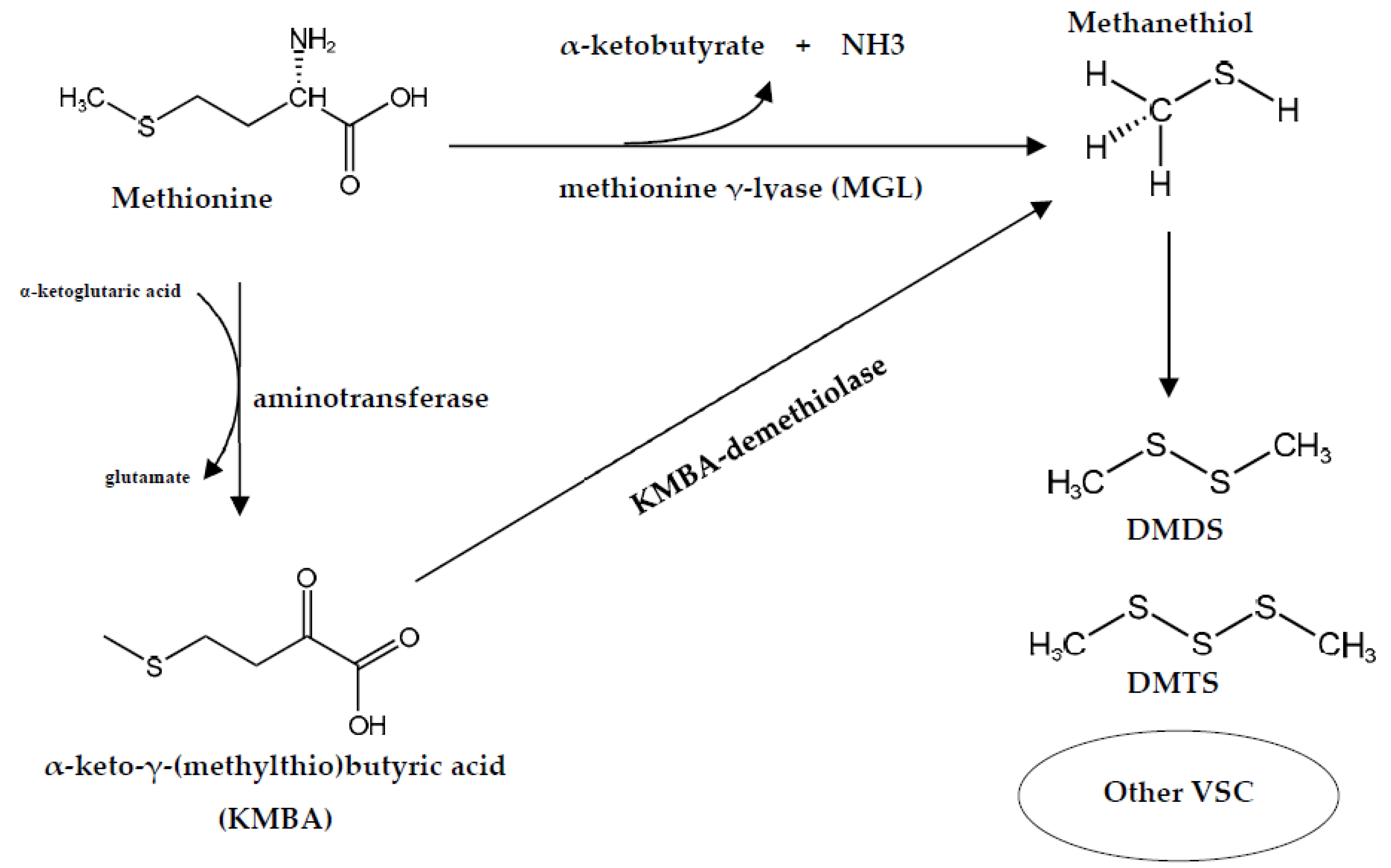
3.1.2. Implication of Arthrobacter spp. in Cheese Flavor

Most flavor development by microorganisms occurs during the cheese-ripening process. The biochemical changes among diverse complex microbial communities involve the conversion of milk fat, carbohydrate, and protein to a very wide range of flavor compounds by three principal metabolic pathways, lipolysis, glycolysis, and proteolysis [68]. These pathways require several different enzymes that can be obtained from milk endogenous enzymes, clotting enzymes, and ripening microbial enzymes [69]. Cheeses produced from raw milk generally exhibit a more complex microbiota than cheeses made of pasteurized milk; as a consequence, a large quantity of aromatic compounds has been found in raw milk cheeses [70]. The degradation of amino acid by microbial enzymes, principally deaminase, transaminase, decarboxylase, lyase, and dehydratase, yields aldehydes, amines, acids, alcohols, and sulfur compounds; while the breakdown of fatty acid will deliver methyl ketones, secondary alcohols, and esters [71,72]. Depending on a variety of microflora presented during the ripening process, the substrates will be metabolized by several pathways to numerous compounds contributing to cheese flavor or off-flavors [68].
Among the microorganisms found on the surface of cheeses, a small number have been evaluated for their ability to produce flavor compounds, especially bacteria belonging to the genus Arthrobacter. Several strains of A. arilaitensis isolated from French smear-ripened cheeses were used to study the production of volatile aroma compounds, and it was found that various volatile compounds, e.g., aldehydes, esters, ketones, alcohols, and sulfur compounds, were produced by two strains, A. arilaitensis Mu107 and A. arilaitensis Po102 [69]. In a comparison study in lab-produced cheese between a typical old-young starter and a defined starter consisting of fungi and bacterial strains, a strain isolated from German Tilsit cheese, A. nicotianae, was the most important in the development of a typical smell—a sulfury volatile flavor [41]. The same study also reported that the sulfury volatile flavor formed by the defined starter on a pilot scale was very weak compared to the old-young smeared cheese. These results are consistent with a previous experiment that grew the strain in a liquid model system with only these two species [73].
A group of volatile sulfur compounds (VSCs) has been recognized as responsible for cheese flavor [74]. Most of these compounds are derivatives of the degradation of the sulfur-carbon bond of methionine to form methanethiol (MTL). The biosynthetic and catabolic pathways of methionine leading to MTL vary among microorganisms depending on their ability to synthesize methionine γ-lyase (MGL) [75]. In the cheese-ripening bacterium B. linens, two volatile sulfur compounds, MTL and α-ketobutyrate, are degraded-methionine products via this enzyme, and later MTL is converted to minor compounds including dimethyl disulfide (DMDS), dimethyl trisulfide (DMTS), and thioesters, while α-ketobutyrate is condensed with active acetaldehyde derived from pyruvate, and metabolized to 2,3-pentanedione (acetyl propionyl) [76]. In addition to B. linens, MGL is also considered to be present in several strains grown on the surface of cheeses such as Micrococcus spp., Staphylococcus spp., and Arthrobacter spp. [77]. A review of the Arthrobacter strain and its ability to produce numerous enzymes have revealed the potential of this genus for the synthesis of MGL [78,79].
The production of MTL is generally associated with the one-step degradation of methionine by MGL; however, it can also originate in a two-step degradation pathway through two other enzymes, aminotransferase and α-keto-γ-(methylthio)-butyric acid demethiolase [80]. A study on the enzymatic pathways of methionine degradation to methanethiol in Actinomycetes bacteria isolated from ripened cheese found that the Arthrobacter sp. strain produced MTL, DMDS, and DMTS from both methionine and α-keto-γ-(methylthio)-butyric acid (KMBA) by enzymatic conversion through one-step and two-step degradation pathways, respectively [77]. The possibility of the enzymatic degradation of methionine via both pathways is shown in Figure 1 In addition, the ability of smear cheese-ripening bacteria to produce VSC from methionine is possibly influenced by cysteine as it appears likely that part of methionine can be converted into cysteine by the transsulfuration pathway through two major enzymes, i.e., cystathionine γ-synthase (CGS) and cystathionine β-lyase (CBL) [81]. CGL has been identified in bacteria as being responsible for hydrogen sulfide (H2S) while CBL releases homocysteine from cystathionine, which can either be involved in the biosynthesis of methionine or hydrolyzed by the CGL resulting in the production of H2S. Consequently, these two enzymes could reduce the amount of methionine, which would reduce VSC production [82]. The reduction of methionine catabolism by cysteine in Arthrobacter spp. could be caused by a reduction in MGL activity due either to a direct effect of high concentrations of cysteine or to other enzymes [74]
Nowadays, many Arthrobacter strains isolated from a variety of cheeses have been discovered. Not only the yellow-pigmented Arthrobacter spp. but a large proportion of grey-white or cream-colored Arthrobacter isolates were also found on the surface of cheeses [41,83,84]. These pale-colored isolates are assumed to be responsible not for the cheese coloration but for the development of flavor and probably play a major role in cheese flavor formation. To achieve a better knowledge of the evolution of cheese flavor, the investigation mentioned that bacteria of the genus Arthrobacter may be required. The utilization in the cheese industry of bacteria belonging to this genus with specific flavor-forming abilities will be later considered as a promising tool to respond to the developing and controlling quality for organoleptic properties of cheese products.

3.2. Association of Arthrobacter spp. Producing Enzymes with Milk and Dairy Products

In the dairy industry, enzymes are widely used for the production and improvement of product quality, as well as for the innovation of new products. The industrial demand for new enzyme sources with different characteristics and low-cost production increases the number of studies related to the isolation and selection of new microorganisms. Among a diversity of microorganisms, psychrophilic bacteria are currently being intensively investigated due to their synthesis of cold-active enzymes. With a high possibility to be applied at low temperatures, especially in the food industry, such as in the processing of milk and dairy products, these enzymes are gaining commercial interest. Although several cold-active enzymes have been discovered, β-galactosidase, commonly known as lactase, which hydrolyzes lactose to glucose and galactose, seems to be a significant food-industrial enzyme because of its potential to degrade lactose for several purposes [85,86].
The principal industrial application of β-galactosidase is in the production of lactose-free milk and other related products—e.g., cheeses, yogurt, butter, and ice cream, for people who are lactose intolerant [87,88]. Generally, enzymatic lactose hydrolysis in milk is conducted under refrigerated conditions for approximately 24 h using commercial β-d-galactosidase extracted from mesophilic yeast Kluyveromyces spp., mainly Kluyveromyces lactis. The major disadvantage of the application of this enzyme in the dairy industry is its low activity under process conditions since it is optimally active at above 30 °C depending on the species of Kluyveromyces used as an enzyme producer [89,90]. Another significant application of β-d-galactosidase is the synthesis of galacto-oligosaccharides (GOS) from lactose. GOS are non-digestible oligosaccharides comprising 2 to 20 molecules of galactose and 1 molecule of glucose [91]. Recent studies ascribed several health benefits to these oligosaccharides [92]. GOS display functional prebiotic properties due to their stimulation of the proliferation of intestinal lactic acid bacteria and bifidobacteria [88]. GOS stimulates Bifidobacterium proliferation in the colon, which suppresses the activity of decomposing bacteria by antagonistic effect and diminishes the formation of toxic metabolites [93,94]. GOS are commercially available as prebiotics, with a low calorific value, and are reported to be capable of promoting satiety and reducing food intake [95].
The production of GOS from lactose through microbial β-galactosidases has been achieved using various enzymes deriving from mesophiles, thermophiles, and hyperthermophiles. By contrast, a small number of syntheses of GOS by the cold-active β-galactosidases from psychrophilic and psychrotolerant microorganisms have been studied [96]. Bioprospecting of microorganisms living in different cold environments led to the identification of cold-active β-galactosidases from many microbial species such as Guehomyces, Thalassospira, Erwinia [97,98,99].
Psychrophiles and psychrotolerant bacteria belonging to the genus Arthrobacter, especially strains isolated from extremely cold environments, have shown an ability to produce β-galactosidase [100]. Several Antarctic Arthrobacter strains, e.g., Arthrobacter sp. 20B, Arthrobacter sp. 32cB, Arthrobacter sp. ON14 and Arthrobacter sp. C2-2, Arthrobacter psychrolactophilus strain F2, and other unclassified Arthrobacter spp. were found to produce β-galactosidases, which were active at an optimum temperature range of 0–50 °C [88,101,102,103,104]. As a result of these discoveries, cold-active β-galactosidases produced by A. psychrolactophilus strain F2 are likely suitable enzymes to use under low-temperature conditions due to their activity being highest at 0 °C while their optimum temperature and pH were 10 °C and 8.0, respectively [105].
In addition to GOS, based on the ability of β-galactosidases to promote transglycosylation, isomerization, and transgalactosylation, this enzyme has been also used to produce hetero-oligosaccharides (HOS) by transferring the galactosyl moiety to sugars instead of lactose or its hydrolysis products, glucose, and galactose [87]. Lactulose, a well-known hetero-oligosaccharide, is a non-absorbable ketose disaccharide with prebiotic properties; therefore, like GOS, it is applied as a functional ingredient in the food industry and for pharmaceutical purposes [106]. Some strains of the genus Arthrobacter isolated from various diverse origins, such as Arthrobacter sp. from a fruit garden soil sample from China, Arthrobacter sp. 32cB from an Antarctic soil sample, and Arthrobacter siccitolerans sp. nov. from a dry soil sample from Spain, produce β-galactosidases which stimulates the hydrolysis of lactulose [106,107,108].
Technologically, lactose can easily be crystallized, which limits its applications in certain processes in the dairy industry. The application of β-galactosidases adds additional profitability to lactose-free or low-lactose dairy products as the hydrolysis of lactose will improve the solubility and digestibility of the dairy products [109]. It has been reported that using β-galactosidases to produce lactose-hydrolyzed yogurt can enhance lactic acid production and decrease whey separation as well as improve yogurt’s flavor, body, and texture [110]. In frozen dairy products, lactose hydrolysis by these enzymes improves the smoothness and scoop ability of the products due to the decrease in the freezing point [111]. The application of β-galactosidases in cheese production was found to reduce the ripening time and enrich the organoleptic properties of cheese. Therefore, this seems to be an effective element for reducing manufacturing costs as cheese ripening usually requires a long period of from six months to two years depending on the variety of cheeses [109,112]. In addition, β-galactosidases are considered a tool for adding value to by-products obtained from cheese production processes. Cheese whey is a by-product of the filtration process and it is recognized as a source of valuable molecules such as protein, lactose, and minerals; thus, it has been used as a substrate for the enzymatic production of GOS through the hydrolysis by β-galactosidases [113].
With the demand for appropriate and innovative technologies in the production of milk and milk-derived products as well as the by-product valorization, microbial β-galactosidases have been of increasing interest since they are very useful as catalysts that present several remarkable applications in the dairy industry. Based on the data obtained from previous studies about its thermostability, and thermoacidophilic and thermoresistant properties, the genus Arthrobacter promisingly provides advantages for use as a potential producer of β-galactosidases to fulfill commercial requirements as its enzymes allow dairy products to be treated under mild conditions.

3.3. Effect of Arthrobacter spp. on Nutritional Components in Milk and Dairy Products

Protein, fat, and carbohydrates, represent significant macronutrients in milk, accounting for 3–5, 4–6, and 4–5% of the total milk content, respectively [114]. Nevertheless, genetic, environmental, physiological, and handling variables are generally regarded as factors affecting milk composition. The contamination of Arthrobacter in milk and dairy products is possibly encountered either earlier in raw milk or later in processed products and can cause negative or positive effects on the quality of milk and its products in terms of their nutrition. According to genome sequencing studies, certain strains of bacteria in the genus Arthrobacter exhibit the genes encoding proteolytic, lipolytic, and glycolytic pathways [115,116,117]. These revelations confirm the results of several studies related to such enzyme-producing Arthrobacter bacteria [118,119]. Proteolysis, lipolysis, and glycolysis are normally considered the important elements of biochemical change in milk during fermentation leading to the nutritional value of final products.

3.3.1. Proteolysis

Proteolysis is a hydrolysis reaction of peptide bonds in which proteins break down into smaller peptides and free amino acids. It is responsible for the metabolism of casein present in milk by the milk endogenous proteinase and other proteolytic enzymes of microorganisms present in milk; thus, leads to several biochemical reactions affecting nutritional and non-nutritional compounds—e.g., branched-chain and aromatic amino acids and sulfur-containing compounds. The branched-chain amino acids consist of leucine, isoleucine, and valine, whereas the aromatic amino acids include tryptophan, phenylalanine, and tyrosine [72]. The breakdown of amino acids via different pathways, namely decarboxylation and deamination, results in several compounds. Branched-amino acids can be decarboxylated using aminotransferases into amines, whereas dehydrogenases in deamination will convert these amino acids into carboxylic acids and ammonia [71]. Aromatic amino acids can also undergo decarboxylation and result in α-keto acids such as tryptophan to indole-3-pyruvate, tyrosine to hydroxybenzaldehyde, and phenylalanine to benzaldehyde [120].
Protein degradation generally occurs through extracellular enzymes synthesized by microorganisms during their growth or through intracellular enzymes produced up-on lysis. The synthesized proteinases expectedly involve casein degradation while those produced upon lysis tend to be peptidases and probably do not affect the degradation of casein but rather the breaking down of peptides originated by hydrolysis of proteinases [121]. By genome sequencing techniques, certain strains of Arthrobacter bacteria—e.g., A. arilaitensis, A. aurescens, and A. chlorophenolicus, demonstrate a number of genes encoding putative proteins with proteolytic functions [115,122].
The generation of peptides during microbial fermentation possibly produces the biologically active substances derived from milk protein. Milk-derived bioactive peptides are usually encrypted and inactive forms within the primary structure of milk protein and they are activated by proteolysis of casein namely α-, β-, γ- and κ-casein [123]. Numerous health-beneficial effects have been attributed to milk-derived bioactive peptides such as anti-hypertensive, hypolipidemic, anti-inflammatory, anti-oxidative, anti-microbial, and anti-osteoporotic effects [124,125,126]. In regards to health-promoting effects, milk-derived bioactive peptides are supposedly considered potential ingredients of functional foods.

3.3.2. Lipolysis

Milk fat is primarily composed of triglycerides with phospholipids, cholesterol, and other lipids present at low concentrations. Milk triglycerides can be degraded through lipolytic pathways by endogenous enzymes in milk and microbial enzymes secreted during fermentation. The lipolytic enzymes can hydrolyze substrates to generate different forms of fatty acids, i.e., short- and intermediate-chain and free fatty acids [71]. The formation of these lipid molecules through lipolysis mostly reflects in the ester bonds between triglycerides and fatty acids cleaved by the lipases [127]. Fatty acids constitute a significant energy source for the growth of aerobic microorganisms. Some Arthrobacter including strains isolated from dairy products encode many proteins with putative lipase or esterase activity [115].
In the human body, free fatty acids (FFAs) serve in the first instance as an essential energy substrate and have a role in biological processes. FFAs can affect the gene expression of macrophages, adipocytes, or endothelial cells. In addition, FFAs can modulate the production of chemokines and cytokines and the expression of genes coding for adhesion molecules, and give rise to pro-inflammatory and inflammation-pro-resolving lipid-derived species [128]. High concentrations of FFAs are associated with some physiological conditions, for example, insulin resistance, fatty liver disease, atherosclerosis, and myocardial dysfunction [129]. Unlike FFAs, short-chain fatty acids (SCFAs) and medium-chain fatty acids (MCFAs) have many health benefits. The bioactivities of SCFAs, especially three major components, i.e., acetate, propionate, and butyrate, have been widely investigated. SCFAs demonstrate anti-inflammatory, immunoregulatory, anti-obesity, anti-diabetes, anticancer, cardiovascular protective, hepatoprotective, and neuroprotective effects [130]. For the regulation of MCFAs, these molecules modulate glucose and lipid metabolism. The unique transport and rapid metabolism of MCFAs provide additional health benefits compared to long-chain fatty acids (LCFAs); therefore, the use of MFAs for treating metabolic and neurological disorders has been of increasing interest [131].

3.3.3. Glycolysis

Milk contains carbohydrates mainly in lactose form and trace amounts of monosaccharides and oligosaccharides. Lactose is a reducing disaccharide consisting of glucose and galactose. The metabolism of lactose to lactate is regularly metabolized by the glycolytic pathways, which are commonly associated with the fermentative metabolism in microorganisms. During fermentation, enzymes responsible for breaking down carbohydrates, which are synthesized by several bacteria contaminated in milk, hydrolyze lactose into its monomers, of which glucose will be further degraded. The principal products of lactose metabolism are L- or D-lactate, which are then oxidized into acetate and CO2 while some microbial strains create other products such as alcohol [132].
Cheese ripening is a type of milk fermentation that is probably most relevant to bacteria belonging to the genus Arthrobacter. Generally, the process of glycolysis in the degradation of milk carbohydrates to lactate seems to be responsible for lactic acid bacteria since they are the dominant bacterial species found at the beginning of the ripening process [72]. Arthrobacter strains that are frequently isolated from ripened cheeses may partly display a role in converting lactose to lactate due to their occurrence in the middle and late stages of the ripening process. Cheese-isolated Arthrobacter strains are aerobic respiratory bacteria and thus produced most of their ATP by oxidative phosphorylation. According to the presence of lactate at the beginning of ripening and quinone-dependent lactate dehydrogenases, glycolysis by ripened-cheese Arthrobacter is importantly related to the utilization of lactate as a carbon source [115]. However, several short-chain components—i.e., acetate, acetoin, diacetyl, ethanol, and acetaldehyde—are produced from an intermediate in lactose metabolism, pyruvate, as it is a precursor of these compounds [120]. The identification of gene encoding for enzymes required for the catabolism of carbohydrates sufficiently confirms the ability to utilize lactose as a substrate for glycolytic pathways [115,117].
Lactose-hydrolyzed milk (low-lactose and lactose-free milk) has been used for the preparation of flavored milk, cheese, yogurt, and other dairy products that serve as an important alternative for people who are lactose intolerant. The hydrolysis of lactose in milk for food processing also prevents lactose crystallization in frozen and condensed milk products; thus, it helps in improving the quality of the products [133]. Moreover, the use of hydrolyzed milk in yogurt and cheese manufacture accelerates the acidification process, because lactose hydrolysis is normally the rate-limiting step of the process, which reduces the set time of yogurt and accelerates the development of structure and flavor in cheese [134].

4. Conclusions

Milk is a rich source of many essential nutrients, including protein, fats, carbohydrates, vitamins, minerals, and essential amino acids; as a result, it is quite a suitable medium for bacterial growth and can, therefore, serve as a source of bacterial contamination. Milk typically contains a variety of bacteria, of which some are beneficial such as those preserving milk through products of fermentation, e.g., cheese and yogurt. The genus Arthrobacter is among the major groups of microorganisms commonly found as a contaminant in milk and dairy products as well as being applied in the dairy industry. Almost all Arthrobacter spp. associated with fermented dairy products, especially a variety of cheeses, are considered microflora that enter milk or dairy products from the environment and milking equipment. They are mostly responsible for the coloration and flavor formation of cheeses, whilst a small number of Arthrobacter spp. isolated from extreme environments are employed as producers of β-galactosidase used for lactose hydrolysis in order to produce GOS and other lactose derivatives. Because of these beneficial aspects derived from bacteria belonging to the genus Arthrobacter including pigmentation, flavor formation, and enzyme production, the genus Arthrobacter is a promising bacterial genus for use in the production of products such as cheeses low-lactose and lactose-free dairy products, and functional dairy products (Figure 2). However, a better understanding of its roles related to these aspects is still required to enlarge the available benefits. Meanwhile, the isolation of new strains for use in the production of pigments, flavor compounds, and enzymes will possibly meet the demands of the dairy industry and beyond.

Author Contributions

Conceptualization, N.S. and L.D.; resources, N.S. and S.L.; writing—original draft preparation, N.S.; writing—review and editing, N.S., S.L. and L.D.; supervision, L.D. All authors have read and agreed to the published version of the manuscript.

Funding

This research received no external funding.

Data Availability Statement

Not applicable.

Acknowledgments

The authors thank Piyada Sukdee and Mookwadee Paulette Dupuis for the preparation of Figure 2.

Conflicts of Interest

The authors declare no conflict of interest.

References

  1. Guha, S.; Sharma, H.; Deshwal, G.K.; Rao, P.S. A comprehensive review on bioactive peptides derived from milk and milk products of minor dairy species. Food Prod. Process Nutr. 2021, 3, 2. [Google Scholar] [CrossRef]
  2. Melini, F.; Melini, V.; Luziatelli, F.; Ruzzi, M. Raw and heat-treated milk: From public health risks to nutritional quality. Beverages 2017, 3, 54. [Google Scholar] [CrossRef] [Green Version]
  3. Fusco, V.; Chieffi, D.; Fanelli, F.; Logrieco, A.F.; Cho, G.-S.; Kabisch, J.; Böhnlein, C.; Franz, C.M.A.P. Microbial quality and safety of milk and milk products in the 21st century. Compr. Rev. Food Sci. Food Saf. 2020, 19, 2013–2049. [Google Scholar] [CrossRef] [PubMed]
  4. Issa, A.T.; Tahergorabi, R. Milk bacteria and gastrointestinal tract: Microbial composition of milk. In Dietary Interventions in Gastrointestinal Diseases; Watson, R.R., Preedy, V.R., Eds.; Academic Press: Cambridge, MA, USA, 2019; pp. 265–275. [Google Scholar] [CrossRef]
  5. Shi, C.; Maktabdar, M. Lactic acid bacteria as biopreservation against spoilage molds in dairy products-A review. Front. Microbiol. 2022, 12, 4283. [Google Scholar] [CrossRef]
  6. Oikonomou, G.; Addis, M.F.; Chassard, C.; Nader-Macias, M.E.F.; Grant, I.; Delbès, C.; Bogni, C.I.; Le Loir, Y.; Even, S. Milk microbiota: What are we exactly talking about? Front. Microbiol. 2020, 11, 60. [Google Scholar] [CrossRef] [Green Version]
  7. Sutthiwong, N.; Fouillaud, M.; Valla, A.; Caro, Y.; Dufossé, L. Bacteria belonging to the extremely versatile genus Arthrobacter as novel source of natural pigments with extended hue range. Food Res. Int. 2014, 65, 156–162. [Google Scholar] [CrossRef]
  8. Comi, G.; Carlo, C. Arthrobacter spp. in milk and milk products. In Reference Module in Food Science; Smithers, G., Ed.; Elsevier: Amsterdam, The Netherlands, 2016; pp. 1–7. [Google Scholar] [CrossRef]
  9. Busse, H.-J.; Wieser, M. The Genus Arthrobacter. In The Prokaryotes; Rosenberg, E., DeLong, E.F., Lory, S., Stackebrandt, E., Thompson, F., Eds.; Springer: Berlin/Heidelberg, Germany, 2014; pp. 105–132. [Google Scholar] [CrossRef]
  10. Roy, P.; Kumar, A. Arthrobacter. In Beneficial Microbes in Agro-Ecology. Bacteria and Fungi; Amaresan, N., Kumar, M.S., Annapurna, K., Kumar, K., Sankaranarayanan, A., Eds.; Academic Press: Cambridge, MA, USA, 2020; pp. 3–11. [Google Scholar] [CrossRef]
  11. List of Prokaryotic Names with Standing in Nomenclature-Genus Arthrobacter. Available online: http://www/bacterio/cict.fr/a/arthrobacter.html (accessed on 2 February 2023).
  12. Irlinger, F.; Bimet, F.; Delettre, J.; Lefèvre, M.; Grimont, P.A.D. Arthrobacter bergerei sp. nov. and Arthrobacter arilaitensis sp. nov., novel coryneform species isolated from the surfaces of cheeses. Int. J. Syst. Evol. Microbiol. 2005, 55, 457–462. [Google Scholar] [CrossRef] [Green Version]
  13. Yan, H.; Du, W.; Ji, S.; Guo, C.; Zhang, Y.; Wang, Y.; Cao, Z.; Li, S. Bacterial composition and interactions in raw milk and teat skin of dairy cows. Ferment 2022, 8, 235. [Google Scholar] [CrossRef]
  14. Owusu-Kwarteng, J.; Akabanda, F.; Agyei, D.; Jespersen, L. Microbial safety of milk production and fermented dairy products in Africa. Microorganisms 2020, 8, 752. [Google Scholar] [CrossRef]
  15. Flegler, A.; Runzheimer, K.; Kombeitz, V.; Mänz, A.T.; von Heidler Heilborn, D.; Etzbach, L.; Schieber, A.; Hölzl, G.; Hüttel, B.; Woehle, C.; et al. Arthrobacter bussei sp. nov., a pink-coloured organism isolated from cheese made of cow’s milk. Int. J. Syst. Evol. Microbiol. 2020, 70, 3027–3036. [Google Scholar] [CrossRef]
  16. Lauková, A.; Micenková, L.; Grešáková, Ľ.; Maďarová, M.; Simonová, M.P.; Focková, V.; Ščerbová, J. Microbiome associated with Slovak raw goat milk, trace minerals, and vitamin E content. Int. J. Food Sci. 2022, 30, 4595473. [Google Scholar] [CrossRef] [PubMed]
  17. Cao, H.; Yan, Y.; Wang, L.; Dong, L.; Pang, X.; Tang, S.; Li, A.; Xiang, A.; Zhang, L.; Zheng, B. High-throughput sequencing reveals bacterial diversity in raw milk production environment and production chain in Tangshan city of China. Food Sci. Anim. Resour. 2021, 41, 452. [Google Scholar] [CrossRef] [PubMed]
  18. Frétin, M.; Martin, B.; Rifa, E.; Isabelle, V.-M.; Pomiès, D.; Ferlay, A.; Montel, M.C.; Delbès, C. Bacterial community assembly from cow teat skin to ripened cheeses is influenced by grazing systems. Sci. Rep. 2018, 8, 200. [Google Scholar] [CrossRef] [PubMed] [Green Version]
  19. Hahne, J.; Lipski, A. Growth interferences between bacterial strains from raw cow’s milk and their impact on growth of Listeria monocytogenes and Staphylococcus aureus. J. Appl. Microbiol. 2021, 131, 2019–2032. [Google Scholar] [CrossRef]
  20. Papademas, P.; Kamilari, E.; Aspri, M.; Anagnostopoulos, A.D.; Mousikos, P.; Kamilaris, A.; Tsaltas, D. Investigation of donkey milk bacterial diversity by 16S rDNA high-throughput sequencing on a Cyprus donkey farm. J. Dairy Sci. 2021, 104, 167–178. [Google Scholar] [CrossRef] [PubMed]
  21. Rodríguez, M.F.; Gomez, A.P.; Parra, C.M. Molecular and proteomic identification of Arthrobacter gandavensis isolated from cows with subclinical mastitis in a dairy farm. Malays. J. Microbiol. 2022, 18, 309–314. [Google Scholar] [CrossRef]
  22. Choi, J.; Lee, S.I.; Rackerby, B.; Goddik, L.; Frojen, R.; Ha, S.-D.; Kim, J.H.; Park, S.H. Microbial communities of a variety of cheeses and comparison between core and rind region of cheeses. J. Dairy Sci. 2020, 103, 4026–4042. [Google Scholar] [CrossRef]
  23. Gobbetti, M.; Rizzello, C.G. Arthrobacter. In Encyclopedia of Food Microbiology, 2nd ed.; Batt, C.A., Tortorello, M.L., Eds.; Academic Press: Cambridge, MA, USA, 2014; pp. 69–76. [Google Scholar] [CrossRef]
  24. Chauhan, A.; Pathak, A.; Jaswal, R.; Edwards, B., III; Chappell, D.; Ball, C.; Garcia-Sillas, R.; Stothard, P.; Seaman, J. Physiological and comparative genomic analysis of Arthrobacter sp. SRS-W-1-2016 provides insights on niche adaptation for survival in uraniferous soils. Genes 2018, 9, 31. [Google Scholar] [CrossRef] [Green Version]
  25. Deeth, H.C. Heat treatment of milk: Thermization and pasteurization. In Reference Module in Food Science; Elsevier: Amsterdam, The Netherlands, 2020. [Google Scholar] [CrossRef]
  26. De Souza, P.V.; Zaffari Grecellé, C.B.; Barreto, F.; Ramírez-Castrillon, M.; Valente, M.; da Costa, M. Bacteria and yeasts associated to Colonial cheese production chain and assessment of their hydrolytic potential. Braz. J. Food Technol. 2021, 24, 1–10. [Google Scholar] [CrossRef]
  27. Penland, M.; Falentin, H.; Parayre, S.; Pawtowski, A.; Maillard, M.-B.; Thierry, A.; Mounier, J.; Coton, M.; Deutsch, S.-M. Linking Pélardon artisanal goat cheese microbial communities to aroma compounds during cheese-making and ripening. Int. J. Food Microbiol. 2021, 345, 109130. [Google Scholar] [CrossRef]
  28. Hanlon, M.; Choi, J.; Goddik, L.; Park, S.H. Microbial and chemical composition of Cheddar cheese supplemented with prebiotics from pasteurized milk to aging. J. Dairy Sci. 2022, 105, 2058–2068. [Google Scholar] [CrossRef] [PubMed]
  29. Ritschard, J.S.; Amato, L.; Kumar, Y.; Müller, B.; Meile, L.; Schuppler, M. The role of the surface smear microbiome in the development of defective smear on surface-ripened red-smear cheese. AIMS Microbiol. 2018, 4, 622–641. [Google Scholar] [CrossRef] [PubMed]
  30. Cosetta, C.M.; Wolfe, B.E. Deconstructing and reconstructing cheese rind microbiomes for experiments in microbial ecology and evolution. Curr. Protoc. Microbiol. 2020, 56, 1–30. [Google Scholar] [CrossRef] [PubMed]
  31. Reuben, R.C.; Langer, D.; Eisenhauer, N.; Jurburg, S.D. Universal drivers of cheese microbiomes. iScience 2023, 26, 105744. [Google Scholar] [CrossRef]
  32. Monnet, C.; Loux, V.; Gibrat, J.F.; Spinnler, E.; Barbe, V.; Vacherie, B.; Gavory, F.; Gourbeyre, E.; Siguier, P.; Chandler, M.; et al. The arthrobacter arilaitensis Re117 genome sequence reveals its genetic adaptation to the surface of cheese. PLoS ONE 2010, 5, e15489. [Google Scholar] [CrossRef] [Green Version]
  33. Niccum, B.A.; Kastman, E.K.; Kfoury, N.; Robbat Jr., A.; Wolfe, B.E. Strain-level diversity impacts cheese rind microbiome assembly and function. mSystems 2020, 5, e00149-20. [Google Scholar] [CrossRef]
  34. Sutthiwong, N.; Fouillaud, M.; Dufossé, L. The influence of pH, NaCl, and the deacidifying yeasts Debaryomyces hansenii and Kluyveromyces marxianus on the production of pigments by the cheese-ripening bacteria Arthrobacter arilaitensis. Foods 2018, 7, 190. [Google Scholar] [CrossRef] [Green Version]
  35. Kaczyński, Ł.K.; Cais-Sokolińska, D.; Bielska, P.; Teichert, J.; Biegalski, J.; Yiğit, A.; Chudy, S. The influence of the texture and color of goat’s salad cheese on the emotional reactions of consumers compared to cow’s milk cheese and Feta cheese. Eur. Food Res. Technol. 2023, 1–16. [Google Scholar] [CrossRef]
  36. Speight, K.C.; Schiano, A.N.; Harwood, W.S.; Drake, M.A. Consumer insights on prepackaged cheddar cheese shreds using focus groups, conjoint analysis, and qualitative multivariate analysis. J. Dairy Sci. 2019, 102, 6971–6986. [Google Scholar] [CrossRef]
  37. Giuffrida, D.; Monnet, C.; Laurent, F.; Cacciola, F.; Oteri, M.; Le Piver, M.; Caro, Y.; Donato, P.; Mondello, L.; Roueyre, D.; et al. Carotenoids from the ripening bacterium Brevibacterium linens impart color to the rind of the French cheese, Fourme de Montbrison (PDO). Nat. Prod. Res. 2020, 34, 10–15. [Google Scholar] [CrossRef]
  38. Possas, A.; Bonilla-Luque, O.M.; Valero, A. From cheese-making to consumption: Exploring the microbial safety of cheeses through predictive microbiology models. Foods 2021, 10, 355. [Google Scholar] [CrossRef] [PubMed]
  39. Mayo, B.; Rodríguez, J.; Vázquez, L.; Flórez, A.B. Microbial interactions within the cheese ecosystem and their application to improve quality and safety. Foods 2021, 10, 602. [Google Scholar] [CrossRef] [PubMed]
  40. Gallegos-Acevedo, M.-A.; Chávez-Martíneza, M.; Corral-Luna, A.; Rentería-Monterrubiom, A.-L.; Burrola-Barraza, E.; Lechuga-Valles, R.; Dominguez-Vuveros, J.; Castillo-González, A.-R.; Sánchez-Vega, R. Microbial characterization and diversity of artisanal Ranchero cheese with emphasis in Lactococcus strains. Food Sci. Technol. 2019, 39, 143–148. [Google Scholar] [CrossRef] [Green Version]
  41. Bockelmann, W. Development of defined surface starter cultures for the ripening of smear cheeses. Int. Dairy J. 2002, 12, 123–131. [Google Scholar] [CrossRef]
  42. Comi, G.; Cantoni, C. Psychrotrophic Bacteria: Arthrobacter spp. In Encyclopedia of Dairy Sciences, 2nd ed.; Fuquay, J.W., Ed.; Academic Press: Cambridge, MA, USA, 2011; pp. 372–378. [Google Scholar] [CrossRef]
  43. Jonnala, B.R.Y.; McSweeney, P.L.H.; Cotter, P.D.; Zhong, S.; Sheehan, J.J.; Kopec, R.E. Comparison of the carotenoid profiles of commonly consumed smear-ripened cheeses. Lebensm. Wiss. Technol. 2021, 135, 110241. [Google Scholar] [CrossRef]
  44. López, G.D.; Álvarez-Rivera, G.; Carazzone, C.; Ibáñez, E.; Leidy, C.; Cifuentes, A. Bacterial carotenoids: Extraction, characterization, and applications. Crit. Rev. Anal. Chem. 2021, 16, 1–24. [Google Scholar] [CrossRef]
  45. Meléndez-Martínez, A.J.; Mandić, A.I.; Bantis, F.; Böhm, V.; Borge, G.I.A.; Brnčić, M.; Bysted, A.; Cano, M.P.; Dias, M.G.; Elgersma, A.; et al. A comprehensive review on carotenoids in foods and feeds: Status quo, applications, patents, and research needs. Crit. Rev. Food Sci. Nutr. 2022, 62, 1999–2049. [Google Scholar] [CrossRef]
  46. Giuffrida, D.; Sutthiwong, S.; Dugo, P.; Donato, P.; Cacciola, F.; Girard-Valenciennes, E.; Le Mao, Y.; Monnet, C.; Fouillaud, M.; Caro, Y.; et al. Characterisation of the C50 carotenoids produced by strains of the cheese-ripening bacterium Arthrobacter arilaitensis. Int. Dairy J. 2016, 55, 10–16. [Google Scholar] [CrossRef] [Green Version]
  47. Vila, E.; Hornero-Méndez, D.; Azziz, G.; Lareo, C.; Saravia, V. Carotenoids from heterotrophic bacteria isolated from Fildes Peninsula, King George Island, Antarctica. Biotechnol. Rep. 2019, 21, e00306. [Google Scholar] [CrossRef]
  48. Yabuzaki, J. Carotenoids Database: Structures, chemical fingerprints and distribution among organisms. Database 2017, 2017, bax004. [Google Scholar] [CrossRef] [Green Version]
  49. Flegler, A.; Lipski, A. The C50 carotenoid bacterioruberin regulates membrane fluidity in pink-pigmented Arthrobacter species. Arch. Microbiol. 2022, 204, 70. [Google Scholar] [CrossRef] [PubMed]
  50. Siziya, I.N.; Hwang, C.Y.; Seo, M.-J. Antioxidant potential and capacity of microorganism-sourced C30 carotenoids—A review. Antioxidants 2022, 11, 1963. [Google Scholar] [CrossRef] [PubMed]
  51. Leclercq-Perlat, M.-N.; Sicard, M.; Perrot, N.; Trelea, I.C.; Picque, D.; Corrieu, G. Temperature and relative humidity influence the ripening descriptors of Camembert-type cheeses throughout ripening. J. Dairy Sci. 2015, 98, 1325–1335. [Google Scholar] [CrossRef] [PubMed] [Green Version]
  52. Sharma, P.; Segat, A.; Kelly, A.L.; Sheehan, J.J. Colorants in cheese manufacture: Production, chemistry, interactions, and regulation. Compr. Rev. Food Sci. Food Saf. 2020, 19, 1220–1242. [Google Scholar] [CrossRef] [Green Version]
  53. Mukhia, S.; Khatri, A.; Acharya, V.; Kumar, R. Comparative genomics and molecular adaptational analysis of Arthrobacter from Sikkim Himalaya provided insights into its survivability under multiple high-altitude stress. Genomics 2021, 113, 151–158. [Google Scholar] [CrossRef]
  54. Seel, W.; Baust, D.; Sons, D.; Albers, M.; Etzbach, L.; Fuss, J.; Lipski, D. Carotenoids are used as regulators for membrane fluidity by Staphylococcus xylosus. Sci. Rep. 2020, 10, 330. [Google Scholar] [CrossRef] [Green Version]
  55. Guo, R.; Liu, T.; Guo, C.; Chen, G.; Fan, J.; Zhang, Q. Carotenoid biosynthesis is associated with low-temperature adaptation in Rhodosporidium kratochvilovae. BMC Microbiol. 2022, 22, 319. [Google Scholar] [CrossRef]
  56. Pérez-Gálvez, A.; Viera, I.; Roca, M. Carotenoids and chlorophylls as antioxidants. Antioxidants 2020, 9, 505. [Google Scholar] [CrossRef]
  57. Caferri, R.; Guardini, Z.; Bassi, R.; Dall′Osto, L. Assessing photoprotective functions of carotenoids in photosynthetic systems of plants and green algae. In Methods in Enzymology; Wurtzel, E.T., Ed.; Academic Press: Cambridge, MA, USA, 2022; Volume 674, pp. 53–84. [Google Scholar] [CrossRef]
  58. Chia, G.W.N.; Seviour, T.; Kjelleberg, S.; Hinks, J. Carotenoids improve bacterial tolerance towards biobutanol through membrane stabilization. Environ. Sci. Nano. 2021, 8, 328–341. [Google Scholar] [CrossRef]
  59. Kumar, S.V.; Taylor, G.; Hasim, S.; Collier, C.P.; Farmer, A.T.; Campagna, S.R.; Bible, A.N.; Doktycz, M.J.; Morrell-Falvey, J. Loss of carotenoids from membranes of Pantoea sp. YR343 results in altered lipid composition and changes in membrane biophysical properties. Biochim. Biophys. Acta Biomembr. 2019, 1861, 1338–1345. [Google Scholar] [CrossRef]
  60. Manrique-Moreno, M.; Jemioła-Rzemińska, M.; Múnera-Jaramillo, J.; López, G.-D.; Suesca, E.; Leidy, C.; Strzałka, K. Staphylococcus aureus carotenoids modulate the thermotropic phase behavior of model systems that mimic its membrane composition. Membranes 2022, 12, 945. [Google Scholar] [CrossRef] [PubMed]
  61. Ram, S.; Mitra, M.; Shah, F.; Tirkey, S.R.; Mishra, S. Bacteria as an alternate biofactory for carotenoid production: A review of its applications, opportunities and challenges. J. Funct. Foods 2020, 67, 103867. [Google Scholar] [CrossRef]
  62. Mostofian, B.; Johnson, Q.R.; Smith, J.C.; Cheng, X. Carotenoids promote lateral packing and condensation of lipid membranes. Phys. Chem. Chem. Phys. 2020, 22, 12281–12293. [Google Scholar] [CrossRef] [PubMed]
  63. Daly, D.; McSweeney, P.; Sheehan, J. Pink discolouration defect in commercial cheese: A review. Dairy Sci. Technol. 2012, 92, 439–453. [Google Scholar] [CrossRef] [Green Version]
  64. Jonnala, B.R.Y.; McSweeney, P.L.H.; Cotter, P.D.; Sheehan, J.J. Recreating pink defect in cheese with different strains of Thermus bacteria. Int. J. Dairy Technol. 2021, 74, 700–708. [Google Scholar] [CrossRef]
  65. Quigley, L.; O’Sullivan, D.J.; Daly, D.; O’Sullivan, O.; Burdikova, Z.; Vana, R.; Beresford, T.P.; Paul Ross, R.; Fitzgerald, G.F.; McSweeney, P.L.H.; et al. Thermus and the pink discoloration defect in cheese. mSystems 2016, 1, e00023-16. [Google Scholar] [CrossRef] [Green Version]
  66. Cleary, J.L.; Kolachina, S.; Wolfe, B.E.; Sanchez, L.M. Coproporphyrin III produced by the bacterium Glutamicibacter arilaitensis binds zinc and is upregulated by fungi in cheese rinds. mSystems 2018, 3, e00036-18. [Google Scholar] [CrossRef] [Green Version]
  67. Sutthiwong, N.; Sukdee, P.; Lekhavat, S.; Dufossé, L. Identification of red pigments produced by cheese-ripening bacterial strains of Glutamicibacter arilaitensis using HPLC. Dairy 2021, 2, 396–409. [Google Scholar] [CrossRef]
  68. Blaya, J.; Barzideh, Z.; LaPointe, G. Symposium review: Interaction of starter cultures and nonstarter lactic acid bacteria in the cheese environment. J. Dairy Sci. 2018, 101, 3611–3629. [Google Scholar] [CrossRef]
  69. Cao, W.; Aubert, J.; Maillard, M.-B.; Boissel, F.; Leduc, A.; Thomas, J.-L.; Deutsch, S.-M.; Camier, B.; Kerjouh, A.; Parayre, S.; et al. Fine-tuning of process parameters modulates specific metabolic bacterial activities and aroma Compound production in semi-hard cheese. J. Agric. Food Chem. 2021, 69, 8511–8529. [Google Scholar] [CrossRef]
  70. Coelho, M.C.; Malcata, F.X.; Silva, C.C.G. Lactic acid bacteria in raw-milk cheeses: From starter cultures to probiotic functions. Foods 2022, 11, 2276. [Google Scholar] [CrossRef] [PubMed]
  71. Zheng, X.; Shi, X.; Wang, B. A review on the general cheese processing technology, flavor biochemical pathways and the influence of yeasts in cheese. Front. Microbiol. 2021, 12, 703284. [Google Scholar] [CrossRef] [PubMed]
  72. Khattab, A.R.; Guirguis, H.A.; Tawfik, S.T.; Farag, M.A. Cheese ripening: A review on modern technologies towards flavor enhancement, process acceleration and improved quality assessment. Trends. Food Sci. Technol. 2019, 88, 343–360. [Google Scholar] [CrossRef]
  73. Bockelmann, W.; Hoppe-Seyler, T. The surface flora of bacteria smear-ripened cheese from cow’s and goat’s milk. Int. Dairy J. 2001, 11, 307–314. [Google Scholar] [CrossRef]
  74. Martín, I.; Rodríguez, A.; García, C.; Córdoba, J.J. Evolution of volatile compounds during ripening and final sensory changes of traditional raw ewe’s milk cheese “Torta del Casar” maturated with selected protective lactic acid bacteria. Foods 2022, 11, 2658. [Google Scholar] [CrossRef]
  75. Peralta, G.H.; Wolf, I.V.; Perotti, M.C.; Bergamini, C.V.; Hynes, E.R. Formation of volatile compounds, peptidolysis and carbohydrate fermentation by mesophilic lactobacilli and streptoccocci cultures in a cheese extract. Dairy Sci. Technol. 2016, 96, 603–621. [Google Scholar] [CrossRef] [Green Version]
  76. Lu, Y.; Nawrath, M.; Sun, J.; Liu, S.Q. Effects of physicochemical parameters on volatile sulphur compound formation from L-methionine catabolism by non-growing cells of Kluyveromyces lactis. AMB Expr. 2018, 8, 109. [Google Scholar] [CrossRef] [Green Version]
  77. Jorge, J.M.; de Silva, M.V.D.M.; Brêda, G.C.; de Souza, C.P.; Leão, R.A.C.; Almeida, R.V.; Bornscheuer, U.T.; de Souza, R.O.M.A. α-Ketobutyrate production under continuous-flow conditions catalyzed by immobilized L-methionine γ-lyase. Eur. J. Org. Chem. 2022, 2022, 165–170. [Google Scholar] [CrossRef]
  78. Anastasiou, R.; Kazou, M.; Georgalaki, M.; Aktypis, A.; Zoumpopoulou, G.; Tsakalidou, E. Omics approaches to assess flavor development in cheese. Foods 2022, 11, 188. [Google Scholar] [CrossRef]
  79. Shabnam, W.A. Arthrobacter as biofactory of therapeutic enzymes. Int. J. Pharm. Pharm. Sci. 2018, 10, 1–5. [Google Scholar] [CrossRef]
  80. Gao, P.; Su, Y.; Zhang, W.; Pang, X.; Xie, N.; Zhang, M.; Lv, J.; Zhang, S. Chemical and flavor characteristics of enzyme-modified cheese made by two-stage processing. Gels 2022, 8, 160. [Google Scholar] [CrossRef] [PubMed]
  81. Ahn, D.U.; Lee, E.J.; Feng, X.; Zhang, W.; Lee, J.H.; Jo, C.; Nam, K. Mechanisms of volatile production from sulfur-containing amino acids by irradiation. Radiat. Phys. Chem. 2016, 119, 80–84. [Google Scholar] [CrossRef]
  82. Lee, D.; Jeong, S.; Ahn, J.; Ha, N.-C.; Kwon, A.-R. Crystal structure of bacterial cystathionine Γ-lyase in the cysteine biosynthesis pathway of Staphylococcus aureus. Crystals 2019, 9, 656. [Google Scholar] [CrossRef] [Green Version]
  83. Sen, U.; Givvimani, S.; Abe, O.A.; Lederer, E.D.; Tyagi, S.C. Cystathionine β-synthase and cystathionine γ-lyase double gene transfer ameliorate homocysteine-mediated mesangial inflammation through hydrogen sulfide generation. Am. J. Physiol. Cell Physiol. 2011, 300, 155–163. [Google Scholar] [CrossRef] [Green Version]
  84. Sutthiwong, N.; Dufossé, L. Production of carotenoids by Arthrobacter arilaitensis strains isolated from smear-ripened cheeses. FEMS Microbiol. Lett. 2014, 360, 174–181. [Google Scholar] [CrossRef] [PubMed] [Green Version]
  85. Rutkiewicz, M.; Bujacz, A.; Wanarska, M.; Wierzbicka-Wos, A.; Cieslinski, H. Active site architecture and reaction mechanism determination of cold adapted β-d-galactosidase from Arthrobacter sp. 32cB. Int. J. Mol. Sci. 2019, 20, 4301. [Google Scholar] [CrossRef] [Green Version]
  86. Saqib, S.; Akram, A.; Halim, S.A.; Tassaduq, R. Sources of β-galactosidase and its applications in food industry. 3 Biotech 2017, 7, 79. [Google Scholar] [CrossRef] [Green Version]
  87. Vera, C.; Guerrero, C.; Aburto, C.; Cordova, A.; Illanes, A. Conventional and non-conventional applications of β-galactosidases. Biochim. Biophys. Acta Proteins. Proteom. 2020, 1868, 140271. [Google Scholar] [CrossRef]
  88. Pawlak-Szukalska, A.; Wanarska, M.; Popinigis, A.T.; Kur, J. A novel cold-active β-d-galactosidase with transglycosylation activity from the Antarctic Arthrobacter sp. 32cB–Gene cloning, purification and characterization. Process Biochem. 2014, 49, 2122–2133. [Google Scholar] [CrossRef] [Green Version]
  89. De Albuquerque, T.L.; de Sousa, M.; de Silva, N.C.G.; Neto, C.A.C.G.; Gonçalves, L.R.B.; Fernandez-Lafuente, R.; Rocha, M.V.P. β-galactosidase from Kluyveromyces lactis: Characterization, production, immobilization and applications-A review. Int. J. Biol. Macromol. 2021, 191, 881–898. [Google Scholar] [CrossRef]
  90. De Sousa, C.C.; de Resende, M.M.; Falleiros, L.N.S.S.; Ribeiro, E.J. Synthesis and immobilization of β-galactosidase from Kluyveromyces marxianus using ion exchange resin. Ind. Biotechnol. 2021, 17, 27–37. [Google Scholar] [CrossRef]
  91. Otieno, D.O. Synthesis of β-alactooligosaccharides from lactose using microbial β-galactosidases. Compr. Rev. Food Sci. Food Saf. 2010, 9, 471–482. [Google Scholar] [CrossRef]
  92. Fai, A.E.C.; Silva, J.B.; Andrade, C.J.; Bution, M.L.; Pastore, G.M. Production of prebiotic galactooligosaccharides from lactose by Pseudozyma tsukubaensis and Pichia kluyveri. Biocatal. Agric. Biotechnol. 2014, 3, 343–350. [Google Scholar] [CrossRef]
  93. Shi, X.; Wu, D.; Xu, Y.; Yu, X. Engineering the optimum pH of β-galactosidase from Aspergillus oryzae for efficient hydrolysis of lactose. J. Dairy Sci. 2022, 105, 4772–4782. [Google Scholar] [CrossRef]
  94. Montilla, A.; Megías-Pérez, R.; Olano, A.; Villamiel, M. Presence of galactooligosaccharides and furosine in special dairy products designed for elderly people. Food Chem. 2015, 172, 481–485. [Google Scholar] [CrossRef] [PubMed] [Green Version]
  95. Dominguez, A.L.; Rodrigues, L.R.; Lima, N.M.; Teixeira, J.A. An Overview of the recent developments on fructooligosaccharide production and applications. Food Bioprocess Technol. 2014, 7, 324–337. [Google Scholar] [CrossRef] [Green Version]
  96. Botvynko, A.; Bednářová, A.; Henke, S.; Shakhno, N.; Čurda, L. Production of galactooligosaccharides using various combinations of the commercial β-galactosidases. Biochem. Biophys. Res. Commun. 2019, 517, 762–766. [Google Scholar] [CrossRef]
  97. Duarte, A.W.F.; Dos Santos, J.A.; Vianna, M.V.; Vieira, J.M.F.; Mallagutti, V.H.; Inforsato, F.J.; Wentzel, L.C.P.; Lario, L.D.; Rodrigues, A.; Pagnocca, F.C.; et al. Cold-adapted enzymes produced by fungi from terrestrial and marine Antarctic environments. Crit. Rev. Biotechnol. 2018, 38, 600–619. [Google Scholar] [CrossRef] [Green Version]
  98. Sarmiento, F.; Peralta, R.; Blamey, J.M. Cold and hot extremozymes: Industrial relevance and current trends. Front. Bioeng. Biotechnol. 2015, 3, 148. [Google Scholar] [CrossRef] [Green Version]
  99. Xia, Y.; He, L.; Mao, J.; Fang, P.; Ma, X.; Wang, Z. Purification, characterization, and gene cloning of a new cold-adapted β-galactosidase from Erwinia sp. E602 isolated in northeast China. J. Dairy Sci. 2018, 1, 6946–6954. [Google Scholar] [CrossRef] [Green Version]
  100. Zolnere, K.; Ciprovica, I. The comparison of commercially available β-galactosidase for dairy industry: Review. Res. Rural. Dev. 2017, 1, 215–222. [Google Scholar] [CrossRef]
  101. Mangiagalli, M.; Lotti, M. Cold-active β-galactosidases: Insight into cold adaption mechanisms and biotechnological exploitation. Mar. Drugs 2021, 19, 43. [Google Scholar] [CrossRef]
  102. Rutkiewicz, M.; Wanarska, M.; Bujacz, A. Mapping the transglycosylation relevant sites of cold-adapted β-d-galactosidase from Arthrobacter sp. 32cB. Int. J. Mol. Sci. 2020, 21, 5354. [Google Scholar] [CrossRef] [PubMed]
  103. Hamid, B.; Bashir, Z.; Yatoo, A.M.; Mohiddin, F.; Majeed, N.; Bansal, M.; Poczai, P.; Almalki, W.H.; Sayyed, R.Z.; Shati, A.A.; et al. Cold-active enzymes and their potential industrial applications—A Review. Molecules 2022, 27, 5885. [Google Scholar] [CrossRef] [PubMed]
  104. Luan, S.; Duan, X.A. Novel thermal-activated β-galactosidase from Bacillus aryabhattai GEL-09 for lactose hydrolysis in milk. Foods 2022, 11, 372. [Google Scholar] [CrossRef]
  105. Nakagawa, T.; Fujimoto, Y.; Ikehata, R.; Miyaji, T.; Tomizuka, N. Purification and molecular characterization of cold-active beta-galactosidase from Arthrobacter psychrolactophilus strain F2. Appl. Microbiol. Biotechnol. 2006, 72, 720–725. [Google Scholar] [CrossRef] [PubMed]
  106. Panesar, P.S.; Bali, V. Prebiotics. In Encyclopedia of Food and Health; Caballero, B., Finglas, P.M., Toldrá, F., Eds.; Academic Press: Cambridge, MA, USA, 2016; pp. 464–471. [Google Scholar] [CrossRef]
  107. De Santi, C.; Altermark, B.; de Pascale, D.; Willassen, N.P. Bioprospecting around Arctic islands: Marine bacteria as rich source of biocatalysts. J. Basic Microbiol. 2016, 56, 238–253. [Google Scholar] [CrossRef] [PubMed] [Green Version]
  108. Díez-Municio, M.; Herrero, M.; Olano, A.; Moreno, F.J. Synthesis of novel bioactive lactose-derived oligosaccharides by microbial glycoside hydrolases. Microb. Biotechnol. 2014, 7, 315–331. [Google Scholar] [CrossRef] [Green Version]
  109. Xavier, J.R.; Ramana, K.V.; Sharma, R.K. β-galactosidase: Biotechnological applications in food processing. J. Food Biochem. 2018, 42, e12564. [Google Scholar] [CrossRef]
  110. Dekker, P.J.T. Enzyme exogenous to milk in dairy technology: β-d-galactosidases. In Encyclopedia of Dairy Sciences, 2nd ed.; Fuquay, J.W., Ed.; Academic Press: Cambridge, MA, USA, 2011; pp. 276–283. [Google Scholar]
  111. Dekker, P.J.T.; Koenders, D.; Bruins, M.J. Lactose-free dairy products: Market developments, production, nutrition and health benefits. Nutrients 2019, 11, 551. [Google Scholar] [CrossRef] [Green Version]
  112. Alshanberi, A.M.; Al-Shaeri, M.A.; Ansari, S.A. Overviewing the application of β-galactosidase “Immobilized on nanoparticles” in dairy industries. Braz. Arch. Biol. Technol. 2021, 64, 1–20. [Google Scholar] [CrossRef]
  113. Orrego, D.; Klotz-Ceberio, B. Enzymatic synthesis of galacto-oligosaccharides from concentrated sweet whey permeate and its application in a dairy product. Appl. Sci. 2022, 12, 10229. [Google Scholar] [CrossRef]
  114. Nayik, G.A.; Jagdale, Y.D.; Gaikwad, S.A.; Devkatte, A.N.; Dar, A.H.; Ansari, M.J. Nutritional profile, processing and potential aroducts: A comparative review of goat milk. Dairy 2022, 3, 622–647. [Google Scholar] [CrossRef]
  115. Crovadore, J.; Grizard, D.; Chablais, R.; Cochard, B.; Blanc, P.; Lefort, F. Whole-genome sequences of two Arthrobacter sp. strains, 4041 and 4042, potentially usable in agriculture and environmental depollution. Microbiol. Resour. Announc. 2018, 7, e01054-18. [Google Scholar] [CrossRef] [Green Version]
  116. Niewerth, H.; Schuldes, J.; Parschat, K.; Kiefer, P.; Vorholt, J.A.; Daniel, R.; Fetzner, S. Complete genome sequence and metabolic potential of the quinaldine-degrading bacterium Arthrobacter sp. Rue61a. BMC Genom. 2012, 13, 534. [Google Scholar] [CrossRef] [Green Version]
  117. Han, S.R.; Kim, B.; Jang, J.H.; Park, H.; Oh, T.J. Complete genome sequence of Arthrobacter sp. PAMC25564 and its comparative genome analysis for elucidating the role of CAZymes in cold adaptation. BMC Genom. 2021, 22, 403. [Google Scholar] [CrossRef]
  118. Chen, C.; Deng, J.; Lv, X.; Li, J.; Du, G.; Li, H.; Liu, L. Biocatalytic synthesis of lactosucrose using a recombinant thermostable β-fructofuranosidase from Arthrobacter sp. 10138. Bioengineered 2020, 1, 416–427. [Google Scholar] [CrossRef] [Green Version]
  119. Solihin, J.; Waturangi, D.E.; Purwadaria, T. Induction of amylase and protease as antibiofilm agents by starch, casein, and yeast extract in Arthrobacter sp. CW01. BMC Microbiol. 2021, 21, 232. [Google Scholar] [CrossRef]
  120. McAuliffe, O.; Kilcawley, K. Symposium review: Genomic investigations of flavor formation by dairy microbiota. J. Dairy Sci. 2019, 102, 909–922. [Google Scholar] [CrossRef] [Green Version]
  121. Kieliszek, M.; Pobiega, K.; Piwowarek, K.; Kot, A.M. Characteristics of the proteolytic enzymes produced by lactic acid bacteria. Molecules 2021, 26, 1858. [Google Scholar] [CrossRef]
  122. Gabriel, H.-F.; Beatriz, G.; Manuel, C.; Laura, C.; Luis, G.J. Transcriptional response of the xerotolerant Arthrobacter sp. Helios strain to PEG-induced drought stress. Front. Microbiol. 2022, 13, 1–16. [Google Scholar] [CrossRef]
  123. Marcone, S.; Belton, O.; Fitzgerald, D.J. Milk-derived bioactive peptides and their health promoting effects: A potential role in atherosclerosis. Br. J. Clin. Pharmacol. 2017, 83, 152–162. [Google Scholar] [CrossRef] [PubMed] [Green Version]
  124. Adams, C.; Sawh, F.; Green-Johnson, J.M.; Jones Taggart, H.; Strap, J.L. Characterization of casein-derived peptide bioactivity: Differential effects on angiotensin-converting enzyme inhibition and cytokine and nitric oxide production. J. Dairy Sci. 2020, 103, 5805–5815. [Google Scholar] [CrossRef] [PubMed]
  125. Lee, S.; Youn, B. Hypolipidemic roles of casein-derived peptides by regulation of trans-intestinal cholesterol excretion and bile acid synthesis. Nutrients 2020, 12, 3058. [Google Scholar] [CrossRef] [PubMed]
  126. Samtiya, M.; Samtiya, S.; Badgujar, P.C.; Puniya, A.K.; Dhewa, T.; Aluko, R.E. Health-promoting and therapeutic attributes of milk-derived bioactive peptides. Nutrients 2022, 14, 3001. [Google Scholar] [CrossRef] [PubMed]
  127. Akarca, G. Lipolysis and aroma occurrence in Erzincan Tulum cheese, which is produced by adding probiotic bacteria and ripened in various packages. Food Sci. Technol. 2020, 40, 102–116. [Google Scholar] [CrossRef] [Green Version]
  128. Nomura, S.O.; Karger, A.B.; Weir, N.L.; Duprez, D.A.; Tsai, M.Y. Free fatty acids, cardiovascular disease, and mortality in the multi-ethnic study of atherosclerosis. J. Clin. Lipidol. 2020, 14, 531–541. [Google Scholar] [CrossRef]
  129. Xiong, R.G.; Zhou, D.D.; Wu, S.X.; Huang, S.Y.; Saimaiti, A.; Yang, Z.J.; Shang, A.; Zhao, C.N.; Gan, R.Y.; Li, H.B. Health benefits and side effects of short-chain fatty acids. Foods 2022, 11, 2863. [Google Scholar] [CrossRef]
  130. Huang, L.; Gao, L.; Chen, C. Role of medium-chain fatty acids in healthy metabolism: A clinical perspective. Trends. Endocrinol. Metab. 2021, 32, 351–366. [Google Scholar] [CrossRef]
  131. Ibrahim, A.H. Impact of hydrolyzed lactose by β-galactosidase enzyme on the physicochemical and organoleptic properties of fermented camel milk. Emir. J. Food Agric. 2018, 30, 778–790. [Google Scholar] [CrossRef]
  132. Bintsis, T. Lactic acid bacteria as starter cultures: An update in their metabolism and genetics. AIMS Microbiol. 2018, 4, 665–684. [Google Scholar] [CrossRef] [PubMed]
  133. Dominici, S.; Marescotti, F.; Sanmartin, C.; Macaluso, M.; Taglieri, I.; Venturi, F.; Zinnai, A.; Facioni, M.S. Lactose: Characteristics, food and drug-related applications, and its possible substitutions in meeting the needs of people with lactose intolerance. Foods 2022, 11, 1486. [Google Scholar] [CrossRef] [PubMed]
  134. Pachekrepapol, U.; Somboonchai, N.; Krimjai, W. Physicochemical, rheological, and microbiological properties of lactose-free functional yogurt supplemented with fructooligosaccharides. J. Food Process Preserv. 2020, 45, 404–419. [Google Scholar] [CrossRef]
Figure 1. Methionine degradation pathways related to flavor formation in cheese by bacteria belonging to the genus Arthrobacter.
Foods 12 01270 g001
Figure 2. Potential origins of bacteria belonging to the genus Arthrobacter in milk and significant benefits of Arthrobacter spp. in the dairy industry.
Foods 12 01270 g002
Disclaimer/Publisher’s Note: The statements, opinions and data contained in all publications are solely those of the individual author(s) and contributor(s) and not of MDPI and/or the editor(s). MDPI and/or the editor(s) disclaim responsibility for any injury to people or property resulting from any ideas, methods, instructions or products referred to in the content.

