Article

Antiproliferative Cancer Cell and Fungicidal Effects of Yellow and Red Araçá (Psidium cattleianum Sabine) Fruit Extract

by
Félix Roman Munieweg
1,
Ana Luisa Reetz Poletto
1,
Jean Ramos Boldori
1,
Cheila Denise Ottonelli Stopiglia
1,
Felipe Barbosa de Carvalho
1,
Sandra Elisa Haas
1,
Nathane Rosa Rodrigues
1,
Márcia Vizzotto
2 and
Cristiane Casagrande Denardin
1,*
1
Campus Uruguaiana, Universidade Federal Do Pampa, BR 472, Km 592, Uruguaiana 97501-970, Brazil
2
Embrapa Clima Temperado, Pelotas 96010-971, Brazil
*
Author to whom correspondence should be addressed.
Foods 2023, 12(23), 4307; https://doi.org/10.3390/foods12234307
Submission received: 18 October 2023 / Revised: 14 November 2023 / Accepted: 22 November 2023 / Published: 29 November 2023

Abstract

:
Araçá is a native Brazil fruit, and has two morphological types, yellow and red; however, it is still little consumed by the population. Although there are few studies on the araçá fruit, some phytochemical propriety benefits have been described for this plant, such as antioxidant effects. To explore the benefits of araçá fruit, the physicochemical characteristics and in vitro toxicological effects of red and yellow araçá fruit were evaluated. In this work, the toxicity of araçá extracts in NIH/3T3 cell lines, the antiproliferative effects in cancer cell lines (C6, HT-29, and DU149), and the overall antifungal effects were evaluated. The irritant potential of araçá extracts was assessed by the HET-CAM test. The results demonstrated that the fruits are rich in fiber content and showed high phenols content. In addition, the araçá extracts had no present toxicity effects in cell lines; however, the red araçá extracts showed antiproliferative effects in HT-29 cancer cells at 50 mg/mL. The antifungal effects of araçá extract were promising in 23 isolates of Candida spp., and both araçá extracts showed no irritant effects. Therefore, this study demonstrated that red and yellow araçá fruit extract has promising biological and pharmacological effects that should be further explored.

Graphical Abstract

1. Introduction

The araçá fruit (Psidium cattleianum Sabine) is a small berry of the Myrtaceae family [1,2] occurring naturally in the area between Bahia and Rio Grande do Sul states (Brazil), easily adapted to a variety of climates [3,4]. Araçá is present in two varieties when ripe, namely yellow and red, which suggests the existence of two morphotypes [5,6]. The yellow and red fruits of “EMBRAPA Clima Temperado” refer to the cultivars “Ya-cy” and “Irapuã”, respectively [7,8]. Fruits can be consumed either in natura or processed as sweets or juices [3,4].
Studies have shown that araçá has a high antioxidants content such as ascorbic acid, phenolic compounds, flavonoids, carotenoids, and terpenoids [1,9,10,11,12,13,14,15]. Some pharmacological activities of araçá leaves extracts have been reported in the literature, such as antimicrobial activity, mainly an antifungal agent [10,13,14,16,17], in addition to the analgesic effect [18], the potential to prevent the tooth enamel demineralization in situ [9], and anti-inflammatory and hepatoprotective effects [19]. Some studies also demonstrate the biological effects of araçá fruit extracts, mainly as antimicrobials in some classes of bacteria and antineoplastic activity in the cell cultures of MCF-7 and Caco-2 [20]. However, it should be considered that most of these studies are concerned with the use of leaves and few studies evaluate the fruits. In addition, there is still limited scientific information available regarding the effects of araçá fruits, in addition to a lack of studies aiming at its use in the form of extract, both for pharmacological and toxicological evaluation.
Based on the previously reported antifungal activities of the araçá leaves, the possible effect of the fruit extract against fungi such as Candida spp. is of great pharmacological interest. Candida albicans is the main etiologic agent of opportunistic fungal infections [21] and vulvovaginal candidiasis (VVC) is the most common infectious disease of the genital tract, affecting about 75% of women of reproductive age [22,23]. In Brazil, although there are no concrete epidemiological data, the prevalence of VVC is estimated at 18–39% of women, which could be much higher due to underreporting of the disease [24]. In the United States, VVC represents the second most common cause of vaginal infections, affecting 70–75% of women and resulting in an annual treatment cost of at least USD 368 million [25]. In addition, the few points of selective fungal toxicity greatly limit therapeutic options and lead to the topical use of antifungals, contributing to fungal resistance and difficulties in treatment [26,27,28].
Studies evaluating the antifungal effect of araçá fruit extracts are scarce in the literature, making this relevant because of the wealth of bioactive compounds and antioxidant capacity of these fruits. In addition, the search for natural compounds that have pharmacological potential against different types of cancer is of great importance. Therefore, the objective of this work was to evaluate the physicochemical composition, as well as the toxicity of the fruits of yellow and red araçá, and to investigate the potential antiproliferative of cancer cells and the antifungal effects of their extracts.

2. Materials and Methods

2.1. Chemicals

Chlorogenic acid; Dulbecco’s modified minimum essential medium (DMEM); HEPES buffer; antibiotics penicillin G and streptomycin; MTT (3-(4,5-dimethylthiazolyl)-2,5-diphenyl-2H-tetrazolium bromide); CTT 2,3,5-triphenyltetrazolium chloride—0.5%; and dimethylsulfoxide were purchased from Sigma-Aldrich (St. Louis, MO, USA). Fetal bovine serum was purchased from GIBCO (Carlsbad, CA, USA). All other reagents were of analytical grade.

2.2. Plant Material and Preparation of the Extracts

Yellow and red araçá fruits from the cultivars “Ya-cy” and “Irapuã”, respectively, were harvested at the ripe stage at EMBRAPA-CPACT-Pelotas/RS, Brazil, and were washed and subsequently frozen for transport in coolers with ice. After arrival, the fruits remained frozen at −20 °C until they were used for the preparation of the extract and the physicochemical analyses.
To obtain the extracts, fruits were thawed and smashed, and samples of pulp and peels from the fruits were taken. For the extraction of the phenolic compounds, three hundred grams of frozen samples were homogenized with 900 mL of an ethanolic solution (95%) protected from light, using an ultra-turrax mixer (IKA, Campinas, SP, Brazil) for 5 min, then placed on a magnetic stirrer for 30 min. After centrifugation for 5 min at 3000 rpm, the supernatant was collected. The residue was subjected to further extraction as described above, mixing its supernatant with the previous one. The recovered supernatant was evaporated in an evaporator route (39–41 °C, pressure 105 Pa, and 50 rpm). The dry extract obtained was resuspended in milli-q water, being used for the determination of total phenolic compounds [29]. The results of total phenolic compounds were expressed as chlorogenic acid equivalents/mL (CAE/mL) using a standard chlorogenic acid curve.

2.3. Physicochemical Analysis of Fruits

The physicochemical analyses of dry matter, ash, and crude protein were carried out according to AOAC methods [30]. Dry matter analysis was carried out using an oven at 105 °C (overnight), and moisture was calculated via difference; ash (mineral residue) analyses were carried out using a muffle furnace (550 °C for 4 h); proteins were analyzed using the Kjeldhal method [30]; lipids were analyzed via the Bligh and Dyer method [31]; pH were analyzed using potentiometry and total acidity [32]. Carbohydrates were calculated using difference as non-nitrogen extracts in the proximate composition of the fruits, i.e., carbohydrates = 100 − (moisture + ash + proteins + lipids).

2.4. Toxicological Evaluation of Araçá Extracts

NIH/3T3 cell lines were obtained from the ATCC. The toxicological effects of araçá extracts were evaluated in NIH/3T3 cell lines and were used as a control. Cells were routinely maintained in Dulbecco’s modified minimum essential medium (DMEM) supplemented with 10% fetal bovine serum and 2 g/L HEPES buffer, pH 7.4, containing antibiotics penicillin G 100 U/mL and streptomycin 100 g/mL, maintained at 37 °C and 5% CO2 conditions. The cells were plated (2 × 10⁵ cells) in 24-well plates and cultured for 24 h to reach 60–70% of confluence before the treatment with araçá extracts. Araçá extracts were diluted in culture medium to final concentrations of 2.5, 5, 10, 25, 50, 100, 200, 300, 400, and 500 mg CAE/mL, the cell lines were treated with extract concentrations for 24 h. Each concentration group included three or four wells. The control group cell was treated with the only standard medium.
To assess the cytotoxicity of araçá extracts, the MTT (3-(4,5-dimethylthiazolyl)-2,5-diphenyl-2H-tetrazolium bromide) assay was performed. MTT assay is a yellow tetrazolium salt that is reduced to purple formazan crystals, widely used for the assessment of cytotoxicity, cell viability, and proliferation studies in cell biology [33]. After the finish of treatment, cells were incubated with 1 mg/mL MTT for 2 h at 37 °C. Purple crystals were dissolved in dimethylsulfoxide. The absorbance was measured using a spectrophotometric microtiter plate reader (Spectra Max M5; Molecular Devices, Sunnyvale, CA, USA) at 570 nm and 630 nm.

2.5. Antiproliferative Activity

Cancer cell lines were obtained from the ATCC. The antiproliferative effect of araçá extracts was evaluated in cancer cell lines glioma C6 (ATCC CCL-107TM), colorectal adenocarcinoma HT-29 (ATCC HTB-38TM), and prostate carcinoma DU-145 (ATCC HTB-81TM). Cells were routinely maintained in Dulbecco’s modified minimum essential medium (DMEM) or RPMI 1640 supplemented with 10% fetal bovine serum and 2 g/L HEPES buffer, pH 7.4, containing antibiotics penicillin G 100 U/mL, and streptomycin 100 g/mL, maintained at 37 °C and 5% CO2 conditions. The cells were plated (2 × 10⁵ cells) in 24-well plates and cultured for 24 h to reach 60–70% confluence before treatment with araçá extracts. According to data observed in toxicological evaluation, the final concentrations of araçá extract treatment were 2.5, 5, 10, 25, and 50 mg CAE/mL diluted in a culture medium, and the cell lines were treated for 24 h. Each sample group included three or four wells. The control group for each cell line was treated with the only standard medium. After the treatment period, the MTT assay was performed, according to the method described above.

2.6. Antifungal Activity

The antifungal activity was identified using the broth microdilution method, according to the M60 protocol of the Clinical and Laboratory Standards and Institute [34]. Twenty-three clinical isolates from the oral microbiota and three ATCC strains of the genus Candida are resistant to fluconazole: twenty-two C. albicans and three non-albicans Candida (C. krusei, C. glabrata, and C. dubliniensis) were evaluated against red and yellow araçá extracts in the concentration range of 1250 to 2.44 μg CAE/mL in 96-well plates. Fluconazole was used as a control drug at the maximum concentration of 32 mg/mL used in the clinic. The plates were incubated at 35 °C for 24 h and interpretation was performed visually. The minimum inhibitory concentration (MIC) was determined as the lowest concentration evaluated capable of totally inhibiting fungal growth. The minimum fungicide concentration (MFC) was determined using 2,3,5-triphenyltetrazolium chloride—0.5% CTT, after determining the MIC, according to [35] with modifications.

2.7. Irritability Test against Vascular Response (HET-CAM)

HET-CAM was performed following the ICCVAM-recommended test method protocol [36]. So, sixty-six fertilized Gallus domesticus eggs were incubated in an automatic egg incubator (Pantanal®—topchok) at a temperature of 37.8 ± 0.3 °C, a relative humidity of 58 ± 2%, and daily movement. Eggs were divided in 12 groups: negative control (0.6% NaCl), positive control (1 N NaOH), and 10 groups of different concentrations of each araçá extract diluted with insulin (2.44, 4.88, 9.76, 19.53, 39.06, 78.12, 156.25, 312.5, 625, and 1250 µg CAE/mL) (n = 3 eggs/group). On the 9th day of incubation, eggs had their chorioallantoic membrane exposed to 300 µL of each extract concentration. The vascularization was evaluated for 5 min for hyperemia, hemorrhage, and/or coagulation. The endpoints were recorded with photographs in 0.5, 2, and 5 min. The numerical time-dependent score values, considering the possible vascular changes and the time-dependent evaluation, were used to generate unique numerical values [37]. Depending on the value obtained, a score below 1 indicated a non-irritating substance, between 1.0 and 4.9 was considered a mild irritant, between 5.0 and 8.9 characterized moderate irritation, and above 9.0 represented severe irritability.
This model does not conflict with ethical and legal obligations since the central nervous system of the chick is too incomplete to suffer or perceive pain. HET-CAM has regulatory acceptance in several countries [38].

2.8. Statistical Analysis

Data were reported as mean ± standard deviation (mean ± SD). The results of the physicochemical data were analyzed using the paired t-test using significance (p < 0.05). The data on antiproliferative activity were submitted to one-way ANOVA followed by Tukey’s test (p < 0.05). All analyses and graphs were performed using the GraphPad PRISM (version 7.0 for Windows; Graph Pad Software).

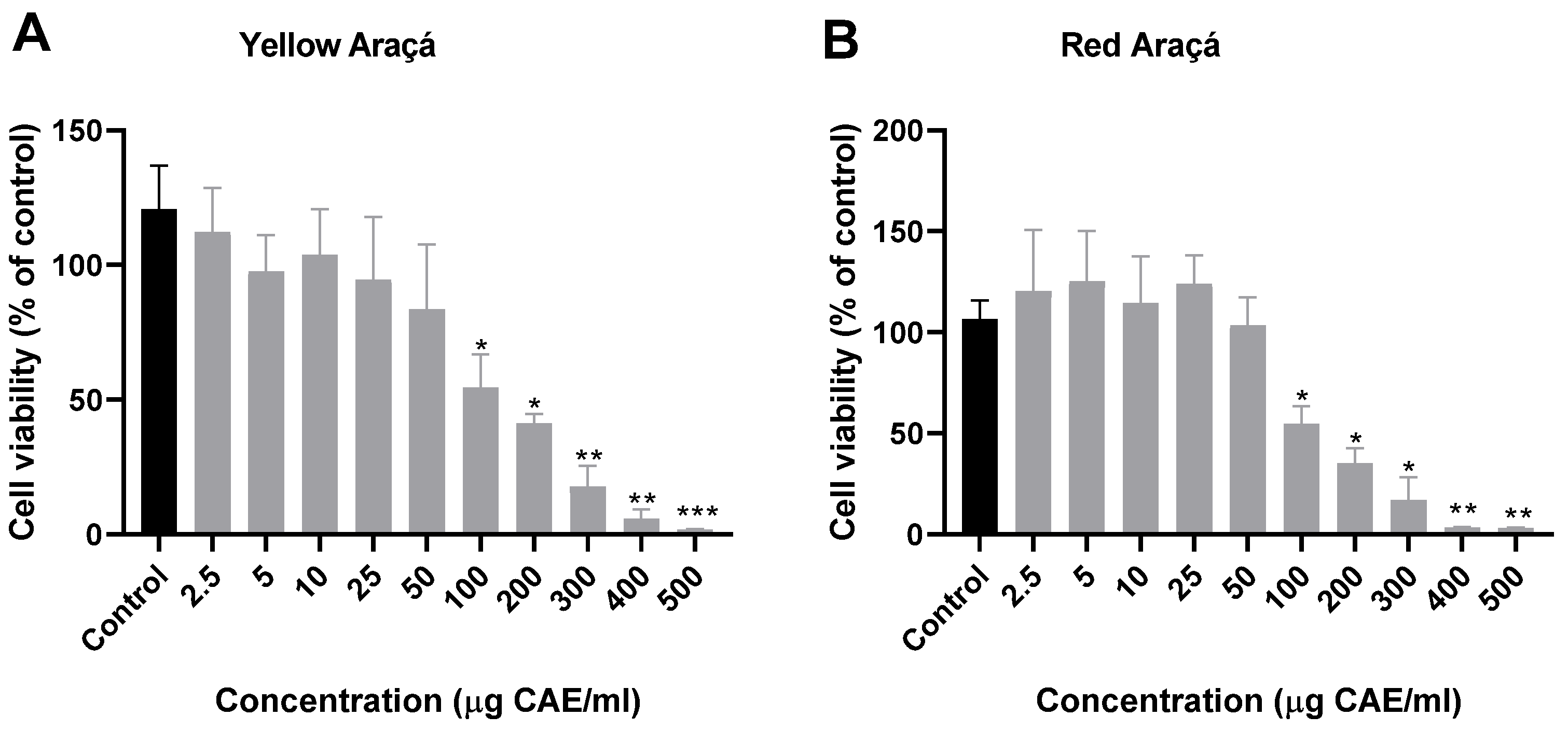
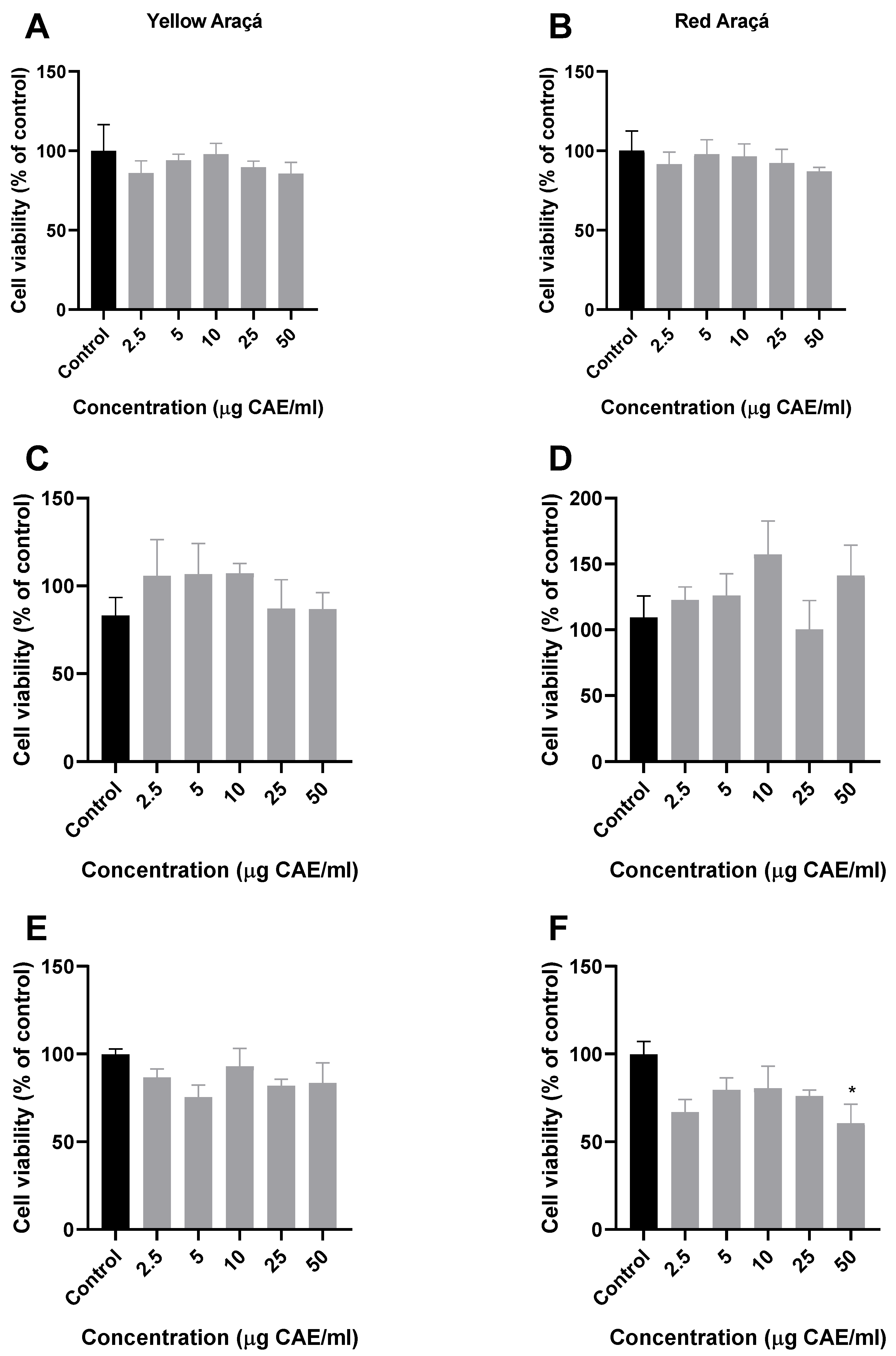
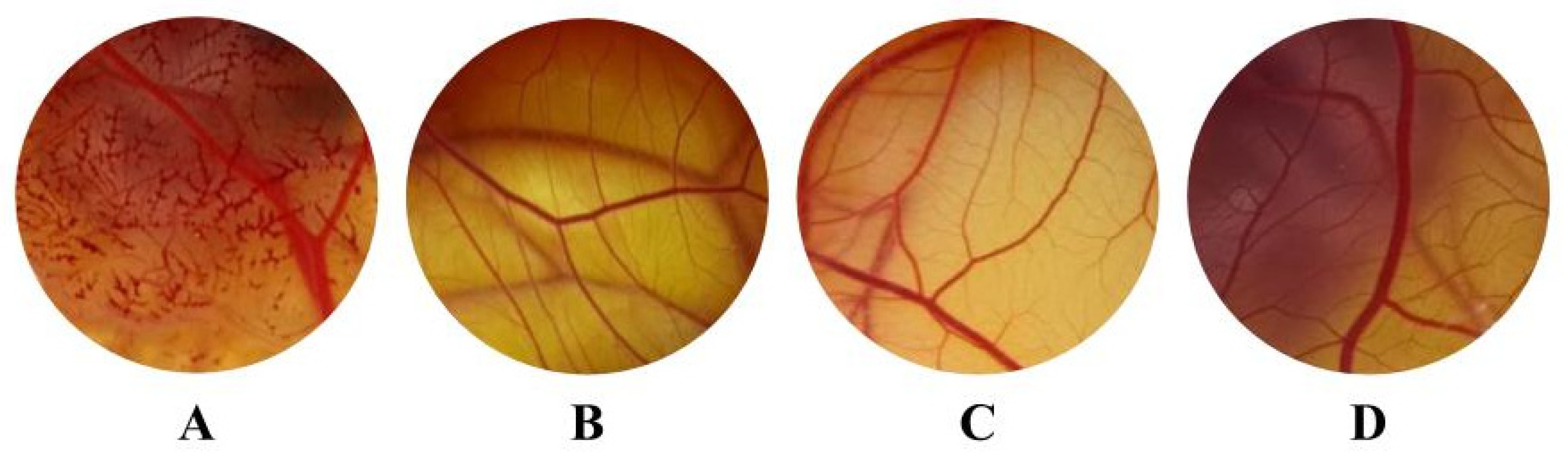
3. Results and Discussion

Araçá is a plant native to southern Brazil, but it is a little-consumed fruit due to the population’s unfamiliarity with it. Araçazeiro leaves are widely used in traditional medicine in the form of teas and stews, and there are already several studies evaluating their pharmacological properties [1,2,3,5,9,10,13,14,16,17,18]. On the other hand, studies evaluating the composition and biological effects of araçá fruits are still scarce; therefore, it is essential to undertake research to stimulate the consumption of araçá fruits and promote their potential and promising pharmacological applications.
In this work, we observed that although the yellow and red araçá fruits are the same species, they present marked differences in physicochemical composition (Table 1). In general, we observed that the araçá is a fruit rich in nutrients and antioxidant compounds, especially dietary fiber. Our results show that red araçá has a significantly higher content of dry matter, carbohydrates, total and insoluble dietary fiber, protein, caloric value, pH, and total soluble solids, when compared to yellow araçá. However, yellow araçá showed a greater amount of minerals (ash), water, and soluble fiber (Table 1). We emphasize that although they are the same species, the fruits present marked differences in chemical composition and can thus be used for different nutritional goals. A study that reviewed the composition and bioactive compounds of araçá demonstrated that in 100 g of araçá fresh fruit there is 81.73–84.9 g of water, 0.75–1.03 g of protein, 0.63–1.50 g of minerals, 4.32–10.01 g of carbohydrate, 0.42–0.55 g of lipid, 3.87–6.14 g of fiber, and 26.8 kcal of energy [4], which is in agreement with results observed in our study. Therefore, we can demonstrate that red araçá fruits are an excellent source of dietary fiber, especially concerning insoluble fiber, and that both araçá fruits are acid (pH around 3.6), with low caloric content and rich in total phenolic compounds, which could enhance their consumption by the population.
The phytochemical evaluation demonstrated that the red and yellow araçá had similar amounts of total phenolic compounds, around 5209–5517 µg CAE/mL, respectively (Table 1). A similar profile of araçá phenolic compounds was observed in previous work performed by our laboratory, where yellow araçá showed a larger amount of total phenols, followed by flavonoid content, than red araçá [39]. On the other hand, a different profile was found by Biegelmeyer and colleagues when comparing the red and yellow araçá and demonstrated that red araçá total content of phenolic compounds, expressed in fresh weight, was more abundant than yellow (501.33–292.03 mg of GAE/100 g, respectively) [7]. Other studies demonstrated that the total content of phenolic compounds did not differ between yellow and red genotypes (5372 mg and 5638 mg of gallic acid equivalent (GAE)/100 g and 603 mg and 606 mg of CAE/100 g, respectively) [40,41], which are very similar to those observed by our group. Although we did not observe significant differences in the amount of total phenolic compounds between the yellow and red fruits, previous work by our group on the characterization of yellow and red araçá extracts showed differences in the composition of major compounds. Both fruits showed remarkably similar amounts of hydroxybenzoate derivatives and flavonols; however, the yellow araçá extract showed a significant number of hydroxycinnamate derivatives while the red araçá had a higher anthocyanin content [39]. These characteristics suggest the pharmacologically beneficial effects of araçá fruits.
To assess the possible cytotoxicity of yellow and red araçá fruit extracts, the cell viability test using the NIH/3T3 fibroblast cell line was performed. We observed a significant reduction in cell viability from the concentration of 100 µg CAE/mL for both yellow and red araçá extracts in 24 h of treatment (Figure 1). Based on these results, the inhibitory concentration of 50% (IC50) was obtained at 73.60 µg CAE/mL for yellow and 83.38 µg CAE/mL for red araçá extracts. A study demonstrated that aqueous and acetonic extracts of red and yellow araçá did not affect the survival of control cells (NIH/3T3 rat embryonic fibroblasts) at concentrations of 40, 60, and 80 µg/mL. However, the same study showed that araçá extracts reduced the proliferation of cancer cell lines MCF-7 (mammary cancer cells) and Caco-2 (colon cancer cells) [20]. The reduction in viability observed in our work may be related to the concentration of extract used in the cells, since different studies have used concentrations expressed in ug/mL of extract and we used concentrations expressed in phenolic compound content. We always choose to express the concentrations used as phenolic compound content to avoid variations due to harvest, post-harvest, and fruit processing factors. This ensures that we always have the same number of bioactive compounds acting in the sample.
Cancer is one of the main causes of morbidity and mortality worldwide [42,43,44]. To evaluate the potential effect of red and yellow araçá ethanolic extract in cancer cell lines, we used cell lines of Rattus norvergicus brain glioma (C6), human prostate carcinoma (DU-145), and human colorectal adenocarcinoma (HT-29) (Figure 2). Based on the IC50 observed in this study, concentrations less than 50 µg EAC/mL of the extracts were chosen to evaluate antiproliferative effects in cancer strains. Among all the concentrations tested, only the extract of red araçá showed a significant reduction in cell viability at a concentration of 50 µg EAC/mL in the HT-29 strain (Figure 2F). No reduction in cell viability was observed in the other cell lines evaluated (Figure 2). Chaves and colleagues (2018), evaluated the biological proprieties of Brazil berries, including red and yellow araçá methanolic extract, and the cytotoxic effect on the cancer cells lines A549 (non-small cell lung cancer cells), RD (rhabdomyosarcoma), and DU145 (prostate carcinoma); however, none of the extracts were able to reduce cell viability in all tested cancer cell lines [45]. We suggest that the effect observed in the red araçá extract and not in the yellow one was due to the presence of anthocyanins content in their composition, as was observed in the composition of the majority phenolic compounds of red araçá [39]. Furthermore, other studies using fruit extracts rich in anthocyanins, such as grapes (Vitis vinifera), blueberries (Vaccinium myrtillus L.), and chokeberry (Aronia meloncarpa E.), were able to inhibit the growth of HT-29 cells [46]. Therefore, we believe that red araçá extract has an antiproliferative effect on cancerous cells regardless of its toxicity.
Is important to emphasize that one of the main risk factors for cancer is the reduced consumption of fruits and vegetables by the population. Among the benefits of consuming fruits and vegetables, we highlight their antioxidant effect, which works by stimulating the activity and expression of antioxidant enzymes, providing essential micronutrients and strengthening the immune system. In addition, dietary fibers, present in large quantities in araçá fruits, are able to increase fecal volume and, consequently, intestinal transit, which reduces the absorption of potentially carcinogenic agents [47]. Thus, the Ministry of Health (MS) of Brazil recommends a minimum consumption of fruits and vegetables of 400 g/day [48] as a preventive measure for the appearance of some types of cancers, such as colorectal cancer. Thus, we suggest that the consumption of araçá fruits could be expanded and used as a source of fibers and bioactive compounds beneficial to health.
The search for new drugs or substances with antimicrobial or antifungal potential has grown considerably over the years, due to the indiscriminate use of antimicrobials, both by preventive medicine and agriculture for pest control, thus considerably increasing microbial resistance [26]. Thus, the use of extracts and natural essential oils has been an alternative to the use of antimicrobials, and subsequently, seeking new alternatives that have equivalents results in reference drugs and reduced side effects [14,17,26]. Here, we evaluated the antifungal effects of yellow and red araçá extracts on Candida spp. (Table 2), and for the first time in the literature, we reported a pronounced antifungal effect of araçá fruit extracts on Candida albicans.
The antifungal effect of yellow and red araçá extracts was evaluated in 22 clinical isolates of Candida albicans and 4 clinical isolates of non-albicans Candida in comparison to the reference drug fluconazole (Table 2). We observed that the reference drug, fluconazole, did not have an antifungal effect in any of the Candida spp. tested when used at the maximum clinic concentration of 32 mg/mL, demonstrating that all Candida spp. used were resistant to fluconazole. In general, the results showed that non-albicans Candida proved to be more resistant to both araçá extracts based on MIC and MFC concentrations (Table 2).
Moreover, we observed that araçá extracts, regardless of the variety, showed significant antifungal activity in the Candida albicans isolates. Among the Candida spp. evaluated in this study, only the isolates C. albicans 2836 F, Candida dubliniensis ATCC 7957, and C. glabrata ATCC 2001 were not inhibited by both araçá extracts. However, the red araçá extract showed a more effective inhibition, as it presented lower values of the geometric mean (GM) (MIC 40.6 µg EAC/mL and MFC 41.7 µg EAC/mL) than those of yellow araçá (MIC 44.9 µg EAC/mL and MFC 48.7 µg EAC/mL). The antifungal effect at a concentration less than or equal to 39.06 µg EAC/mL was observed in 18 of the 22 isolates of C. albicans evaluated against the extract of red araçá and 19 of the 22 isolates of C. albicans in relation to yellow araçá. However, at these same concentrations, only the extract of red araçá showed activity for two of the four non-albicans Candida isolates. These same isolates showed inhibition by the extract of yellow araçá but at a higher concentration (78.12 µg EAC/mL) (Table 2).
Other authors have already reported that the araçá leaves have antimicrobial [13,16] and antifungal effects [10,13,14,17]. Scur and colleagues [14] observed that the araçá leaves ethanolic extract showed the highest antimicrobial activity against the microorganisms tested when compared to the aqueous extract. In the ethanolic extract, the MIC ranged from 0.78 to 25 mg/mL for Gram-negative bacteria, from 0.78 to 3.125 mg/mL for Gram-positive bacteria, and exhibited a MIC of 3.125 mg/mL for yeast C. albicans ATCC [14]. In our work, the extracts from araçá fruits were able to inhibit 21 of the 22 isolates of C. albicans with 156 µg/mL, that is, a much lower concentration than that observed for the leaves. We found only one study that evaluated the antimicrobial effect of the araçá fruit extract. In this work, the water and acetone extracts of yellow and red araçá fruits were evaluated. The antimicrobial activity against S. enteritidis was evaluated by looking at the formation of the inhibition halo and determining the minimal inhibitory concentration (MIC) of the extracts. All araçá extracts showed antimicrobial activity and the MIC was 5%, except for the water extract of red araçá (16%) [20].
The antimicrobial activity of plants is mainly related with the presence of phenolic compounds in leaves and fruits [49,50]. Araçá contains flavonoids (i.e., kaempferol and quercetin) and anthocyanidins (cyanidin) that are well recognized as antimicrobial agents. The mode of action of these compounds is related to their reaction with microbial cellular membrane by inactivating essential enzymes or forming complexes with metallic ions, limiting their accessibility to the microbial metabolism [20]. It has been shown that extracts from fruits that are rich in secondary metabolites usually prevent bacterial cell proliferation. Phenolic compounds could act as a destabilizing bacterial cell membrane that is primarily responsible for the respiration of these microorganisms. In yeast, phenolics could act as metal chelators or scavenging free radicals, which are otherwise harmful to the cell. In this case, the cell membrane is not harmed and, consequently, mitochondria, which are vital for respiration, are not affected by the action of phenolic compounds [20,51].
The mechanisms of action of herbal medicine in the treatment of Candida spp. are still poorly studied. Several mechanisms of action have been proposed from the rupture of the cellular membranes, which seems to interrupt the cell cycle through the synthesis of proteins and the alteration of the yeast DNA [52]. We believe that the araçá extracts used in our study had expressive antifungal activity in Candida spp. due to the richness of phenolic compounds in the composition.
Since the antifungal effect under Candida spp. was observed, the irritant potential of araçá extracts was evaluated to examine the development of a formulation for the treatment of candida infections. For this, we used an irritability test in the chorioallantoic membrane (HET-CAM) [37]. The use of the HET-CAM test to evaluate the irritability of plant extracts as sources for substances through topical application was been already described [53]. We observed that all tested concentrations in HET-CAM for both yellow and red araçá extracts had no negative effect on the vascularization of the chorioallantoic membrane, with all considered non-irritating (Figure 3). In addition to safety for ocular applications, evaluation via HET-CAM is also related to the irritant potential in other epitheliums, which can be correlated to the mucous/substrates, such as the vaginal epithelium, demonstrating that its application through different routes of administration are favored [54]. The positive control (NaOH 0.1 N) reached a score of 9, considered a severe irritant, and was different from the negative control that presented a score of 0, being non-irritating (Figure 3), thus validating these assessments, according to what is recommended by the Interagency Coordinating Committee on the Validation of Alternative Methods [37].

4. Conclusions

Yellow and red araçá are small fruits common in southern Brazil and are rich in nutrients, mainly phenolic compounds; vitamin C; and total dietary fiber. Here, we observed that the extract of red araçá fruit showed a cytotoxic effect against colorectal cancer, which is very promising and needs further research, since this type of cancer is one of the most common among men and women. Furthermore, we highlight the antifungal effect in isolates of the genus Candida spp. that are resistant to fluconazole, where both araçá extracts were effective in low concentrations, with emphasis on the red araçá extract. In addition, both araçá extracts did not show irritant effects, suggesting suitability for topical application. Thus, we conclude that the extracts of the yellow and red araçá fruits have promising biological effects. In the future, we aim to explore the mechanisms of action and the pharmacological potential of the extracts as antifungal and antiproliferative agents. Furthermore, we intend to develop and test topical pharmaceutical methods involving the extract of red araçá fruits for the treatment of vaginal candidiasis.

Author Contributions

Conceptualization, F.R.M. and C.C.D.; Methodology, F.R.M., J.R.B. and F.B.d.C.; Formal analysis, F.R.M., A.L.R.P. and J.R.B.; Investigation, F.R.M., A.L.R.P., J.R.B. and N.R.R.; Resources, C.D.O.S. and M.V.; Writing—original draft, F.R.M., J.R.B., C.D.O.S., F.B.d.C., S.E.H. and C.C.D.; Writing—review & editing, C.D.O.S., S.E.H., N.R.R., M.V. and C.C.D.; Visualization, F.B.d.C.; Supervision, C.D.O.S., S.E.H. and C.C.D.; Project administration, C.C.D.; Funding acquisition, C.C.D. All authors have read and agreed to the published version of the manuscript.

Funding

This research was funded by Unipampa, grant number “Inovapampa 2021”. This study was financed in part by the Coordenação de Aperfeiçoamento de Pessoal de Nível Superior—Brasil (CAPES)—Finance Code 001, and the CAPES fellowships (F.R.M).

Data Availability Statement

The data presented in this study are available on request from the corresponding author.

Acknowledgments

We thank EMBRAPA Clima Temperado for supplying the fruits and constant partnership in this group’s work. We would also like to thank NIH for the ImageJ open software (version 1.54f).

Conflicts of Interest

The authors declare no conflict of interest.

References

  1. Im, I.; Park, K.-R.; Kim, S.-M.; Kim, C.; Park, J.H.; Nam, D.; Jang, H.-J.; Shim, B.S.; Ahn, K.S.; Mosaddik, A.; et al. The Butanol Fraction of Guava (Psidium cattleianum Sabine) Leaf Extract Suppresses MMP-2 and MMP-9 Expression and Activity Through the Suppression of the ERK1/2 MAPK Signaling Pathway. Nutr. Cancer 2012, 64, 255–266. [Google Scholar] [CrossRef] [PubMed]
  2. Pino, J.A.; Marbot, R.; Vázquez, C. Characterization of Volatiles in Strawberry Guava (Psidium cattleianum Sabine) Fruit. J. Agric. Food Chem. 2001, 49, 5883–5887. [Google Scholar] [CrossRef] [PubMed]
  3. Egea, M.B.; Pereira-Netto, A.B.; Cacho, J.; Ferreira, V.; Lopez, R. Comparative analysis of aroma compounds and sensorial features of strawberry and lemon guavas (Psidium cattleianum Sabine). Food Chem. 2014, 164, 272–277. [Google Scholar] [CrossRef] [PubMed]
  4. dos Santos Pereira, E.; Vinholes, J.; Franzon, R.C.; Dalmazo, G.; Vizzotto, M.; Nora, L. Psidium cattleianum fruits: A review on its composition and bioactivity. Food Chem. 2018, 258, 95–103. [Google Scholar] [CrossRef] [PubMed]
  5. Rocha, L.D.; Preussler, K.H.; Pegorini, F.; de Farias, V.; Maranho, L.T. Estudo anatômico comparativo da casca do caule do araçá-amarelo e araçá-vermelho, Psidium cattleianum Sabine, Myrtaceae. Acta Bot. Bras. 2008, 22, 1114–1122. [Google Scholar] [CrossRef]
  6. Fetter, M.d.R.; Vizzotto, M.; Corbelini, D.D.; Gonzalez, T.N. Propriedades funcionais de araçá-amarelo, araçá-vermelho (Psidium cattleyanum Sabine) e araçá-pera (P. acutangulum D.C.) cultivados em Pelotas/RS. Braz. J. Food Technol. 2010, 13, 92–95. [Google Scholar] [CrossRef]
  7. Biegelmeyer, R.; Andrade, J.M.M.; Aboy, A.L.; Apel, M.A.; Dresch, R.R.; Marin, R.; Raseira, M.D.C.B.; Henriques, A.T. Comparative Analysis of the Chemical Composition and Antioxidant Activity of Red (Psidium cattleianum) and Yellow (Psidium cattleianum var. lucidum) Strawberry Guava Fruit. J. Food Sci. 2011, 76, C991–C996. [Google Scholar] [CrossRef]
  8. Franzon, R.C.; Campos, L.D.O.; Proença, C.E.B.; Sousa-Silva, J.C. Araçás do Gênero. In Araçás do Gênero Psidium Princ. Espécies, Ocorrência, Descrição e Usos; Embrapa Cerrados: Planaltina, Brazil, 2009; Volume 48. [Google Scholar]
  9. Brighenti, F.L.; Gaetti-Jardim, E.; Danelon, M.; Evangelista, G.V.; Delbem, A.C.B. Effect of Psidium cattleianum leaf extract on enamel demineralisation and dental biofilm composition in situ. Arch. Oral Biol. 2012, 57, 1034–1040. [Google Scholar] [CrossRef]
  10. Castro, M.R.; Victoria, F.N.; Oliveira, D.H.; Jacob, R.G.; Savegnago, L.; Alves, D. Essential oil of Psidium cattleianum leaves: Antioxidant and antifungal activity. Pharm. Biol. 2015, 53, 242–250. [Google Scholar] [CrossRef]
  11. Do Nascimento, K.S.; Sattler, J.A.G.; Macedo, L.F.L.; González, C.V.S.; de Melo, I.L.P.; da Silva Araújo, E.; Granato, D.; Sattler, A.; de Almeida-Muradian, L.B. Phenolic compounds, antioxidant capacity and physicochemical properties of Brazilian Apis mellifera honeys. LWT-Food Sci. Technol. 2018, 91, 85–94. [Google Scholar] [CrossRef]
  12. Ho, R.; Violette, A.; Cressend, D.; Raharivelomanana, P.; Carrupt, P.; Hostettmann, K. Antioxidant potential and radical-scavenging effects of flavonoids from the leaves of Psidium cattleianum grown in French Polynesia. Nat. Prod. Res. 2012, 26, 274–277. [Google Scholar] [CrossRef]
  13. Sangalli, J.; Júnior, E.G.J.; Bueno, C.R.E.; Jacinto, R.C.; Sivieri-Araújo, G.; Filho, J.E.G.; Cintra, L.T.; Junior, E.D. Antimicrobial activity of Psidium cattleianum associated with calcium hydroxide against Enterococcus faecalis and Candida albicans: An in vitro study. Clin. Oral Investig. 2018, 22, 2273–2279. [Google Scholar] [CrossRef] [PubMed]
  14. Scur, M.C.; Pinto, F.G.S.; Pandini, J.A.; Costa, W.F.; Leite, C.W.; Temponi, L.G. Antimicrobial and antioxidant activity of essential oil and different plant extracts of Psidium cattleianum Sabine. Braz. J. Biol. 2016, 76, 101–108. [Google Scholar] [CrossRef] [PubMed]
  15. Vinholes, J.; Reis, S.F.; Lemos, G.; Barbieri, R.L.; de Freitas, V.; Franzon, R.C.; Vizzotto, M. Effect of in vitro digestion on the functional properties of Psidium cattleianum Sabine (araçá), Butia odorata (Barb. Rodr.) Noblick (butiá) and Eugenia uniflora L. (pitanga) fruit extracts. Food Funct. 2018, 9, 6380–6390. [Google Scholar] [CrossRef] [PubMed]
  16. Massunari, L.; Novais, R.Z.; Oliveira, M.T.; Valentim, D.; Junior, E.D.; Duque, C. Antimicrobial Activity and Biocompatibility of the Psidium cattleianum Extracts for Endodontic Purposes. Braz. Dent. J. 2017, 28, 372–379. [Google Scholar] [CrossRef]
  17. Soliman, F.M.; Fathy, M.M.; Salama, M.M.; Saber, F.R. Comparative study of the volatile oil content and antimicrobial activity of Psidium guajava L. and Psidium cattleianum Sabine leaves. Bull. Fac. Pharm. Cairo Univ. 2016, 54, 219–225. [Google Scholar] [CrossRef]
  18. Alvarenga, F.Q.; Mota, B.C.; Leite, M.N.; Fonseca, J.M.; Oliveira, D.A.; Royo, V.d.A.; e Silva, M.L.; Esperandim, V.; Borges, A.; Laurentiz, R.S. In vivo analgesic activity, toxicity and phytochemical screening of the hydroalcoholic extract from the leaves of Psidium cattleianum Sabine. J. Ethnopharmacol. 2013, 150, 280–284. [Google Scholar] [CrossRef]
  19. Saber, F.R.; Abdelbary, G.A.; Salama, M.M.; Saleh, D.O.; Fathy, M.M.; Soliman, F.M. UPLC/QTOF/MS profiling of two Psidium species and the in-vivo hepatoprotective activity of their nano-formulated liposomes. Food Res. Int. 2018, 105, 1029–1038. [Google Scholar] [CrossRef]
  20. Medina, A.L.; Haas, L.I.R.; Chaves, F.C.; Salvador, M.; Zambiazi, R.C.; da Silva, W.P.; Nora, L.; Rombaldi, C.V. Araçá (Psidium cattleianum Sabine) fruit extracts with antioxidant and antimicrobial activities and antiproliferative effect on human cancer cells. Food Chem. 2011, 128, 916–922. [Google Scholar] [CrossRef]
  21. Martínez, A.; Bono, C.; Megías, J.; Yáñez, A.; Gozalbo, D.; Gil, M.L. Systemic Candidiasis and TLR2 Agonist Exposure Impact the Antifungal Response of Hematopoietic Stem and Progenitor Cells. Front. Cell. Infect. Microbiol. 2018, 8, 309. [Google Scholar] [CrossRef]
  22. El-Houssaini, H.H.; Elnabawy, O.M.; Nasser, H.A.; Elkhatib, W.F. Correlation between antifungal resistance and virulence factors in Candida albicans recovered from vaginal specimens. Microb. Pathog. 2019, 128, 13–19. [Google Scholar] [CrossRef]
  23. Zhang, X.; Li, T.; Chen, X.; Wang, S.; Liu, Z. Nystatin enhances the immune response against Candida albicans and protects the ultrastructure of the vaginal epithelium in a rat model of vulvovaginal candidiasis. BMC Microbiol. 2018, 18, 166. [Google Scholar] [CrossRef]
  24. de Oliveira, K.P.; Tanajura, L.M.L.; Nascimento, G.B.; Marques, A.S.; de Souza, L.P.D.; Vieira, T.Q.; dos Reis, E. Candidíase no cenário brasileiro atual: Epidemiologia, prevenção e manejo. Braz. J. Dev. 2022, 8, 79108–79123. [Google Scholar] [CrossRef]
  25. Jafarzadeh, L.; Ranjbar, M.; Nazari, T.; Eshkaleti, M.N.; Gharehbolagh, S.A.; Sobel, J.D.; Mahmoudi, S. Vulvovaginal candidiasis: An overview of mycological, clinical, and immunological aspects. J. Obstet. Gynaecol. Res. 2022, 48, 1546–1560. [Google Scholar] [CrossRef]
  26. Madigan, M.T.; Martinko, J.M.; Bender, K.S.; Buckley, D.H.; Stahl, D.A. Microbiologia de Brock, 14th ed.; Artmed Editora: Porto Alegre, Brazil, 2016. [Google Scholar]
  27. Tortora, G.J.; Funke, B.R.; Case, C.L. Microbiologia, 10th ed.; Artmed Editora: Porto Alegre, Brazil, 2012. [Google Scholar]
  28. Rahayu, R.P.; Prasetyo, R.A.; Purwanto, D.A.; Kresnoadi, U.; Iskandar, R.P.D.; Rubianto, M. The immunomodulatory effect of green tea (Camellia sinensis) leaves extract on immunocompromised Wistar rats infected by Candida albicans. Vet. World 2018, 11, 765–770. [Google Scholar] [CrossRef]
  29. Swain, T.; Hillis, W.E. The phenolic constituents of Prunus domestica. I.—The quantitative analysis of phenolic constituents. J. Sci. Food Agric. 1959, 10, 63–68. [Google Scholar] [CrossRef]
  30. Chemists, A. Official methods of analysis of AOAC International. Assoc. Off. Anal. Chem. Int. 1995. [Google Scholar] [CrossRef]
  31. Bligh, E.G.; Dyer, W.J. A rapid method of total lipid extraction and purification. Can. J. Biochem. Physiol. 1959, 37, 911–917. [Google Scholar] [CrossRef]
  32. Zenebon, O.; Pascuet, N.S.; Tiglea, P. Instituto Adolfo Lutz Métodos Físico-Químicos Para Análise de Alimentos; Instituto Adolfo Lutz: São Paulo, Brazil, 2008; ISBN 9788578110796. [Google Scholar]
  33. Denardin, C.C.; Parisi, M.M.; Martins, L.A.M.; Terra, S.R.; Borojevic, R.; Vizzotto, M.; Perry, M.L.S.; Emanuelli, T.; Guma, F.T.C.R. Antiproliferative and cytotoxic effects of purple pitanga (Eugenia uniflora L.) extract on activated hepatic stellate cells. Cell Biochem. Funct. 2014, 32, 16–23. [Google Scholar] [CrossRef]
  34. Lewis, J.S. Clinical and Laboratory Standards Institute (CLSI) Performance Standards for Antimicrobial Susceptibility Testing, 27th ed.; CLSI Supplement M100: Wayne, PA, USA, 2017; ISBN 1562388045. [Google Scholar]
  35. Veiga, A.; Toledo, M.d.G.T.; Rossa, L.S.; Mengarda, M.; Stofella, N.C.F.; Oliveira, L.J.; Gonçalves, A.G.; Murakami, F.S. Colorimetric microdilution assay: Validation of a standard method for determination of MIC, IC50%, and IC90% of antimicrobial compounds. J. Microbiol. Methods 2019, 162, 50–61. [Google Scholar] [CrossRef]
  36. Lokman, N.A.; Elder, A.S.F.; Ricciardelli, C.; Oehler, M.K. Chick Chorioallantoic Membrane (CAM) Assay as an In Vivo Model to Study the Effect of Newly Identified Molecules on Ovarian Cancer Invasion and Metastasis. Int. J. Mol. Sci. 2012, 13, 9959–9970. [Google Scholar] [CrossRef]
  37. ICCVAM. ICCVAM Test Method Evaluation Report: Current Validation Status of In Vitro Test Methods Proposed for Identifying Eye Injury Hazard Potential of Chemicals and Products; NIH Publication No. 10-7553; National Institute of Environmental Health Sciences: Research Triangle Park, NC, USA, 2010; pp. 1–10. Available online: https://ntp.niehs.nih.gov/sites/default/files/iccvam/docs/ocutox_docs/invitro-2010/tmer-vol2.pdf (accessed on 17 October 2023).
  38. Machado, R.M. Preclinical Performance of Vaginal Semisolid Products: Technological and Safety Evaluations Assuming Physiologic Parameters. Ph.D. Thesis, Universidade da Beira Interior, Covilhã, Portugal, 2018. [Google Scholar]
  39. Moraes, L.d.L.S.; Rodrigues, N.R.; Forno, A.H.D.; Tambara, A.L.; Boldori, J.R.; Vizzotto, M.; Quatrin, A.; Emanuelli, T.; Denardin, C.C. Araçá (Psidium cattleianum Sabine) ethanol extracts increase lifespan and alleviate oxidative stress in Caenorhabditis elegans. J. Agric. Food Res. 2023, 11, 100505. [Google Scholar] [CrossRef]
  40. Luximon-Ramma, A.; Bahorun, T.; Crozier, A. Antioxidant actions and phenolic and vitamin C contents of common Mauritian exotic fruits. J. Sci. Food Agric. 2003, 83, 496–502. [Google Scholar] [CrossRef]
  41. Vinholes, J.; Lemos, G.; Barbieri, R.L.; Franzon, R.C.; Vizzotto, M. In vitro assessment of the antihyperglycemic and antioxidant properties of araçá, butiá and pitanga. Food Biosci. 2017, 19, 92–100. [Google Scholar] [CrossRef]
  42. Alberts, B.; Johnson, A.; Lewis, J.; Raff, M.; Robert, K.; Walter, P. Molecular Biology of the Cell; Garland Science: New York, NY, USA, 2008; ISBN 9780815341055. [Google Scholar]
  43. de Oliveira Santos, M. Estimativa 2018: Incidência de Câncer no Brasil. Rev. Bras. Cancerol. 2018, 64, 119–120. [Google Scholar] [CrossRef]
  44. World Health Organization (WHO). The Top 10 Causes of Death; 2018. Available online: https://www.who.int/news-room/fact-sheets/detail/the-top-10-causes-of-death (accessed on 17 October 2023).
  45. Chaves, V.C.; Boff, L.; Vizzotto, M.; Calvete, E.; Reginatto, F.H.; Simões, C.M.O. Berries grown in Brazil: Anthocyanin profiles and biological properties. J. Sci. Food Agric. 2018, 98, 4331–4338. [Google Scholar] [CrossRef]
  46. Abo El-Ella, D.M.; Bishayee, A. The Epigenetic Targets of Berry Anthocyanins in Cancer Prevention. In Epigenetics of Cancer Prevention; Academic Press: Cambridge, MA, USA, 2019. [Google Scholar]
  47. Poirier, A.E.; Ruan, Y.; Hebert, L.A.; Grevers, X.; Walter, S.D.; Villeneuve, P.J.; Brenner, D.R.; Friedenreich, C.M. Estimates of the current and future burden of cancer attributable to low fruit and vegetable consumption in Canada. Prev. Med. 2019, 122, 20–30. [Google Scholar] [CrossRef]
  48. Monteiro, C.A.; Mondini, L.; Bl Costa, R. Mudanças na composição e adequação nutricional da dieta familiar nas áreas metropolitanas do Brasil (1988–1996). Rev. Saúde Pública. 2000, 34, 251–258. [Google Scholar] [CrossRef]
  49. Duarte, M.C.T.; Figueira, G.M.; Sartoratto, A.; Rehder, V.L.G.; Delarmelina, C. Anti-Candida activity of Brazilian medicinal plants. J. Ethnopharmacol. 2005, 97, 305–311. [Google Scholar] [CrossRef]
  50. Höfling, J.; Anibal, P.; Obando-Pereda, G.; Peixoto, I.; Furletti, V.; Foglio, M.; Gonçalves, R. Antimicrobial potential of some plant extracts against Candida species. Braz. J. Biol. 2010, 70, 1065–1068. [Google Scholar] [CrossRef]
  51. Butassi, E.; Svetaz, L.A.; Zhou, S.; Wolfender, J.-L.; Cortés, J.C.G.; Ribas, J.C.; Díaz, C.; Palacio, J.P.-D.; Vicente, F.; Zacchino, S.A. The antifungal activity and mechanisms of action of quantified extracts from berries, leaves and roots of Phytolacca tetramera. Phytomedicine 2019, 60, 152884. [Google Scholar] [CrossRef] [PubMed]
  52. Rodrigues Santos, V.; Maria Rita Pereira, E. Antifungal Activity of Brazilian Medicinal Plants against Candida Species. In Candida Albicans; IntechOpen: London, UK, 2019. [Google Scholar] [CrossRef]
  53. Wilson, T.D.; Steck, W.F. A modified HET–CAM assay approach to the assessment of anti-irritant properties of plant extracts. Food Chem. Toxicol. 2000, 38, 867–872. [Google Scholar] [CrossRef] [PubMed]
  54. Palmeira-de-Oliveira, R.; Monteiro Machado, R.; Martinez-de-Oliveira, J.; Palmeira-de-Oliveira, A. Testing vaginal irritation with the Hen’s Egg Test-Chorioallantoic Membrane assay. ALTEX-Altern. Anim. Exp. 2018, 35, 495–503. [Google Scholar] [CrossRef] [PubMed]
Figure 1. Toxicological evaluation of araçá fruit extracts using NIH/3T3 cells using MTT assay. (A) yellow araçá extract and (B) red araçá extract. Data correspond to the average of three independent experiments ± SD. * p < 0.05; ** p < 0.01; *** p < 0.001 indicate significant differences between treated and control cell lines (ANOVA with Tukey post-test).
Foods 12 04307 g001
Figure 2. Effect of araçá fruit extracts on viability of cancer cell lines using MTT assay. (A) C6 glioma cell line, (C) DU145 prostate cancer cell line, and (E) HT-29 colorectal cancer cell line exposed to yellow araçá extract (left) and (B) C6 glioma cell line, (D) DU145 prostate cancer cell line, and (F) HT-29 colorectal cancer cell line exposed to red araçá extract (right). Data correspond to the average of three independent experiments ± SD. * p < 0.05 indicates significant differences between treated and control cell lines (ANOVA with Tukey’s post hoc test).
Foods 12 04307 g002
Figure 3. Photograph at 5 min of the chorioallantoic membrane of the positive control (A), negative control (B), and 1250 µg CAE/mL of yellow (C) and red (D) araçá.
Foods 12 04307 g003
Table 1. Physicochemical composition of yellow and red araçá fruits.
Yellow AraçáRed Araçá
Dry matter18.14 ± 0.16 b20.75 ± 0.09 a
Moisture81.86 ± 0.16 a79.25 ± 0.09 b
Ash (mineral residue)0.90 ± 0.01 a0.79 ± 0.01 b
Carbohydrates16.26 ± 0.23 b18.56 ± 0.12 a
Total dietary fiber5.36 ± 1.51 b7.80 ± 1.02 a
Soluble fiber2.43 ± 1.33 a0.66 ± 0.45 b
Insoluble fiber2.93 ± 0.18 b7.48 ± 1.96 a
Crude protein0.74 ± 0.02 b0.92 ± 0.04 a
Total lipids0.67 ± 0.120.56 ± 0.14
Total energy value69.11 ± 1.87 b74.17 ± 0.75 a
pH3.56 ± 0.00 b3.61 ± 0.01 a
Total acidity (100 g of citric acid)24.51 ± 1.0323.02 ± 0.22
Total soluble solids (°Brix)10.01 ± 0.11 b13.50 ± 0.14 a
Phenolic compounds (µg chlorogenic acid equivalent/mL)5517.87 ± 482.985209.61 ± 302.09
Values expressed in % (g/100 g fresh weight) and total energy value (Kcal/100 g fresh weight) of fruits in mean ± SD. The means followed by different letters show significant differences with p < 0.05 (n = 3).
Table 2. Number of isolates with minimum inhibitory concentration (MIC) and minimum fungicide concentration (MFC) of yellow and red araçá fruit extracts.
IsolatedYellow Araçá 1Red Araçá 1Fluconazole 2
MICMFCMICMFCMICMFC
Candida albicans 15 A9.769.769.769.76>320>320
Candida albicans 10 A39.0639.06156.25156.25>320>320
Candida albicans 10 G19.5319.5319.5319.53>320>320
Candida albicans 14 A39.0639.0619.5319.53>320>320
Candida albicans ATCC 10231 78.1278.1239.0639.06>320>320
Candida albicans 24 C9.769.7678.1278.12>320>320
Candida albicans 26 A (n 3)39.0639.0619.5319.53>320>320
Candida albicans 2836 F>1250>1250>1250>1250>320>320
Candida albicans 3 H39.0639.0619.5319.53>320>320
Candida albicans 3 Q39.0639.0619.5319.53>320>320
Candida albicans 49 F9.769.7639.0639.06>320>320
Candida albicans 55 A19.5319.5319.5319.53>320>320
Candida albicans 6 E19.5319.5319.5319.53>320>320
Candida albicans 623139.0639.0639.0639.06>320>320
Candida albicans 66 A9.769.7619.5319.53>320>320
Candida albicans 69 A39.0639.0619.5319.53>320>320
Candida albicans 69 F78.1278.1278.1278.12>320>320
Candida albicans 76 E39.0639.069.769.76>320>320
Candida albicans 8 F39.0678.1239.0678.12>320>320
Candida albicans 91 A39.0639.0619.5319.53>320>320
Candida albicans 49 F19.5319.5319.5319.53>320>320
Candida albicans 55 A19.5319.5319.5319.53>320>320
Candida dubliniensis ATCC 79571250.001250.00625.00625.00>320>320
Candida glabrata 61 B78.1278.1239.0639.06>320>320
Candida glabrata ATCC 2001>1250>1250>1250>1250>320>320
Candida krusei 85 A78.1278.1239.0639.06>320>320
Results expressed in MIC and MFC (n = 3). 1 Concentration of the extract in µg of chlorogenic acid equivalent/mL. 2 Concentration of fluconazole expressed in (µg/mL). Fluconazole was used as a control drug in the maximum concentration of 32 mg/mL used in the clinic. C. albicans (n = 22); non-albicans Candida (n = 4).
Disclaimer/Publisher’s Note: The statements, opinions and data contained in all publications are solely those of the individual author(s) and contributor(s) and not of MDPI and/or the editor(s). MDPI and/or the editor(s) disclaim responsibility for any injury to people or property resulting from any ideas, methods, instructions or products referred to in the content.

