Article

Can Bioactive Compounds in Beetroot/Carrot Juice Have a Neuroprotective Effect? Morphological Studies of Neurons Immunoreactive to Calretinin of the Rat Hippocampus after Exposure to Cadmium

by
Małgorzata Matysek
1,
Edyta Kowalczuk-Vasilev
2,*,
Radosław Szalak
1,
Ewa Baranowska-Wójcik
3,
Marcin B. Arciszewski
1 and
Dominik Szwajgier
3,*
1
Department of Animal Anatomy and Histology, Faculty of Veterinary Medicine, University of Life Sciences, 12 Akademicka St., 20-950 Lublin, Poland
2
Institute of Animal Nutrition and Bromatology, Faculty of Animal Science and Bioeconomy, University of Life Sciences, 13 Akademicka St., 20-950 Lublin, Poland
3
Department of Biotechnology, Microbiology and Human Nutrition, Faculty of Food Science and Biotechnology, University of Life Sciences, 8 Skromna St., 20-704 Lublin, Poland
*
Authors to whom correspondence should be addressed.
Foods 2022, 11(18), 2794; https://doi.org/10.3390/foods11182794
Submission received: 14 July 2022 / Revised: 5 September 2022 / Accepted: 6 September 2022 / Published: 10 September 2022
(This article belongs to the Special Issue Food Components in Health Promotion and Disease Prevention)

Abstract

:
Cadmium ions (Cd2+) penetrate the blood–brain barrier and can, among other effects, influence intracellular calcium metabolism, leading to neurodegeneration. In the presented work, we estimated the effect of Cd2+ on the expression of calretinin in the neurons of the rat hippocampus and analyzed the reverse effect of freshly pressed beetroot/carrot juice in this context. In the 12-week lasting experiment, 32 8-week-old male Wistar rats were divided into four experimental groups (n = 8): the control group (C) received pure tap water; the Cd group (Cd)—received Cd2+ dissolved in tap water (5 mg Cd2+/kg b.w.); and two groups received beetroot/carrot juice: the BCJ group was administered only juice, and the Cd + BCJ group received juice with the addition of Cd2+ (5 mg Cd2+/kg b.w.). The exposition to low doses of Cd2+ caused a significant decrease in calretinin-immunoreactive (Cr-IR) neurons compared to the non-exposed groups. Moreover, the addition of Cd2+ to tap water reduced the numbers and length of Cr-IR nerve fibers. The negative effect of Cd2+ was significantly attenuated by the simultaneous supplementation of beetroot/carrot juice (Cd + BCJ). The study showed that the bioactive compounds in the beetroot/carrot juice can modulate Ca2+ levels in neurons, and thus, potentially act as a neuroprotective factor against neuronal damage.

1. Introduction

Cadmium (Cd2+) is a highly toxic heavy metal that is abundant in the environment. It enters the body through the respiratory or digestive tract, leading to excessive accumulation in internal organs, such as the kidneys, liver, or bones, increasing the risk of organ damage, osteoporosis, hypertension, or diabetes [1,2]. Cd2+ is also a known cause of neuroinflammation and dementia (including Alzheimer’s disease), as essentially pointed out in several excellent reviews (e.g., by Huat et al. [3]; Zhang et al. [4]). It is estimated that a significant part of Cd2+ present in the bloodstream crosses the blood–brain barrier and accumulates in various structures of the central nervous system (CNS) [5]. Within the CNS, the hippocampus is the target structure for Cd2+ toxicity. The hippocampus proper is a part of the limbic system. It plays an important role in the consolidation of information from short-term memory to long-term memory and in spatial navigation. On the one hand, it is a specific center of the brain’s memory and reasoning, and on the other, it is the area of the brain most affected by neurodegenerative diseases, of which dementia is the main symptom [6]. Cd2+ can also influence intracellular calcium (Ca2+) metabolism. Excessive accumulation of Ca2+ in nerve cells is responsible for the activation of processes that lead to neurodegenerative diseases, and ultimately to cell death [7]. Ca2+-binding proteins (CaBPs) provide an important first line of defense due to their ability to buffer incoming calcium, allowing the neurons to rapidly attain homeostasis. Moreover, it is known that Cd2+ is a strong calcium channel blocker—it inhibits Ca2+ uptake by cells, which disrupts the transmission of neuronal signals [8]. Calretinin (Cr) is one of the three main types of CaBPs present in inhibitory GABAergic neurons. The important function of Cr is the regulation of Ca2+ flow; it participates in synaptic plasticity and also influences the excitability of interneurons in the hippocampus by regulating other GABAergic neurons [9,10]. Studies on another heavy metal, Pb2+, have shown some promising effects in terms of the ability of beetroot juice to alleviate some of its toxic impact on the organism, particularly with regard to antioxidant and neurological functions [11,12,13].
The beet (Beta vulgaris L.) is a valuable source of unique natural compounds exerting antioxidant properties that may be beneficial to human health, as comprehensively presented in selected review papers, e.g., by [14] or [15]. It was confirmed that the consumption of beetroot may improve cerebral blood flow, and consequently, the cognitive function [16]. Beetroot extract showed anti-anxiety and antidepressant properties [17]. The carrot (Daucus carota L., Apiaceae family) is another highly appreciated, edible fleshy root, widely discussed in some excellent reviews, e.g., by Ahmad et al. [18] or Aćimović [19]. It is a source of α- and β-carotene, anthocyanins, lycopene, and phenolic acids [20], compounds known to have antioxidant properties (e.g., Mazewski et al. [21]). In our previous paper, we showed that beetroot/carrot juice was a rich source of bioactive compounds (mainly polyphenols, nitric pigments, and saponins). The juice revealed strong antioxidant activity, as confirmed using three experimental in vitro methods [22]. It has been reported before (e.g., in [3]), that Cd2+ can contribute to the development of Alzheimer’s disease. Although the presented paper focused in the first place on the ability of the juice to reduce the general toxicity of Cd2+ in the brain, a possible relationship between Cd2+ toxicity and acetylcholine-related enzymes was assessed as markers of cognitive function.
Based on the above, the present study aimed to evaluate the potential neuroprotective properties of bioactive compounds (present in beetroot/carrot juice), as a protective factor against neurodegenerative diseases, and also determine the effect of Cd2+ on neurons in the CNS, sensitive to Cd2+-induced memory disorders, with a particular focus on Cr immunoreactivity in rat hippocampal neurons.

2. Materials and Methods

2.1. Preparation of Beetroot/Carrot Juice (BCJ)

The beetroot/carrot juice (BCJ) was prepared essentially as described in our previous work [22]. In short, freshly pressed juices were obtained from thoroughly washed vegetables (Opolski beetroot and Nantejska carrot; in tap water), using an Angel 750 low-pressure screw press (Angel Co. Ltd., Naarden, The Netherlands). A single raw juice blend was prepared by mixing the beetroot and carrot juices at a ratio of 4:1 v/v (with pH corrected to 4.0 by the addition of 0.25 g of citric acid per 100 mL of juice). The final juice was freshly prepared and administered to rats within 2 days.

2.2. Qualitative Characteristics of Juice

The composition of the juice, as well as its total polyphenolic content and antioxidant activity were given in our previous work [22]. The anticholinesterase activity of BCJ (against acetyl- and butyrylcholinesterase) was studied as described by Studzińska-Sroka et al. [23] using juice prepared as described above and not otherwise processed in any way prior to analysis. To check the pro-cognitive efficiency of the juice, the ability to inhibit acetyl- and butyrylcholinesterase activity was tested as described earlier [23], expressed as the equivalent concentration of donepezil, a well-known cholinesterase inhibitor.

2.3. Animals

This experiment was approved by the 2nd Local Ethical Committee at the University of Life Sciences in Lublin, Poland (ref. no. 105/2015). Thirty-two (n = 32) male, 8-week-old Wistar rats were kept in single cages under a 12 h light–dark cycle with the light on at 06:00 a.m., at a room temperature of 21 ± 1 °C and a relative humidity of 55 ± 10%, with the cages ventilated every 4 min. The rats received the standard commercial rodent food (LSM; Agropol, Motycz, Poland; containing 12.06 MJ/kg ME (metabolic energy); crude protein: min. 16.00%; crude fat: min. 2.8%; crude ash: max. 7.00%; crude fiber: max. 5.00%; calcium min. 1.10%; phosphorus min. 0.70%; sodium max. 0.22 %; and vit. A: 8000 IU/kg; Vit. D3: 1000 IU/kg; Vit. E: 50 mg/kg) in the amount of 210 g per rat per week. Cadmium chloride of very high purity (99.999%) was used (Sigma-Aldrich, Poznań, Poland, cat. no 439800). The animals were acclimatized to the laboratory for at least one week before being used in the experiment. The rats were randomly divided into four experimental groups (n = 8): the control group (C) received 100 mL of tap water; the Cd group (Cd) received Cd2+ dissolved in 100 mL tap water/day (equivalent to 5 mg Cd2+/kg b.w.); and two groups received beetroot/carrot juice: the BCJ group was administered only 100 mL of juice per day, and the Cd + BCJ group received juice with the addition of Cd2+ ions (Cd2+ dissolved in 100 mL juice/day (equivalent to 5 mg Cd2+/kg b.w.). The experimental dose of Cd2+ was based on literature data [24,25,26]. The feed and fluid intake was controlled daily. The experiment lasted for 12 weeks.

2.4. Experimental Procedures and Analyses

After euthanasia (decapitation), the rats’ brains were stored in 10% buffered formalin (pH = 7) for 12 h at 4 °C, hydrated in decreasing concentrations of ethyl alcohol, and embedded in paraffin blocks in accordance with the previously described method [27]. Briefly, the paraffin blocks were cut into 5 μm-thick sections which were placed on silanized glass-slides (SuperFrostPlus, Thermo Fisher Scientific, Braunschweig, Germany). To block the endogenous peroxidase activity, the sections were chilled and washed in 3% hydrogen peroxidase (20 min). The slides were then flushed twice with PBS (pH 7.4) (15 min each time) and incubated in 2.5% normal goat serum (ImPRESSTM; MP-7451, Vector Labs, Burlingame, CA, USA) at room temperature (RT) for 20 min. The sections were incubated for 24 h at 4 °C with primary monoclonal mouse antibodies raised against Cr (1:2000; C7479, Sigma, Taufkirchen, Germany). The next day, the slides were washed in a washing buffer (2 × 15 min) and covered with anti-mouse/rabbit Ig (ImPRESSTM; MP-7500 Vector Labs, Newark, CA, USA) for 1 h. The specificity of the antibodies used was verified using a negative control in which primary antibodies were replaced with the same concentrations of appropriate non-immune IgG. 3,3′-diaminobenzidine chromogen (ImmPACTTMDAB, SK-4105, Vector Labs, Newark, CA, USA) was used to visualize the primary antisera. A working solution of DAB was applied onto the sections, and the process was monitored under a light microscope. Finally, the slides were rinsed with distilled water. Moreover, counterstaining (for 20 min) with Mayer’s hematoxylin was performed. After washing in distilled water, the sections were dehydrated in increasing concentrations of ethyl alcohol, cleared in xylene, mounted in DPX (Sigma-Aldrich, St. Louis, MO, USA), and cover slipped. The slides were viewed under a light microscope (Axiolab, Zeiss, Jena, Germany) connected to a digital camera (Olympus Color View III, Tokio, Japan). From each animal, approx. 25–30 sections immunostained for Cr were examined. The Cr-IR neurons were assessed by analyzing and counting no less than one hundred neurons immunoreactive (IR) to Cr in the CA1 field of the hippocampus of each group (the control and experimental) using Cell D software (Olympus, Tokio, Japan). Image J software (ImageJ 1.53 k; National Institute of Health, Bethesda, MD, USA) was used to quantify and statistically compare the length of Cr-IR nerve fibers. At least two independent observers were involved in quantification analyses, and the results obtained by them were averaged.

2.5. Statistical Analysis

The collected data were analyzed with Statistica software ver. 13.1 ((StatSoft, Kraków, Poland). Normality was assessed using the Kolmogorov–Smirnov test, and Levene’s homogeneity of variance test was applied to examine the equality of variances. One-way ANOVA and Tukey’s post-hoc tests were performed to compare all the experimental groups individually, whereas the two-way ANOVA was used to determine the impact of experimental factors: Cd2+ exposure and beetroot/carrot juice treatment and their interaction. Significant differences between the groups were identified at p ≤ 0.05 and p ≤  0.01.

3. Results

3.1. Anticholinesterase Activity of the Studied Juice

In our previous paper [22], we presented the composition of BCJ (using high-resolution, qualitative, and quantitative HPLC-ESIQTOF- MS) and identified the main bioactive components present (betanin, isobetanin, vulgaxanthin I and II, indicaxanthin, neobetanin, dexarboxyneobetanin, and decarboxybetanin). The juice showed significant total polyphenolic content, high antioxidant activity (tested using three experimental models), and positive effects towards human colon epithelial normal and cancer cells [22], so it was a promising research material in the context of the presented work. In the present work, we tested the ability of the juice to decrease the activity of acetyl- and butyrylcholinesterase and we report that the activity towards both enzymes was high (equal to donepezil applied at the concentration of 0.05 ± 0.00 µg/mL and 0.09 ± 0.01 µg/mL, respectively).

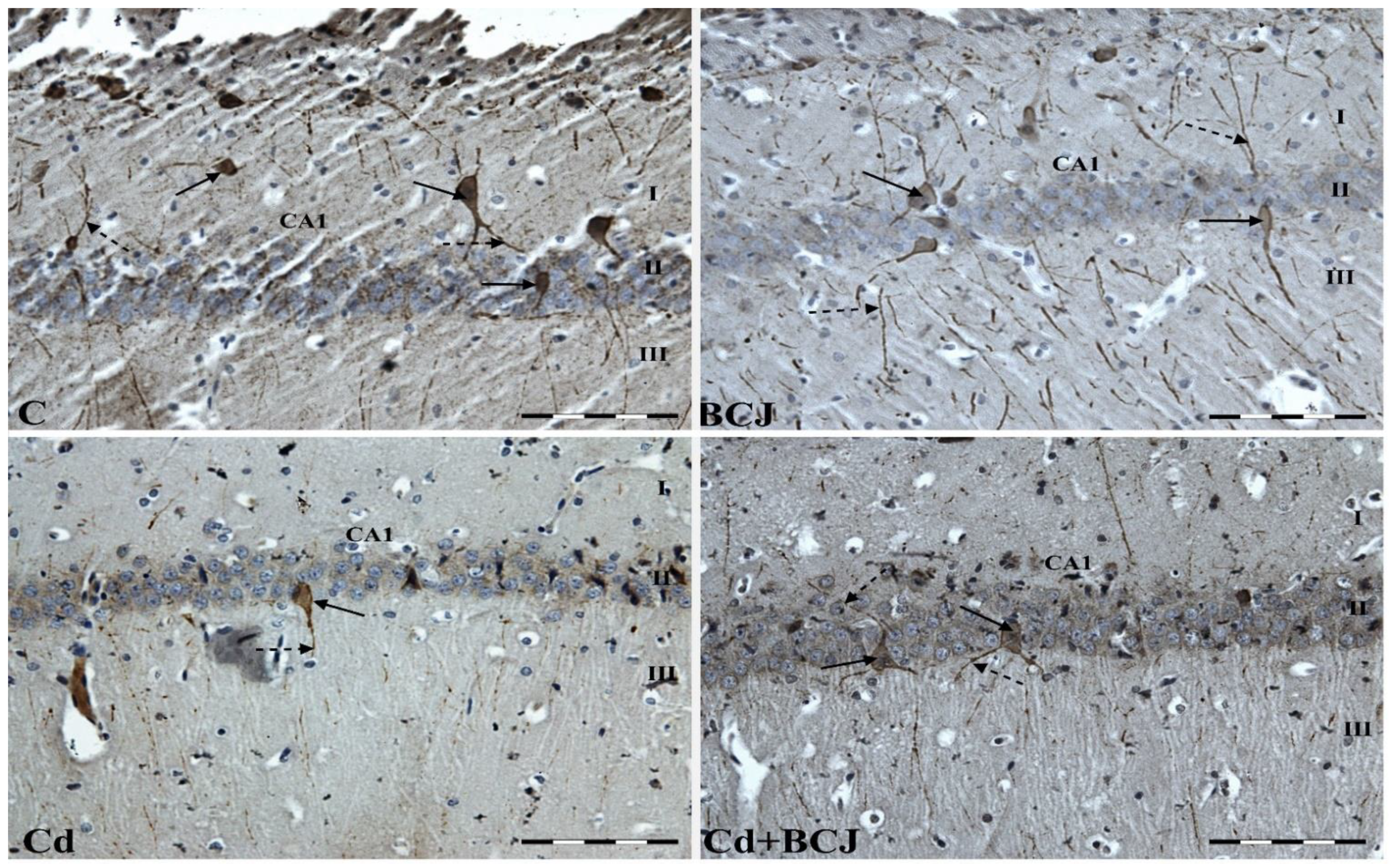
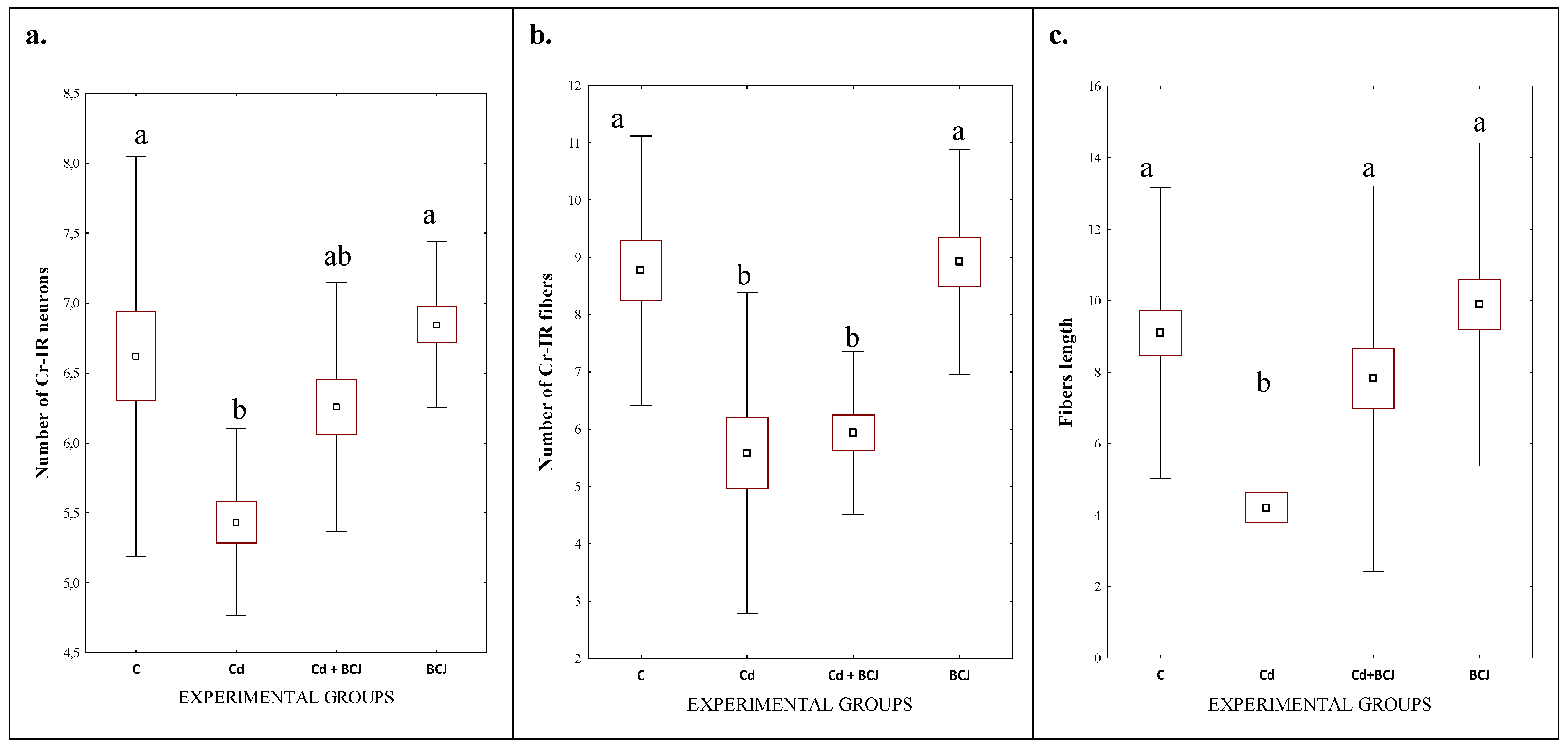
3.2. Cr Expression in the CA1 Field of the Hippocampus

The hippocampus consists of the Ammon’s horn (CA1–CA3) and the dentate gyrus (DG), but in the present study, we focused on the CA1 field of the rat hippocampus due to the high sensitivity of the nerve cells in this area to damage, as compared to CA2 and CA3 fields of the hippocampus [28]. The dorsal portion of the hippocampus was examined. In all the studied groups, multiform (oval, round, triangular, and fusiform) Cr-IR neurons were observed, unevenly distributed in all layers (the marginal, pyramidal, and multiform) of the CA1 field of the rat hippocampus. The neurons were characterized by the presence of cytoplasmic and nuclear reactions (Figure 1). The average numbers of Cr-IR neurons in the CA1 field of the rat hippocampus were estimated at 6.62 ± 0.72 in the control group, while the analogous neuronal populations in the BCJ group were calculated at 6.85 ± 0.30 (Figure 2a). There were no statistically significant differences in the mean numbers of Cr-IR neurons between the control group receiving tap water and the BCJ group (Figure 2a). In both cases, the neurons showed intense (+++) or moderate (++) nuclear/cytoplasmic reactions to Cr (Figure 1 C and BCJ).
Although collectively in both groups exposed to Cd2+, a decrease in the average number of Cr-IR neurons was stated in comparison to non-exposed groups (p < 0.001; Figure 2a; Table 1), and a statistically significant decrease (p ≤ 0.01) in the average number of Cr-IR neurons was only observed in the group receiving Cd2+ with tap water (Cd) as compared to control group (6.62 ± 0.72 vs. 5.43 ± 0.33, respectively) (Figure 2a). On the other hand, a positive trend was stated in case of the BCJ treated group. The mean number of Cr-IR neurons was higher in the brains of rats receiving Cd2+ dissolved in beetroot/carrot juice (Cd + BCJ group) as compared to the Cd group, although the differences were not confirmed statistically (Figure 2a). BCJ treatment significantly affected the numbers of Cr-IR neurons as compared to non-treated groups (6.55 ± 0.47 vs. 6.03 ± 0.82; p = 0.025; Table 1). In the Cd2+-treated group (Cd), the nuclear/cytoplasmic reaction was weak (+) and individual Cr-IR neurons were located only in the layer of pyramidal cells of the CA1 fields (Figure 1 Cd). In contrast, in the Cd2+ + BCJ group, weak (+) to moderate (++) nuclear/cytoplasmic reactions in Cr-IR neurons were recorded, and neurons IR to Cr were present in all layers of the hippocampal CA1 fields (Figure 1 Cd + BCJ).
The immunoreactivity to Cr was also observed in the nerve fibers of the hippocampal CA1 field. In the control and BCJ groups, the reaction to Cr in the nerve fibers was intense (+++) or moderate (++) (Figure 1 C and BCJ). The Cr-IR nerve fibers were numerous (the average numbers of Cr-IR nerve fibers were 8.77 ± 1.17 in the control group and 8.92 ± 0.98 in the BCJ group; Figure 2b) and long (9.10 µm and 9.89 µm, respectively; Figure 2b). There were no statistically significant differences in terms of the mean number of nerve fibers between those groups (Figure 2b). This is in contrast to the Cd2+-treated groups (Table 1). In the Cd2+-treated group (Cd), receiving Cd2+ dissolved in tap water, Cr-IR nerve fibers were less numerous (5.58 ± 1.40; Figure 2b), singular, with a weak (+) Cr-IR reaction (Figure 1). Moreover, in many areas of the CA1 field, no Cr-IR nerve fibers were observed (Figure 1 Cd). Exposition to Cd2+ had a significant impact on both the number, as well on the length, of the nerve fibers (p < 0.0001; Table 1). The length of the nerve fibers in the Cd2+ group was the shortest among all of the experimental groups (Figure 2c; Table 1). The supplementation of the BCJ juice attenuated the negative impact of cadmium ions on the length of the nerve fibers (4.20 µm in Cd groups vs. 7.82 µm in Cd+BCJ group; Figure 2c; p = 0.002; Table 1). The statistical analysis confirmed a significant interaction between both experimental factors (Cd and BCJ addition) on the length of the nerve fibers (p = 0.044; Table 1).

4. Discussion

The presented results show that chronic exposure to even low doses of Cd2+ can be associated with neurodegenerative disorders characterized by memory impairment or dementia. As it reaches the brain, Cd2+ inhibits neurogenesis, including in the hippocampus [29], while also producing free radicals that can damage neurons [30]. Based on the findings reported by Lopez et al. [31], it can be argued that Cd2+ affects the morphology of nerve cells—the changes mainly concern nerve projections (axons and dendrites), which completely disappear after prolonged exposure to Cd2+. Likewise, the results of the present study show that there is a relationship between the consumption of Cd2+ in tap water and a weak Cr immunoreactivity observed in the nerve fibers of the hippocampal CA1 field. As compared to the control group, the groups of rats receiving Cd2+ (Cd and Cd + BCJ) had significantly fewer nerve fibers. Not only was the number of nerve fibers lower, the analysis of images confirmed that the nerve fibers were also significantly shorter. There were no statistically significant differences in the average number of nerve fibers between the groups receiving Cd2+, irrespective of the solvent used (Figure 2c; Table 1).
In the present study, Cr immunoreactivity in the CA1 field of the rat hippocampus was assessed to investigate the effect of beetroot/carrot juice on neurons present in the CNS structure sensitive to memory disorders due to administration of Cd2+. Previously, it was shown that beetroot consumption may improve the cognitive functions of the brain by facilitating better cerebral blood flow [32]. Olasehinde et al. [33] observed an improvement of cognitive functions (decreased by scopolamine) in rats receiving beetroot powder (2 and 4% in feed). In another study, beetroot extract (100 μg d.m. mL−1) lowered the activity of acetylcholinesterase (by 93.3% as compared to 94.2% observed for the standard enzyme inhibitor donepezil at the same concentration) [34]. The consumption of raw beet (100 g a day for 8 weeks) significantly improved antioxidant activity as well as cognitive functions in a group of patients suffering from type II diabetes [35]. It was previously shown that beetroot can exert a positive action against toxic heavy metal ions in the context of neurodegeneration and cognitive deficits. The administration of lead (Pb acetate, 40 mg/kg b.w.) intensified lipid oxidation and reduced glutathione levels and antioxidant capacity in cerebral tissues of rats. However, the administration of beet juice (8 mL kg−1 b.w.) increased the level of glutathione levels (15 vs. 25 mg GSH/g cerebral tissue). Moreover, acetylcholinesterase activity in the cerebral tissue was elevated by approximately 15% as compared to the group receiving Pb only [12]. Similarly, acetylcholinesterase activity levels were reduced in the blood of farmers who consumed a beet-based beverage for two months (2 months, 2 × 500 mL daily) [13].
Calcium is an omnipresent intracellular ion that acts as a signaling mediator in many cellular processes, including proliferation, differentiation, and cell survival/death. It is also involved in long-term processes such as memory acquisition, which are mediated by the interaction of Ca2+ with intracellular CaBPs [36]. Cr is a good neuronal marker due to its intracellular Ca2+ buffering properties. It is probable that a change in the level of CaBPs or their modification may lead to an impairment of neuronal calcium homeostasis and, consequently, cause pathological reactions and even cell death. Such changes may result from diabetes mellitus, neurodegenerative diseases, but also from the toxic effects of heavy metals. According to Xu et al. [37] and Yuan et al. [38], exposure to cadmium disrupts intracellular Ca2+ homeostasis, thereby inducing apoptotic morphological changes in neurons. In the present study, Cr-IR neurons were observed in all layers of the CA1 field of the rat hippocampus. The mean number of Cr-IR neurons in the control group and the group receiving beetroot/carrot juice was similar; no statistically significant differences were found. In both groups receiving Cd2+, a decrease in the average number of Cr-IR neurons was shown as compared to the control group; however, in the group receiving Cd2+ with tap water the number of Cr-IR neurons was lower than in the group receiving Cd2+ dissolved in beetroot/carrot juice, which confirms the protective capacity of bioactive components present in the beetroot. The study also confirmed that Cd2+ intake is associated with the weak response of Cr-expressing hippocampal neurons. Accordingly, exposure to Cd2+ reduces the number of Cr-expressing nerve cells in the hippocampus.

5. Conclusions

The obtained results may suggest that chronic exposure to even low doses of Cd2+ can be associated with neurodegenerative disorders characterized by memory impairment or dementia. Cd2+ had a significant impact on the number of neurons, and also on the morphology of nerve fibers, as well as the immunoreactivity to Cr in rat hippocampal neurons, which impaired intracellular Ca2+ homeostasis.
However, the negative impact of Cd2+ was probably reduced with the inclusion of beetroot/carrot juice. The beneficial effects of beetroot/carrot juice may be related to the action of bioactive compounds that induce an increase of intracellular Ca2+ in the hippocampal neurons, thus preventing the toxic influence of heavy metals on the CNS structures and a protective factor against neurodegenerative diseases. The above data may suggest that the regulation of Cd-induced Ca2+ homeostasis may be a good strategy in the prevention of diseases affecting the CNS structures and provide the basis for further research into the possible neuroprotective role of bioactive compounds.

Author Contributions

Conceptualization, M.M.; Methodology, M.M., E.K.-V., E.B.-W., R.S. and D.S.; Software, E.K.-V. and R.S.; Validation: D.S.; Formal analysis, M.M., D.S., and E.K.-V.; Investigation, M.M., E.K.-V., E.B.-W., R.S. and D.S.; Resources: M.M., D.S. and E.B.-W.; Data curation, M.M. and E.K.-V.; Writing—original draft preparation, M.M., E.K.-V. and D.S.; Writing—review and editing, M.M., D.S., E.K.-V., R.S., E.B.-W. and M.B.A.; Visualization, M.M. and E.K.-V.; Supervision, D.S., M.B.A.; Project administration, M.M.; Funding acquisition, D.S. and M.B.A. All authors have read and agreed to the published version of the manuscript.

Funding

This research received no external funding.

Institutional Review Board Statement

This experiment was approved by the 2nd Local Ethical Committee at the University of Life Sciences in Lublin, Poland (ref. no. 105/2015).

Informed Consent Statement

Not applicable.

Data Availability Statement

Data is contained within the article.

Conflicts of Interest

The authors declare no conflict of interest.

References

  1. Andjelkovic, M.; Buha Djordjevic, A.; Antonijevic, E.; Antonijevic, B.; Stanic, M.; Kotur-Stevuljevic, J.; Spasojevic-Kalimanovska, V.; Jovanovic, M.; Boricic, N.; Wallace, D.; et al. Toxic Effect of Acute Cadmium and Lead Exposure in Rat Blood, Liver, and Kidney. Int. J. Environ. Res. Public. Health 2019, 16, 274. [Google Scholar] [CrossRef] [PubMed]
  2. Satarug, S. Dietary Cadmium Intake and Its Effects on Kidneys. Toxics 2018, 6, 15. [Google Scholar] [CrossRef] [PubMed]
  3. Huat, T.J.; Camats-Perna, J.; Newcombe, E.A.; Valmas, N.; Kitazawa, M.; Medeiros, R. Metal Toxicity Links to Alzheimer’s Disease and Neuroinflammation. J. Mol. Biol. 2019, 431, 1843–1868. [Google Scholar] [CrossRef] [PubMed]
  4. Zhang, H.; Dong, X.; Zhao, R.; Zhang, R.; Xu, C.; Wang, X.; Liu, C.; Hu, X.; Huang, S.; Chen, L. Cadmium Results in Accumulation of Autophagosomes-Dependent Apoptosis through Activating Akt-Impaired Autophagic Flux in Neuronal Cells. Cell. Signal. 2019, 55, 26–39. [Google Scholar] [CrossRef]
  5. Zhang, T.; Xu, Z.; Wen, L.; Lei, D.; Li, S.; Wang, J.; Huang, J.; Wang, N.; Durkan, C.; Liao, X.; et al. Cadmium-Induced Dysfunction of the Blood-Brain Barrier Depends on ROS-Mediated Inhibition of PTPase Activity in Zebrafish. J. Hazard. Mater. 2021, 412, 125198. [Google Scholar] [CrossRef]
  6. Feng, J.; Chen, S.; Wang, Y.; Liu, Q.; Yang, M.; Li, X.; Nie, C.; Qin, J.; Chen, H.; Yuan, X.; et al. Maternal Exposure to Cadmium Impairs Cognitive Development of Male Offspring by Targeting the Coronin-1a Signaling Pathway. Chemosphere 2019, 225, 765–774. [Google Scholar] [CrossRef]
  7. Forcella, M.; Lau, P.; Oldani, M.; Melchioretto, P.; Bogni, A.; Gribaldo, L.; Fusi, P.; Urani, C. Neuronal Specific and Non-Specific Responses to Cadmium Possibly Involved in Neurodegeneration: A Toxicogenomics Study in a Human Neuronal Cell Model. Neurotoxicology 2020, 76, 162–173. [Google Scholar] [CrossRef]
  8. Marchetti, C. Role of Calcium Channels in Heavy Metal Toxicity. ISRN Toxicol. 2013, 2013, 184360. [Google Scholar] [CrossRef]
  9. Kovács-Öller, T.; Szarka, G.; Ganczer, A.; Tengölics, Á.; Balogh, B.; Völgyi, B. Expression of Ca2+-Binding Buffer Proteins in the Human and Mouse Retinal Neurons. Int. J. Mol. Sci. 2019, 20, 2229. [Google Scholar] [CrossRef]
  10. Saffari, R.; Grotefeld, K.; Kravchenko, M.; Zhang, M.; Zhang, W. Calretinin+-Neurons-Mediated GABAergic Inhibition in Mouse Prefrontal Cortex. Prog. Neuropsychopharmacol. Biol. Psychiatry 2019, 94, 109658. [Google Scholar] [CrossRef]
  11. Deseva, I.; Stoyanova, M.; Petkova, N.; Mihaylova, D. Red Beetroot Juice Phytochemicals Bioaccessibility: An In Vitro Approach. Pol. J. Food Nutr. Sci. 2020, 70, 45–53. [Google Scholar] [CrossRef]
  12. Shaban, N.Z.; Abd El-Kader, S.E.; Mogahed, F.A.K.; El-Kersh, M.A.L.; Habashy, N.H. Synergistic Protective Effect of Beta Vulgaris with Meso-2,3-Dimercaptosuccinic Acid against Lead-Induced Neurotoxicity in Male Rats. Sci. Rep. 2021, 11, 252. [Google Scholar] [CrossRef]
  13. Wulandari, D.D.; Santoso, A.P.R.; Wulansari, D.D. The Effect of Beetroot (Beta vulgaris L.) Juiceon Cholinesterase Activity in Farmers Exposed to Ogranophosphate Pesticides. Indones. J. Med. Lab. Sci. Technol. 2019, 1, 80–87. [Google Scholar] [CrossRef]
  14. Chhikara, N.; Kushwaha, K.; Sharma, P.; Gat, Y.; Panghal, A. Bioactive Compounds of Beetroot and Utilization in Food Processing Industry: A Critical Review. Food Chem. 2019, 272, 192–200. [Google Scholar] [CrossRef] [PubMed]
  15. Liliana, C.; Oana-Viorela, N. Red Beetroot: Composition and Health Effects—A Review. J. Nutr. Med. Diet Care 2020, 6, 43. [Google Scholar] [CrossRef]
  16. Wightman, E.L.; Haskell-Ramsay, C.F.; Thompson, K.G.; Blackwell, J.R.; Winyard, P.G.; Forster, J.; Jones, A.M.; Kennedy, D.O. Dietary Nitrate Modulates Cerebral Blood Flow Parameters and Cognitive Performance in Humans: A Double-Blind, Placebo-Controlled, Crossover Investigation. Physiol. Behav. 2015, 149, 149–158. [Google Scholar] [CrossRef] [PubMed]
  17. Sulakhiya, K.; Patel, V.; Saxena, R.; Dashore, J.; Srivastava, A.; Rathore, M. Effect of Beta Vulgaris Linn. Leaves Extract on Anxiety- and Depressive-like Behavior and Oxidative Stress in Mice after Acute Restraint Stress. Pharmacogn. Res. 2016, 8, 1. [Google Scholar] [CrossRef] [PubMed]
  18. Ahmad, T.; Cawood, M.; Iqbal, Q.; Ariño, A.; Batool, A.; Tariq, R.M.S.; Azam, M.; Akhtar, S. Phytochemicals in Daucus carota and Their Health Benefits—Review Article. Foods 2019, 8, 424. [Google Scholar] [CrossRef]
  19. Aćimović, M.G. Nutraceutical potential of Apiaceae. In Bioactive Molecules in Food; Mérillon, J.-M., Ramawat, K.G., Eds.; Reference Series in Phytochemistry; Springer International Publishing: Cham, Swizerland, 2019; pp. 1311–1341. [Google Scholar] [CrossRef]
  20. Arscott, S.A.; Tanumihardjo, S.A. Carrots of Many Colors Provide Basic Nutrition and Bioavailable Phytochemicals Acting as a Functional Food. Compr. Rev. Food Sci. Food Saf. 2010, 9, 223–239. [Google Scholar] [CrossRef]
  21. Mazewski, C.; Liang, K.; Gonzalez de Mejia, E. Comparison of the Effect of Chemical Composition of Anthocyanin-Rich Plant Extracts on Colon Cancer Cell Proliferation and Their Potential Mechanism of Action Using in Vitro, in Silico, and Biochemical Assays. Food Chem. 2018, 242, 378–388. [Google Scholar] [CrossRef]
  22. Szwajgier, D.; Baranowska-Wójcik, E.; Sosnowska, B.; Kukula-Koch, W.; Paduch, R.; Sokołowska, B.; Waśko, A.; Solarska, E. High-Pressure Processing at Increased Temperatures Provides Enhanced Removal of Indigenous Microbial Contamination in Beet/Carrot Juice without Damaging the Bioactive Components. Int. J. Food Sci. Technol. 2022, 57, 276–289. [Google Scholar] [CrossRef]
  23. Studzińska-Sroka, E.; Majchrzak-Celińska, A.; Zalewski, P.; Szwajgier, D.; Baranowska-Wójcik, E.; Żarowski, M.; Plech, T.; Cielecka-Piontek, J. Permeability of Hypogymnia Physodes Extract Component—Physodic Acid through the Blood–Brain Barrier as an Important Argument for Its Anticancer and Neuroprotective Activity within the Central Nervous System. Cancers 2021, 13, 1717. [Google Scholar] [CrossRef] [PubMed]
  24. Buha, A.; Bulat, Z.; Đukić-Ćosić, D.; Matović, V. Effects of Oral and Intraperitoneal Magnesium Treatment against Cadmium-Induced Oxidative Stress in Plasma of Rats. Arch. Ind. Hyg. Toxicol. 2012, 63, 247–254. [Google Scholar] [CrossRef] [PubMed]
  25. Elkhadragy, M.F.; Kassab, R.B.; Metwally, D.; Almeer, R.S.; Abdel-Gaber, R.; Al-Olayan, E.M.; Essawy, E.A.; Amin, H.K.; Abdel Moneim, A.E. Protective Effects of Fragaria Ananassa Methanolic Extract in a Rat Model of Cadmium Chloride-Induced Neurotoxicity. Biosci. Rep. 2018, 38, BSR20180861. [Google Scholar] [CrossRef]
  26. Yuan, G.; Dai, S.; Yin, Z.; Lu, H.; Jia, R.; Xu, J.; Song, X.; Li, L.; Shu, Y.; Zhao, X. Toxicological Assessment of Combined Lead and Cadmium: Acute and Sub-Chronic Toxicity Study in Rats. Food Chem. Toxicol. 2014, 65, 260–268. [Google Scholar] [CrossRef]
  27. Matysek, M.; Borowiec, K.; Szwajgier, D.; Szalak, R.; Arciszewski, M. Insulin Receptors in the CA1 Field of Hippocampus and Selected Blood Parameters in Diabetic Rats Fed with Bilberry Fruit. Ann. Agric. Environ. Med. 2021, 28, 430–436. [Google Scholar] [CrossRef]
  28. Cordner, Z.A.; Khambadkone, S.G.; Boersma, G.J.; Song, L.; Summers, T.N.; Moran, T.H.; Tamashiro, K.L.K. Maternal High-Fat Diet Results in Cognitive Impairment and Hippocampal Gene Expression Changes in Rat Offspring. Exp. Neurol. 2019, 318, 92–100. [Google Scholar] [CrossRef]
  29. Wang, H.; Zhang, L.; Abel, G.M.; Storm, D.R.; Xia, Z. Cadmium Exposure Impairs Cognition and Olfactory Memory in Male C57BL/6 Mice. Toxicol. Sci. 2018, 161, 87–102. [Google Scholar] [CrossRef]
  30. Almazan, G.; Liu, H.-N.; Khorchid, A.; Sundararajan, S.; Martinez-Bermudez, A.K.; Chemtob, S. Exposure of Developing Oligodendrocytes to Cadmium Causes HSP72 Induction, Free Radical Generation, Reduction in Glutathione Levels, and Cell Death. Free Radic. Biol. Med. 2000, 29, 858–869. [Google Scholar] [CrossRef]
  31. López, E.; Figueroa, S.; Oset-Gasque, M.J.; González, M.P. Apoptosis and Necrosis: Two Distinct Events Induced by Cadmium in Cortical Neurons in Culture: Cadmium Induces Apoptosis and Necrosis in Neurons. Br. J. Pharmacol. 2003, 138, 901–911. [Google Scholar] [CrossRef]
  32. Presley, T.D.; Morgan, A.R.; Bechtold, E.; Clodfelter, W.; Dove, R.W.; Jennings, J.M.; Kraft, R.A.; Bruce King, S.; Laurienti, P.J.; Jack Rejeski, W.; et al. Acute Effect of a High Nitrate Diet on Brain Perfusion in Older Adults. Nitric Oxide 2011, 24, 34–42. [Google Scholar] [CrossRef] [PubMed]
  33. Olasehinde, T.A.; Oyeleye, S.I.; Ibeji, C.U.; Oboh, G. Beetroot Supplemented Diet Exhibit Anti-Amnesic Effect via Modulation of Cholinesterases, Purinergic Enzymes, Monoamine Oxidase and Attenuation of Redox Imbalance in the Brain of Scopolamine Treated Male Rats. Nutr. Neurosci. 2022, 25, 1011–1025. [Google Scholar] [CrossRef] [PubMed]
  34. Rehman, S.; Shah, S.; Mehmood Butt, A.; Masood Shah, S.; Jabeen, Z.; Nadeem, A. Biochemical Profiling and Elucidation of Biological Activities of Beta Vulgaris L. Leaves and Roots Extracts. Saudi J. Biol. Sci. 2021, 28, 592–602. [Google Scholar] [CrossRef] [PubMed]
  35. Aliahmadi, M.; Amiri, F.; Bahrami, L.S.; Hosseini, A.F.; Abiri, B.; Vafa, M. Effects of Raw Red Beetroot Consumption on Metabolic Markers and Cognitive Function in Type 2 Diabetes Patients. J. Diabetes Metab. Disord. 2021, 20, 673–682. [Google Scholar] [CrossRef] [PubMed]
  36. Schwaller, B.; Meyer, M.; Schiffmann, S. “New” Functions for “Old” Proteins: The Role of the Calcium-Binding Proteins Calbindin D-28k, Calretinin and Parvalbumin, in Cerebellar Physiology. Studies with Knockout Mice. Cerebellum 2002, 1, 241–258. [Google Scholar] [CrossRef]
  37. Xu, B.; Chen, S.; Luo, Y.; Chen, Z.; Liu, L.; Zhou, H.; Chen, W.; Shen, T.; Han, X.; Chen, L.; et al. Calcium Signaling Is Involved in Cadmium-Induced Neuronal Apoptosis via Induction of Reactive Oxygen Species and Activation of MAPK/MTOR Network. PLoS ONE 2011, 6, e19052. [Google Scholar] [CrossRef]
  38. Yuan, Y.; Jiang, C.; Xu, H.; Sun, Y.; Hu, F.; Bian, J.; Liu, X.; Gu, J.; Liu, Z. Cadmium-Induced Apoptosis in Primary Rat Cerebral Cortical Neurons Culture Is Mediated by a Calcium Signaling Pathway. PLoS ONE 2013, 8, e64330. [Google Scholar] [CrossRef]
Figure 1. Cr-IR neurons and Cr-IR fibers in the hippocampal CA1 fields of experimental rats’ brains in the respective groups: C—the control group received 100 mL of tap water; Cd group—received Cd2+ dissolved in 100 mL tap water/day (equivalent to 5 mg Cd2+/kg b.w.); BCJ group—received 100 mL of juice per day; Cd+BCJ group—received juice with the addition of Cd2+ ions (Cd2+ dissolved in 100 mL juice/day (equivalent to 5 mg Cd2+/kg b.w.); I: the marginal layer, II: the pyramidal layer, III: the multiform layer; the arrows indicate Cr-IR neurons (solid one) and Cr-IR fibers (dotted one) of the hippocampus. Scale bars = 20 μm.
Foods 11 02794 g001
Figure 2. The figures present: the average numbers of Cr-IR neurons (a) and the average number of Cr-IR fibers (b) in the CA1 fields of rat hippocampus and the average length of nerve fibers (c). The data are expressed as means ± SEM (standard error of the mean; box) and standard deviation (whiskers); a,b—different letters indicate significant differences between the experimental groups at p ≤ 0.01.
Foods 11 02794 g002
Table 1. The impact of experimental factors: Cd exposition and BCJ treatment on average numbers of Cr-IR neurons and Cr-IR nerve fibers and length of nerve fibers (µm) in the CA1 field of rat hippocampus in the experimental groups.
ParameterCd ExpositionBCJ SupplementationTwo-Way ANOVA
Impact of
++Cd ExpositionBCJ TreatmentCd × BCJ
No of Cr-IR neurons6.73 a ± 0.535.85 b ± 0.576.03 B ± 0.826.55 A ± 0.47<0.0010.0250.178
No of Cr-IR fibers8.85 a ± 1.025.76 b ± 1.067.18 ± 2.087.43 ± 1.77<0.000010.6200.835
Length of nerve fibers9.50 a ± 2.136.01 b ± 2.786.65 b ± 3.028.86 a ± 2.64<0.00010.0020.044
Explanations: The data are expressed as means ± SD (standard deviation); a,b—values in the rows with different letters differ significantly at p ≤ 0.01; A,B—values in the rows with different letters differ significantly at p ≤ 0.05.
Publisher’s Note: MDPI stays neutral with regard to jurisdictional claims in published maps and institutional affiliations.

