Review

Control Measurements of Escherichia coli Biofilm: A Review

1
Engineering Research Centre of Fujian-Taiwan Special Marine Food Processing and Nutrition, Ministry of Education, Fuzhou 350002, China
2
Department of Cardiovascular Sciences, Leicester Diabetes Centre, College of Life Sciences, University of Leicester, Leicester LE1 7RH, UK
3
Diabetes Research Centre, Leicester General Hospital, Leicester LE5 4PW, UK
*
Author to whom correspondence should be addressed.
Foods 2022, 11(16), 2469; https://doi.org/10.3390/foods11162469
Submission received: 30 June 2022 / Revised: 4 August 2022 / Accepted: 11 August 2022 / Published: 16 August 2022
(This article belongs to the Special Issue Women’s Special Issue Series: Food Science)

Abstract

:
Escherichia coli (E. coli) is a common pathogen that causes diarrhea in humans and animals. In particular, E. coli can easily form biofilm on the surface of living or non-living carriers, which can lead to the cross-contamination of food. This review mainly summarizes the formation process of E. coli biofilm, the prevalence of biofilm in the food industry, and inhibition methods of E. coli biofilm, including chemical and physical methods, and inhibition by bioactive extracts from plants and animals. This review aims to provide a basis for the prevention and control of E. coli biofilm in the food industry.

1. Introduction

Escherichia coli (E. coli), a member of Enterobacteriaceae, was discovered by Escherich in 1885. It is 0.5 × 1-3 μm in size with peritrichous flagellae [1]. E. coli can also ferment a variety of sugars to produce acid and gas. Certain special serotypes of E. coli were recognized as pathogenic and capable of causing human- and animal-infections by the mid of 20th century. In 1982, E. coli was first defined as an intestinal pathogen and an important source of food-borne diseases. These bacteria can cause serious diseases, such as hemorrhagic proctitis, hemolytic uremic syndrome, and acute renal failure. Additionally, E. coli can easily form biofilms, which also affects human health [2,3].
At present, despite plenty of research for strengthening the prevention and control of E. coli’s pollution, the incidents of food poisoning by the contamination of E. coli are still common, and have caused serious damage to human health, as well as huge losses to the food industry and economy [4]. In 2018, there was an E. coli outbreak in 13 states in North America, which caused multiple infections [5]. In 2011, Germany experienced the spread of enterohemorrhagic E. coli (EHEC) disease. Consumers ate contaminated bean sprouts, which caused diarrhea, hemolytic uremic syndrome and even death [6,7]. In 1996, a large outbreak of E. coli O157 food poisoning occurred in dozens of middle schools and kindergartens in Okayama, Hiroshima, and other areas of Japan, and the number of infections reached nearly 10,000 [8].
Notably, the biofilm-formation capability of E. coli can greatly increase its resistance to environmental stress and often results in sterilization failures. Biofilm is composed of microbial colonies and self-produced extracellular polymeric substances (EPS), where the microorganisms of the same or different species are spontaneously wrapped in their self-secreted EPS matrix, which provides resistance to the external environment, and attachment to the surface of living or non-living carriers [9,10,11]. Therefore, the purpose of this article is to briefly review the prevalence, formation process, and control measures (chemical, physical, biological components) of E. coli biofilm, which could provide novel insights into the control of E. coli biofilm in the food industry.

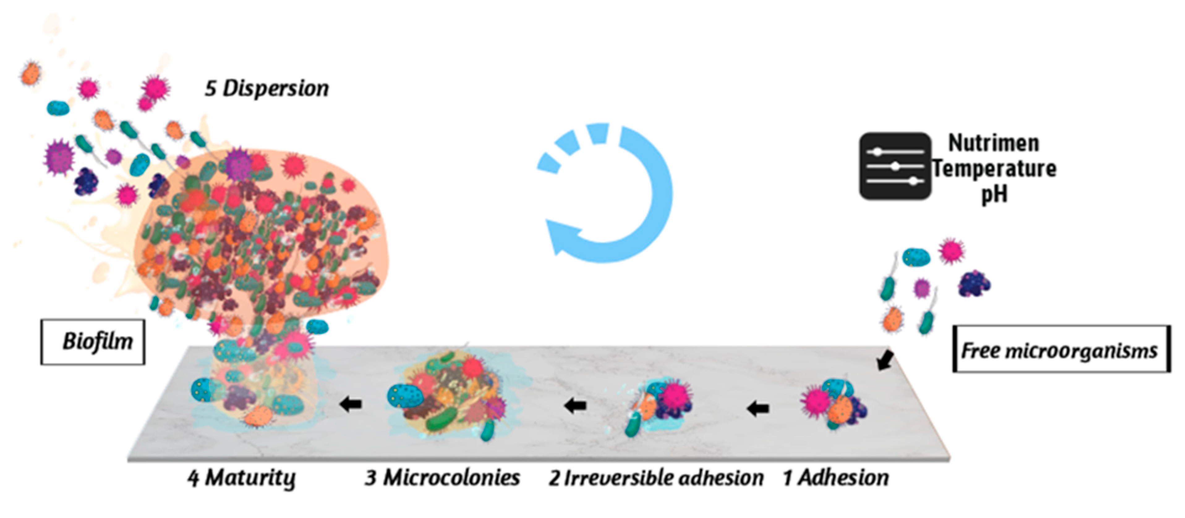
2. Stages of the Formation of E. coli Biofilm

The formation of microbial biofilm is a universal phenomenon. As long as the conditions permit, microorganisms often form biofilm in the environment [12]. The formation of biofilm depends upon the changes in nutrients, temperature, osmotic pressure, pH, and other factors in the environment. The formation of biofilm provides protection for microorganisms. The microorganisms in biofilm exhibit stronger environmental resistance than the migratory microorganisms and can survive in harsh environmental conditions.
Since the observation of microbes and biofilms by Antony van Leeuwenhoek in dental plaques using a microscope in 1676, studies about the mechanisms of biofilm formation have evolved rapidly [13]. To date, the formation of E. coli biofilm is generally divided into five stages, including reversible adhesion, irreversible adhesion, colony formation, biofilm maturation, and dispersion, as shown in Figure 1.

2.1. Reversible Adhesion between Microorganisms and Carriers

The free microorganisms receive environmental signals (such as nutrients), and use their extracellular organelles (such as flagella, cilia, and curly fimbriae) and outer membrane proteins in order to adhere to the carrier surface.

2.2. Irreversible Adhesion between Microorganisms and Carriers

Microorganisms secrete EPS, which enhances the adhesion of microorganisms with the carrier surface. EPS is composed of nucleic acids, proteins, lipids, lipopolysaccharides, and other substances, and forms a protective barrier for microorganisms by restricting and preventing the antibacterial agents from reaching the biological membrane of target organisms [14,15]. Notably, the latest findings also reveal that the bacterial organelles play important roles in the microbial irreversible attachment during the biofilm formation. For instance, the bacterial cell wall often deforms at this stage, which could enhance the adhesion between microorganisms and carriers [16].

2.3. Colony Formation Stage

The microorganisms, adhering to the carrier surface, grow and multiply to form colonies. Then, the continuous proliferation of microorganisms makes their biological growth space crowded with a lack of nutrients and accumulation of toxic substances. At this stage, the quorum-sensing (QS) signals, a cell-to-cell communication mechanism in bacteria, could induce the E. coli into the biofilm lifestyle by regulating the synthesis of biofilm matrix compounds [17]. During this process, EPS is significantly increased, forming a gel phase on the surface of the carrier to protect the microorganisms [18].

2.4. Biofilm Maturation Stage

E. coli microcolonies continue to accumulate, which further increases the thickness of the film and results in the formation of mushroom-like or columnar subunits. Then, the biofilm grows in a three-dimensional manner and eventually forms a viable three-dimensional structure [19].

2.5. Biofilm Dispersion Stage

The dispersion of biofilm can be divided into active dispersion and passive dispersion. The active dispersion refers to a decrease in the level of cyclic diguanylate (c-di-GMP) in cells, which results in the production of enzymes that degrade the matrix of biofilm and promote its dispersion. The passive dispersion depends on the external factors, such as physical triggers (wear, fluid shear, etc.) and enzymatic degradation (extracellular hydrolase) [20]. The dispersion of microorganisms from biofilm is beneficial for their reproduction and biofilm recombination, thereby forming a cycle of wandering microorganisms and biofilm [12].
In conclusion, the formation of E. coli biofilm is a strong survival support for E. coli in unfavorable living environmental conditions, protects the growth and reproduction of E. coli to the greatest extent, and provides favorable conditions for E. coli to wreak havoc on and pollute food, eventually causing infection and affecting human health.

3. Prevalence of E. coli Biofilm in the Food Industry

Hygienically, E. coli is an indicator of fecal pollution, and a serious threat to the life and health of consumers. Once the content of E. coli in a food item exceeds the standard, it is withdrawn from the shelves, resulting in huge economic losses.
Moreover, the biofilm-making ability of E. coli may pose a greater threat to the processing and production of food. Studies show that once the microbial biofilm is formed, the resistance of bacteria to disinfectants is increased by 500 times, suggesting that, in the presence of biofilms, the germicidal time and concentration of disinfectants must be increased to 10–100 times to kill bacteria [21,22]. Another study also proposes that the EPS of biofilm can inhibit antibiotics and bactericides from entering cells, and reduce the permeability, resulting in a significant increase in the concentration of antibiotics and bactericides in the presence of biofilm [23].
At present, a couple of studies have shown that the E. coli biofilm exists in nearly all aspects of the processing and production of all kinds of food (brewing industry, dairy products, fresh food, meat products, etc.), and may cause pipeline biological pollution and equipment damage (water system, cooling tower, and other technical failures), thereby contaminating food items [18,24,25,26,27].
Food ingredients can also be easily contaminated by E. coli, as it grows in a natural environment. This may introduce E. coli into the food industry. Studies have reported the isolation of E. coli from peppers, tomatoes, cantaloupes, and other crops in Nuevo Leon and Coavira, Mexico. A total of 341 strains of E. coli have been isolated, of which 76% of strains have the ability to form biofilms. Among them, 34% of E. coli strains form strong biofilms [5], which pose a potential risk to endanger consumers’ health. At the same time, due to the pathogenicity of E. coli to cause human and animal diseases, a great importance has been given to the detection of E. coli in poultry products. At present, Brazil is the largest exporter of chicken. Among the 88 strains of E. coli isolated from the chicken meat of four retail chicken companies in Brazil, 31 strains could form a strong biofilm, while only four strains could not form biofilm [28]. In addition, E. coli can easily form biofilms in processing equipment, such as stainless-steel metal products [29,30], polyethylene [31], walls and drains [32], due to improper cleaning. Nancy et al. isolated E. coli from fresh-cut processing equipment. The co-culturing of E. coli with the isolated Burkholderia caryophylli and Ralstonia insidiosa increases biofilm biomass by 180% and 63%, respectively, posing a great challenge to eliminating the foodborne pathogens in fresh-cut processing equipment [33].

4. Control Measures for the Prevention of E. coli Biofilm Formation

E. coli can form biofilm on the surface of food items, pipes, and processing equipment, which greatly increases its resistance to environmental conditions and reduces the effectiveness of disinfectants [34]. The biofilm increases the possibility of cross-infection of E. coli in food items and harms the health of consumers. Therefore, it is urgent to take effective measures to reduce or control the formation of E. coli biofilm during processing, as shown in Table 1, to reduce the risk of microbial contamination.

4.1. Chemical Methods

At present, the disinfectants, commonly used in food processing and slaughterhouses, include chlorine disinfectants, quaternary ammonium chloride (QAC), and lactic acid, which inhibit the formation of E. coli biofilm to a certain extent, but cannot completely inactivate it [56]. When studying the inhibitory effects of chlorine dioxide (CD), neutral oxygen potential water (NEOW), and sodium hypochlorite (SH) on E. coli biofilm, the inhibitory effects of NEOW and CD on E. coli biofilm biomass were significantly higher than that of SH and exhibited a certain degradation effect on E coli biofilm. Among them, NEOW was the most stable at a low temperature (5 °C) with the lowest chlorine loss rate and a wider bacteriostatic temperature range [35]. The comparative study of the inhibitory effect of 2-hydroxypropyl-3-piperazinyl-quinoline carboxylic acid methacrylate (HPQM) and silver-substituted zeolite (ZEOMIC) on linear low-density polyethylene (LLDPE) E. coli biofilm suggested that the HPQM exhibited comparatively better inhibitory properties and bacteriostatic effects on LLDPE with low roughness than ZEOMIC [36]. At the same time, a study has shown that different materials can affect the effectiveness of disinfectants. For example, ClO2 and SH were found to show the highest anti-biofilm activity against E. coli on stainless steel, followed by glass, plastic, and wood [37]. This information is useful in food processing for the selection of correct bactericides according to the actual application and different contact surfaces in food processing. However, a study demonstrated that the SH and lauroyl arginate ethyl (LAE) could inhibit the biofilm of E. coli on the surface of Hami melon, but a 2000-μg/mL concentration of SH and arginine LAE had no obvious effects on the inhibition of E. coli biofilm, and the biofilm showed high resistance to disinfectants [38]. Therefore, for the conventional chemical disinfectants, it is difficult to penetrate and spread into the biofilm in order to kill microorganisms and can no longer meet the standards of controlling microorganisms in food processing. In addition, using chemical methods for microbial control, such as chemical preservatives, not only easily produces adverse effects on food sensory properties and nutrition, but also easily causes allergies and other problems. Therefore, there is an urgent need to find new antibacterial methods or antimicrobial agents to reduce the occurrence of microbial cross-contamination. With the deepening of the research on microbial sterilization, anti-microbial photodynamic technology, to a certain extent, makes up for the shortcomings of general chemical bactericides, and shows great advantages in the pharmaceutical and food industries. A study has shown that riboflavin-mediated photodynamic sterilization technology has a significant inhibitory effect on E. coli biofilm and the mixed biofilm of E. coli and Staphylococcus aureus. Its inhibitory effect is mainly through the rise of reactive oxygen species, leading to bacterial oxidative stress and damaging the respiratory system [39].

4.2. Physical Methods

For the inhibition of E. coli biofilm in food processing and production, physical methods (washing, high-temperature sterilization, etc.) are often used in addition to chemical methods to reduce the occurrence of food’s cross-contamination. High-temperature sterilization is the most common physical method to control microorganisms. Saturated steam (SS) and superheated steam (SHS) can inactivate polyvinyl chloride (PVC) in food processing facilities and inhibit the formation of E. coli biofilm on the surface of stainless steel. SHS showed higher lethality and better inactivation of the cells in E. coli biofilm [40]. To prevent the formation of bacterial biofilm, it is a good method to suppress the biofilm adhesion on a solid surface by vibration. A study has shown that nano-vibration on the surface of materials can effectively inhibit the formation of E. coli biofilm. When the amplitude of vibration on the surface is greater than 21nm, the greater the amplitude, the less the number of viable bacteria. It can provide a reference for some precise and difficult methods to clean mechanical surfaces in food or medicine [57]. In addition, new physical sterilization technologies, including low-temperature plasma sterilization, not only show good anti-biofilm activities, but also retain the original sensory properties of food items to the greatest extent [58]. The low-temperature plasma sterilization also showed a good inhibitory effect on the formation of E. coli biofilm and a reduction in E. coli’s abundance under the biofilm to 5log10. At the same time, the low-temperature plasma sterilization, in combination with water spray, can not only inhibit bacteria, but also improve the efficiency of antifouling [41]. Similarly, with the extension of the study, Kadri et al. also found that cold atmospheric plasma treatment with a flow rate of 5 L/min could significantly inhibit E. coli biofilm and Listeria biofilm, while this inhibitory effect slightly decreased as biofilm growth age increased. Afterwards, a mixed culture of the two bacteria showed that the production of EPS in E. coli biofilm may contribute to the increases in the resistance of cold atmospheric plasma, which provides a new target to inactivate bacteria on the complex solid surface [59]. In addition, Adator et al. found that when the temperature was lower than 10 °C, the Shiga toxigenic E. coli (STEC) in the biofilm on stainless steel would not transfer to lettuce. However, when the temperature is 25 °C, STEC will transfer from the biofilm to stainless steel. It is suggested that keeping a low temperature in a food processing environment is helpful to control the transfer of E. coli in biofilm, resulting in microbial cross-contamination [60].
In the actual food processing and production, food industries often use the combination of chemical and physical sterilization methods to control microorganisms, and the synergistic effect between them maximizes the removal or inhibition of E. coli biofilm. The combination of lactic acid (LA) with water vapors can effectively reduce the abundance of E. coli on the surface of PVC and stainless steel, and exhibits lethal effects on the microorganisms in biofilm [42]. The combination of cold nitrogen plasma (CNP) with clove oil also showed significant synergistic inhibitory effects on E. coli biofilm [43].

4.3. Biological Components

In recent years, due to the decrease in the sensitivity of microorganisms in biofilm to conventional disinfectants, the germicidal efficacy of conventional disinfectants cannot reach an ideal state, which increases the risk of food microbial safety. Therefore, there is an urgent need to seek new active germicidal substances and improve their efficiency. Biological extracts are natural compounds, which avoid the problem of chemical disinfectant residues and have attracted wide attention because of their higher level of safety. Therefore, the inhibitory effects of biological extracts on E. coli biofilm have been widely reported.
With the attitude of safety, environmental protection, economy, and turning waste into treasure, animal extracts such as the scavenging effects of scallop shells on E. coli biofilm have attracted the attention of researchers. The main component of scallop shell is calcium carbonate (CaCO3), which is easily decomposed into calcium oxide (CaO) by heating, which then exhibits broad-spectrum antibacterial properties [61,62]. Studying the scallop shell powder (SSP) for its anti-biofilm activity, it was found that only 0.25% and 0.5% of scallop shell powder could inhibit E. coli biofilm on the surface of stainless steel in whey powder solution and meat processing plant washing water by 4 and 6 log CFU/cm2 and 3 and 5 log CFU/cm2, respectively. This inhibitory effect was positively correlated with the concentration of scallop shell powder, suggesting that it can be a good candidate for application in the food industry [44].
In addition, the concept of using bacteriophages, the natural predators of bacteria, as a novel strategy to prevent and eliminate biofilms has gradually attracted the attention of researchers. The treatment with phage AZO145A significantly inhibited the biofilm formation of STEC on the surface of stainless-steel plate, and significantly inhibited their migration at 24 °C in the biofilm formed in beef, which was reduced to 3.1 log10 CFU, thereby reducing the risk of food cross infection [45]. At the same time, with the in-depth study of bacteriophages, increasingly more bacteriophages were found to have a good inhibitory effect on E. coli biofilm, such as bacteriophage FP43, Daica, and135 [27,46]. In the bacteriostatic effect of phage, phage cocktails have a better inhibition effect on biofilm than that of single phage. The main inhibition process is to decompose biofilm into weak biofilm or non-biofilm to reduce bacteria [47].
The inhibitory effects of plant extracts on the biofilm of E. coli have been a hot topic. The plant extracts can be directly used in food processing equipment or food items to inhibit the E. coli biofilm and prolong the shelf-life of food items. Phenols are widely studied for their inhibitory effects on E. coli biofilm. Four components were obtained after extracting the active components from burdock leaves with ethanol and diluted with water to form different concentrations of ethanol (20%, 34%, and 70%). It was found that the active components obtained by different ethanol elution could significantly inhibit the E. coli biofilm, where the inhibitory effects were correlated with the phenol contents in the burdock leaves. The 70% ethanol elution could seriously damage the structure of E. coli biofilm, which no longer showed the state of a multi-layer growth [48]. The comparative study of nine catechol compounds, including catechol (CAT); veratrylalcohol (VER); guaiacol (GUA); 2-ethoxyphenol (ETH); 4-methylcatechol (MEC); 4-tert-butyl catechol (TEB); pyrogallol (PYR); 3-methoxycatechol (MET); and o-phenylene-phosphochloridite (OPP), suggested that the ETH, MEC, TEB, and PYR had the best inhibitory effects on E. coli biofilm [49]. The low-concentration carvanol revealed significant inhibitory effects on E. coli biofilm, which were proportional to the time, and could completely remove the biofilm after 15 min of exposure [50]. The characteristics of carvanol, such as low concentration and long time, can meet the requirements of disinfection in the food industry. Similarly, 50 μg/mL of coumarin could also inhibit more than 80% of E. coli biofilm without affecting the growth of bacteria [51]. In addition, the clove oil, lectins, and other substances also showed inhibitory effects on E. coli biofilm. The combination of clove oil and liposome, making solid liposomes (SLPs), can avoid the disadvantage of volatilization under light and at a high temperature, and improve stability and the utilization rate. The SLPs treatment dispersed the E. coli biofilm from the whole to a loose state, which seriously damaged its structure, decreased the biomass under membrane, and saw some cells collapse. In addition, the application of SLPs to the surface of cucumber and lettuce also showed a good inhibitory effect on biofilm and prolonged the shelf life of vegetables [52]. The antibacterial lectin, extracted from pomegranate peel, was found to have inhibitory effects on all the drug-resistant E. coli biofilms, with an inhibition rate of 50% at 6.25 g/mL or more [53].
The inhibitory effects of plant extracts on E. coli biofilm mainly depend on the inhibition of cell metabolic activities; gene expression; reduction in extracellular polysaccharides, bacterial motility, and cell hydrophobicity; and directly preventing the initial adhesion stage of biofilm formation. The treatment of E. coli with SLPs suggested a negative correlation of the SLPs concentration of clove oil with the bacterial metabolic activities, extracellular polysaccharides, and protein contents, thereby weakening the protective barrier and adhesion of bacteria [52]. The extract of Coptis chinensis could also inhibit the metabolic activities of E. coli biofilm [54]. Coumarin, umbelliferone, and aescin significantly inhibited the expression of CSG operon, involved in curl formation, and movement genes; reduced the formation of fimbriae; prevented the adhesion of E. coli to the surface; and inhibited the formation of biofilm [51]. Studying the inhibition of E. coli biofilm with triterpenoids (oleanolic acid and ursolic acid) suggested that oleanolic acid analogues could change the expression of genes involved in the regulation of type IV pili [63]. CAT, TEB, and PYR could significantly reduce the mobility of E. coli by inhibiting the formation of biofilm, and ETH, MEC, TEB, and PYR could significantly reduce the surface hydrophobicity of E. coli cells and hinder the adhesion stage during the formation of E. coli biofilm [49]. In the inhibition of vitamin C on E. coli, vitamin C can down-regulate the signal transduction genes and regulatory genes of biofilm by more than 27 times [4]. In the study of the inhibitory effect of cinnamon, marjoram, and thyme on the biofilm of E. coli and Listeria on the surface of polypropylene, it was also found that the inhibitory effect of cinnamon, marjoram, and thyme could be achieved by penetrating the biofilm and cell membrane, further changing the fluidity and permeability of the membrane, condensing the protons of the cytoplasm to form the weak mitochondria [55].

5. Conclusions

The formation of microbial biofilm is a kind of self-protection behavior of microorganisms in unfavorable environmental conditions, which helps them in avoiding environmental stresses. Therefore, the E. coli biofilm has become a hidden danger of microbial contamination in the food industry. The formation of E. coli biofilm is a complex process, which is affected by a variety of regulatory networks. At present, there is an urgent need for studies on the regulatory mechanism of biofilm maturation, as well as the prevention methods of biofilm formation. Meanwhile, studying the mechanisms of the inhibition of E. coli biofilm is of great significance for the development of efficient and safe germicide to reduce E. coli biofilm in the food industry.

Author Contributions

F.Z.: Writing—Original Draft; D.W.: Writing—Original Draft; J.H.: Writing—Review and Editing; Y.Z.: Supervision, Funding Acquisition; B.K.T.: Writing—Review and Editing; S.L.: Supervision, Funding Acquisition. All authors have read and agreed to the published version of the manuscript.

Funding

The study was financially supported by the National Natural Science Foundation of China (No. 31801649), “13th Five Year Plan” National Key Research and Development Project (2016YFD0400403); Natural Science Foundation of Fujian Province (2020I0010); Science and Technology Major Project of Zhangzhou (ZZ2019ZD18); Special Funds for Science and Technology Innovation of FAFU (CXZX2018063; CXZX2019100S; CXZX2019101S).

Institutional Review Board Statement

Not applicable.

Informed Consent Statement

Not applicable.

Data Availability Statement

Not applicable.

Conflicts of Interest

The authors declare no conflict of interest.

References

  1. Friedmann, H. Escherich and Escherichia. Adv. Appl. Microbiol. 2006, 60, 133–196. [Google Scholar] [CrossRef] [PubMed]
  2. Wells, J.G.; Davis, B.R.; Wachsmuth, I.K.; Riley, L.W.; Morris, G.K. Laboratory investigation of hemorrhagic colitis outbreaks associated with a rare Escherichia coli serotype. J. Clin. Microbiol. 1983, 18, 512–520. [Google Scholar] [CrossRef] [PubMed]
  3. Doyle, M.P. Escherichia coli O157:H7 and its significance in foods. Int. J. Food Microbiol. 1991, 12, 289–301. [Google Scholar] [CrossRef]
  4. Shivaprasad, D.P.; Taneja, N.K.; Lakra, A.; Sachdev, D. In vitro and in situ abrogation of biofilm formation in E. coli by vitamin C through ROS generation, disruption of quorum sensing and exopolysaccharide production. Food Chem. 2020, 341, 128171. [Google Scholar] [CrossRef]
  5. Corzo-Ariyama, H.A.; García-Heredia, A.; Heredia, N.; García, S.; León, J.; Jaykus, L.; Solís-Soto, L. Phylogroups, pathotypes, biofilm formation and antimicrobial resistance of Escherichia coli isolates in farms and packing facilities of tomato, jalapeño pepper and cantaloupe from Northern Mexico. Int. J. Food Microbiol. 2019, 290, 96–104. [Google Scholar] [CrossRef] [PubMed]
  6. Frank, C.; Werber, D.; Cramer, J.; Askar, M.; Faber, M.; an der Heiden, M.; Bernard, H.; Fruth, A.; Prager, R.; Spode, A.; et al. Epidemic Profile of Shiga-Toxin-Producing Escherichia coli O104:H4 Outbreak in Germany. N. Engl. J. Med. 2011, 365, 1771–1780. [Google Scholar] [CrossRef]
  7. Buchholz, U.; Bernard, H.; Werber, D.; Böhmer, M.M.; Remschmidt, C.; Wilking, H.; Deleré, Y.; an der Heiden, M.; Adlhoch, C.; Dreesman, J.; et al. German Outbreak of Escherichia coli O104:H4 Associated with Sprouts. N. Engl. J. Med. 2011, 365, 1763–1770. [Google Scholar] [CrossRef]
  8. Mermin, J.H.; Griffin, P.M. Invited Commentary: Public Health in Crisis: Outbreaks of Escherichia coli O157: H7 Infections in Japan. Am. J. Epidemiol. 1999, 150, 797–803. [Google Scholar] [CrossRef]
  9. Branda, S.S.; Vik, Å.; Friedman, L.; Kolter, R. Biofilms: The matrix revisited. Trends Microbiol. 2005, 13, 20–26. [Google Scholar] [CrossRef]
  10. Flemming, H.C.; Neu, T.R.; Wozniak, D.J. The EPS Matrix: The “House of Biofilm Cells”. J. Bacteriol. 2007, 189, 7945–7947. [Google Scholar] [CrossRef]
  11. Carpio, A.; Cebrián, E.; Vidal, P. Biofilms as poroelastic materials. Int. J. Non-Linear Mech. 2019, 109, 1–8. [Google Scholar] [CrossRef]
  12. Watnick, P.; Kolter, R. Biofilm, City of Microbes. J. Bacteriol. 2000, 182, 2675. [Google Scholar] [CrossRef] [PubMed]
  13. Costerton, J.W.; Stewart, P.S.; Greenberg, E.P. Bacterial Biofilms: A Common Cause of Persistent Infections. Science 1999, 284, 1318. [Google Scholar] [CrossRef] [PubMed]
  14. Sutherland, I.W. The biofilm matrix-an immobilaized but dynamic microbial environment. Trends Microbiol. 2001, 9, 222–227. [Google Scholar] [CrossRef]
  15. Simes, M.; Bennett, R.N.; Rosa, E.A.S. Understanding antimicrobial activities of phytochemicals against multidrug resistant bacteria and biofilms. Cheminform 2009, 26, 746–757. [Google Scholar] [CrossRef]
  16. Uneputty, A.; Dávila-Lezama, A.; Garibo, D.; Oknianska, A.; Bogdanchikova, N.; Hernández-Sánchez, J.F.; Susarrey-Arce, A. Strategies applied to modify structured and smooth surfaces: A step closer to reduce bacterial adhesion and biofilm formation. Colloid Interface Sci. Commun. 2022, 46, 100560. [Google Scholar] [CrossRef]
  17. Harmsen, M.; Yang, L.; Pamp, S.N.J.; Tolker-Nielsen, T. An update on Pseudomonas aeruginosa biofilm formation, tolerance, and dispersal. Pathog. Dis. 2010, 59, 253–268. [Google Scholar] [CrossRef]
  18. Simões, M.; Simões, L.C.; Vieira, M.J. A review of current and emergent biofilm control strategies. LWT-Food Sci. Technol. 2010, 43, 573–583. [Google Scholar] [CrossRef]
  19. Danese, N.P.; Pratt, A.L.; Kolter, R. Exopolysaccharide production is required for development of Escherichia coli K-12 biofilm architecture. J. Bacteriol. 2000, 182, 2399–3593. [Google Scholar] [CrossRef]
  20. Wille, J.; Coenye, T. Biofilm dispersion: The key to biofilm eradication or opening Pandora’s box? Biofilm 2020, 2, 100027. [Google Scholar] [CrossRef]
  21. Sofos, J.N.; Geornaras, I. Overview of current meat hygiene and safety risks and summary of recent studies on biofilms, and control of Escherichia coli O157:H7 in nonintact, and Listeria monocytogenes in ready-to-eat, meat products. Meat Ence 2010, 86, 2–14. [Google Scholar] [CrossRef] [PubMed]
  22. Fletcher, C.G.C. Assessing manufacturers’ recommended concentrations of commercial sanitizers on inactivation of Listeria monocytogenes. Food Control 2012, 26, 194–199. [Google Scholar] [CrossRef]
  23. Helgadóttir, S.; Pandit, S.; Mokkapati, V.R.S.S.; Westerlund, F.; Apell, P.; Mijakovic, I. Vitamin C Pretreatment Enhances the Antibacterial Effect of Cold Atmospheric Plasma. Front. Cell. Infect. Microbiol. 2017, 7, 43. [Google Scholar] [CrossRef] [PubMed]
  24. Gilbert, P.; McBain, A.J.; Rickard, A.H. Formation of microbial biofilm in hygienic situations: A problem of control. Int. Biodeterior. Biodegrad. 2003, 51, 245–248. [Google Scholar] [CrossRef]
  25. Kang, J.W.; Lee, H.Y.; Kang, D.H. Synergistic bactericidal effect of hot water with citric acid against Escherichia coli O157:H7 biofilm formed on stainless steel. Food Microbiol. 2020, 95, 103676. [Google Scholar] [CrossRef]
  26. Ma, Z.; Bumunang, E.W.; Stanford, K.; Bie, X.; Niu, Y.D.; Mcallister, T.A. Biofilm Formation by Shiga Toxin-Producing Escherichia coli on Stainless Steel Coupons as Affected by Temperature and Incubation Time. Microorganisms 2019, 7, 95. [Google Scholar] [CrossRef]
  27. Milho, C.; Silva, M.; Sillankorva, S.; Harper, D.R. Biofilm Applications of Bacteriophages. Bacteriophages 2019, 1–35. [Google Scholar] [CrossRef]
  28. Crecencio, R.B.; Brisola, M.C.; Bitner, D.; Frigo, A.; Stefani, L.M. Antimicrobial susceptibility, biofilm formation and genetic profiles of Escherichia coli isolated from retail chicken meat. Infect. Genet. Evol. 2020, 84, 104355. [Google Scholar] [CrossRef]
  29. Skandamis, P.N.; Stopforth, J.D.; Ashton, L.V.; Geornaras, I.; Kendall, P.A. Escherichia coli O157:H7 survival, biofilm formation and acid tolerance under simulated slaughter plant moist and dry conditions. Food Microbiol. 2009, 26, 112–119. [Google Scholar] [CrossRef]
  30. Hood, S.K.; Zottola, E.A. Adherence to stainless steel by foodborne microorganisms during growth in model food systems. Int. J. Food Microbiol. 1997, 37, 145–153. [Google Scholar] [CrossRef]
  31. Dourou, D.; Beauchamp, C.S.; Yoon, Y.; Geornaras, I.; Belk, K.E.; Smith, G.C.; Nychas, G.J.E.; Sofos, J.N. Attachment and biofilm formation by Escherichia coli O157:H7 at different temperatures, on various food-contact surfaces encountered in beef processing. Int. J. Food Microbiol. 2011, 149, 262–268. [Google Scholar] [CrossRef] [PubMed]
  32. Lin, S.; Yang, L.; Chen, G.; Li, B.; Chen, D.; Li, L.; Xu, Z. Pathogenic features and characteristics of food borne pathogens biofilm: Biomass, viability and matrix. Microb. Pathog. 2017, 111, 285–291. [Google Scholar] [CrossRef]
  33. Liu, N.T.; Nou, X.; Lefcourt, A.M.; Shelton, D.R.; Lo, Y.M. Dual-species biofilm formation by Escherichia coli O157:H7 and environmental bacteria isolated from fresh-cut processing facilities. Int. J. Food Microbiol. 2014, 171, 15–20. [Google Scholar] [CrossRef] [PubMed]
  34. Duc, H.M.; Son, H.M.; Honjoh, K.I.; Miyamoto, T. Isolation and application of bacteriophages to reduce Salmonella contamination in raw chicken meat. LWT-Food Sci. Technol. 2018, 91, 353–360. [Google Scholar] [CrossRef]
  35. Meireles, A.; Ferreira, C.; Melo, L.; Simes, M. Comparative stability and efficacy of selected chlorine-based biocides against Escherichia coli in planktonic and biofilm states. Food Res. Int. 2017, 102, 511–518. [Google Scholar] [CrossRef] [PubMed]
  36. Pin-Ngoen, K.; Kositchaiyong, A.; Prapagdee, B.; Sombatsompop, N. Formation of Escherichia coli biofilm on LLDPE sheets by incorporation of 2-hydroxypropyl-3-piperazinyl-quinoline carboxylic acid methacrylate or silver-substituted zeolite. Int. Biodeterior. Biodegrad. 2016, 109, 211–222. [Google Scholar] [CrossRef]
  37. Bang, J.; Hong, A.; Kim, H.; Beuchat, L.R.; Rhee, M.S.; Kim, Y.; Ryu, J.-H. Inactivation of Escherichia coli O157:H7 in biofilm on food-contact surfaces by sequential treatments of aqueous chlorine dioxide and drying. Int. J. Food Microbiol. 2014, 191, 129–134. [Google Scholar] [CrossRef]
  38. Fu, Y.; Deering, A.J.; Bhunia, A.K.; Yao, Y. Biofilm of Escherichia coli O157:H7 on cantaloupe surface is resistant to lauroyl arginate ethyl and sodium hypochlorite. Int. J. Food Microbiol. 2017, 260, 11–16. [Google Scholar] [CrossRef]
  39. Banerjee, S.; Ghosh, D.; Vishakha, K.; Das, S.; Mondal, S.; Ganguli, A. Photodynamic antimicrobial chemotherapy (PACT) using riboflavin inhibits the mono and dual species biofilm produced by antibiotic resistant Staphylococcus aureus and Escherichia coli. Photodiagnosis Photodyn. Ther. 2020, 32, 102002. [Google Scholar] [CrossRef]
  40. Ban, G.-H.; Yoon, H.; Kang, D.-H. A comparison of saturated steam and superheated steam for inactivation of Escherichia coli O157:H7, Salmonella Typhimurium, and Listeria monocytogenes biofilms on polyvinyl chloride and stainless steel. Food Control 2014, 40, 344–350. [Google Scholar] [CrossRef]
  41. Kovalova, Z.; Leroy, M.; Kirkpatrick, M.J.; Odic, E.; Machala, Z. Corona discharges with water electrospray for Escherichia coli biofilm eradication on a surface. Bioelectrochemistry 2016, 112, 91–99. [Google Scholar] [CrossRef] [PubMed]
  42. Ban, G.H.; Park, S.H.; Kim, S.O.; Ryu, S.; Kang, D.H. Synergistic effect of steam and lactic acid against Escherichia coli O157:H7, Salmonella Typhimurium, and Listeria monocytogenes biofilms on polyvinyl chloride and stainless steel. Int. J. Food Microbiol. 2012, 157, 218–223. [Google Scholar] [CrossRef]
  43. Cui, H.; Ma, C.; Lin, L. Synergetic antibacterial efficacy of cold nitrogen plasma and clove oil against Escherichia coli O157:H7 biofilms on lettuce. Food Control 2016, 66, 8–16. [Google Scholar] [CrossRef]
  44. Bodur, T.; Cagri-Mehmetoglu, A. Removal of Listeria monocytogenes, Staphylococcus aureus and Escherichia coli O157:H7 biofilms on stainless steel using scallop shell powder. Food Control 2012, 25, 1–9. [Google Scholar] [CrossRef]
  45. Wang, C.; Hang, H.; Zhou, S.B.; Niu, Y.D.; Du, H.C.; Kim, S.; Tim, A.M. Bacteriophage biocontrol of Shiga toxigenic Escherichia coli (STEC) O145 biofilms on stainless steel reduces the contamination of beef. Food Microbiol. 2020, 92, 103572. [Google Scholar] [CrossRef] [PubMed]
  46. Zhang, Y.; Shigemura, K.; Duc, H.M.; Shen, C.; Huang, H.-H.; Sato, J.; Masuda, Y.; Honjoh, K.-I.; Miyamoto, T. Effects of bacteriophage on inhibition and removal of mixed biofilm of enterohemorrhagic Escherichia coli O157:H7 and O91:H. LWT-Food Sci. Technol. 2020, 134, 109945. [Google Scholar] [CrossRef]
  47. Montso, P.K.; Mlambo, V.; Ateba, C.N. Efficacy of novel phages for control of multi-drug resistant Escherichia coli O177 on artificially contaminated beef and their potential to disrupt biofilm formation. Food Microbiol. 2021, 94, 103647. [Google Scholar] [CrossRef]
  48. Lou, Z.; Song, X.; Hong, Y.; Wang, H.; Lin, Y. Separation and enrichment of burdock leaf components and their inhibition activity on biofilm formation of E. coli. Food Control 2013, 32, 270–274. [Google Scholar] [CrossRef]
  49. Baptista, J.; Simes, M.; Borges, A. Effect of plant-based catecholic molecules on the prevention and eradication of Escherichia coli biofilms: A structure activity relationship study. Int. Biodeterior. Biodegrad. 2018, 141, 101–113. [Google Scholar] [CrossRef]
  50. Campana, R.; Baffone, W. Carvacrol efficacy in reducing microbial biofilms on stainless steel and in limiting re-growth of injured cells. Food Control 2018, 90, 10–17. [Google Scholar] [CrossRef]
  51. Lee, J.H.; Kim, Y.G.; Cho, H.S.; Ryu, S.Y.; Cho, M.H.; Lee, J. Coumarins reduce biofilm formation and the virulence of Escherichia coli O157:H7. Phytomed. Int. J. Phytother. Phytopharm. 2014, 21, 1037–1042. [Google Scholar] [CrossRef] [PubMed]
  52. Cui, H.; Zhang, C.; Li, C.; Lin, L. Inhibition of Escherichia coli O157:H7 biofilm on vegetable surface by solid liposomes of clove oil. LWT-Food Sci. Technol. 2019, 117, 108656. [Google Scholar] [CrossRef]
  53. Silva, P.M.; Baldry, M.; Peng, P.; Oliveira Silva, J.N.; Soares, T.; Brayner, F.A.; Alves, L.C.; Feitosa, A.P.S.; Paiva, P.M.G.; Ingmer, H.; et al. Punica granatum sarcotesta lectin (PgTeL) impairs growth, structure, viability, aggregation, and biofilm formation ability of Staphylococcus aureus clinical isolates. Int. J. Biol. Macromol. 2019, 123, 600–608. [Google Scholar] [CrossRef]
  54. Mohammadi, M.; Masoumipour, F.; Hassanshahian, M.; Jafarinasab, T. Study the antibacterial and antibiofilm activity of Carum copticum against antibiotic-resistant bacteria in planktonic and biofilm forms. Microb. Pathog. 2019, 129, 99–105. [Google Scholar] [CrossRef] [PubMed]
  55. Vidács, A.; Kerekes, E.; Rajkó, R.; Petkovits, T.; Alharbi, N.S.; Khaled, J.M.; Vágvölgyi, C.; Krisch, J. Optimization of essential oil-based natural disinfectants against Listeria monocytogenes and Escherichia coli biofilms formed on polypropylene surfaces. J. Mol. Liq. 2018, 255, 257–262. [Google Scholar] [CrossRef]
  56. Wang, R.; Kalchayanand, N.; King, D.A.; Luedtke, B.E.; Bosilevac, J.M.; Arthur, T.M. Biofilm Formation and Sanitizer Resistance of Escherichia coli O157:H7 Strains Isolated from “High Event Period” Meat Contamination. J. Food Prot. 2014, 77, 1982–1987. [Google Scholar] [CrossRef]
  57. Lin, F.; Yuan, S.; Han, W. Effective prevention of Escherichia coli biofilm on materials by nano-vibration. Colloids Surf. A Physicochem. Eng. Asp. 2021, 608, 125610. [Google Scholar] [CrossRef]
  58. Zhu, Y.; Li, C.; Cui, H.; Lin, L. Feasibility of cold plasma for the control of biofilms in food industry. Trends Food Sci. Technol. 2020, 99, 142–151. [Google Scholar] [CrossRef]
  59. Kadri, H.; Costello, K.M.; Thomas, P.; Wantock, T.; Velliou, E.G. The antimicrobial efficacy of remote cold atmospheric plasma effluent against single and mixed bacterial biofilms of varying age. Food Res. Int. 2021, 141, 110126. [Google Scholar] [CrossRef]
  60. Adator, E.H.; Cheng, M.; Holley, R.; Mcallister, T.; Narvaez-Bravo, C. Ability of Shiga toxigenic Escherichia coli to survive within dry-surface biofilms and transfer to fresh lettuce. Int. J. Food Microbiol. 2018, 269, 52–59. [Google Scholar] [CrossRef]
  61. Cagri-Mehmetoglu, A. Inhibition of Listeria monocytogenes and Salmonella enteritidis on chicken wings using scallop-shell powder. Poultry Science 2011, 20, 2600–2605. [Google Scholar] [CrossRef] [PubMed]
  62. Bae, D.H.; Yeon, J.H.; Park, S.Y.; Lee, D.H.; Ha, S.D. Bactericidal effects of CaO (scallop-shell powder) on foodborne pathogenic bacteria. Arch. Pharmacal Res. 2006, 29, 298–301. [Google Scholar] [CrossRef] [PubMed]
  63. Lawrence, J.A.; Huang, Z.; Rathinavelu, S.; Hu, J.-F.; Garo, E.; Ellis, M.; Norman, V.L.; Buckle, R.; Williams, R.B.; Starks, C.M.; et al. Optimized plant compound with potent anti-biofilm activity across gram-negative species. Bioorg. Med. Chem. 2020, 28, 115229. [Google Scholar] [CrossRef] [PubMed]
Figure 1. Schematic diagram of microbial biofilm formation. When the environmental conditions (nutrients, temperature, pH, etc.) are adverse, the free microorganism actively and reversibly adheres to the carrier surface, and then secretes EPS (blue) to further enhance the adhesion between the microorganism and the carrier by forming irreversible adhesion. Then, the microorganism grows and propagates to form colonies, and biofilm (yellow) is formed on the surface. After the increase in microorganisms, the biofilm is mature, becoming mushroom-like and with a three-dimensional structure. With the increase in microorganisms in the biofilm, they disperse from the biofilm to form new free microorganisms. As the cycle continues, microorganisms can survive in harsh environments.
Foods 11 02469 g001
Table 1. Inhibition effect of different sterilization methods on E. coli biofilm.
Bacteriostatic ModeBacteriaCarrier MaterialAntibacterial SubstanceConcentrationProcess TimeReduction (logCFU·cm−2)
or Bacteriostatic Rate
Reference
Chemical methodsE. coli CECT 434Stainless steel AISI 316Neutral oxygen potential water50 ppm20 min3.26 [35]
Chlorine dioxide50 ppm20 min3.20
Sodium dichloroisocyanurate50 ppm20 min3.20
Sodium hypochlorite50 ppm20 min2.46
E. coli ATCC 25922Linear low-density poly-ethylene2-hydroxypropyl-3-piperazinyl-quinoline carboxylic acid methacrylate1500–2500 ppm3-5 day99% [36]
E. coli O157:H7Stainless steelSodium hypochlorite (NaOCl)200 μg/mL15 min7.7 [37]
Aqueous chlorine dioxide (ClO2)200 μg/mL15 minND
GlassNaOCl200 μg/mL15 min8.2
ClO2200 μg/mL15 minND
PlasticNaOCl200 μg/mL15 min3.3
ClO2200 μg/mL15 minND
WoodNaOCl200 μg/mL15 min1.3
ClO2200 μg/mL15 min1.5
E. coli O157:H7 B6-914Glass cover SlidesLauroyl arginate ethyl200 μg/mL24 h0.46 [38]
Sodium hypochlorite200 μg/mL24 h0.59
Multi drug resistant Escherichia coli glass slidesPhotodynamic antimicrobial chemotherapy50 μM10min34% [39]
Physical methodsE. coli O157:H7Polyvinyl chlorideSaturated steam/5 s1.21 [40]
Superheated steam/5 s1.26
Stainless steelSaturated steam/5 s1.52
Superheated steam/5 s1.84
E. coli strain BW25113 F+Glass cover slidesPositive corona/15 min5.28 [41]
Negative corona 5.4
E. coli O157:H7
(ATCC 35150,
ATCC 43889,
ATCC 43890)
Polyvinyl chlorideLactic acid and water vapors0.5%–2%5 s0.76–3.78 [42]
Stainless steelLactic acid and water vapors0.5%–2%5 s1.64–3.92
E. coli EHEC O157:H7 CICC 21530Stainless steelClove oil1 mg/mL30 min3.32 [43]
Cold nitrogen plasma/3 min2.23
Biological components componentsE. coli O157:H7 NCTC 12900Stainless steelScallop shell powder0.25%1 min4-6 [44]
0.5%3-5
STEC O145:H25Stainless steelBacteriophage AZO145A2 × 1010 pfu/mL3 h3.1 [45]
Escherichia coli O157:H7 and O91:H-96-well platesBacteriophage FP431010 pfu/mL6 h2.85 [46]
E. coli O17796-well
polystyrene plates
Phage cocktail stock1 × 108 pfu/mL24 hND [47]
E. coli ATCC25922Silicone disksComponents of burdock leaves0.017 mg/mL24 h50% [48]
E. coli CECT43496-well microtiter plates2-ethoxyphenol7 mM24 h58.0 ± 15.0% [49]
4-methylcatechol3.5 mM24 h61.0 ± 10.0%
4-tert-butyl catechol1.6 mM24 h77.0 ± 0.0%
pyrogallol5 mM24 h73.0 ± 4.0%
E. coli
O157:H7 ATCC 35150
Stainless steelCarvacrol1%5 min6.04 [50]
E. coli O157:H7 ATCC4389596-well polystyrene platesCoumarins50 µg/mL24 habove 80% [51]
E. coli O157:H748-well plateSolid liposomes0.5 mg/mL24 h65.74% [52]
E. coli 2011-60-1493-3, 2011/25/62, C24716 C-2603696-well microtiter platePunica granatum sarcotesta lectin ≥6.25 g/mL24 h≥50% [53]
Escherichia coli (ATCC 35218)96-well poly-
styrene microtiter plate
Ethanol extract of Carum coptis chinensis25 mg/mL24 h≥70% [54]
E. coli EMC1796-well polystyrene plateVitamin C30 mM24 h50% [4]
Escherichia coli SZMC 0582Polypropylene spatulaCinnamomum Zeylanicum1.2 mg/mL10 min9 ± 5.45% [55]
Origanum majorana4.5 mg/mL10 min100 ± 0.00%
Thymus vulgaris3.8 mg/mL10 min100 ± 0.00%
HC-DPE (active ingredients:
15% peracetic-acid, 20% total peroxide)
0.1%10 min100 ± 0.00%
Sodium hypochlorite0.84%10 min100 ± 0.00%
ND: below the detection limit, not detected.
Publisher’s Note: MDPI stays neutral with regard to jurisdictional claims in published maps and institutional affiliations.

