Two Rare Cases of Non-Syndromic Paramolars with Family Occurrence and a Review of Literature


Abstract
:1. Introduction
2. Case Presentation
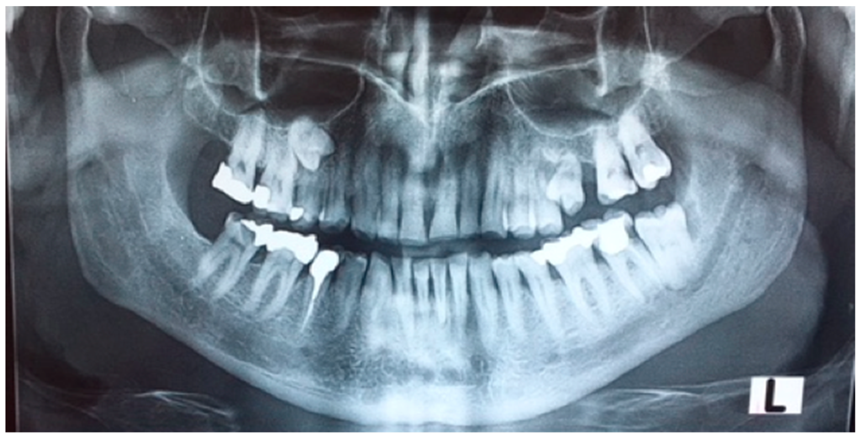
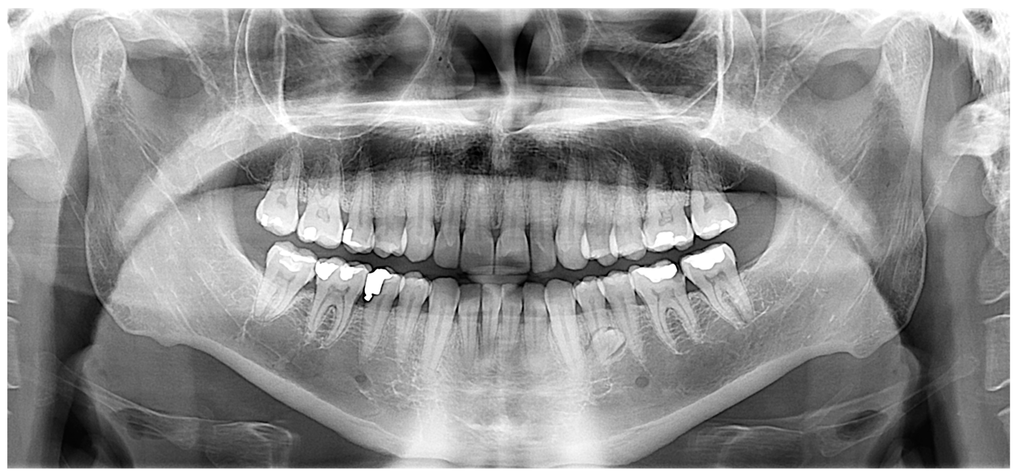
2.1. Case 1
Case 1 Presentation
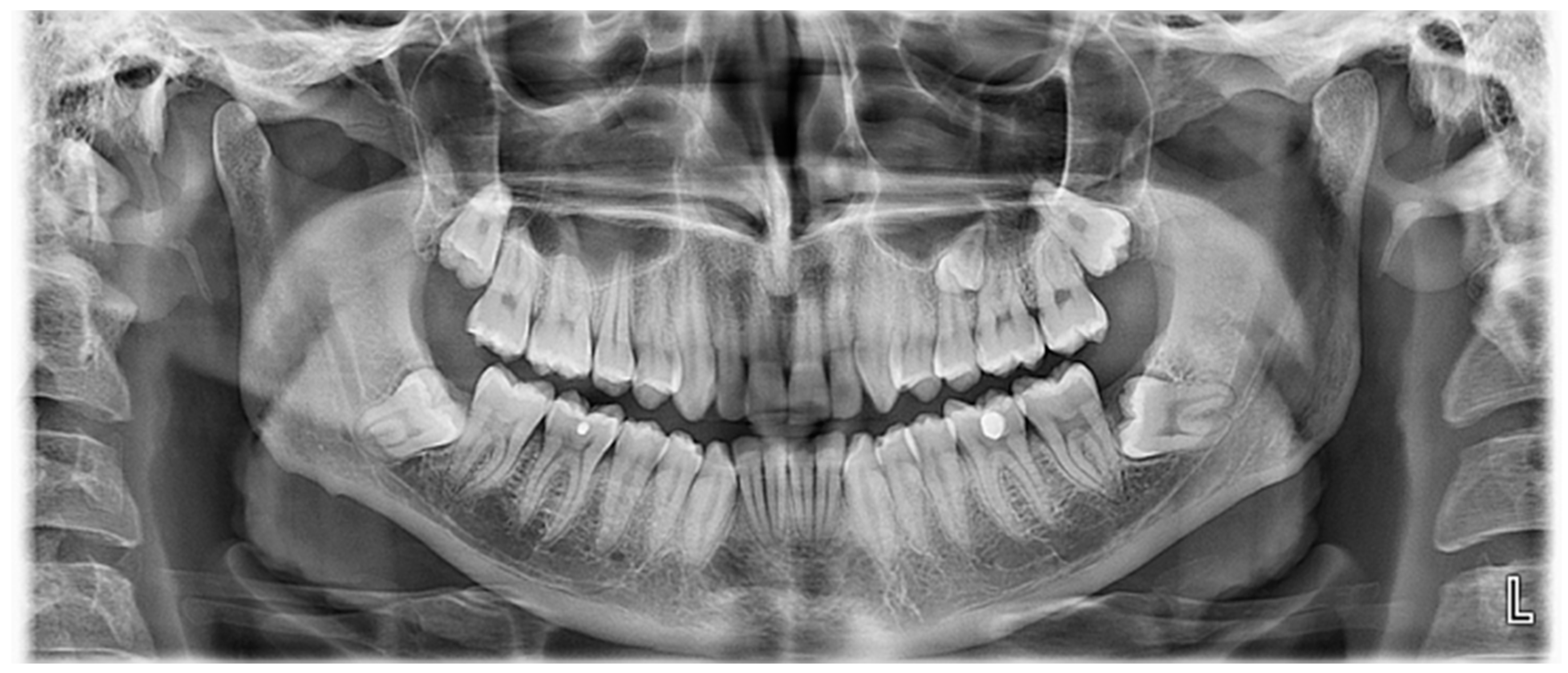
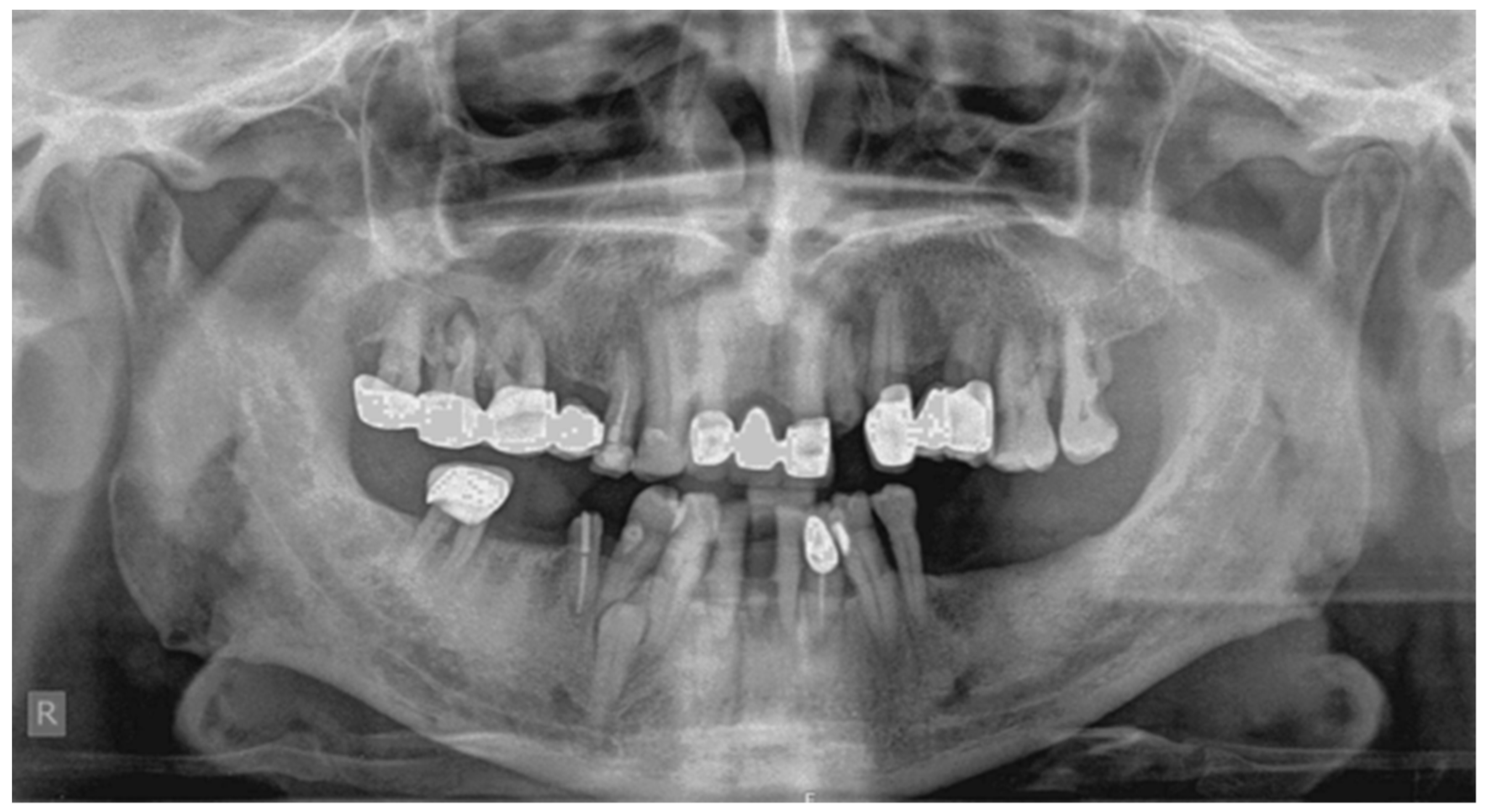
2.2. Case 2
Case 2 Presentation
3. Discussion
Author Contributions
Funding
Conflicts of Interest
References
- Anthonappa, R.P.; King, N.M.; Rabie, A.B. Diagnostic tools used to predict the prevalence of supernumerary teeth: A meta-analysis. Dentomaxillofac. Radiol. 2012, 41, 444–449. [Google Scholar] [CrossRef]
- Munshi, A.; Munshi, A.K. Midline space closure in the mixed dentition: A case report. J. Indian Soc. Pedod. Prev. Dent. 2001, 19, 57–60. [Google Scholar] [PubMed]
- Díaz, A.; Orozco, J.; Fonseca, M. Multiple hyperodontia: Report of a case with 17 supernumerary teeth with non syndromic association. Med. Oral. Patol. Oral Cir. Bucal. 2009, 14, E229–E231. [Google Scholar] [PubMed]
- Pippi, R. Odontomas and supernumerary teeth: Is there a common origin? Int. J. Med. Sci. 2014, 11, 1282–1297. [Google Scholar] [CrossRef] [PubMed]
- Rajab, L.D.; Hamdan, M.A. Supernumerary teeth: Review of the literature and a survey of 152 cases. Int. J. Paediatr. Dent. 2002, 12, 244–254. [Google Scholar] [CrossRef] [PubMed]
- Lubinsky, M.; Kantaputra, P.N. Syndromes with supernumerary teeth. Am. J. Med. Genet. A 2016, 170, 2611–2616. [Google Scholar] [CrossRef]
- Subasioglu, A.; Savas, S.; Kucukyilmaz, E.; Kesim, S.; Yagci, A.; Dundar, M. Genetic background of supernumerary teeth. Eur. J. Dent. 2015, 9, 153–158. [Google Scholar] [PubMed]
- Garvey, M.T.; Barry, H.J.; Blake, M. Supernumerary teeth an overview of the classification, diagnosis and treatment. J. Can. Dent. Assoc. 1999, 65, 612–616. [Google Scholar]
- Dubuk, A.N.; Selvig, K.A.; Tellefsen, G.; Wikesjö, U.M. Atypically located paramolar. Report of a rare case. Eur. J. Oral Sci. 1996, 104, 138–140. [Google Scholar] [CrossRef] [PubMed]
- Toureno, L.; Park, J.H.; Hwang, E.H.; Cederberg, R.A.; Shin, J.W. Identification of Supernumerary Teeth in 2D and 3D: Review of Literature and a Proposal. J. Dent. Educ. 2013, 77, 43–50. [Google Scholar] [PubMed]
- Kakolewska–Maczyńska, J.; Zyszko, A. Paramolar and distomolar teeth. Czas Stomatol. 1990, 43, 232–237. [Google Scholar]
- Mitchell, L. Supernumerary teeth. Dent. Update 1989, 16, 65–69. [Google Scholar] [PubMed]
- Primosch, R.E. Anterior supernumerary teeth—Assessment and surgical intervention in children. Pediatr. Dent. 1981, 3, 204–215. [Google Scholar]
- Ata-Ali, F.; Ata-Ali, J.; Peñarrocha-Oltra, D.; Peñarrocha-Diago, M. Prevalence, etiology, diagnosis, treatment and complications of supernumerary teeth. J. Clin. Exp. Dent. 2014, 6, e414–e418. [Google Scholar] [CrossRef] [PubMed]
- Srivatsan, P.; Aravindha Babu, N. Mesiodens with an unusual morphology and multiple impacted supernumerary teeth in a non-syndromic patient. Ind. J. Dent. Res. 2003, 18, 138–140. [Google Scholar] [CrossRef]
- Scheiner, M.A.; Sampson, W.J. Supernumerary teeth: A review of literature and four case reports. Aust. Dent. J. 1997, 42, 160–165. [Google Scholar] [CrossRef]
- Munne, P.M.; Felszeghy, S.; Jussila, M.; Suomalainen, M.; Thesleff, I.; Jernvall, J. Splitting placodes: Effects of bone morphogenetic protein and Activin on the patterning and identity of mouse incisors. Evol. Dev. 2010, 12, 383–392. [Google Scholar] [CrossRef]
- Fleming, P.S.; Xavier, G.M.; DiBiase, A.T.; Cobourne, M.T. Revisiting the supernumerary: The epidemiological and molecular basis of extra teeth. Br. Dent. J. 2010, 208, 25–30. [Google Scholar] [CrossRef]
- Brook, A.H. A unifying etiological explanation for anomalies of human tooth number and size. Arch. Oral Biol. 1984, 29, 373–378. [Google Scholar] [CrossRef]
- Gallas, M.M.; Garcia, A. Retention of permanent incisors by mesiodens: A family affair. Br. Dent. J. 2000, 188, 63–64. [Google Scholar] [CrossRef]
- Rao, P.V.; Chidzonga, M.M. Supernumerary teeth: Literature review. Cent. Afr. J. Med. 2001, 47, 22–26. [Google Scholar]
- Nayak, G.; Shetty, S.; Singh, I.; Pitalia, D. Paramolar—A supernumerary molar: A case report and an overview. Dent. Res. J. 2012, 9, 797–803. [Google Scholar]
- Khambete, N.; Kumar, R. Genetics and presence of non-syndromic supernumerary teeth: A mystery case report and review of literature. Contemp. Clin. Dent. 2012, 3, 499–502. [Google Scholar] [CrossRef]
- Verma, P.; Sachdeva, J.; Verma, K. Non-syndromic familial hyperdontia: Two case reports and review of literature. J. Indian Acad. Oral Med. Radiol. 2010, 22, 105–108. [Google Scholar] [CrossRef]
- Inchingolo, F.; Tatullo, M.; Abenavoli, F.M.; Marrelli, M.; Inchingolo, A.D.; Gentile, M.; Inchingolo, A.M.; Dipalma, G. Non-syndromic multiple supernumerary teeth in a family unit with a normal karyotype: Case report. Int. J. Med. Sci. 2010, 7, 378–384. [Google Scholar] [CrossRef]
- Orhan, A.I.; Özer, L.; Orhan, K. Familial Occurrence of Nonsyndromal Multiple Supernumerary Teeth. Angle Orthod. 2006, 76, 891–897. [Google Scholar]
- Batra, P.; Duggal, R.; Parkash, H. Non-syndromic multiple supernumerary teeth transmitted as an autosomal dominant trait. J. Oral Pathol. Med. 2005, 34, 621–625. [Google Scholar] [CrossRef]
- Cassia, A.; El-Toum, S.; Feki, A.; Megarbane, A. Five mandibular incisors: An autosomal recessive trait? Br. Dent. J. 2004, 197, 307–309. [Google Scholar] [CrossRef]
- Sharma, A. A rare non-syndrome case of concomitant multiple supernumerary teeth and partial anodontia. J. Clin. Pediatr. Dent. 2001, 25, 167–169. [Google Scholar] [CrossRef]
- Umweni, A.A.; Osunbor, G.E. Non-syndrome multiple supernumerary teeth in Nigerians. Odontostomatol. Trop. 2002, 25, 43–48. [Google Scholar]
- Marya, C.M.; Kumar, B.R. Familial occurrence of mesiodentes with unusual findings: Case reports. Quintessence Int. 1998, 29, 49–51. [Google Scholar]
- Seddon, R.P.; Johnstone, S.C.; Smith, P.B. Mesiodentes in twins: A case report and a review of the literature. Int. J. Paediatr. Dent. 1997, 7, 177–184. [Google Scholar] [CrossRef]
- Almeida, J.D.; Guimarães Cabral, L.A.; Martins Gomes, A.P.; Moraes, E. Supernumerary mesiodentes with familial character: A clinical report. Quintessence Int. 1995, 26, 343–345. [Google Scholar]
- Liu, D.G.; Zhang, W.L.; Zhang, Z.Y.; Wu, Y.T.; Ma, X.C. Three-dimensional evaluations of supernumerary teeth using cone-beam computed tomography for 487 cases. Oral. Surg. Oral Med. Oral Pathol. Oral Radiol. Endod. 2007, 103, 403–411. [Google Scholar] [CrossRef]
- Bayrak, S.; Dalci, K.; Sari, S. Case report: Evaluation of supernumerary teeth with computerized tomography. Oral Surg. Oral Med. Oral Pathol. Oral Radiol. Endod. 2005, 100, e65–e69. [Google Scholar] [CrossRef]
- Lu, X.; Yu, F.; Liu, J.; Cai, W.; Zhao, Y.; Zhao, S.; Liu, S. The epidemiology of supernumerary teeth and the associated molecular mechanism. Organogenesis 2017, 13, 71–82. [Google Scholar] [CrossRef]
- Takahashi, M.; Hosomichi, K.; Yamaguchi, T.; Yano, K.; Funatsu, T.; Adel, M.; Haga, S.; Maki, K.; Tajima, A. Whole-exome sequencing analysis of supernumerary teeth occurrence in Japanese individuals. Hum. Genome Var. 2017, 4, 16046. [Google Scholar] [CrossRef]


| Reference | Publication Year | Family Members Affected |
|---|---|---|
| Present case | 2018 | (1) Father, son and daughter (2) Father and son |
| Khambete and Kumar | 2012 | Father, son and two grandsons |
| Verma et al. | 2010 | (1) Two siblings (2) Father and son |
| Inhingolo et al. | 2010 | Three siblings |
| Orhan et al. | 2006 | (1) Mother and son (2) Mother and son |
| Batra et al. | 2005 | (1) Two siblings and father (2) Two siblings and mother |
| Cassia et al. | 2004 | Five members of a family |
| Shama | 2003 | Father and daughter |
| Umweni and Osunbor | 2002 | Two brothers and daughter |
| Galas and Garcia | 2000 | Two sisters |
| Marya and Kumar | 1998 | Two brothers |
| Seddon et al. | 1997 | Twins |
| Almeida et al. | 1995 | Three siblings |
© 2019 by the authors. Licensee MDPI, Basel, Switzerland. This article is an open access article distributed under the terms and conditions of the Creative Commons Attribution (CC BY) license (http://creativecommons.org/licenses/by/4.0/).
Share and Cite
Palikaraki, G.; Vardas, E.; Mitsea, A. Two Rare Cases of Non-Syndromic Paramolars with Family Occurrence and a Review of Literature. Dent. J. 2019, 7, 38. https://doi.org/10.3390/dj7020038
Palikaraki G, Vardas E, Mitsea A. Two Rare Cases of Non-Syndromic Paramolars with Family Occurrence and a Review of Literature. Dentistry Journal. 2019; 7(2):38. https://doi.org/10.3390/dj7020038
Chicago/Turabian StylePalikaraki, Georgia, Emmanouel Vardas, and Anastasia Mitsea. 2019. "Two Rare Cases of Non-Syndromic Paramolars with Family Occurrence and a Review of Literature" Dentistry Journal 7, no. 2: 38. https://doi.org/10.3390/dj7020038
APA StylePalikaraki, G., Vardas, E., & Mitsea, A. (2019). Two Rare Cases of Non-Syndromic Paramolars with Family Occurrence and a Review of Literature. Dentistry Journal, 7(2), 38. https://doi.org/10.3390/dj7020038