Review

Dental Procedure-Related Emphysema: A Scoping Review of Clinical Presentation, Anatomical Distribution, Management, and Outcomes

1
Department of Oral Surgery, University Clinic of Dentistry, Medical University of Vienna, Sensengasse 2a, 1090 Vienna, Austria
2
Department of Oral Biology, University Clinic of Dentistry, Medical University of Vienna, Sensengasse 2a, 1090 Vienna, Austria
3
Austrian Cluster for Tissue Regeneration, 1200 Vienna, Austria
4
Department of Periodontology, School of Dental Medicine, University of Bern, Freiburgstrasse 7, 3010 Bern, Switzerland
*
Authors to whom correspondence should be addressed.
Dent. J. 2026, 14(9), 602; https://doi.org/10.3390/dj14090602
Submission received: 5 August 2026 / Revised: 4 September 2026 / Accepted: 9 September 2026 / Published: 17 September 2026
(This article belongs to the Section Oral and Maxillofacial Surgery)

Abstract

Background/Objectives: Dental procedure-related emphysema is an infrequently reported complication of dental care that may extend from superficial facial tissues into deep cervical, mediastinal, orbital, thoracic, intracranial, or vascular compartments. This revised scoping review aimed to map contemporary published cases using an expanded multi-database search and to characterize procedures, anatomical distribution, clinical presentation, diagnostic assessment, management, complications, and outcomes. Methods: PubMed/MEDLINE, Scopus, and Web of Science Core Collection were searched for English-language reports published from 1 January 2016 through 26 August 2026. Records were deduplicated and screened using predefined eligibility criteria; data were charted descriptively in accordance with PRISMA-ScR. Results: The expanded searches yielded 642 database records. After application of the publication window and deduplication, 246 unique records were screened. The synthesis included 109 studies describing 129 patients. The case literature remained dominated by extraction-related events, while restorative/prosthodontic treatment, dental cleaning/air-polishing, and endodontic procedures were also represented. Facial and cervical emphysema remained the most frequently reported distributions, and mediastinal and deep fascial-space extension were prominent among published cases. CT and antibiotics were frequently reported, but the case-report evidence does not establish their comparative effectiveness. Conclusions: Dental procedure-related emphysema is represented by a heterogeneous case literature with strong potential for publication bias. Sudden swelling with crepitus after dental treatment should prompt immediate assessment, with imaging and escalation guided by suspected anatomical extent and clinical severity. Reported frequencies should not be interpreted as incidence, procedure-specific risk, or evidence of treatment effectiveness.

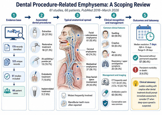
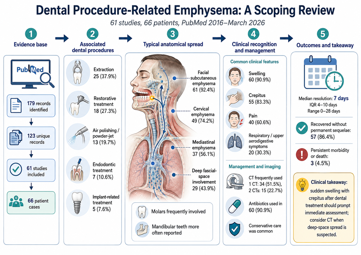
Graphical Abstract

1. Introduction

Dental procedure-related emphysema is an infrequently reported but clinically important iatrogenic complication caused by the introduction of pressurized air or gas into facial or cervical soft tissues. Air may remain localized or dissect along connected fascial planes into the parapharyngeal, retropharyngeal, mediastinal, orbital, thoracic, intracranial, or vascular compartments. Rare published complications include pneumothorax, airway compromise, visual or neurologic involvement, air embolism, cardiovascular collapse, and death. Sudden swelling during or shortly after dental treatment, particularly when accompanied by palpable crepitus, is therefore an important diagnostic clue [1,2,3].
The anatomical pattern of spread is determined by the point of entry and the continuity of the cervicofacial spaces. Air introduced through an extraction socket, periodontal or peri-implant pocket, root canal, mucosal laceration, or operative wound can enter the buccal, sublingual, submandibular, masticator, parapharyngeal, and retropharyngeal spaces. Posterior cervical pathways may communicate with the mediastinum, while maxillary and infratemporal routes can extend toward the pterygopalatine region and orbit. The resulting presentation may therefore range from isolated facial swelling to dysphagia, dysphonia, chest discomfort, dyspnea, orbital symptoms, or neurologic deterioration [1,2,3].
Dental emphysema has been described after tooth extraction, restorative and endodontic treatment, periodontal therapy, implant procedures, sinus-related dental surgery, and professional dental cleaning or air-polishing. High-speed air turbines, air–water syringes, air-driven handpieces, and subgingival air-polishing systems are frequently implicated, although cases can occur without a forward-venting air instrument when postoperative pressure-generating behaviors or other routes of air entry are present [1,2,3,4].
Jones et al. systematically reviewed 135 cases reported from 1993 to 2020 and characterized dental causes, anatomical locations, implicated devices, and management [1]. Subsequent reviews have focused on mediastinal extension, cervicofacial emphysema, and endodontic mechanisms [2,3,4]. The present scoping review was designed as a contemporary extension that maps cases from 2016 onward and integrates patient-level information on clinical presentation, anatomical extent, diagnostic investigations, management, complications, and outcomes. The scoping design was selected because the evidence is heterogeneous and predominantly descriptive; the objective is evidence mapping rather than estimation of incidence, comparative risk, or treatment effects.

2. Materials and Methods

2.1. Study Design and Reporting

This scoping review was conducted and reported in accordance with the Preferred Reporting Items for Systematic Reviews and Meta-Analyses Extension for Scoping Reviews (PRISMA-ScR) [5]. The protocol was not prospectively registered. The review was designed to map the contemporary case literature, characterize recurring clinical patterns, and identify evidence gaps rather than to estimate incidence, procedure-specific risk, or treatment effectiveness.

2.2. Information Sources and Search Strategy

PubMed/MEDLINE, Scopus, and Web of Science Core Collection were searched for reports published from 1 January 2016 through 26 August 2026. The PubMed strategy combined terms for subcutaneous, cervicofacial, mediastinal, surgical, and orbital emphysema with dental, oral-surgical, extraction, endodontic, periodontal, implant, restorative, air-polishing, air-turbine, and high-speed handpiece terms in title/abstract fields. Equivalent topic searches were adapted for Scopus (TITLE-ABS-KEY) and Web of Science Core Collection (TS). No case-report publication-type filter was applied because inconsistent indexing could reduce sensitivity. The complete reproducible search strings and database-specific processing are provided in Supplementary Table S1. Reference lists of relevant reviews and included reports were checked for eligible records.

2.3. Eligibility Criteria

Dental procedure-related emphysema was operationally defined as clinically or radiologically demonstrated air in facial, cervical, mediastinal, orbital, thoracic, intracranial, vascular, intraosseous, pericardial, or named deep fascial compartments occurring during or after dental or dental-related care. Only English-language literature was eligible. Case reports and case series were included when sufficient patient-level information was available to establish the dental exposure and clinical diagnosis/course. Dental-related procedures included oral surgery, implant treatment, endodontic and periodontal procedures, restorative/prosthodontic treatment, and dental cleaning or air-polishing. Reviews without new patients, editorials, conference abstracts without a full report, experimental studies, duplicate publications of the same patient, non-English full texts, and reports unrelated to dental care were excluded.

2.4. Record Management and Study Selection

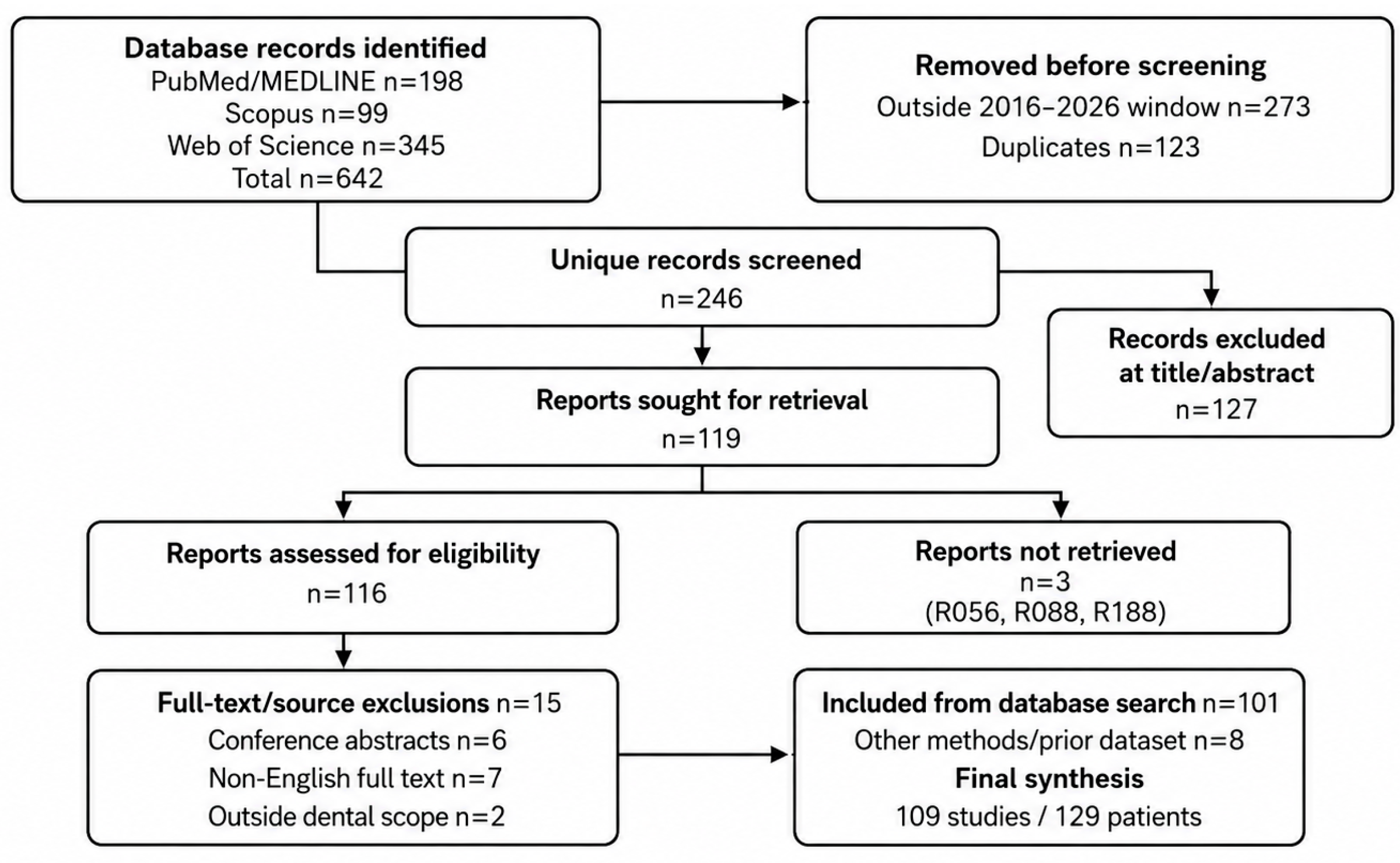
The three database exports were combined and processed against the prespecified publication window. PubMed yielded 198 records, Scopus 99, and Web of Science 345 (642 database records in total). The exported Scopus and Web of Science sets contained records outside the review period; 273 records were removed on application of the 2016–2026 window. A further 123 duplicate or overlapping database records were removed, leaving 246 unique records for title/abstract screening. Reports considered potentially eligible underwent full-text/source assessment. Duplicate publications and potential reports of the same patient were compared using DOI, title, authorship, institution, age/sex, procedure, treated tooth, and clinical chronology. One investigator (L.S.) screened titles and abstracts and assessed potentially eligible reports against the predefined criteria. Uncertain eligibility decisions and potential duplicate publications were reviewed with the coauthors and resolved by consensus. The same eligibility assessment was applied to reports identified from the previous review dataset or by supplementary source checking.

2.5. Data Charting

One investigator (L.S.) charted patient-level data in a standardized spreadsheet using predefined variables. Variables included age, sex, relevant medical history, procedure, jaw and tooth group, documented or possible air source, self-induced pressure-generating mechanisms, anatomical distribution, swelling, crepitus, pain, respiratory or upper aerodigestive symptoms, imaging, antibiotic use, complications, follow-up, complete resolution time, and persistent morbidity or death. Missing or unavailable information was coded as not reported. Procedure categories were coded as multiple-response variables; when more than one relevant procedure was reported for an individual patient, all applicable categories were retained. Ambiguous source descriptions were discussed among the authors and resolved by consensus. Generative AI was used only to assist with administrative organization of screening records, standardization of tabular material, flagging possible duplicate or internally inconsistent entries, arithmetic checks of summary counts, and preparation of graphical materials. It did not make final eligibility decisions or independently determine patient-level data. Final inclusion/exclusion decisions, patient-level data charting/extraction, and verification of the values used in the synthesis were performed by the authors; all AI-assisted checks and summaries were reviewed and, where necessary, corrected by the authors.

2.6. Anatomical Classification

Because source terminology was heterogeneous, an author-developed descriptive classification was used. Facial involvement included subcutaneous air in cheek, buccal, perioral, temporal, infraorbital, or other facial soft tissues; cervical involvement comprised subcutaneous air in the neck; and deep fascial-space involvement comprised named spaces such as submandibular, sublingual, masticator, parapharyngeal, retropharyngeal, infratemporal, or pterygopalatine spaces. Mediastinal, orbital, thoracic subcutaneous, intracranial, vascular, intraosseous, pericardial, and scalp categories followed the source descriptions. Individual patients could be assigned to more than one anatomical category.

2.7. Data Synthesis and Critical Appraisal

Data were synthesized descriptively. Patient-level frequencies and percentages use the denominator of 129 patients unless otherwise specified, and multiple-response categories may therefore sum to more than 100%. Resolution time was summarized at the episode level using the numerical time to complete resolution. No meta-analysis was performed because the evidence consisted of heterogeneous noncomparative case reports and case series. Consistent with the mapping purpose of this scoping review and the predominantly descriptive case-report evidence, no formal risk-of-bias assessment was undertaken. Frequencies were interpreted as patterns in the published literature rather than incidence or risk estimates.

3. Results

3.1. Study Selection

The expanded database searches identified 642 records (PubMed/MEDLINE, n = 198; Scopus, n = 99; Web of Science Core Collection, n = 345). Application of the prespecified 2016–2026 publication window removed 273 records, and deduplication removed 123 additional records, leaving 246 unique records for screening. Title/abstract screening excluded 127 records, and 119 reports were sought for retrieval. Three reports could not be retrieved. Of 116 reports assessed for eligibility, 15 were excluded at the source/full-text stage (6 conference abstracts, 7 non-English full texts, and 2 reports outside the predefined dental-procedure scope). One hundred and one eligible reports arose from the expanded database corpus. Eight additional eligible reports were retained from the previous review dataset or identified by supplementary source checking, resulting in a final synthesis of 109 studies describing 129 patients (Figure 1; Supplementary Tables S1 and S4) [5].

3.2. Patient and Study Characteristics

The published sample comprised 129 patients. Female sex was reported for 74/129 patients (57.4%), male sex for 47/129 (36.4%), and sex was not reported for 8 patients (6.2%). Reported ages ranged from 2.5 to 84 years. Included publications originated from multiple clinical disciplines and settings, including oral and maxillofacial surgery, restorative dentistry, endodontics, periodontology, implant dentistry, emergency medicine, otolaryngology, ophthalmology, and anesthesiology. The 109 included studies are listed in Supplementary Table S2 and are cited in the reference list [2,6,7,8,9,10,11,12,13,14,15,16,17,18,19,20,21,22,23,24,25,26,27,28,29,30,31,32,33,34,35,36,37,38,39,40,41,42,43,44,45,46,47,48,49,50,51,52,53,54,55,56,57,58,59,60,61,62,63,64,65,66,67,68,69,70,71,72,73,74,75,76,77,78,79,80,81,82,83,84,85,86,87,88,89,90,91,92,93,94,95,96,97,98,99,100,101,102,103,104,105,106,107,108,109,110,111,112,113].

3.3. Associated Procedures and Tooth Distribution

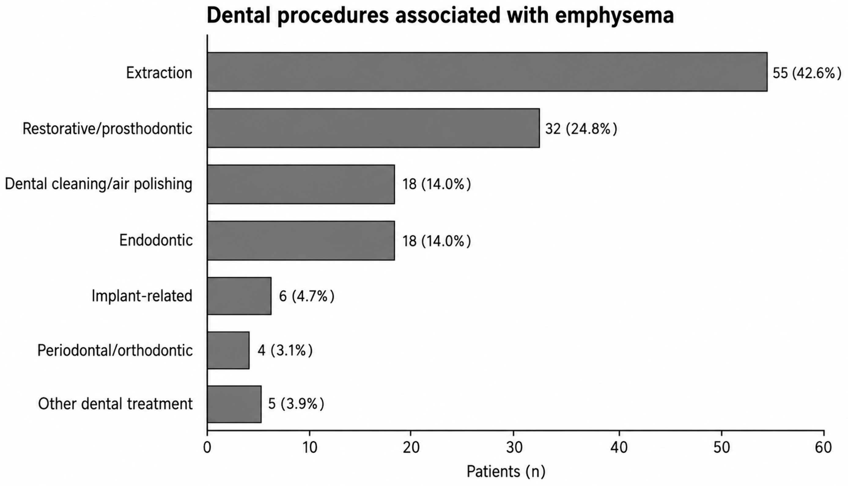
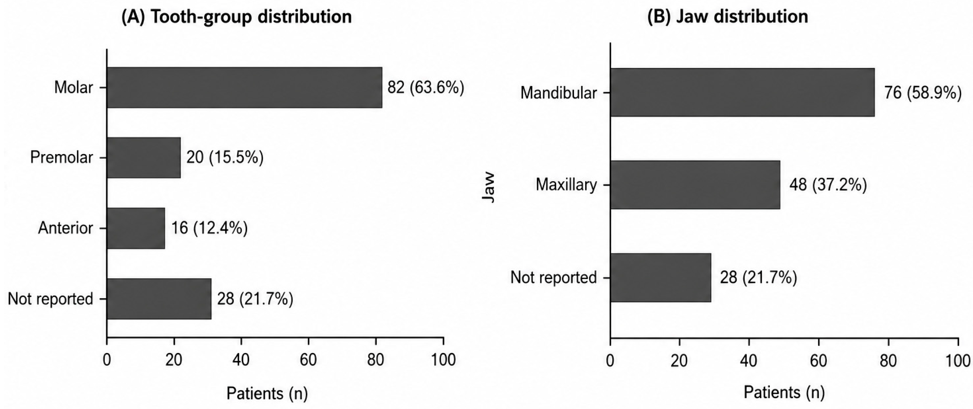
Figure 2 shows that extraction remained the most frequently represented procedure category (55/129, 42.6%), followed by restorative/prosthodontic treatment (32/129, 24.8%), dental cleaning or air-polishing (18/129, 14.0%), endodontic treatment (18/129, 14.0%), implant-related procedures (6/129, 4.7%), and periodontal/orthodontic or other dental procedures. Mandibular involvement was reported in 76 patients (58.9%) and maxillary involvement in 48 (37.2%). Molars were the most frequently represented tooth group (82/129, 63.6%; Figure 3). These frequencies describe the published cases and should not be interpreted as procedure-, jaw-, or tooth-specific risk [6,18,20,21,24,29,30,44,59,75,85,104,108].

3.4. Anatomical Distribution and Clinical Presentation

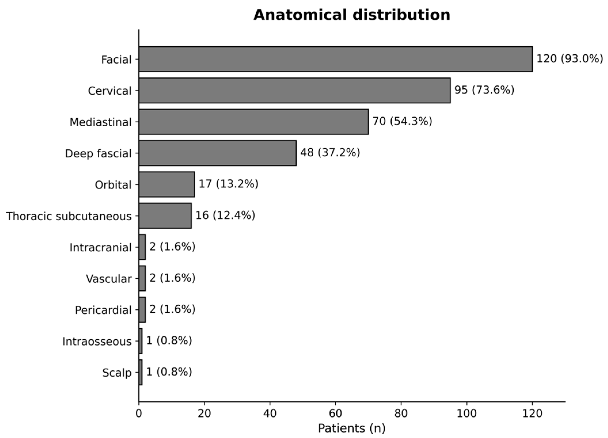
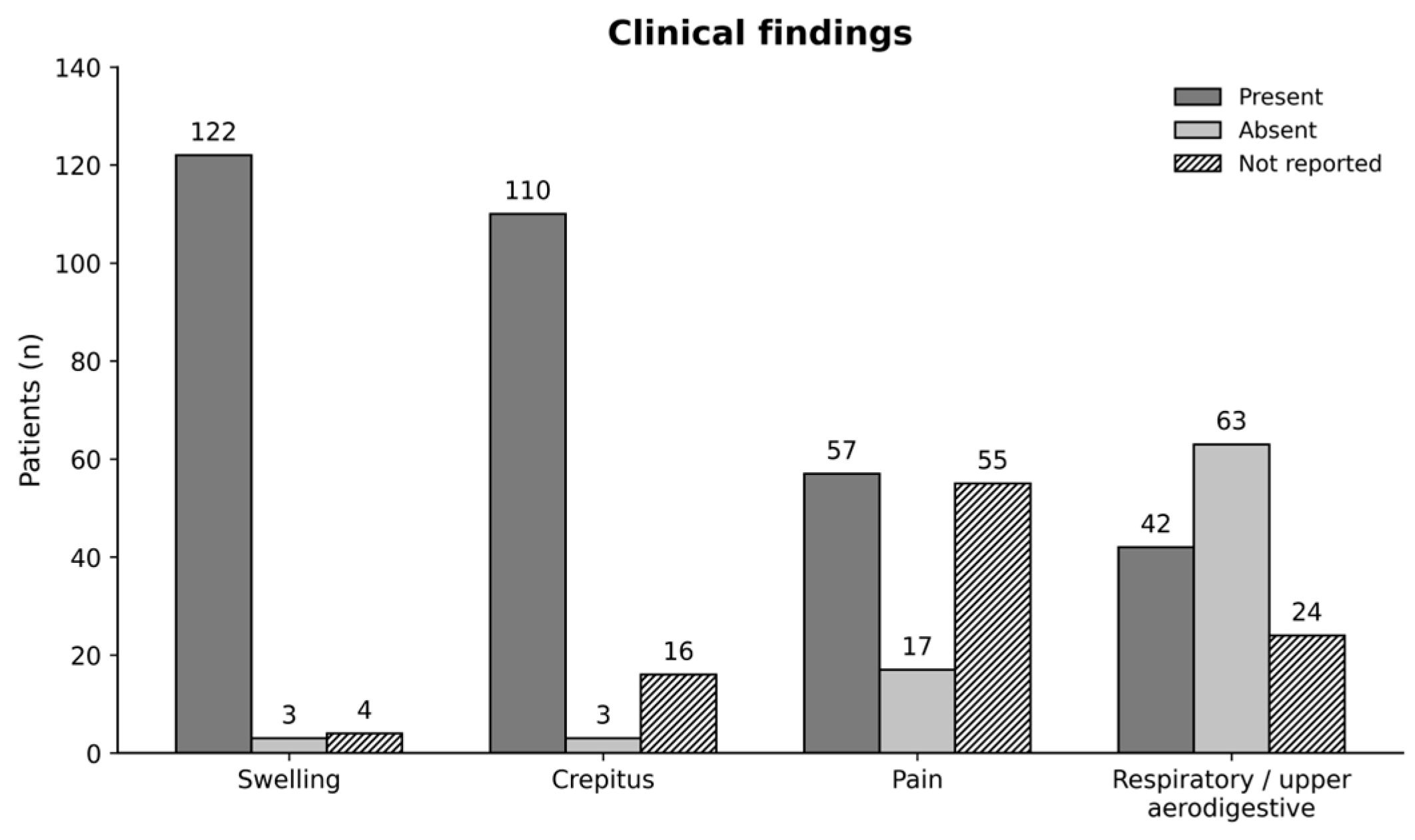
Figure 4 summarizes that facial subcutaneous emphysema was reported in 120 patients (93.0%), cervical emphysema in 95 (73.6%), mediastinal emphysema in 70 (54.3%), and named deep fascial-space involvement in 48 (37.2%). Orbital involvement was reported in 17 patients (13.2%). Less frequent reported compartments included thoracic subcutaneous tissues, intracranial or spinal/extradural spaces, the vascular system, pericardium, intraosseous sites, and scalp. Clinical findings reported swelling in 122 patients, crepitus in 110, pain in 57, and respiratory or upper aerodigestive symptoms in 42 (Figure 5) [7,11,31,32,33,35,39,42,73,77,90,102,105].

3.5. Diagnostic Assessment and Implicated Mechanisms

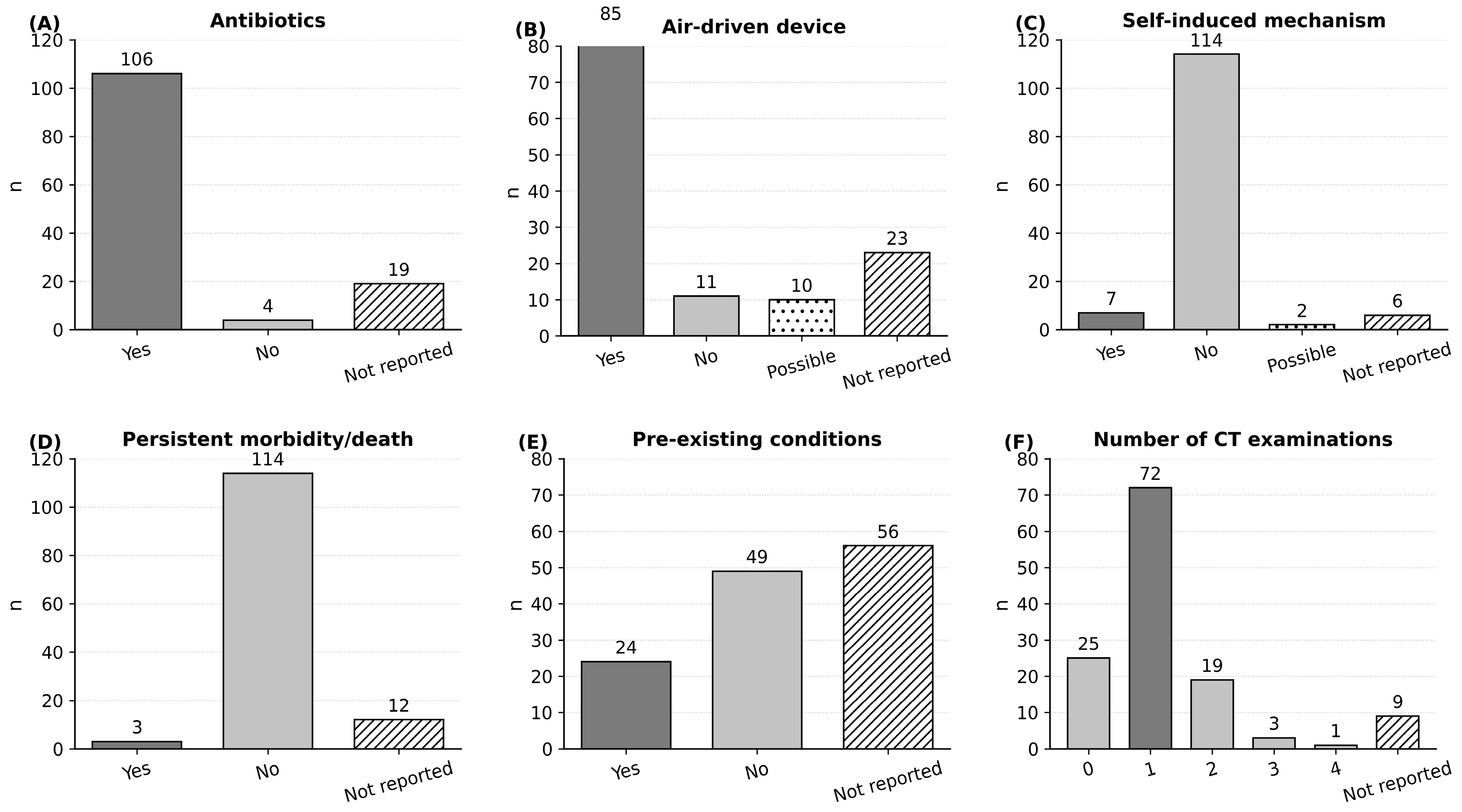
CT was frequently used to confirm diagnosis and delineate anatomical extent (Figure 6). Overall, 95/129 patients (73.6%) underwent at least one CT examination: 72 underwent one CT examination, 19 underwent two, 3 underwent three, and 1 underwent four. Twenty-five patients were coded as having no CT examination, and CT frequency was not reported for 9. An air-driven device was explicitly documented in 85 patients, explicitly absent in 11, considered possible in 10, and not reported in 23. Pressure-generating patient behavior was documented in 7 cases and considered possible in 2. These variables distinguish documented mechanisms from plausible or inferred routes of air entry [2,12,16,35,75,80,85,103,104,108].

3.6. Management and Outcomes

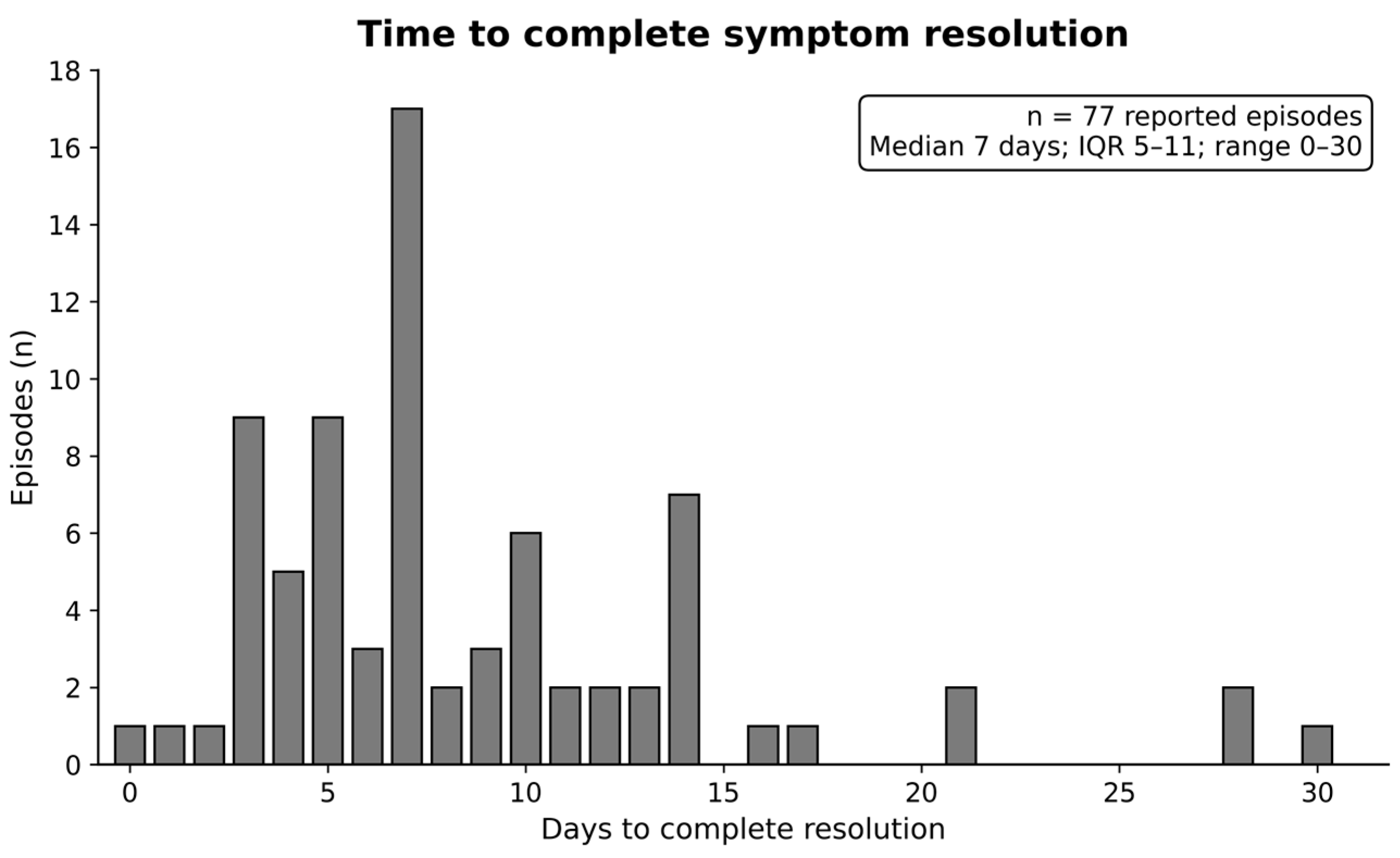
Antibiotic therapy was reported in 106 patients (82.2%), was explicitly not used in 4, and was not reported in 19. Reported management also included observation, analgesia, oxygen, avoidance of pressure-increasing activities, hospital monitoring, specialist consultation, and, in selected severe cases, invasive airway/thoracic management or hyperbaric oxygen. Persistent morbidity or death was documented in 3 patients; no persistent morbidity was reported in 114, while long-term status was not reported in 12 (Figure 6). Across 77 episodes with an exact reported time to complete resolution, the median was 7 days (interquartile range, 5–11 days; range, 0–30 days) (Figure 7). These descriptive management frequencies cannot establish the effectiveness of antibiotics, oxygen, imaging, hospital admission, or conservative care [1,2,3,4,6,7,8,9,90,102].

4. Discussion

4.1. Principal Findings

This expanded scoping review maps 109 studies describing 129 patients with dental procedure-related emphysema published from 2016 through August 2026. Broadening the information sources from PubMed alone to PubMed/MEDLINE, Scopus, and Web of Science substantially increased the screened evidence base and identified additional case reports and case series that were not captured by the original search. Extraction remained the most frequently represented context in the published literature, but restorative/prosthodontic procedures, dental cleaning or air-polishing, endodontic treatment, implant therapy, and other procedures were also represented. Facial and cervical involvement predominated, while mediastinal and deep fascial-space extension remained prominent among published cases. The expanded corpus also contains rare orbital, intracranial, vascular, pericardial, and thoracic complications.

4.2. Added Value Relative to Previous Reviews

The present review complements rather than replaces earlier historical syntheses. Jones et al. examined 135 cases reported from 1993 to 2020 and focused on causes, anatomical location, devices, and management [1]. The current scoping review uses a contemporary 2016–2026 window, an expanded multi-database search, and a broader patient-level charting framework covering presenting symptoms, detailed anatomical spread, diagnostic investigations, management patterns, complications, resolution time, and persistent outcomes. The new search also captures recently reported air-polishing, peri-implant, orbital, neurologic, and air-embolism presentations that post-date or were outside earlier evidence syntheses.

4.3. Publication Bias and Interpretation of Frequencies

The published sample is strongly susceptible to publication and selection bias. Extensive, unusual, or life-threatening events are more likely to be written up than mild episodes that resolve rapidly in routine dental practice. Database indexing, English-language restriction, and inconsistent terminology also influence retrieval. Consequently, the observed frequencies of mediastinal extension, CT use, antibiotic treatment, or particular procedures are properties of the published case literature, not estimates of incidence, comparative risk, or typical clinical distribution.

4.4. Mechanisms and Anatomical Spread

The reported anatomical patterns are consistent with the continuity of cervicofacial fascial spaces, but the mechanism of air entry was not always directly documented. A tissue defect together with a pressurized air source is a recurring pattern: extraction sockets, periodontal or peri-implant pockets, root canals, crestal perforations, gingival lacerations, and surgical wounds can provide access to loose connective-tissue planes. Air may pass through submandibular, sublingual, masticator, parapharyngeal, or retropharyngeal spaces toward the mediastinum, while maxillary/infratemporal pathways can reach the orbit [1,2,3,4]. Several recent cases additionally show that electric handpieces, postoperative emesis, nose blowing, or procedures without a classic forward-venting turbine can still be associated with emphysema [12,85,103]. These observations support distinguishing directly documented devices/actions from inferred anatomical routes.

4.5. Clinical Recognition and Imaging

Sudden swelling during or shortly after dental care should prompt palpation for crepitus, review of the procedure and instruments used, assessment of vital signs, and attention to airway, swallowing, voice, chest, ocular, and neurologic symptoms. Differential diagnoses include allergic reaction, angioedema, hematoma, infection, postoperative edema, pneumoparotitis, and necrotizing fasciitis. CT is particularly informative when symptoms extend beyond a localized superficial area, when cervical, mediastinal, orbital, thoracic, or neurologic involvement is suspected, or when the diagnosis is uncertain [1,2,3,4]. However, case reports cannot establish that every clinically stable localized case requires CT or repeated imaging; imaging should be individualized to the suspected anatomical extent and clinical severity.

4.6. Management and Escalation of Care

Antibiotics were frequently administered in published cases, but the available evidence cannot demonstrate prophylactic benefit or define evidence-based indications. The same limitation applies to oxygen therapy, hospital admission, serial CT, corticosteroids, and other interventions. Clinical escalation is nevertheless warranted when airway stability is uncertain, symptoms are progressive, deep-space or mediastinal spread is suspected, systemic inflammatory findings occur, or orbital, neurologic, thoracic, or vascular complications are present. Reports of tension pneumothorax, air embolism, neurologic compromise, and death show why severe presentations require multidisciplinary emergency assessment [6,8,9,90,102].

4.7. Prevention and Clinical Implications

The case patterns support plausible clinical precautions rather than comparative evidence-based preventive recommendations. These include avoiding direct pressurized air near extraction sockets, periodontal/peri-implant defects, root canals with compromised apical barriers, mucosal lacerations, and fresh surgical wounds; using non-air-driven or appropriately vented systems when feasible; and adhering to device-specific instructions for subgingival air-polishing [1,2,3,4,10,104,108]. Patients should be advised to avoid forceful nose blowing or other pressure-generating behaviors when a tissue or sinus communication is possible. These implications are biologically plausible but have not been tested in comparative prevention studies.

4.8. Strengths and Limitations

A major strength of the revision is the expanded search across three major bibliographic databases with a documented deduplicated screening corpus of 246 unique records. The review also provides structured patient-level mapping across procedures, anatomical sites, clinical findings, investigations, management, and outcomes. Limitations remain substantial. The evidence is almost entirely case reports and small case series; mild events are likely underreported; clinical variables are incompletely and inconsistently described; and causal attribution is uncertain when several instruments or behaviors are plausible. The English-language restriction may exclude relevant non-English cases. The 2016–2026 window intentionally prioritizes contemporary practice but excludes older evidence. Three potentially relevant reports could not be retrieved. Finally, the anatomical categories are author-developed descriptive groupings. Screening and data charting were performed by one investigator, with coauthor discussion of uncertain cases, which may have increased the risk of selection or extraction error.

4.9. Future Research

Multicenter registries or structured surveillance systems are needed to estimate incidence and procedure-specific risk using denominators for the number and type of dental procedures performed. Standardized case reporting should document the exact instrument and air source, route of tissue entry, timing of onset, anatomical extent, airway/respiratory/ocular/neurologic findings, rationale for imaging, management decisions, microbiology when relevant, and short- and long-term outcomes. Comparative studies are needed before firm recommendations can be made regarding prophylactic antibiotics, imaging thresholds, oxygen therapy, or admission criteria.

5. Conclusions

Dental procedure-related emphysema is represented by a heterogeneous and publication-biased case literature and the present review cannot estimate its incidence. Published cases show that air introduced during or after dental care can remain localized or extend into deep cervical, mediastinal, orbital, thoracic, intracranial, or vascular compartments. Sudden swelling with palpable crepitus should prompt immediate clinical assessment and cessation of pressurized-air use. CT may be considered when deeper extension or complications are suspected, while progressive symptoms, airway concern, systemic illness, orbital or neurologic findings, thoracic complications, or suspected air embolism require urgent escalation. Preventive measures should be understood as clinically plausible precautions derived from recurring case patterns rather than proven interventions.

Supplementary Materials

The following supporting information can be downloaded at: https://www.mdpi.com/article/10.3390/dj14090602/s1, Table S1: Complete database search strategies and search log; Table S2: Study-level inclusion list; Table S3: Patient-level charted dataset; Table S4: Full-text/source exclusions and reports not retrieved [114,115,116,117,118,119,120,121,122,123,124,125,126,127,128,129,130,131]; completed PRISMA-ScR checklist.

Author Contributions

Conceptualization, U.K.; methodology, U.K. and R.G.; investigation, L.S.; data curation, L.S.; formal analysis, L.S.; supervision, U.K.; writing—original draft preparation, U.K.; writing—review and editing, U.K., L.S. and R.G. All authors have read and agreed to the published version of the manuscript.

Funding

This research received no external funding.

Institutional Review Board Statement

Not applicable. This review used only data from previously published reports and did not involve new participant recruitment, intervention, tissue collection, or access to unpublished identifiable data.

Informed Consent Statement

Not applicable. No newly collected individual patient data are presented.

Data Availability Statement

All data generated or analyzed in this scoping review are included in the article and its Supplementary Materials. The complete database search strategies and search log, study-level inclusion list, patient-level charted dataset, and full-text/source exclusions and reports not retrieved are provided in Supplementary Tables S1–S4.

Acknowledgments

During preparation of this manuscript, the authors used ChatGPT (OpenAI, GPT-5.6 Sol; accessed August–September 2026) for language editing, manuscript organization, administrative organization and data checking, and preparation of graphical materials. All AI-assisted output was reviewed and revised by the authors.

Conflicts of Interest

The authors declare no conflicts of interest.

Abbreviations

CT, computed tomography; PRISMA-ScR, Preferred Reporting Items for Systematic Reviews and Meta-Analyses Extension for Scoping Reviews; SE, subcutaneous emphysema.

References

  1. Jones, A.; Stagnell, S.; Renton, T.; Aggarwal, V.R.; Moore, R. Causes of subcutaneous emphysema following dental procedures: A systematic review of cases 1993–2020. Br. Dent. J. 2021, 231, 493–500. [Google Scholar] [CrossRef] [Scilit] [PubMed]
  2. Spille, J.; Wagner, J.; Spille, D.C.; Naujokat, H.; Gülses, A.; Wiltfang, J.; Kübel, P. Pronounced mediastinal emphysema after restorative treatment of the lower left molar-a case report and a systematic review of the literature. Oral Maxillofac. Surg. 2023, 27, 533–541. [Google Scholar] [CrossRef] [Scilit] [PubMed]
  3. Kanaparthi, K.; Talwar, A.; Khan, S.; Talwar, A.; Capozzi, B.; Talwar, A. Cervicofacial emphysema: A systematic review. Intractable Rare Dis. Res. 2024, 13, 208–212. [Google Scholar] [CrossRef] [Scilit] [PubMed]
  4. Fasoulas, A.; Boutsioukis, C.; Lambrianidis, T. Subcutaneous emphysema in patients undergoing root canal treatment: A systematic review of the factors affecting its development and management. Int. Endod. J. 2019, 52, 1586–1604. [Google Scholar] [CrossRef] [Scilit] [PubMed]
  5. Tricco, A.C.; Lillie, E.; Zarin, W.; O’Brien, K.K.; Colquhoun, H.; Levac, D.; Moher, D.; Peters, M.D.J.; Horsley, T.; Weeks, L.; et al. PRISMA Extension for Scoping Reviews (PRISMA-ScR): Checklist and Explanation. Ann. Intern. Med. 2018, 169, 467–473. [Google Scholar] [CrossRef] [Scilit] [PubMed]
  6. Sepsick, H.; Ma, B.; Zeng, Q.; Castro-Núñez, J. Pneumomediastinum with Mediastinitis Following Third Molar Extraction with a High-Speed Air Handpiece: A Case Report and Literature Review. Cureus 2025, 17, e84695. [Google Scholar] [CrossRef] [Scilit] [PubMed]
  7. North, L.; Sulman, C. Subcutaneous emphysema and vocal fold paresis as a complication of a dental procedure. Int. J. Pediatr. Otorhinolaryngol. 2019, 124, 76–78. [Google Scholar] [CrossRef] [Scilit] [PubMed]
  8. Fukuda, H.; Fukushima, K.; Takahashi, Y.; Hayakawa, A.; Tokue, H.; Obana, Y.; Iwahara, K.; Tsuzuki, T.; Yokoo, S.; Sano, R. Unexpected death from air embolism during routine dental Procedure: A case report. Leg. Med. 2025, 77, 102674. [Google Scholar] [CrossRef] [Scilit] [PubMed]
  9. Abe, S.; Wakamatsu, K.; Takahashi, K.; Sato, H.; Yoshida, K.; Yamazaki, S.; Kawaai, H. Pulmonary Air Embolism Caused by an Air Turbine During Oral Surgery Under General Anesthesia: A Case Report. Int. Med. Case Rep. J. 2025, 18, 1439–1446. [Google Scholar] [CrossRef] [Scilit] [PubMed]
  10. Bruckmann, C.; Bruckmann, L.; Gahleitner, A.; Stavropoulos, A.; Bertl, K. Pneumocephalus as result of nonsurgical peri-implantitis treatment with an air-polishing device for submucosal debridement—A case report. Clin. Exp. Dent. Res. 2022, 8, 632–639. [Google Scholar] [CrossRef] [Scilit] [PubMed]
  11. Henin, C.; Watelet, L.; Wauters, V. A Rare Complication of Dental Care: A Case of Subcutaneous Emphysema, Pneumomediastinum, and Pneumopericardium. Cureus 2025, 17, e85035. [Google Scholar] [CrossRef] [Scilit] [PubMed]
  12. Chang, V.K.O.; Tsai, H.H. Patient-induced progressive facial subcutaneous emphysema masquerading as odontogenic abscess. BMJ Case Rep. 2021, 14, e243489. [Google Scholar] [CrossRef] [Scilit] [PubMed]
  13. Attia, R.; Nabil, N.; Anis, M. Subcutaneous emphysema during surgical gingival depigmentation: A case report. J. Med. Case Rep. 2024, 18, 514. [Google Scholar] [CrossRef] [Scilit] [PubMed]
  14. Almeida, S.I.; Faustino, J.; Duarte Armindo, R.; Mendonça, V. Subcutaneous facial emphysema secondary to a dental procedure. BMJ Case Rep. 2021, 14, e242300. [Google Scholar] [CrossRef] [Scilit] [PubMed]
  15. Peters, M.; Shall, F.; Evrard, L. Pneumomediastinum after Third Molar Extraction: Case Report, Physiopathology, and Literature Review. Case Rep. Dent. 2023, 2023, 4562710. [Google Scholar] [CrossRef] [Scilit] [PubMed]
  16. Mordi, V.; Leung, B.; Peyman, A.; El Kininy, W. An unsuspecting presentation of extensive pneumomediastinum following dental air-polishing. Br. Dent. J. 2025, 239, 549–553. [Google Scholar] [CrossRef] [Scilit] [PubMed]
  17. Kim, Y.S.; Huh, J.; Myoung, H.; Kim, S.M.; Seo, M.H. Severe bilateral subcutaneous emphysema after prophylactic treatment: A case report. J. Dent. Anesth. Pain Med. 2024, 24, 421–425. [Google Scholar] [CrossRef] [Scilit] [PubMed]
  18. Mascarenhas, R.J. Management of subcutaneous facial emphysema secondary to a class V dental restoration. Clin. Case Rep. 2019, 7, 1025–1030. [Google Scholar] [CrossRef] [Scilit] [PubMed]
  19. Chien, P.Y.H. Iatrogenic subcutaneous facial emphysema secondary to a Class V dental restoration: A case report. Aust. Dent. J. 2019, 64, 43–46. [Google Scholar] [CrossRef] [Scilit] [PubMed]
  20. Lee, S.T.; Subu, M.G.; Kwon, T.G. Emphysema following air-powder abrasive treatment for peri-implantitis. Maxillofac. Plast. Reconstr. Surg. 2018, 40, 12. [Google Scholar] [CrossRef] [Scilit] [PubMed]
  21. Alonso, V.; Garcia-Caballero, L.; Couto, I.; Diniz, M.; Diz, P.; Limeres, J. Subcutaneous emphysema related to air-powder tooth polishing: A report of three cases. Aust. Dent. J. 2017, 62, 510–515. [Google Scholar] [CrossRef] [Scilit] [PubMed]
  22. La Monaca, G.; Pranno, N.; Annibali, S.; Vozza, I.; Cristalli, M.P. Subcutaneous Facial Emphysema Following Open-Flap Air-Powder Abrasive Debridement for Peri-Implantitis: A Case Report and an Overview. Int. J. Environ. Res. Public Health 2021, 18, 13286. [Google Scholar] [CrossRef] [Scilit] [PubMed]
  23. Bocchialini, G.; Ambrosi, S.; Castellani, A. Massive Cervicothoracic Subcutaneous Emphysema and Pneumomediastinum Developing during a Dental Hygiene Procedure. Case Rep. Dent. 2017, 2017, 7016467. [Google Scholar] [CrossRef] [Scilit] [PubMed]
  24. Padmanaban, K.; Revathy, V.; Rajiah, D.; Raghavan, P. Subcutaneous Emphysema of Periorbital and Canine Space Following Endodontic Treatment. Cureus 2024, 16, e56307. [Google Scholar] [CrossRef] [Scilit] [PubMed]
  25. Dodge, A.; Kreh, K.; Kofina, V.; Rawal, S.Y. Sublingual emphysema following alveoloplasty: A case report. Clin. Case Rep. 2020, 8, 2051–2054. [Google Scholar] [CrossRef] [Scilit] [PubMed]
  26. Cuccia, A.M.; Geraci, A. Cervicofacial and mediastinal emphysema after dental extraction. Dent. Med. Probl. 2019, 56, 203–207. [Google Scholar] [CrossRef] [Scilit] [PubMed]
  27. Khoshkhounejad, M.; Hashemi Nasab, M.S.; Aminsobhani, M.; Moayeri, R.S. Challenging Diagnosis of Severe Bilateral Cervicofacial Subcutaneous Emphysema following Root Perforation in a Maxillary Lateral Incisor: A Case Report. Iran. Endod. J. 2019, 14, 220–224. [Google Scholar] [CrossRef] [Scilit] [PubMed]
  28. Nizar, M.A.M.; Nabil, S. Facial Emphysema following Closure of Oroantral Fistulae. Case Rep. Dent. 2021, 2021, 5001266. [Google Scholar] [CrossRef] [Scilit] [PubMed]
  29. Farina, R.; Zaetta, A.; Minenna, L.; Trombelli, L. Orbital and Periorbital Emphysema Following Maxillary Sinus Floor Elevation: A Case Report and Literature Review. J. Oral Maxillofac. Surg. 2016, 74, 2192.e1–2192.e7. [Google Scholar] [CrossRef] [Scilit] [PubMed]
  30. El Hage, M.; Nurdin, N.; Bischof, M.; Nedir, R. Emphysema after Sinus Grafting: Importance of Patient’s Information, Early Diagnosis, and Management. Case Rep. Dent. 2020, 2020, 1525673. [Google Scholar] [CrossRef] [Scilit] [PubMed]
  31. Chang, C.H.; Lien, W.C. Palpebral emphysema following a dental procedure. Am. J. Emerg. Med. 2018, 36, 908.e1–908.e2. [Google Scholar] [CrossRef] [Scilit] [PubMed]
  32. Moschetta, L.; Termote, K.; Ten Tusscher, M. Emphysema of the periorbital region and mediastinum in a young child following a dental procedure. J. Am. Assoc. Pediatr. Ophthalmol. Strabismus 2021, 25, 190–192. [Google Scholar] [CrossRef] [Scilit] [PubMed]
  33. Chang, J.R.; Rajaii, F.; McCulley, T.J. Delayed Orbital Emphysema Mimicking Orbital Cellulitis: An Uncommon Complication of Dental Surgery. Middle East. Afr. J. Ophthalmol. 2019, 26, 175–177. [Google Scholar] [CrossRef] [Scilit] [PubMed]
  34. Obara, T.; Nojima, T.; Nakatsuji, K.; Hongo, T.; Yumoto, T.; Nakao, A. Subcutaneous and Periorbital Emphysema Following a Dental Procedure. Cureus 2025, 17, e83089. [Google Scholar] [CrossRef] [Scilit] [PubMed]
  35. Busuladzic, A.; Patry, M.; Fradet, L.; Turgeon, V.; Bussieres, M. Cervicofacial and mediastinal emphysema following minor dental procedure: A case report and review of the literature. J. Otolaryngol. Head Neck Surg. 2020, 49, 61. [Google Scholar] [CrossRef] [Scilit] [PubMed]
  36. Shimizu, R.; Sukegawa, S.; Sukegawa, Y.; Hasegawa, K.; Ono, S.; Fujimura, A.; Yamamoto, I.; Ibaragi, S.; Sasaki, A.; Furuki, Y. Subcutaneous Emphysema Related to Dental Treatment: A Case Series. Healthcare 2022, 10, 290. [Google Scholar] [CrossRef] [Scilit] [PubMed]
  37. Lau, H.K.; Pothiawala, S. Cervicofacial Emphysema Complicating a Dental Procedure. J. Emerg. Med. 2020, 59, 586–589. [Google Scholar] [CrossRef] [Scilit] [PubMed]
  38. Carter, N.J. Essential Role of Rapid Diagnosis in Subcutaneous Emphysema Following Dental Procedures. Cureus 2025, 17, e93298. [Google Scholar] [CrossRef] [Scilit] [PubMed]
  39. Fink, P.; Doyle, L. Progressive subcutaneous emphysema and compressive optic neuropathy following dental analgesia. J. Am. Coll. Emerg. Physicians Open 2020, 1, 1278–1280. [Google Scholar] [CrossRef] [Scilit] [PubMed]
  40. Fuentes-Martín, Á.; Loucel Bellino, M.A.; Cilleruelo-Ramos, Á.; Soro-García, J.; Matilla González, J.M. Hamman’s sign after a dental procedure? Cir. Esp. 2023, 101, 579–581. [Google Scholar] [CrossRef] [Scilit] [PubMed]
  41. Deskins, S.J.; Brunner, N.E.; Brunner, M. A Case Report on Subcutaneous Emphysema and Pneumomediastinum Following a Routine Dental Procedure. Cureus 2023, 15, e41177. [Google Scholar] [CrossRef] [Scilit] [PubMed]
  42. Nożewski, J.B.; Dura, M.L.; Kłopocka, M.; Kwiatkowska, M.M.; Konieczny, J.P.; Nicpoń-Nożewska, K.K. A 39-Year-Old Woman with Cervicofacial and Mediastinal Emphysema 10 Hours After an Elective Lower Molar Dental Extraction. Am. J. Case Rep. 2021, 22, e931793. [Google Scholar] [CrossRef] [Scilit] [PubMed]
  43. Tegenbosch, C.; Wellekens, S.; Meysman, M. A swollen face and neck after dental surgery: Think of subcutaneous emphysema and pneumomediastinum. Respir. Med. Case Rep. 2023, 46, 101926. [Google Scholar] [CrossRef] [Scilit] [PubMed]
  44. Lee, S.W.; Huh, Y.H.; Cha, M.S. Iatrogenic subcutaneous cervicofacial emphysema with pneumomediastinum after class V restoration. J. Korean Assoc. Oral Maxillofac. Surg. 2017, 43, 49–52. [Google Scholar] [CrossRef] [Scilit] [PubMed]
  45. Zhang, X.; Zhou, M.; Zhu, X.; Qin, Z.; Ling, H.; Zhu, Z.; Gan, X. Subcutaneous emphysema secondary to autogenous bone grafting: A case report. BMC Oral Health 2025, 25, 506. [Google Scholar] [CrossRef] [Scilit] [PubMed]
  46. Rawlinson, R.D.; Negmadjanov, U.; Rubay, D.; Ohanisian, L.; Waxman, J. Pneumomediastinum After Dental Filling: A Rare Case Presentation. Cureus 2019, 11, e5593. [Google Scholar] [CrossRef] [Scilit] [PubMed]
  47. Chrysovitsiotis, G.; Boutsikou, A.; Kollia, P.; Chrysovergis, A. Cervicofacial subcutaneous emphysema following dental procedure. BMJ Case Rep. 2021, 14, e243256. [Google Scholar] [CrossRef] [Scilit] [PubMed]
  48. Gandhi, R.K.; Brown, T.S. Iatrogenic Cervicofacial Subcutaneous Emphysema: A Rare Dental Complication. Dermatol. Surg. 2021, 47, 845–846. [Google Scholar] [CrossRef] [Scilit] [PubMed]
  49. Fleagle, J. Subcutaneous emphysema following dental care of a patient with ventriculoperitoneal shunt: Case report. Spec. Care Dent. 2020, 40, 488–492. [Google Scholar] [CrossRef] [Scilit] [PubMed]
  50. Kaliszewski, K.; Cendal, I.; Krassowska, M.; Szwed, D.; Wojtczak, B.; Rudnicki, J. Pneumomediastinum and subcutaneous emphysema after dental extraction. Pol. Arch. Intern. Med. 2020, 130, 244–245. [Google Scholar] [CrossRef] [Scilit] [PubMed]
  51. Singh, V.; Ganti, L.; Haupt, J.D.; Marshall, B. Iatrogenic post-pulpectomy cervicofacial subcutaneous emphysema in a paediatric patient. BMJ Case Rep. 2020, 13, e234074. [Google Scholar] [CrossRef] [Scilit] [PubMed]
  52. Pan, Y. Massive emphysema after tooth extraction. Am. J. Emerg. Med. 2020, 38, 164.e1–164.e3. [Google Scholar] [CrossRef] [Scilit] [PubMed]
  53. Fehrle, C.; Gillissen, A. Mediastinal and Cutaneous Emphysema Following Dental Extraction. Dtsch. Ärztebl. Int. 2019, 116, 212. [Google Scholar] [CrossRef] [Scilit] [PubMed]
  54. Boggess, W.J.; Ronan, J.; Panchal, N. Orbital, Mediastinal and Cervicofacial Subcutaneous Emphysema after Dental Rehabilitation in a Pediatric Patient. Pediatr. Dent. 2017, 39, 465–467. [Google Scholar] [PubMed]
  55. Akra, G.A.; Yousif, K. Cervicofacial and mediastinal emphysema complicating tooth extraction in an elderly patient: A preventable complication. BMJ Case Rep. 2017, 2017, bcr-2017-219245. [Google Scholar] [CrossRef] [Scilit] [PubMed]
  56. Thompson, C.; Gohil, R. Pneumatic dental extractions: An unusual cause of extensive cervical surgical emphysema. BMJ Case Rep. 2017, 2017, bcr-2016-218677. [Google Scholar] [CrossRef] [Scilit] [PubMed]
  57. Tan, S.; Nikolarakos, D. Subcutaneous emphysema secondary to dental extraction: A case report. Aust. Dent. J. 2017, 62, 95–97. [Google Scholar] [CrossRef] [Scilit] [PubMed]
  58. Mañas Vera, M.R.; Lara Rosales, R.; Sánchez González, A. Pneumomediastinum and subcutaneous emphysema: A rare complication of dental extraction. Arch. Bronconeumol. 2016, 52, 104–105. [Google Scholar] [CrossRef] [Scilit] [PubMed]
  59. Ozenbas, C.; Aydin, S. Rare complication of a common procedure: Pneumomediastinum following an endodontic procedure. Ulus. Travma Acil Cerrahi Derg. 2025, 31, 490–492. [Google Scholar] [CrossRef] [Scilit] [PubMed]
  60. Bai, Y.P.; Sha, J.J.; Chai, C.C.; Sun, H.P. With two episodes of right retromandibular angle subcutaneous emphysema during right upper molar crown preparation: A case report. World J. Clin. Cases 2023, 11, 4698–4706. [Google Scholar] [CrossRef] [Scilit] [PubMed]
  61. Ye, L.Y.; Wang, L.F.; Gao, J.X. Pneumomediastinum and subcutaneous emphysema secondary to dental extraction: Two case reports. World J. Clin. Cases 2022, 10, 9904–9910. [Google Scholar] [CrossRef] [Scilit] [PubMed]
  62. Işık, H.; Sapmaz, E.; Özbey, M.; Çaylak, H.; Şengül İnan, M. A rare cause of pneumomediastinum and subcutaneous emphysema: Tooth extraction. Turk. Gogus Kalp Damar Cerrahisi Derg. 2019, 28, 390–393. [Google Scholar] [CrossRef] [Scilit] [PubMed]
  63. Rad, M.V.; Chan, E.K.Y.; Ahmed, I.H. Cervicofacial subcutaneous emphysema and pneumomediastinum secondary to dental treatment in a young man. Respir. Med. Case Rep. 2019, 28, 100918. [Google Scholar] [CrossRef] [Scilit] [PubMed]
  64. Ramnarine, M.; Dubin, Z. Cervicofacial and mediastinal emphysema due to a dental procedure. J. Emerg. Trauma Shock 2017, 10, 34–36. [Google Scholar] [CrossRef] [Scilit] [PubMed]
  65. Önal, Ö.; Hasdıraz, L.; Oğuzkaya, F. Iatrogenic pneumomediastinum and subcutaneous emphysema after mandibular left first molar tooth extraction. Erciyes Med. J. 2016, 38, 38–40. [Google Scholar] [CrossRef] [Scilit]
  66. Ocakcioglu, I.; Koyuncu, S.; Kupeli, M.; Bol, O. Pneumomediastinum after Tooth Extraction. Case Rep. Surg. 2016, 2016, 4769180. [Google Scholar] [CrossRef] [Scilit] [PubMed]
  67. Ban, C.; Uribe-Rivera, A.; Krishnan, D. Severe Cervicofacial Emphysema and Pneumomediastinum Caused by a Dental Handpiece: A Case Report and Guidelines for Management for the Dental Practitioner. N. Y. State Dent. J. 2016, 82, 22–25. [Google Scholar] [PubMed]
  68. Ishikawa, K.; Omori, K.; Ohsaka, H.; Yanagawa, Y. A Pregnant Woman with Pneumomediastinum after Tooth Treatment. J. Emerg. Trauma Shock 2017, 10, 162–163. [Google Scholar] [CrossRef] [Scilit] [PubMed]
  69. Tenore, G.; Palaia, G.; Ciolfi, C.; Mohsen, M.; Battisti, A.; Romeo, U. Subcutaneous emphysema during root canal therapy: Endodontic accident by sodium hypoclorite. Ann. Stomatol. 2017, 8, 117–122. [Google Scholar] [CrossRef] [Scilit] [PubMed]
  70. Van Tubergen, E.A.; Tindle, D.; Fox, G.M. Sudden Onset of Subcutaneous Air Emphysema After the Application of Air to a Maxillary Premolar Located in a Nonsurgical Field. Oper. Dent. 2017, 42, E134–E138. [Google Scholar] [CrossRef] [Scilit] [PubMed]
  71. Gowans, K.; Patel, M.; Lewis, K. Surgical Emphysema: A Rare Complication of a Simple Surgical Dental Extraction Without the Use of an Air-Driven Rotor. Dent. Update 2017, 44, 217–220. [Google Scholar] [CrossRef] [Scilit] [PubMed]
  72. Liu, C.C.; Lin, M.Y. Diffuse soft tissue emphysema after dental procedure. Can. J. Emerg. Med. 2018, 20, S38–S39. [Google Scholar] [CrossRef] [Scilit] [PubMed]
  73. Tay, Y.B.E.; Loh, W.S. Extensive subcutaneous emphysema, pneumomediastinum, and pneumorrhachis following third molar surgery. Int. J. Oral Maxillofac. Surg. 2018, 47, 1609–1612. [Google Scholar] [CrossRef] [Scilit] [PubMed]
  74. Zaheer, S.; Srinivasan, S.; Othman, M.I. Image challenge: Acute chest pain after tooth extraction. Emerg. Med. J. 2018, 35, 332. [Google Scholar] [CrossRef] [Scilit] [PubMed]
  75. Jeong, C.-H.; Yoon, S.; Chung, S.-W.; Kim, J.-Y.; Park, K.-H.; Huh, J.-K. Subcutaneous emphysema related to dental procedures. J. Korean Assoc. Oral Maxillofac. Surg. 2018, 44, 212–219. [Google Scholar] [CrossRef] [Scilit] [PubMed]
  76. Marek, E.; Kot, K.; Kozłowski, A.; Lipski, M. Air emphysema after drying the cavity with an air syringe. J. Stomatol. 2019, 71, 444–448. [Google Scholar] [CrossRef] [Scilit]
  77. Paschos, K.A.; Chatzigeorgiadis, A. Cervicofacial Emphysema, Pneumomediastinum and Pneumothorax Caused by a Dental Procedure. J. Coll. Physicians Surg. Pak. 2019, 29, 191–192. [Google Scholar] [CrossRef] [Scilit] [PubMed]
  78. Ohta, K.; Yoshimura, H.; Ryoke, T.; Matsuda, S.; Yoshida, H.; Omori, M.; Yamamoto, S.; Ueno, T.; Sano, K. Investigation of the electric handpiece-related pneumomediastinum and cervicofacial subcutaneous emphysema in third molar surgery. J. Hard Tissue Biol. 2019, 28, 79–86. [Google Scholar] [CrossRef] [Scilit]
  79. Kuromatsu, Y.; Nakano, H.; Inoue, K.; Ogura, A.; Omori, M.; Nakajima, Y.; Ueno, T. A case of mediastinal emphysema caused by root canal treatment using Er: YAG laser. J. Oral Maxillofac. Surg. Med. Pathol. 2020, 32, 124–128. [Google Scholar] [CrossRef] [Scilit]
  80. Michel, J.; Aiken, A.; Abramowicz, S. Iatrogenic cervicofacial emphysema after dental procedures: A case series and radiographic review. Oral Surg. Oral Med. Oral Pathol. Oral Radiol. 2020, 130, e106–e111. [Google Scholar] [CrossRef] [Scilit] [PubMed]
  81. Shashidhar, C.; Sarda, P.N.; Parmar, J.; Patil, S. Iatrogenic periorbital subcutaneous emphysema after endodontic treatment—A case report. Open Access Maced. J. Med. Sci. 2020, 8, 15–18. [Google Scholar] [CrossRef] [Scilit]
  82. Ozdemir, S.; Eroglu, S.E. Iatrogenic subcutaneous emphysema and pneumomediastinum due to dental extraction procedure. North. Clin. Istanb. 2020, 7, 398–399. [Google Scholar] [CrossRef] [Scilit] [PubMed]
  83. Brzycki, R.M. Case Report: Subcutaneous Emphysema and Pneumomediastinum Following Dental Extraction. Clin. Pr. Cases Emerg. Med. 2021, 5, 58–61. [Google Scholar] [CrossRef] [Scilit] [PubMed]
  84. Malekpour, M. Extensive subcutaneous emphysema and pneumomediastinum on the dental chair. Emerg. Med. J. 2021, 38, 393–397. [Google Scholar] [CrossRef] [Scilit] [PubMed]
  85. Chiou, B.J.; Chou, S.J.; Ho, D.C.; Fang, C.Y. Extraction with an electric motor-driven handpiece may still cause subcutaneous emphysema—A case report. J. Dent. Sci. 2021, 16, 1033–1034. [Google Scholar] [CrossRef] [Scilit] [PubMed]
  86. Chen, K.C.; Chiu, W.C.; Lo, W.L.; Huang, T.T. Iatrogenic orbital, cervicofacial, and mediastinal emphysema through endodontic crestal perforation. J. Dent. Sci. 2021, 16, 549–551. [Google Scholar] [CrossRef] [Scilit] [PubMed]
  87. Al-Kaisy, M.A.; Al-Shaibani, N.I.; Fazallulah, M. Pneumomediastinum following a dental procedure. J. Fac. Med. Baghdad 2021, 63, 176–178. [Google Scholar] [CrossRef] [Scilit]
  88. Musha, A.; Ohno, T. Subcutaneous Emphysema Caused by Third Molar Extraction. OTO Open 2021, 5, 2473974X211004527. [Google Scholar] [CrossRef] [Scilit] [PubMed]
  89. Sarfi, D.; Haitami, S.; Farouk, M.; Ben Yahya, I. Subcutaneous emphysema during mandibular wisdom tooth extraction: Cases series. Ann. Med. Surg. 2021, 72, 103039. [Google Scholar] [CrossRef] [Scilit] [PubMed]
  90. Ya, S.; Semkovych, M.; Synoverska, O.; Vovk, Z.; Kuzenko, O. A Case of Spontaneous Pneumomediastinum and Tension Pneumothorax in a Pediatric Patient after Elective Dental Procedure. Paediatr. East. Eur. 2022, 10, 168–174. [Google Scholar] [CrossRef] [Scilit]
  91. Loureiro, R.M.; Soares, C.R. Cervicofacial and mediastinal emphysema following dental extraction. Diagn. Interv. Imaging 2022, 103, 443–444. [Google Scholar] [CrossRef] [Scilit] [PubMed]
  92. Buescher, G.; Klose, H.; Schultze, A. Cutaneous and Mediastinal Emphysema Following Dental Cleaning. Dtsch. Arztebl. Int. 2022, 119, 112. [Google Scholar] [CrossRef] [Scilit] [PubMed]
  93. Brito, D.; Medeiros, C.; Caley, L. Subcutaneous Emphysema after a Dental Procedure. Eur. J. Case Rep. Intern. Med. 2022, 9, 003153. [Google Scholar] [CrossRef] [Scilit] [PubMed]
  94. Melkote, D.R.; Rao, N.R. Subcutaneous Emphysema of Dental Origin—Two Case Reports with a Comprehensive Review of Literature from 1965 to 2020, with an Emphasis on Timely Management. Indian J. Dent. Sci. 2022, 14, 144–153. [Google Scholar] [CrossRef] [Scilit] [PubMed]
  95. Dilworth, N.; Abrahim, M.; Starratt, C. Air of caution: Sudden unilateral cervicofacial swelling amidst dental procedure? Think emphysema. Can. J. Emerg. Med. 2023, 25, 93–95. [Google Scholar] [CrossRef] [Scilit] [PubMed]
  96. Shah, N.; Sharma, B.; Campbell, N.; Shafi, A. Extensive subcutaneous emphysema secondary to endodontic treatment. Br. Dent. J. 2023, 234, 88–91. [Google Scholar] [CrossRef] [Scilit] [PubMed]
  97. Gamble, E.; Chami, P. Dental surgical emphysema following bridge sectioning. Br. Dent. J. 2024, 237, 859–860. [Google Scholar] [CrossRef] [Scilit] [PubMed]
  98. Mikhail, S.; Mina, G.; Wray, A.; Matonis, D. A Case Report of Facial Swelling and Crepitus Following a Dental Procedure. J. Educ. Teach. Emerg. Med. 2025, 10, V8–V12. [Google Scholar] [CrossRef] [Scilit] [PubMed]
  99. Ulas, A.B.; Aydin, Y.; Egilmez, M.Z. A Woman with Facial, Neck, and Chest Swelling During Dental Cleaning. JAMA 2025, 333, 2199–2200. [Google Scholar] [CrossRef] [Scilit] [PubMed]
  100. Liu, X.L.; Ji, G.Y.; Li, J.L. Cervicothoracic subcutaneous and mediastinal emphysema following wisdom tooth extraction under general anesthesia: A case report. Asian J. Surg. 2025, 48, 776–777. [Google Scholar] [CrossRef] [Scilit] [PubMed]
  101. Fung, V.C.K.; Lee, G.C.C.; Chow, V.L.Y. Life-Threatening Pneumomediastinum Following Dental Scaling and Prophylaxis—Case Report. Oral Surg. 2024, 18, 217–219. [Google Scholar] [CrossRef] [Scilit]
  102. Ge, B.; Zhu, X.; Ju, Y.; Ma, Y.; Song, Y. Arterial cerebral air embolism occurred during root canal treatment: A case report and literature review. Asian Biomed. 2026, 20, 142–146. [Google Scholar] [CrossRef] [Scilit] [PubMed]
  103. Ming, N.; Ghouleh, T.; Ford, B.; Wang, S.; He, P. Case report: Subcutaneous emphysema following postoperative nausea. SAGE Open Med. Case Rep. 2026, 14, 2050313X261458036. [Google Scholar] [CrossRef] [Scilit] [PubMed]
  104. Law, B.; Omar, S.N.S.; Soh, H.Y. Extensive Cervicofacial and Mediastinal Emphysema Following Subgingival Air-Polishing for Peri-Implantitis: A Case Report. Aust. Dent. J. 2026, early view. [Google Scholar] [CrossRef] [Scilit] [PubMed]
  105. Lynton, Z.; Al-Ani, A.; Florido, A.; Steinke, R.; Chow, B.; Punja, K.G. From oral cavity to orbit: A rare case of intraorbital emphysema following dental extraction. Can. J. Ophthalmol. 2026, 61, 800–802. [Google Scholar] [CrossRef] [Scilit] [PubMed]
  106. Khan, K.; Mathew, N.; Sybil, D.; Khan, I. Massive acute cervicofacial emphysema following attempted third molar extraction: A case report in effective clinical intervention. Natl. J. Maxillofac. Surg. 2026, 17, 374–376. [Google Scholar] [CrossRef] [Scilit] [PubMed]
  107. Ulloa Luchsinger, C.; Tapia Corón, S.; Ramírez Skinner, H. Pneumomediastinum After Third Molar Surgery: A Case Report. Cureus 2026, 18, e112649. [Google Scholar] [CrossRef] [Scilit] [PubMed]
  108. Hussein, R.M.; El Rahman Amer, S.A.; Moussa, S.G. Subcutaneous Emphysema Following Subgingival Polishing Using an Air Polishing System with Subgingival Nozzle: A Case Report. Int. J. Dent. Hyg. 2026, early view. [Google Scholar] [CrossRef] [Scilit] [PubMed]
  109. Matsushita, R.; Ehara, J.; Yamaba, T.; Nasu, K.; Sugita, Y.; Hiraoka, E. Subcutaneous Emphysema and Pneumomediastinum Following a Dental Procedure Without Tooth Extraction: A Case Report and Literature Review. Intern. Med. 2026, 65, 1074–1078. [Google Scholar] [CrossRef] [Scilit] [PubMed]
  110. Rajabian, M.S.; Naseri, R.; Mahram, H.; Mardani, P. Subcutaneous emphysema following a minor dental procedure: A case report. J. Med. Case Rep. 2026, 20, 440. [Google Scholar] [CrossRef] [Scilit] [PubMed]
  111. Majcen, L.; Tarle, M.; Raos, M.; Biocic, J.; Peric, B.; Brajdic, D.; Danic, P.; Salaric, I. Voice Change Following Third Molar Alveolectomy: A Rare and Preventable Complication. Dent. J. 2026, 14, 232. [Google Scholar] [CrossRef] [Scilit] [PubMed]
  112. Petroni, G.; Zaccheo, F.; Passaretti, A.; Bhandi, S.H.; Di Nardo, D.; Testarelli, L.; Cicconetti, A. Management of Facial Subcutaneous Emphysema during Third Molar Surgery: A Case Report. World J. Dent. 2022, 13, 172–175. [Google Scholar] [CrossRef] [Scilit]
  113. Dervan, L.; Dervan, K.; Renton, B.J. Subcutaneous emphysema: A rare complication of dental extraction. Br. J. Hosp. Med. 2022, 83, 1. [Google Scholar] [CrossRef] [Scilit] [PubMed]
  114. Yepez-Ramos, D.; Dajer-Fadel, W.; Ramírez-Castaneda, S.; Flores-Calderon, O.; Latorre-Davila, C.; Arguero-Sanchez, R. Severe Subcutaneous Emphysema and Pneumomediastinum After a Molar Extraction: A Rare Case in Mexico. Chest 2016, 150, 61A. [Google Scholar] [CrossRef] [Scilit]
  115. Miyamoto, Y.; Tanaka, K.; Harada, J. Admission to Intensive Care Unit after the Development of Subcutaneous Emphysema and Mediastinal Emphysema during Oral Surgery Performed under General Anesthesia. J. Jpn. Dent. Soc. Anesthesiol. 2018, 46, 142–144. [Google Scholar] [CrossRef] [PubMed]
  116. Zaigham, S.; Doraiswamy, M.; Dy, P.K.; Patton, C.D. Delayed Subcutaneous Emphysema, Pneumomediastinum and Pneumothorax Post Dental Extraction. Am. J. Respir. Crit. Care Med. 2018, 197, A3244. [Google Scholar]
  117. Miller, J.M.; Lapinel, N.; Creek, G.; Kantrow, S.P. Pneumomediastinum After Mandibular Molar Extraction. Am. J. Respir. Crit. Care Med. 2018, 197, A1800. [Google Scholar]
  118. Ferreira Simões, A.; Rodrigues, J.B.; Úria, S.; Nunes da Silva, G. Subcutaneous Emphysema and Pneumomediastinum During a Tooth Extraction. Acta Med. Port. 2018, 31, 435–439. [Google Scholar] [CrossRef] [Scilit] [PubMed]
  119. Vonderlin, N.; Gießen-Jung, C. Acute Facial Swelling Following Dental Procedure—an Allergic Reaction? Anasthesiol. Intensivmed. Notfallmed. Schmerzther. 2019, 54, 435–438. [Google Scholar] [CrossRef] [Scilit] [PubMed]
  120. Iyer, D.; Ramanathan, R.; Datta, D. A Rare Case of Pyriform Sinus Laceration Caused by Wisdom Tooth Extraction Leading to Severe Pneumomediastinum. Am. J. Respir. Crit. Care Med. 2020, 201, A7059. [Google Scholar] [CrossRef] [Scilit]
  121. Sendino, Á.R.; Rico, O.C.; Moles, M.J.D.; Sendino, J.I.R. Is dentofacial orthopedics harmless? Pediatr. Aten. Primaria 2020, 22, 161–163. [Google Scholar]
  122. Brolet, E.; Bato, O.; Goffin, P. Chest pain after extraction of a wisdom tooth: About a case of pneumomediastinum. Rev. Med. Liege 2021, 76, 648–650. [Google Scholar] [PubMed]
  123. Maslova, N.N.; Miloserdov, M.A.; Korneva, Y.S.; Shelyakin, S.Y.; Dedova, N.V.; Evseev, A.V. Paradoxical air embolism of intracerebral arteries as a complication of tooth extraction. Zhurnal Nevrol. Psikhiatrii Im. SS Korsakova 2021, 121, 53–57. [Google Scholar] [CrossRef] [Scilit] [PubMed]
  124. Choung, H.-W.; Choi, Y.-J.; Kang, H.; Lee, U.-L. Four Different Reasons of Subcutaneous Emphysema with or Without Pneumomediastinum and Pneumothorax after Facial Bone Surgery. J. Craniofac. Surg. 2022, 33, E616–E620. [Google Scholar] [CrossRef] [Scilit] [PubMed]
  125. Kubík, J.; Kubíková, V. Mediastinal emphysema and subcutaneous emphysema of the face and neck following a common dental procedure (a case report). Stud. Pneumol. Phthiseol. 2022, 82, 211–216. [Google Scholar]
  126. De Luna, D.; Cuello, L.N.; Walgamage, T.; Walgamage, M. Pneumomediastinum: A Rare Complication of Dental Extraction. Am. J. Respir. Crit. Care Med. 2024, 209, A3683. [Google Scholar] [CrossRef] [Scilit]
  127. Luyt, K.S. Unusual presentation of cervicofacial and mediastinal emphysema secondary to routine dental treatment in a pediatric patient. Am. J. Med. Sci. 2026, 371, S275. [Google Scholar] [CrossRef] [Scilit]
  128. Gao, Q.Q.; Ge, Z.P.; Liu, M.H. Subcutaneous emphysema in multiple head and neck spaces induced by subgingival air-polishing: A case report. Zhonghua Kou Qiang Yi Xue Za Zhi 2026, 61, 412–415. [Google Scholar] [CrossRef] [PubMed]
  129. Vargo, R.J.; Potluri, A.; Yeung, A.Y.; Aldojain, A.; Bilodeau, E.A. Cervicofacial subcutaneous emphysema: A clinical case and review of the literature. Gen. Dent. 2016, 64, 68–71. [Google Scholar] [PubMed]
  130. Ahn, S.-H.; Seo, M.H. Cervicofacial Subcutaneous Emphysema and Pneumomediastinum Following Class V Resin Restoration: A Case Report. Oper. Dent. 2024, 49, 247–252. [Google Scholar] [CrossRef] [Scilit] [PubMed]
  131. Chen, P.; Phan, S.; Arvier, J.; Durward, C. Management of cervico-facial subcutaneous emphysema following third molar surgery. In Case Reports in Dentistry; Nova Science Publishers: Hauppauge, NY, USA, 2018; pp. 113–118. [Google Scholar]
Figure 1. Study selection after the expanded multi-database search. The searches yielded 642 records; after date restriction and deduplication, 246 unique records were screened. Of 119 reports sought, 116 were retrieved and 101 met the eligibility criteria; eight additional eligible reports were identified through the previous dataset or supplementary source checking. The final synthesis included 109 studies describing 129 patients.
Dentistry 14 00602 g001
Figure 2. Dental procedures associated with dental procedure-related emphysema. Horizontal bars show the number and percentage of patients according to the dental procedure temporally associated with the development of emphysema. Tooth extraction was the most frequently represented procedure, followed by restorative/prosthodontic treatment, dental cleaning or air-polishing, and endodontic treatment. Implant-related, periodontal/orthodontic, and other dental procedures were reported less frequently. Percentages are calculated using the total cohort of 129 patients and therefore should be interpreted as frequencies within the published case literature rather than estimates of procedure-specific incidence or risk.
Dentistry 14 00602 g002
Figure 3. Tooth group and jaw distribution of dental procedures associated with emphysema. (A) Horizontal bars show the number and percentage of patients according to the tooth group involved in the reported dental procedure. Molars were the most frequently involved tooth group, followed by premolars and anterior teeth. The category “Not reported” indicates that the affected tooth group was not reported in the source publication. More than one tooth group could be involved in an individual patient. (B) Horizontal bars show the number and percentage of patients according to jaw location (mandible, maxilla, or not reported). Mandibular procedures were represented more frequently than maxillary procedures in the published case literature. Not reported indicates that the jaw location was not clearly specified in the original report. Percentages in both panels are calculated using the total cohort of 129 patients. Because categories were not always mutually exclusive and because of missing reporting in some studies, percentages should be interpreted as descriptive frequencies within the published case literature rather than as epidemiologic estimates.
Dentistry 14 00602 g003
Figure 4. Anatomical distribution of dental procedure-related emphysema. Horizontal bars show the number and percentage of patients in whom air was reported within each anatomical region. Facial subcutaneous emphysema was the most frequently reported distribution, followed by cervical involvement, mediastinal emphysema, and involvement of named deep fascial spaces. Less frequent anatomical extensions included the orbit, thoracic subcutaneous tissues, intracranial compartment, vascular system, pericardium, intraosseous sites, and scalp. Anatomical categories were derived from the terminology used in the individual source reports and represent an author-developed descriptive classification. Because air frequently extended across several connected anatomical compartments, individual patients could be assigned to multiple categories. Percentages are based on 129 patients and therefore do not sum to 100%.
Dentistry 14 00602 g004
Figure 5. Clinical findings reported in patients with dental procedure-related emphysema. Grouped bars show whether swelling, palpable crepitus, pain, and respiratory or upper aerodigestive symptoms were reported as present, absent, or not reported. Swelling and palpable crepitus were the most consistently documented clinical findings. Pain and respiratory or upper aerodigestive symptoms were less uniformly reported and showed a larger proportion of missing information. “Not reported” indicates that the respective finding could not be determined from the source publication. Values represent patient counts within the total cohort of 129 patients.
Dentistry 14 00602 g005
Figure 6. (AF) Diagnostic assessment, management, implicated mechanisms, and outcomes in patients with dental procedure-related emphysema. The six panels summarize selected patient-level variables across the 129 included patients: antibiotic use (A), documented use of an air-driven device (B), self-induced or pressure-generating (C) mechanisms, persistent morbidity (D) or death, pre-existing medical conditions (E), and the number of computed tomography (CT) examinations (F). Categories distinguish explicitly documented findings from absent, possible, or unreported information where applicable. Antibiotic administration and use of an air-driven device were frequently reported (A,B), whereas self-induced mechanisms and persistent morbidity or death were uncommon (C). CT was the principal cross-sectional imaging modality used to establish anatomical extent and to evaluate deep cervical, mediastinal, thoracic, orbital, or other complications (F). These frequencies describe management and reporting patterns within published cases and should not be interpreted as evidence of treatment effectiveness or diagnostic necessity.
Dentistry 14 00602 g006
Figure 7. Time to complete symptom resolution in reported episodes of dental procedure-related emphysema. Bars show the frequency distribution of the reported time from onset to complete clinical resolution among 77 episodes for which a numerical resolution time was available. The median time to complete resolution was 7 days, with an interquartile range of 5–11 days and an observed range of 0–30 days. Resolution time is presented at the episode level because individual patients could experience more than one temporally distinct episode. Cases without a numerical resolution time were excluded from this distribution. The figure describes the course of published cases and should not be interpreted as a population-based estimate of recovery time.
Dentistry 14 00602 g007
Disclaimer/Publisher’s Note: The statements, opinions and data contained in all publications are solely those of the individual author(s) and contributor(s) and not of MDPI and/or the editor(s). MDPI and/or the editor(s) disclaim responsibility for any injury to people or property resulting from any ideas, methods, instructions or products referred to in the content.