Article

Bonding Orthodontic Attachments to 3D-Printed Photosensitive Definitive Resin: An In Vitro Study

by
Omaika Victoria Criollo-Barrios
1,
Carlos Roberto Luna-Domínguez
1,
Carlos Alberto Luna-Lara
1,
Ricardo de Jesus Figueroa-López
1,
Ronaldo Câmara Cozza
2 and
Jorge Humberto Luna-Domínguez
1,*
1
Faculty of Dentistry, Autonomous University of Tamaulipas, Av. Universidad esq. con Blvd. Adolfo López Mateos s/n, Tampico C.P., Tamaulipas 89337, Mexico
2
CEETEPS—State Center of Technological Education “Paula Souza”, Department of Mechanical Manufacturing, Av. Antônia Rosa Fioravante 804, Mauá 09390-120, SP, Brazil
*
Author to whom correspondence should be addressed.
Dent. J. 2025, 13(8), 341; https://doi.org/10.3390/dj13080341
Submission received: 8 May 2025 / Revised: 8 July 2025 / Accepted: 8 July 2025 / Published: 24 July 2025
(This article belongs to the Section Dental Materials)

Abstract

Background/Objectives: The increasing clinical integration of 3D-printed definitive resins requires a comprehensive understanding of their physicochemical properties and adhesive behavior. However, there is limited evidence regarding the optimal surface treatment and bonding strategies for clear aligner composite attachments on these materials. This study aimed to characterize a 3D-printed definitive resin, evaluate the effects of surface treatments on its surface topography, and compare the shear bond strength (SBS) of the bonded attachments using different adhesive systems, both before and after thermocycling. Methods: A total of 120 rectangular specimens were fabricated from a 3D printed dental resin (Crowntec®, SAREMCO Dental AG—Mexico City, Mexico). For physicochemical characterization, six samples underwent scanning electron microscopy/energy-dispersive spectroscopy, X-ray diffraction, and thermogravimetric analysis. To evaluate surface topography, 42 polished specimens were assigned to three groups: untreated (control), etched with 4% hydrofluoric acid (HFA), or sandblasted with 50 µm Al2O3 (AA). Each group was subdivided for SEM observation and surface roughness (Ra) measurement. For SBS testing, 72 additional samples received the same surface treatments and were further subdivided according to the adhesive system: Transbond™ XT Primer (TXT) or Single Bond Universal (SBU). Results: The AA group showed the highest Ra (2.21 ± 0.30 µm), followed by HFA (0.81 ± 0.20 µm) and control (0.07 ± 0.30 µm) (p < 0.001). The highest SBS was observed in the AA + SBU group, followed by AA + TXT. Conclusions: Sandblasting with Al2O3 particles, combined with a universal adhesive, significantly improved bond strength, suggesting a viable protocol for 3D printed definitive composites in aligner attachment applications.

1. Introduction

Contemporary dentistry has experienced a profound digital transformation through the integration of CAD/CAM systems that employ subtractive and additive manufacturing methods for the fabrication of prosthetic restorations [1]. Dental milling machines have been extensively validated in the literature for their ability to fabricate ceramic, resin, and hybrid restorations with excellent replication of tooth morphology, dimensional accuracy, internal fit, and efficient manufacturing times [1,2,3]. As a result, they represent a reliable and widely adopted method for producing a variety of dental prostheses, including crowns, bridges, veneers, inlays, and onlays [2,3]. Despite these advantages, subtractive manufacturing inherently involves considerable material wastage (up to 70% of the original milling block) and constraints on geometric complexity due to the limited accessibility of milling burs [4,5].
On the contrary, additive manufacturing (AM) by VAT photopolymerization enables the layer-by-layer construction of complex structures by selectively curing liquid resin through targeted light-activated polymerization, guided by a digital model [6]. This approach allows a more efficient use of materials and substantially reduces production times [6,7]. Recent investigations have shown that the marginal and internal fit of 3D-printed crowns can be comparable to those produced by subtractive methods, thus meeting clinical standards for precise adaptation [8,9,10].
Within this context, definitive photosensitive composite resins have emerged as an innovative alternative to 3D printed permanent restorations, encompassing crowns, inlays, veneers, and even prosthetic teeth [11]. Among these resins, Crowntec® (SAREMCO Dental AG—Mexico City, Mexico) is notable for comprising a methacrylate-based monomer matrix reinforced with inorganic fillers (30–50% by weight). This formulation confers more than 150 MPa flexural strength, dimensional stability after photopolymerization, favorable optical characteristics, and clinically acceptable esthetics, largely attributable to its translucency and color stability after artificial ageing [12,13,14,15].
In parallel, the growing number of adult patients seeking orthodontic treatment, many of whom have preexisting prosthetic restorations, requires bonding orthodontic device—such as buttons, brackets, or auxiliary attachments—to natural tooth surfaces and a variety of restorative substrates (CAD/CAM ceramics, porcelains, metals, and more recently, 3D printed resins) [16,17,18,19]. In the specific context of clear aligner therapy, the secure adhesion of attachments to these substrates is critical to achieve precise control over tooth movement [20,21,22,23].
Given this scenario, a comprehensive understanding of the chemical composition and microstructure of 3D-printed resins is indispensable to select surface conditioning strategies that favor mechanical microretention and stable chemical bonding. In materials with high glass content (e.g., silica), surfaces are commonly etched with hydrofluoric acid (HFA) and subsequently treated with a silane primer, creating microretentive features in the vitreous phase and siloxane bonds with the adhesive resin. In contrast, for predominantly organic matrices or those containing metal oxides, airborne particle abrasion with aluminum oxide (Al2O3) is commonly used followed by the application of primers incorporating functional monomers such as 10-methacryloyloxydecyl dihydrogen phosphate (MDP) [11,24,25,26].
Universal adhesives offer a promising approach to address the heterogeneous nature of these substrates. By incorporating multiple functional monomers (MDP, silane, and hydrophilic and hydrophobic acrylates) into a single formulation, these products facilitate effective bonding to a wide variety of materials, including enamel, dentin, metals, ceramics, and resins, thus streamlining clinical procedures compared to conventional adhesives [27,28,29]. In fact, several investigations report that universal adhesives can achieve bond strengths comparable to or surpassing those of conventional orthodontic bonding systems, particularly in ceramic materials such as lithium disilicate and zirconia [30,31,32].
Despite recent advances, evidence on adhesive protocols specifically developed for definitive 3D-printed resins remains limited. To the best of our knowledge, this study stands as one of the pioneering efforts to integrate an extensive physicochemical characterization, including microstructural, elemental, and thermal aspects—of a definitive 3D printed composite resin designed for long-term intraoral applications with a mechanism-driven evaluation of bonding methodologies for clear aligner attachments. By explicitly associating the material’s hybrid organic–inorganic architecture with two standard surface-conditioning techniques (airborne particle abrasion and hydrofluoric acid etching) and two clinically available adhesive systems (a universal adhesive and a conventional orthodontic primer), this study transcends the empirical protocols previously reported. Given that clear aligner therapy is increasingly dependent on reliable attachment bonding to permanent 3D printed crowns and bridges, the development of quantitative, clinically oriented guidance for these novel restorative substrates is both innovative and urgently required. This study aimed to characterize the physicochemical properties of a definitive photosensitive 3D-printed resin, evaluate the effects of surface treatments (4% hydrofluoric acid etching and airborne-particle abrasion with 50 µm Al2O3) on its surface topography, and assess the bond strength of orthodontic attachments using two adhesive systems (3MTM Single Bond Universal—Mexico City, Mexico and Primer 3MTM TransbondTM XT—Mexico City, Mexico) under thermocycling. The findings are expected to inform the development of reliable and clinically effective bonding protocols for aligner-based orthodontic treatment that involves 3D printed permanent restorations.

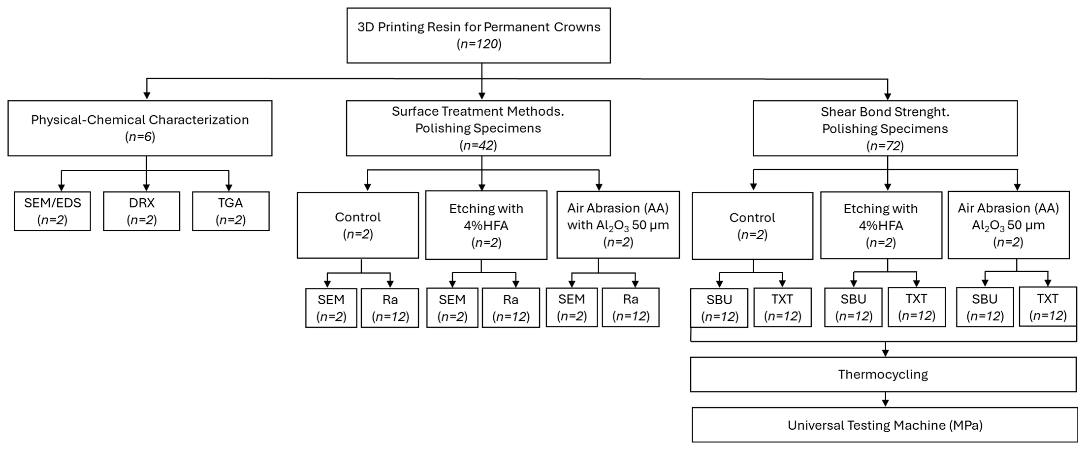
2. Materials and Methods

2.1. Materials Used In Vitro Study

The materials used in this in vitro study are shown in Table 1: 3D printed composite resins, bonding agents and composite resin.

2.2. Preparation of 3D Printed Samples

A total of 120 rectangular specimens (10 mm × 5 mm × 1 mm) were designed using Computer-Aided Design software (Fusion 360, Autodesk Inc.—Mexico City, Mexico). The files were exported in standard tessellation language (STL) format and subsequently processed using 3D SprintTM software (3D Systems, Inc.—Mexico City, Mexico), arranging the samples in groups of ten at a 0° orientation relative to the build platform [33,34]. A 3D printed composite resin for definitive restoration (Crowntec, SAREMCO Dental AG—Mexico City, Mexico) was used for specimen fabrication. The resin was homogenized in a dedicated mixer (LC-3DMixer, 3D Systems, Inc.—Mexico) for 5 min, then transferred to the resin tray of a digital light processing (DLP) 3D Printer (NextDent 5100, 3D Systems, Inc.—Mexico). Printing was initiated by transferring the STL files via USB interface. After that, the printed specimens were thoroughly rinsed in 99% ethyl alcohol using an ultrasonic bath in combination with a soft bristle brush to remove uncured resin residues. Post-curing was carried out in a UV polymerization chamber (LC-3DPrint Box, 3D Systems, Inc.—Mexico) for 30 min according to the manufacturer’s instructions. Subsequently, all samples were trimmed with a diamond disc under water cooling and allocated for further analysis, as illustrated in Figure 1.

2.3. Physical–Chemical Characterization

Six unpolished Crowntec resin specimens were assigned to three groups (n = 2 per group) for physicochemical characterization using complementary analytical techniques.

2.3.1. Scanning Electron Microscopy and Energy-Dispersive X-Ray Spectroscopy (SEM/EDS)

Specimens were mounted on aluminum stubs and examined using a field-emission scanning electron microscope (JSM-7401F, JEOL Ltd.—Tokyo, Japan) equipped with an energy-dispersive X-ray spectroscopy detector (EDAX, AMETEK Inc.—Mahwah, NJ, USA). SEM imaging and elemental EDS analysis were performed at an accelerating voltage of 15 kV under high vacuum.

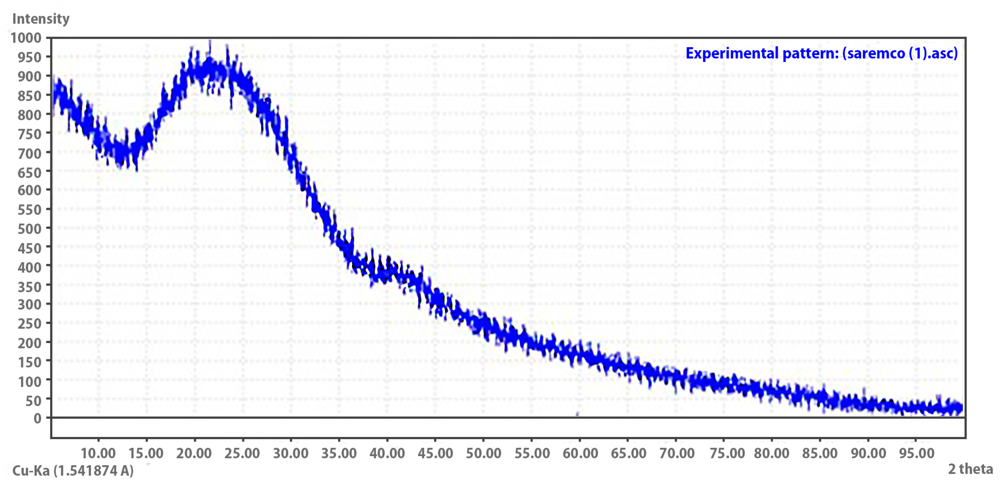
2.3.2. X-Ray Diffraction (XRD)

Crystalline phase analysis was performed using PANalytical X’Pert Pro MPD—Materials Powder Diffractometer (Malvern Panalytical Ltd.—Almelo, The Netherlands). Specimens were placed on a standard sample holder and diffraction patterns were recorded in a 2θ range of 5° to 10°, with a step size of 0.016° and a counting time of 300 s per step, using X’Celerator Solid State Detector (Malvern Panalytical Ltd.—Almelo, The Netherlands).

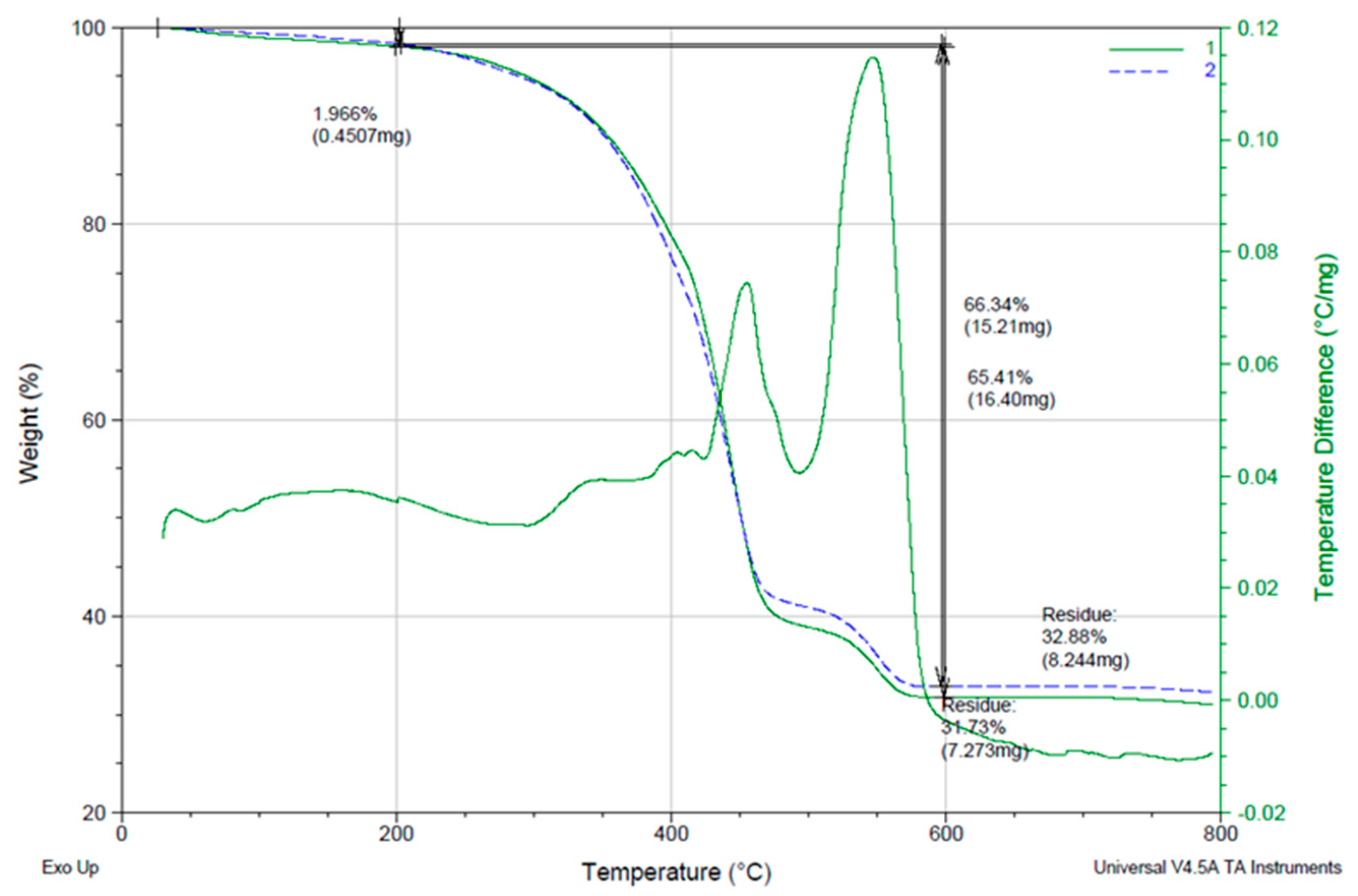
2.3.3. Thermogravimetric Analysis (TGA)

Thermal stability and compositional analysis were performed using a simultaneous TGA/DSC analysis (Universal V4.5A Analyzer, TA Instruments—New Castle, DE, USA). The specimens were heated from 25 °C to 800 °C at a constant rate of 10 °C/min in an air atmosphere and mass loss was continuously recorded to evaluate the organic/inorganic content.

2.4. Surface Treatment Methods—Surface Morphology and Surface Roughness (Ra)

A total of 42 Crowntec resin-fabricated specimens were polished using a porcelain polishing system (DIATECH, COLTENE-Whaledent AG Inc.—Altstätten, Switzerland) and randomly assigned to three experimental groups according to the surface treatment applied: (1) no treatment (control); (2) chemical etching with 4% HFA gel (BISCO Inc.—Schaumburg, IL, USA) for 60 s [35,36]; and (3) airborne particle abrasion (AA) with 50 μm Al2O3 particles for 10 s at 0.2 MPa pressure and a working distance of 10 mm [13,37,38]. After surface treatment, all samples were rinsed with sterile distilled water for 5 s and dried with oil-free compressed air.
Each surface treatment group was subdivided into two types of analysis: quantitative surface roughness measurement (Ra) and qualitative surface morphology evaluation. For surface roughness analysis, a priori, the sample size calculation was performed using G*Power 3.1 software (Heinrich Heine University—Düsseldorf, Germany). Based on a statistical power of 0.85, an alpha error probability of 0.05, and an effect size of 0.70 (Cohen’s f), the required sample size was determined to be twelve specimens per group (n = 12). A contact profilometer (Portable Surface Roughness Tester Surftest SJ-410 Series, Mitutoyo America Corporation, Aurora, IL, USA) was used to measure surface roughness (Ra) according to Standard ISO 21920-2:2021 [39].
For qualitative evaluation of surface morphology, two additional specimens per group (n = 2) were subjected to scanning electron microscopy (SEM). Before imaging, the samples were inspected under a stereomicroscope, mounted on aluminum stubs, sprayed with gold (SCD 050 Sputter Coater—BalTec AG, Pfäffikon, Switzerland), and examined using a field-emission SEM (JSM-7401F, JEOL Ltd.—Tokyo, Japan) operated at an acceleration voltage of 15 kV.

2.5. Shear Bond Strength (SBS) Test

2.5.1. Attachment Fabrication

Rectangular attachments (5.0 mm × 2.0 mm × 2.0 mm) were digitally designed using computer-aided design software (Fusion 360, Autodesk Inc.—Mexico) and printed using a model resin. From the printed models, custom matrices were made with 0.020″ thermoplastic sheets (ZenduraTM FLX, Bay Materials LLC—Fremont, CA, USA) using a pressure molding thermoforming unit (MINISTAR S®, SCHEU-DENTAL GmbH, Iserlohn, Germany), following the manufacturer’s recommended settings. These matrices were used to standardize the shape and dimensions of composite attachments fabricated from nanohybrid composite resin (FiltekTM Z350 XT, 3MTM ESPETM—Mexico City, Mexico).

2.5.2. Preparation and Grouping of Specimens

Seventy-two rectangular specimens (10 mm × 5 mm × 1 mm) of the definitive Crowntec resin printed in 3D were polished using a porcelain polishing system (DIATECH, COLTENE-Whaledent AG Inc.—Altstätten, Switzerland). The samples were randomly assigned to three surface treatment groups (n = 24 per group): (1) no treatment (control), and (2) etching with 4% HFA (BISCO Inc.—Schaumburg, IL, USA) for 60 s and (3) AA with 50 μm Al2O3 particles for 10 s at 0.2 MPa and 10 mm distance. All samples were rinsed with sterile distilled water for 5 s and air dried with oil-free compressed air.
Each surface treatment group was further divided into two adhesive subgroups (n = 12) according to the bonding protocol:
  • TXT—TransbondTM XT Light Cure Adhesive Primer (3MTM UnitekTM—Mexico City, Mexico).
  • SBU—Single BondTM Universal Adhesive (3MTM ESPETM—Mexico City, Mexico).
In the TXT subgroup, a thin layer of primer was applied directly to the printed resin surface without precuring, as per the manufacturer’s instructions. The composite attachment was immediately positioned and light-cured for 10 s on both the mesial and distal surfaces. In the SBU subgroup, the adhesive was actively applied to the bonding surface for 20 s, followed by gentle air drying for 5 s and light cure for 10 s prior to attachment placement. The composite attachments were then placed and light cured in the same manner.

2.5.3. Thermocycling and Mechanical Testing

All bonded specimens were subjected to artificial aging by thermocycling for 500 cycles between 5 °C and 55 °C, with a dwell time of 30 s and a transfer interval of 5 s [25,40,41]. This thermocycling regimen was selected to simulate the early stage thermal fatigue experienced by orthodontic attachments under clinical conditions. According to Standard ISO/TS 11405:2015 [42], 500 cycles represent the standard in vitro protocol to assess the initial durability of the bond. Following thermocycling, each specimen was embedded in autopolymerizing acrylic resin blocks (Líquido Normal Nictone 250 mL Monómero, MDC Dental—Zapopan, Jalisco, Mexico), with the bonded surface orientated parallel to the direction of the applied force.
The shear bond strength was measured using a universal testing machine (Alliance RT/30, MTS Systems Corporation, Eden Prairie, MN, USA) equipped with a chisel-edge blade. The load was applied at a crosshead speed of 1 mm/min until debonding occurred. The maximum force required to dislodge each attachment was recorded in Newtons (N) and converted to shear bond strength (MPa) by dividing by the bonded surface area.

2.6. Statistical Analysis

For the Ra data, one-way analysis of variance (ANOVA) was used to compare surface roughness between the three treatment groups. For SBS, a two-way ANOVA was performed to evaluate the main effects of surface treatment and adhesive system, as well as their interaction on bond strength. Post hoc pairwise comparisons were performed using the Games–Howell test (p < 0.05) to address unequal variances and sample sizes. All statistical analyses were performed with SPSS 25 software (IBM Co., New York, NY, USA), and statistical significance was established at α = 0.05.

3. Results

3.1. Physical–Chemical Characterization

3.1.1. Surface Morphology and Elemental Composition (SEM/EDS)

The elemental composition of the unpolished Crowntec resin surface was evaluated using energy-dispersive X-ray spectroscopy (EDS), revealing a matrix predominantly composed of carbon (46.28 wt%) and oxygen (44.87 wt%), with minor concentrations of silicon (5.99 wt%), aluminum (1.74 wt%), barium (0.73 wt%) and titanium (0.38 wt%) (Figure 2). The corresponding EDS spectrum exhibited intense peaks for carbon and oxygen, consistent with the polymeric organic matrix of the material. Peaks for silicon, aluminum, barium, and titanium were present at lower intensities, reflecting their role as part of the resin’s inorganic filler phase.

3.1.2. Crystalline Structure Analysis (XRD)

The X-ray diffraction (XRD) pattern of the 3D-printed Crowntec resin exhibited a broad diffuse hump between approximately 10° and 35° (2θ), without the presence of sharp or well-defined Bragg peaks (Figure 3). This pattern is indicative of an amorphous structure, suggesting the absence of long-range atomic order or crystalline phases detectable by XRD. The lack of diffraction peaks confirms that the resin matrix is predominantly amorphous, which is consistent with the expected molecular arrangement of cross-linked, light-cured polymeric systems used in photopolymerizable dental materials.

3.1.3. Thermal Stability and Composition (TGA)

Thermogravimetric analysis (TGA) of Crowntec resin 3D printed revealed a major mass loss event of approximately 66.34% occurring between 200 °C and 600 °C, corresponding to thermal degradation of the organic polymer matrix (Figure 4). A minimal initial weight loss of 1.97% was observed below 200 °C, likely associated with the evaporation of adsorbed moisture or residual volatiles.
A major weight loss of ≈66% occurred between 200 °C and 600 °C, corresponding to the degradation of the organic matrix. A stable inorganic residue of ≈33% remained above 600 °C, confirming the hybrid organic-inorganic composition of the material.
Beyond 600 °C, the mass stabilized, with a final residual weight of 32.88%, indicative of a non-volatile inorganic filler fraction. The presence of this stable residue supports the hybrid nature of the material, comprising a polymerizable organic matrix reinforced by inorganic components, as previously suggested by EDS and XRD findings. The onset of significant degradation above 200 °C confirms that the material exhibits adequate thermal stability for intraoral conditions and standard processing protocols.

3.2. Surface Treatment Methods

3.2.1. Surface Morphology (SEM Analysis)

Scanning electron microscopy (SEM) analysis at 50× and 1500× magnifications revealed distinct surface features among the three conditioning protocols applied to the Crowntec resin specimens (Figure 5A–F). The control group (no treatment) exhibited a smooth and homogeneous surface with minimal irregularities and low surface roughness, indicative of limited micromechanical retention capacity. On the contrary, specimens treated with 4% HFA displayed finely distributed microporosities, suggesting superficial etching likely caused by selective dissolution of the inorganic filler phase. This microtextured pattern may facilitate moderate adhesive penetration without compromising structural integrity. The most pronounced surface modification was observed after AA with 50 µm Al2O3 particles, which produced a highly irregular topography characterized by deep craters, grooves, and surface roughening. This morphological alteration significantly increased the available bonding area and provided well-defined microretentive features that were conducive to enhanced mechanical interlocking with adhesive systems.

3.2.2. Surface Roughness Analysis (Ra Values)

Quantitative surface roughness measurements revealed significant differences among surface treatment groups (Figure 6). The AA group exhibited the highest mean roughness (Ra = 2.21 ± 0.30 µm), followed by the etching group treated with 4% HFA (Ra = 0.81 ± 0.20 µm). The control group (without surface treatment) showed the lowest roughness values (Ra = 0.07 ± 0.03 µm).
Statistical analysis confirmed that the differences in Ra between all groups were statistically significant (p < 0.05). Post hoc comparisons indicated that each group differed significantly from the others, as denoted by distinct superscript letters.

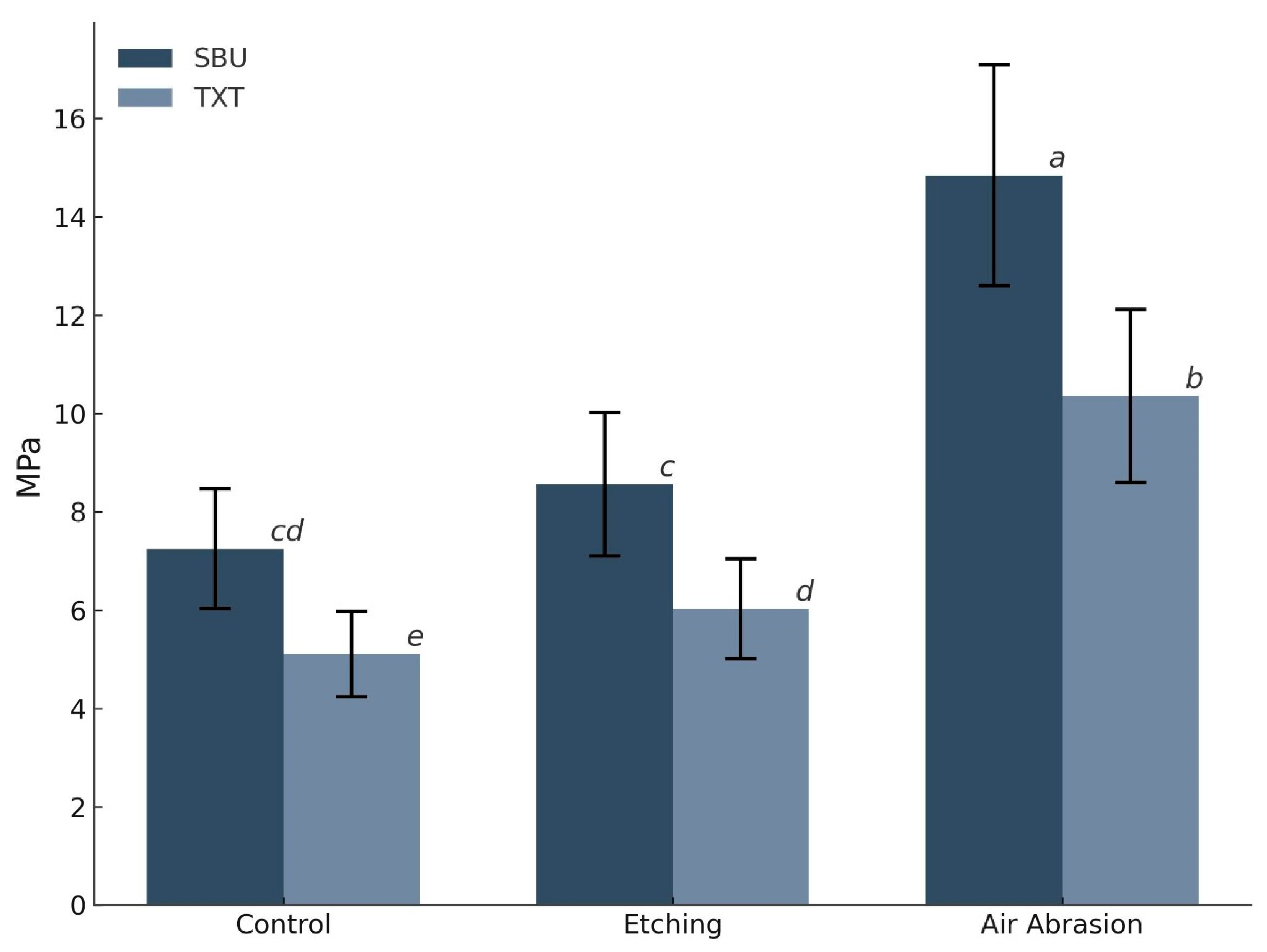
3.3. Shear Bond Strength (SBS) Analysis

A two-way ANOVA demonstrated statistically significant main effects for both surface treatment (F = 125.41; p < 0.001) and adhesive system (F = 74.35; p < 0.001), as well as a significant interaction between the two factors (F = 4.19; p = 0.019) (Table 2).
Among all groups, airborne particle abrasion yielded the highest bond strength values, with SBU achieving 14.84 ± 2.24 MPa and TXT achieving 10.36 ± 1.76 MPa. This was followed by the HFA-etched group, with SBS values of 8.56 ± 1.46 MPa for SBU and 6.03 ± 1.02 MPa for TXT. The control group exhibited the lowest bond strength: 7.25 ± 1.21 MPa for SBU and 5.11 ± 0.87 MPa for TXT (Figure 7).
Air abrasion produced the highest bond strengths for both adhesives, with SBU consistently outperforming TXT under all conditions.
Pairwise comparisons using the Games–Howell test revealed that air abrasion produced significantly higher SBS values than both etching and control treatments in adhesives (p < 0.001). SBU consistently outperformed TXT within each surface treatment group, with the greatest difference observed under air abrasion conditions (Δ = 4.48 MPa; p < 0.001). However, no statistically significant differences were observed between SBU in the control vs. etching groups (p = 0.545), or between TXT in the same groups (p = 0.759 and 0.999), reflecting a similarly moderate bond strength under these conditions (Table 3).
Failure mode analysis indicated that adhesive failure was the predominant mode in all groups, occurring in 80% of samples, regardless of surface treatment or adhesive type.

4. Discussion

This study evaluated the physicochemical characteristics and bonding performance of a definitive 3D printed resin (Crowntec) subjected to different surface treatments and adhesive systems. The findings confirm that the material exhibits a hybrid composition—primarily a cross-linked methacrylate-based polymer matrix reinforced with amorphous inorganic fillers—typical of nanohybrid or microfilled composite resins used for permanent dental restorations. These properties have critical implications for surface conditioning protocols and adhesive selection, particularly when bonding orthodontic attachments to 3D printed substrates.
Energy-dispersive X-ray spectroscopy (EDS) analysis of the 3D-printed definitive resin (Crowntec) revealed a predominantly organic composition, with carbon and oxygen comprising approximately 46% and 45% by weight, respectively, along with minor amounts of silicon, aluminum, barium, and titanium. This elemental profile is characteristic of hybrid methacrylate-based composites reinforced with barium–aluminum silicate glass fillers and radiopaque oxides such as titania, consistent with other studies evaluating DLP-printed materials for definitive restorations [12,13,14,38]. These findings align with previous reports by Donmez et al. [12] and Çakmak et al. [10], who documented similar filler compositions in printed composite crowns intended for long-term intraoral use. Complementary structural analysis by X-ray diffraction (XRD) demonstrated a broad amorphous halo between 10° and 35° (2θ), with no discernible Bragg peaks, confirming the lack of long-range crystalline order. This amorphous pattern is typical of crosslinked photopolymerized resin matrices and has been similarly observed in CAD/CAM and 3D-printed hybrid composites evaluated by Di Fiore et al. [14] and Mao et al. [15], who emphasized the role of filler distribution and polymer network structure in determining both mechanical and adhesive performance. Furthermore, thermogravimetric analysis (TGA) revealed a mass loss of approximately 66% between 200 °C and 600 °C, associated with the decomposition of the organic matrix, and a stable inorganic residue of 32.88% beyond 600 °C. These results are in agreement with previous TGA findings in 3D printed definitive resins, confirming the manufacturer’s reported filler content (30–50 wt%) and supporting the hybrid classification of the material [13,14,38]. Taken together, the EDS, XRD, and TGA data demonstrate that Crowntec behaves as a hybrid restorative composite featuring a highly filled amorphous polymer matrix interspersed with glass-based inorganic particles.
A predominantly polymer-based matrix requires surface treatments that enhance micromechanical retention, which is critical to achieving durable adhesion. In the present study, airborne particle abrasion using 50 μm Al2O3 at 0.2 MPa and a working distance of 10 mm resulted in the highest surface roughness and, correspondingly, the highest shear bond strength (SBS). These findings are consistent with previous research that shows that air abrasion creates an irregular topography with craters and grooves that increase the effective bonding area and provide undercuts for adhesive infiltration [13,15,36]. Studies by Kang et al. [13], Mao et al. [15] and Ersöz et al. [38] have further confirmed that this surface morphology promotes improved bond durability in definitive 3D-printed resins such as Crowntec, particularly under thermocycling. Di Fiore et al. [14] also reported that this treatment increases surface free energy, facilitating uniform adhesive wetting. Importantly, when the abrasion parameters used in this study are applied, no damage is induced to the mechanical integrity of the 3D printed resin. This is supported by previous studies that demonstrate that such settings do not generate microcracks or internal defects, nor do they reduce flexural strength or bonding performance in polymer-based printed composites [14,15,38]. Collectively, the evidence supports airborne particle abrasion as a safe and highly effective surface conditioning method for clinical use with 3D printed definitive resins. The higher standard deviation observed in the air abrasion group can be explained by the multifactorial and inherently stochastic nature of the particle projection. Although the nominal parameters were maintained constant, minor clinical variations in jet angulation, dynamic distance, and local exposure of inorganic fillers may result in heterogeneous surface topographies across specimens. Each particle impact generates variable-depth craters and ridges, producing surfaces that combine highly retentive areas with relatively smooth regions. This heterogeneity increases both the surface roughness (Ra) and the dispersion of the adhesive strength values. Controlled investigations have shown that minor adjustments, as minor as 3°–5° in angulation or 2–3 mm in distance, can modify Ra by up to 40% and result in a double increase in the standard deviation of SBS compared to chemically conditioned surfaces [13,43,44]. These findings substantiate the notion that while air abrasion is effective in establishing micromechanical retention, it inherently exhibits variability. Therefore, the use of 3D printed positioning guides to standardize angulation (90°) and distance (10 mm) is a possible alternative to reduce dispersion while preserving bonding performance. In the case of etching with 4% hydrofluoric acid (HFA), it produced a measurable increase in surface roughness and bond strength compared to the control group; however, its overall effectiveness was still lower than that achieved by air abrasion. These findings can be attributed to the selective action of HFA in silica-based fillers; the acid partially dissolves exposed glass particles, creating microporosities that increase surface energy and promote adhesive wetting [26,32]. However, as noted by Çakmak et al. [10] and Donmez et al. [12], the reaction remains inherently limited in 3D printed resin specimens such as Crowntec, whose formulation contains a reduced fraction of etchable glass and incorporates oxides such as Al2O3 and TiO2 that are less susceptible to acid attack [10,11,26].
The adhesive behavior observed in this study was affected by the bonding system used, reflecting the chemical composition of the substrate. Our research indicates that the latest generation universal adhesive (Single Bond Universal) outperformed a traditional orthodontic primer (Transbond XT), regardless of whether the specimens had undergone surface conditioning. This result can be attributed to the presence of functional molecules within the universal adhesive, specifically 10-methacryloyloxydecyl dihydrogen phosphate (10-MDP) and the silane coupling agent, both of which promote chemical interaction with the inorganic components of the 3D-printed resin. The silane coupling agent enhances adhesion by forming siloxane bonds with exposed silica (SiO2) particles following acid etching, thereby strengthening the chemical integration with the resin’s filler phase [4,13,28]. In parallel, the phosphate functional group of 10-MDP has a high affinity for metal oxides such as alumina and titania, enabling additional chemical bonding at the adhesive–substrate interface [28,30,38]. In comparison, a typical primer, which is largely composed of methacrylic resin, relies predominantly on micromechanical retention without providing this improved chemical connectivity. This limitation is particularly evident on smooth, untreated surfaces, where bond strength tends to be significantly reduced—a finding that has also been reported in previous studies evaluating the performance of conventional primers on 3D-printed resins [13,38,43]. The superiority of combining air abrasion with a universal adhesive thus reflects this synergy: the optimal surface roughness achieved by air abrasion, together with the chemistry of a universal adhesive that can effectively bond to the printed resin, not only ensures a bond strength above the threshold required to prevent debonding during treatment, but also supports a controlled and predictable removal of the attachment at the end of therapy. Importantly, recent in vitro evidence suggests that the use of fine rotary instrumentation, rather than conventional mechanical debonding techniques, allows selective removal of composite attachments without inducing cohesive failure on the 3D-printed resin substrate [45]. This minimally invasive approach preserves the structural and aesthetic integrity of the definitive restoration, which is particularly relevant in adult patients who undergo interdisciplinary orthodontic-prosthetic treatment where the prosthetic element must remain intact after orthodontic therapy.
This in vitro study did not account for all intraoral conditions like chewing forces, moisture levels, or enzymatic breakdown, which might influence the long-term performance of adhesives. Consequently, future studies should incorporate in vivo research to evaluate the clinical longevity of bonded attachments in 3D printed definitive restorations that experience functional stresses and thermal cycling over time. It is also essential to investigate how attachment removal affects the surface condition of 3D printed prostheses. As these restorations might remain post-orthodontic treatment, reducing surface damage during removal is crucial to maintaining their appearance and functionality. Clinically, the results endorse the use of universal and air abrasion adhesives as an effective bonding method for clear aligner attachments on definitive 3D printed crowns, particularly for adult patients receiving orthodontic treatments involving prosthetic components.

5. Conclusions

Based on the limitations of this in vitro study, the following conclusions were reached.
  • The tested 3D printed definitive resin (Crowntec) demonstrated a hybrid structure composed of an amorphous polymer matrix reinforced with approximately 33% inorganic fillers, as confirmed by EDS, XRD and TGA analyses.
  • Among the evaluated protocols, airborne particle abrasion with 50 µm Al2O3 followed by the application of a universal adhesive (containing 10-MDP and silane) achieved the highest shear bond strength, exceeding clinically accepted thresholds for orthodontic retention.
  • Polished, untreated resin surfaces exhibited inadequate adhesion for clinical use, highlighting the need for substrate pretreatment in aligner-based orthodontic workflows.

Author Contributions

Methodology, O.V.C.-B.; Software, O.V.C.-B.; Validation, C.R.L.-D.; Formal analysis, C.A.L.-L.; Investigation, R.d.J.F.-L.; Resources, J.H.L.-D.; Data curation, R.d.J.F.-L.; Writing—original draft, J.H.L.-D.; Writing—review & editing, R.C.C.; Supervision, R.C.C. and J.H.L.-D.; Project administration, R.C.C. and J.H.L.-D. All authors have read and agreed to the published version of the manuscript.

Funding

The APC was funded by Prof. Jorge Humberto Luna-Domínguez.

Informed Consent Statement

Not applicable.

Data Availability Statement

The original contributions presented in this study are included in the article. Further inquiries can be directed to the corresponding author.

Conflicts of Interest

The authors declare no conflict of interest.

References

  1. Tian, Y.; Chen, C.; Xu, X.; Wang, J.; Hou, X.; Li, K.; Lu, X.; Shi, H.; Lee, E.-S.; Jiang, H.B.; et al. A Review of 3D Printing in Dentistry: Technologies, Affecting Factors, and Applications. Scanning 2021, 2021, 9950131. [Google Scholar] [CrossRef] [PubMed]
  2. Alharbi, N.; Wismeijer, D.; Osman, R. Additive Manufacturing Techniques in Prosthodontics: Where Do We Currently Stand? A Critical Review. Int. J. Prosthodont. 2017, 30, 474–484. [Google Scholar] [CrossRef] [PubMed]
  3. Revilla-León, M.; Özcan, M. Additive Manufacturing Technologies Used for Processing Polymers: Current Status and Potential Application in Prosthetic Dentistry. J. Prosthodont. 2019, 28, 146–158. [Google Scholar] [CrossRef] [PubMed]
  4. Diken Türksayar, A.A.; Demirel, M.; Donmez, M.B.; Olcay, E.O.; Eyüboğlu, T.F.; Özcan, M. Comparison of wear and fracture resistance of additively and subtractively manufactured screw-retained, implant-supported crowns. J. Prosthet. Dent. 2024, 132, 154–164. Available online: https://www.thejpd.org/action/showFullText?pii=S0022391323004183 (accessed on 26 February 2025). [CrossRef] [PubMed]
  5. Prause, E.; Malgaj, T.; Kocjan, A.; Beuer, F.; Hey, J.; Jevnikar, P.; Schmidt, F. Mechanical properties of 3D-printed and milled composite resins for definitive restorations: An in vitro comparison of initial strength and fatigue behavior. J. Esthet. Restor. Dent. 2024, 36, 391–401. [Google Scholar] [CrossRef] [PubMed]
  6. Daher, R.; Ardu, S.; di Bella, E.; Krejci, I.; Duc, O. Efficiency of 3D printed composite resin restorations compared with subtractive materials: Evaluation of fatigue behavior, cost, and time of production. J. Prosthet. Dent. 2024, 131, 943–950. [Google Scholar] [CrossRef] [PubMed]
  7. Alhotan, A.; Fouda, A.M.; Al-Johani, H.; Yoon, H.I.; Matinlinna, J.P. Physical and mechanical properties of various resins for additively manufactured definitive fixed dental restorations: Effect of material type and thermal cycling. J. Prosthet. Dent. 2025, 134, 238.e1–238.e8. [Google Scholar] [CrossRef] [PubMed]
  8. Donmez, M.B.; Okutan, Y. Marginal gap and fracture resistance of implant-supported 3D-printed definitive composite crowns: An in vitro study. J. Dent. 2022, 124, 104216. [Google Scholar] [CrossRef] [PubMed]
  9. Çakmak, G.; Rusa, A.M.; Borga Donmez, M.; Akay, C.; Gdem Kahveci, Ç.; Schimmel, M.; Yilmaz, B. Trueness of crowns fabricated by using additively and subtractively manufactured resin-based CAD-CAM materials. J. Prosthet. Dent. 2024, 131, 951–958. [Google Scholar] [CrossRef] [PubMed]
  10. Çakmak, G.; Donmez, M.B.; Molinero-Mourelle, P.; Kahveci, Ç.; Abou-Ayash, S.; Peutzfeldt, A.; Yilmaz, B. Fracture resistance of additively or subtractively manufactured resin-based definitive crowns: Effect of restorative material, resin cement, and cyclic loading. Dent. Mater. 2024, 40, 1072–1077. [Google Scholar] [CrossRef] [PubMed]
  11. Küçükekenci, A.S.; Dönmez, M.B.; Dede, D.Ö.; Çakmak, G.; Yilmaz, B. Bond strength of recently introduced computer-aided design and computer-aided manufacturing resin-based crown materials to polyetheretherketone and titanium. J. Prosthet. Dent. 2024, 132, 1066.e1–1066.e8. [Google Scholar] [CrossRef] [PubMed]
  12. Donmez, M.B.; Çakmak, G.; Yılmaz, D.; Schimmel, M.; Abou-Ayash, S.; Yilmaz, B.; Peutzfeldt, A. Bond strength of additively manufactured composite resins to dentin and titanium when bonded with dual-polymerizing resin cements. J. Prosthet. Dent. 2023, 132, 1067.e1–1067.e8. [Google Scholar] [CrossRef] [PubMed]
  13. Kang, Y.J.; Park, Y.; Shin, Y.; Kim, J.H. Effect of Adhesion Conditions on the Shear Bond Strength of 3D Printing Resins after Thermocycling Used for Definitive Prosthesis. Polymers 2023, 15, 1390. [Google Scholar] [CrossRef] [PubMed]
  14. Di Fiore, A.; Stellini, E.; Alageel, O.; Alhotan, A. Comparison of mechanical and surface properties of two 3D printed composite resins for definitive restoration. J. Prosthet. Dent. 2024, 132, 839.e1–839.e7. Available online: https://www.sciencedirect.com/science/article/abs/pii/S0022391324004529 (accessed on 23 April 2025). [CrossRef] [PubMed]
  15. Mao, Z.; Schmidt, F.; Beuer, F.; Yassine, J.; Hey, J.; Prause, E. Effect of surface treatment strategies on bond strength of additively and subtractively manufactured hybrid materials for permanent crowns. Clin. Oral. Investig. 2024, 28, 371. [Google Scholar] [CrossRef] [PubMed]
  16. Shahin, S.Y.; AlQahtani, N.; Abushowmi, T.H.; Siddiqui, I.A.; Akhtar, S.; Nassar, E.A.; Gad, M.M. The effect of surface treatment and thermal aging on the bonding of clear aligner attachments to provisional resin-based material: Shear bond strength analysis. Front. Oral Health 2024, 5, 1449833. [Google Scholar] [CrossRef] [PubMed]
  17. Chen, W.; Qian, L.; Qian, Y.; Zhang, Z.; Wen, X. Comparative study of three composite materials in bonding attachments for clear aligners. Orthod. Craniofac Res. 2021, 24, 520–527. [Google Scholar] [CrossRef] [PubMed]
  18. Alsaud, B.A.; Hajjaj, M.S.; Masoud, A.I.; Abou Neel, E.A.; Abuelenain, D.A.; Linjawi, A.I. Bonding of Clear Aligner Composite Attachments to Ceramic Materials: An In Vitro Study. Materials 2022, 15, 4145. [Google Scholar] [CrossRef] [PubMed]
  19. Biadsee, A.; Rosner, O.; Khalil, C.; Atanasova, V.; Blushtein, J.; Levartovsky, S. Comparative evaluation of shear bond strength of orthodontic brackets bonded to three-dimensionally-printed and milled materials after surface treatment and artificial aging. Korean J. Orthod. 2023, 53, 45–53. [Google Scholar] [CrossRef] [PubMed]
  20. Bichu, Y.M.; Alwafi, A.; Liu, X.; Andrews, J.; Ludwig, B.; Bichu, A.Y.; Zou, B. Advances in orthodontic clear aligner materials. Bioact. Mater. 2023, 22, 384–403. [Google Scholar] [CrossRef] [PubMed]
  21. Weir, T. Clear aligners in orthodontic treatment. Aust. Dent. J. 2017, 62, 58–62. [Google Scholar] [CrossRef] [PubMed]
  22. Jedliński, M.; Mazur, M.; Greco, M.; Belfus, J.; Grocholewicz, K.; Janiszewska-Olszowska, J. Attachments for the Orthodontic Aligner Treatment—State of the Art—A Comprehensive Systematic Review. Int. J. Environ. Res. Public Health 2023, 20, 4481. [Google Scholar] [CrossRef] [PubMed]
  23. Jindal, P.; Juneja, M.; Siena, F.L.; Bajaj, D.; Breedon, P. Mechanical and geometric properties of thermoformed and 3D printed clear dental aligners. Am. J. Orthod. Dentofac. Orthop. 2019, 156, 694–701. [Google Scholar] [CrossRef] [PubMed]
  24. Çokakoğlu, S.; Nalçacı, R.; Altıntaş, S.H.; Atıcı, F. Can a Self-etching Primer be Effective in Bonding Aligner Attachments to Different Types of Ceramics? Turk. J. Orthod. 2024, 37, 30–35. [Google Scholar] [CrossRef] [PubMed]
  25. Ahmed, T.; Fareen, N.; Alam, M.K. The effect of surface treatment and thermocycling on the shear bond strength of orthodontic brackets to the Y-TZP zirconia ceramics: A systematic review. Dental Press. J. Orthod. 2021, 26, 212118. [Google Scholar] [CrossRef] [PubMed]
  26. Alqerban, A. Lithium di silicate ceramic surface treated with Er,Cr:YSGG and other conditioning regimes bonded to orthodontic bracket. Saudi Dent. J. 2021, 33, 188–193. [Google Scholar] [CrossRef] [PubMed]
  27. Bourgi, R.; Kharouf, N.; Cuevas-Suárez, C.E.; Lukomska-Szymanska, M.; Haikel, Y.; Hardan, L. A Literature Review of Adhesive Systems in Dentistry: Key Components and Their Clinical Applications. Appl. Sci. 2024, 14, 8111. [Google Scholar] [CrossRef]
  28. Yu, J.; Zhang, Z.; Yang, H.; Wang, Y.; Muhetaer, A.; Lei, J.; Huang, C. Effect of universal adhesive and silane pretreatment on bond durability of metal brackets to dental glass ceramics. Eur. J. Oral. Sci. 2021, 129, e12772. [Google Scholar] [CrossRef] [PubMed]
  29. Niknam, O.; Shamohammadi, M.; Ataei, Z.; Rakhshan, V. Combined Effects of Different Bracket Bonding Adhesives and Different Resin Removal Methods on Enamel Discoloration: A Preliminary Study. Int. J. Dent. 2023, 2023, 8838264. [Google Scholar] [CrossRef] [PubMed]
  30. Lima, R.B.W.; de Araújo Ferreira Muniz, I.; Campos, D.E.S.; Murillo-Gómez, F.; de Andrade, A.K.M.; Duarte, R.M.; de Souza, G.M. Effect of universal adhesives and self-etch ceramic primers on bond strength to glass-ceramics: A systematic review and meta-analysis of in vitro studies. J. Prosthet. Dent. 2024, 131, 392–402. Available online: https://www.sciencedirect.com/science/article/abs/pii/S0022391322000555 (accessed on 23 April 2025). [CrossRef] [PubMed]
  31. Kutalmıs Buyuk, S.; Kucukekenci, A.S. Effects of different etching methods and bonding procedures on shear bond strength of orthodontic metal brackets applied to different CAD/CAM ceramic materials. Angle Orthod. 2018, 88, 221–226. [Google Scholar] [CrossRef] [PubMed]
  32. Pédemay, C.; François, P.; Fouquet, V.; Abdel-Gawad, S.; Attal, J.P.; Dantagnan, C.A. Shear bond strength and ARI scores of metal brackets to glazed glass ceramics and zirconia: An in vitro study investigating surface treatment protocols. BMC Oral Health 2024, 24, 1567. [Google Scholar] [CrossRef] [PubMed]
  33. Farkas, A.Z.; Galatanu, S.V.; Nagib, R. The Influence of Printing Layer Thickness and Orientation on the Mechanical Properties of DLP 3D-Printed Dental Resin. Polymers 2023, 15, 1113. [Google Scholar] [CrossRef] [PubMed]
  34. Alsarani, M.M. Influence of aging process and restoration thickness on the fracture resistance of provisional crowns: A comparative study. Saudi Dent. J. 2023, 35, 939–945. [Google Scholar] [CrossRef] [PubMed]
  35. May, M.M.; Fraga, S.; May, L.G. Effect of milling, fitting adjustments, and hydrofluoric acid etching on the strength and roughness of CAD-CAM glass-ceramics: A systematic review and meta-analysis. J. Prosthet. Dent. 2022, 128, 1190–1200. [Google Scholar] [CrossRef] [PubMed]
  36. Murillo-Gómez, F.; Hernández-Víquez, J.R.; Sauma-Montes de Oca, J.R.; Vargas-Vargas, C.; González-Vargas, N.; Vega-Baudrit, J.R.; Chavarría-Bolaños, D. Mechanical, Adhesive and Surface Properties of a Zirconia-Reinforced Lithium Silicate CAD/CAM Ceramic Exposed to Different Etching Protocols. Materials 2024, 17, 5039. [Google Scholar] [CrossRef] [PubMed]
  37. Kang, Y.J.; Kim, H.; Lee, J.; Park, Y.; Kim, J.H. Effect of airborne particle abrasion treatment of two types of 3D-printing resin materials for permanent restoration materials on flexural strength. Dent. Mater. 2023, 39, 648–658. [Google Scholar] [CrossRef]
  38. Ersöz, B.; Aydın, N.; Ezmek, B.; Karaoğlanoğlu, S.; Çal, İ.K. Effect of Surface Treatments Applied to 3D Printed Permanent Resins on Shear Bond Strength. J. Clin. Exp. Dent. 2024, 16, e1059–e1066. [Google Scholar] [CrossRef] [PubMed]
  39. ISO 21920-2:2021; Geometrical Product Specifications (GPS)—Surface Texture: Profile—Part 2: Terms, Definitions and Surface Texture Parame-ters. Available online: https://www.iso.org/ (accessed on 30 April 2025).
  40. Fotovat, F.; Shishehian, A.; Alijani, S.; Alafchi, B.; Parchami, P. Comparison of shear bond strength of orthodontic stainless-steel brackets on temporary crowns fabricated by three different methods: An in vitro study. Int. Orthod. 2022, 20, 100641. [Google Scholar] [CrossRef] [PubMed]
  41. Al Jabbari, Y.S.; Al Taweel, S.M.; Rifaiy MAl Alqahtani, M.Q.; Koutsoukis, T.; Zinelis, S. Effects of surface treatment and artificial aging on the shear bond strength of orthodontic brackets bonded to four different provisional restorations. Angle Orthod. 2014, 84, 649–655. [Google Scholar] [CrossRef] [PubMed]
  42. ISO/TS 11405:2015; Dentistry—Testing of Adhesion to Tooth Structure. Available online: https://www.iso.org/ (accessed on 30 April 2025).
  43. Lankes, V.; Reymus, M.; Liebermann, A.; Stawarczyk, B. Bond strength between temporary 3D printable resin and conventional resin composite: Influence of cleaning methods and air-abrasion parameters. Clin. Oral Investig. 2023, 27, 31–43. [Google Scholar] [CrossRef] [PubMed]
  44. Hammamy, M.; Rueda, S.R.; Pio, A.; Rizzante, F.A.P.; Lawson, N.C. Effect of Air Particle Abrasion and Primers on Bond Strength to 3D-Printed Crown Materials. Materials 2025, 18, 265. [Google Scholar] [CrossRef] [PubMed]
  45. Gorassini, F.; Fiorillo, L.; Marrapodi, M.M.; D’Amico, C.; Basile, M.; Cicciù, M.; Cervino, G. Debonding issues in orthodontics: An RCTs systematic review. Explor. Med. 2024, 5, 477–491. [Google Scholar] [CrossRef]
Figure 1. Experiment design.
Dentistry 13 00341 g001
Figure 2. Energy-dispersive X-ray spectroscopy (EDS) spectrum of the Crowntec resin surface. Dominant peaks for carbon and oxygen reflect the polymeric organic matrix, while lower intensity peaks for silicon, aluminum, barium, and titanium indicate the presence of dispersed inorganic fillers.
Dentistry 13 00341 g002
Figure 3. X-ray diffraction (XRD) pattern of the 3D printed Crowntec resin. The broad diffuse halo between 10° and 35° (2θ) without distinct Bragg peaks indicates an amorphous structure characteristic of polymer-based restorative materials.
Dentistry 13 00341 g003
Figure 4. Thermogravimetric analysis (TGA) curve of the 3D-Printed Crowntec resin.
Dentistry 13 00341 g004
Figure 5. SEM micrographs of Crowntec® resin specimens after different surface treatments—topographical differences observed for each surface conditioning protocol: (A) control—50×, (B) control—1500×, (C) etching 4% HFA—50×, (D) etching 4% HFA—1500×, (E) AA with 50 µm Al2O3—50× and (F) AA with 50 µm Al2O3—1500×.
Dentistry 13 00341 g005
Figure 6. Mean surface roughness (Ra, µm) of Crowntec resin specimens after air abrasion, hydrofluoric acid etching, and no surface treatment (control). Error bars represent standard deviation. Statistically significant differences identified between all groups (one-way ANOVA, p < 0.001), with distinct superscript letters indicating significance in post hoc comparisons (Games–Howell, p < 0.05).
Dentistry 13 00341 g006
Figure 7. Shear bond strength (MPa) of Crowntec resin specimens as a function of surface treatment and adhesive system. The error bars represent the standard deviation. Different superscript letters denote statistically significant differences between groups (p < 0.05, Games–Howell post hoc test).
Dentistry 13 00341 g007
Table 1. Materials used 3D printed composite resins for definitive restoration.
MaterialChemical CompositionManufacturer
3D Printed composite resins for definitive restoration
Crowntec® (additively manufactured composite resin)Esterification products of 4.4′-isopropylphenol, ethoxylated and 2-methylprop-2enoic acid, silanized dental glass, pyrogenic silica, initiator. Total content of inorganic fillers: 30–50 wt%SAREMCO Dental AG
Surface Conditioning Methods
Porcelain etchant
(4% HFA)
Hydrofluoric Acid 5–10%
Sodium Fluoride < 1
BISCO Inc.
Rhino (Al2O3)Aluminum oxide particles White Alumina WA-200
(50 μm)
MDC Dental
Bonding agents
Single bond universal adhesive (SBU)MDP phosphate monomer, Dimethacrylate resins, HEMA, Vitrebond copolymer, filler, ethanol, water, initiators, silane.3MTM ESPETM
Transbond XT Light Cure Adhesive PrimerBisphenol a diglycidyl ether dimethacrylate, Triethylene glycol dimethacrylate (TEGDMA)3MTM UnitekTM
Composite resin
3M FiltekTM Z350 XT compositeMatrix: Bis-GMA, UDMA, Bis-EMA
Filler: Silica, zirconia nanoparticles (20 µm)
(72.5 wt%/55.9 vol%)
3MTM ESPETM
Table 2. Two-way ANOVA for the effects of surface treatment and adhesive system on shear bond strength (SBS).
Type III Sum of SquaresdfMean SquareF
Surface Treatment564.92282.45125.41
Adhesive167.451167.4574.35
Surface Treatment x Adhesive Interaction18.8629.434.19
Error148.65662.25
Table 3. Pairwise comparisons of shear bond strength (SBS) values between treatment–adhesive combinations using the Games–Howell post hoc test. ΔMPa indicates the difference in the mean SBS between the groups. The p < 0.05 denote statistically significant differences.
ComparisonΔMPap-Value
SBU (Air Abrasion)—TXT (Air Abrasion)4.48<0.001
SBU (Air Abrasion)—SBU (Etching)6.28<0.001
SBU (Air Abrasion)—SBU (Control)7.59<0.001
TXT (Air Abrasion)—TXT (Etching)4.33<0.001
TXT (Air Abrasion)—TXT (Control)5.25<0.001
SBU (Etching)—TXT (Etching)2.53<0.001
SBU (Control)—TXT (Control)2.14<0.001
SBU (Etching)—SBU (Control)1.310.545
TXT (Etching)—TXT (Control)0.920.759
Disclaimer/Publisher’s Note: The statements, opinions and data contained in all publications are solely those of the individual author(s) and contributor(s) and not of MDPI and/or the editor(s). MDPI and/or the editor(s) disclaim responsibility for any injury to people or property resulting from any ideas, methods, instructions or products referred to in the content.

