
Impact of Incorporating Nanoparticles to Adhesive Resin on the Demineralization of Enamel: A Systematic Review


Abstract
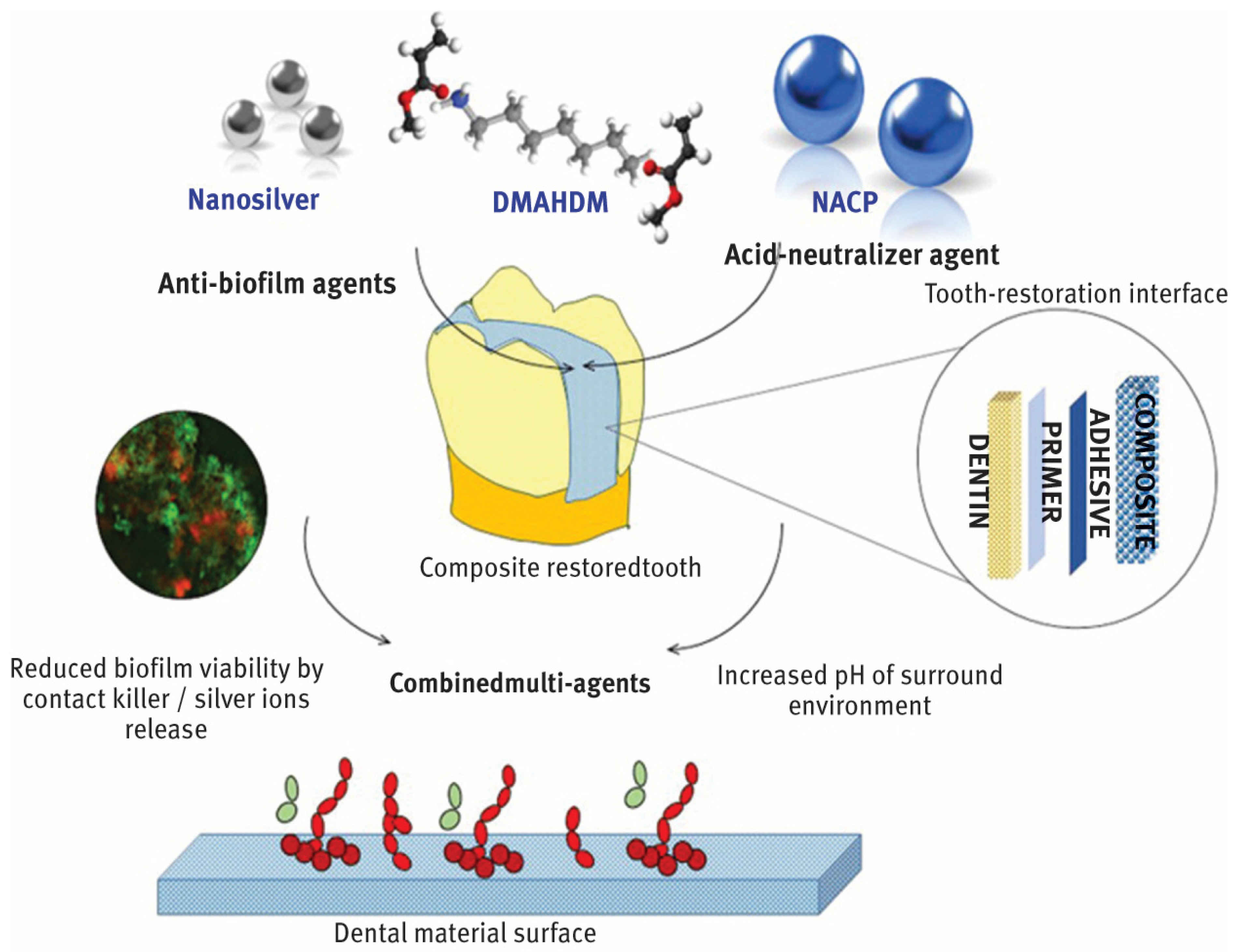
1. Introduction
2. Material and Methods
2.1. Research Question
2.2. Search Strategy
2.3. Eligibility Criteria for Literature Search
2.4. Exclusion Criteria
3. Results
3.1. General Characteristics of Included Studies
3.2. General Outcomes of Included Studies
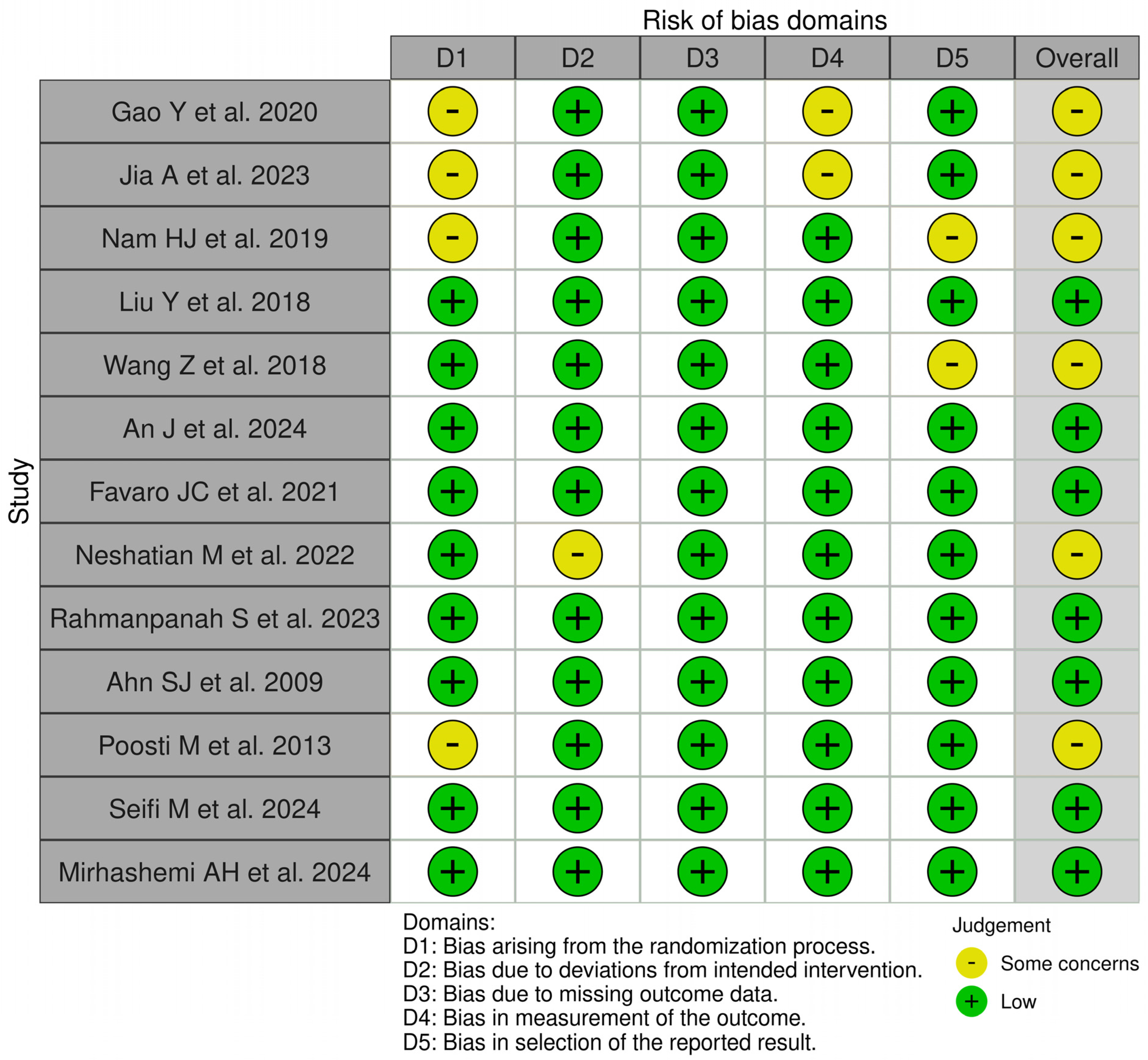
3.3. Risk of Bias Assessment
4. Discussion
5. Conclusions
Funding
Acknowledgments
Conflicts of Interest
References
- Altammar, K.A. A review on nanoparticles: Characteristics, synthesis, applications, and challenges. Front. Microbiol. 2023, 14, 1155622. [Google Scholar] [CrossRef] [PubMed]
- Joudeh, N.; Linke, D. Nanoparticle classification, physicochemical properties, characterization, and applications: A comprehensive review for biologists. J. Nanobiotechnol. 2022, 20, 262. [Google Scholar] [CrossRef] [PubMed] [PubMed Central]
- Yao, Y.; Zhou, Y.; Liu, L.; Xu, Y.; Chen, Q.; Wang, Y.; Wu, S.; Deng, Y.; Zhang, J.; Shao, A. Nanoparticle-Based Drug Delivery in Cancer Therapy and Its Role in Overcoming Drug Resistance. Front. Mol. Biosci. 2020, 7, 193. [Google Scholar] [CrossRef] [PubMed] [PubMed Central]
- Gavas, S.; Quazi, S.; Karpiński, T.M. Nanoparticles for Cancer Therapy: Current Progress and Challenges. Nanoscale Res. Lett. 2021, 16, 173. [Google Scholar] [CrossRef] [PubMed] [PubMed Central]
- Payal; Pandey, P. Role of Nanotechnology in Electronics: A Review of Recent Developments and Patents. Recent Pat. Nanotechnol. 2022, 16, 45–66. [Google Scholar] [CrossRef] [PubMed]
- Gulati, R.; Sharma, S.; Sharma, R.K. Antimicrobial textile: Recent developments and functional perspective. Polym. Bull 2022, 79, 5747–5771. [Google Scholar] [CrossRef] [PubMed] [PubMed Central]
- Rybka, M.; Mazurek, Ł.; Konop, M. Beneficial Effect of Wound Dressings Containing Silver and Silver Nanoparticles in Wound Healing-From Experimental Studies to Clinical Practice. Life 2022, 13, 69. [Google Scholar] [CrossRef] [PubMed] [PubMed Central]
- Krishnan, P.D.; Banas, D.; Durai, R.D.; Kabanov, D.; Hosnedlova, B.; Kepinska, M.; Fernandez, C.; Ruttkay-Nedecky, B.; Nguyen, H.V.; Farid, A.; et al. Silver Nanomaterials for Wound Dressing Applications. Pharmaceutics 2020, 12, 821. [Google Scholar] [CrossRef] [PubMed] [PubMed Central]
- Herranz, F.; Almarza, E.; Rodríguez, I.; Salinas, B.; Rosell, Y.; Desco, M.; Bulte, J.W.; Ruiz-Cabello, J. The application of nanoparticles in gene therapy and magnetic resonance imaging. Microsc. Res. Tech. 2011, 74, 577–591. [Google Scholar] [CrossRef] [PubMed] [PubMed Central]
- Melo, M.A.; Orrego, S.; Weir, M.D.; Xu, H.H.; Arola, D.D. Designing Multiagent Dental Materials for Enhanced Resistance to Biofilm Damage at the Bonded Interface. ACS Appl. Mater. Interfaces 2016, 8, 11779–11787. [Google Scholar] [CrossRef] [PubMed]
- Moraes, G.; Zambom, C.; Siqueira, W.L. Nanoparticles in Dentistry: A Comprehensive Review. Pharmaceuticals 2021, 14, 752. [Google Scholar] [CrossRef] [PubMed] [PubMed Central]
- Malik, S.; Waheed, Y. Emerging Applications of Nanotechnology in Dentistry. Dent. J. 2023, 11, 266. [Google Scholar] [CrossRef] [PubMed]
- Nasiri, K.; Masoumi, S.M.; Amini, S.; Goudarzi, M.; Tafreshi, S.M.; Bagheri, A.; Yasamineh, S.; Alwan, M.; Arellano, M.T.C.; Gholizadeh, O. Recent advances in metal nanoparticles to treat periodontitis. J. Nanobiotechnol. 2023, 21, 283. [Google Scholar] [CrossRef]
- Vasiliu, S.; Racovita, S.; Gugoasa, I.A.; Lungan, M.-A.; Popa, M.; Desbrieres, J. The Benefits of Smart Nanoparticles in Dental Applications. Int. J. Mol. Sci. 2021, 22, 2585. [Google Scholar] [CrossRef] [PubMed] [PubMed Central]
- Budi, H.S.; Jameel, M.F.; Widjaja, G.; Alasady, M.S.; Mahmudiono, T.; Mustafa, Y.F.; Fardeeva, I.; Kuznetsova, M. Study on the role of nano antibacterial materials in orthodontics (a review). Braz. J. Biol. Rev. Brasleira Biol. 2022, 84, e257070. [Google Scholar] [CrossRef] [PubMed]
- Gao, Y.; Liang, K.; Weir, M.D.; Gao, J.; Imazato, S.; Tay, F.R.; Lynch, C.D.; Oates, T.W.; Li, J.; Xu, H.H. Enamel remineralization via poly(amido amine) and adhesive resin containing calcium phosphate nanoparticles. J. Dent. 2020, 92, 103262. [Google Scholar] [CrossRef]
- Jia, A.; Wang, P.; Tong, F.; Chen, Z.; Deng, Y.; Yao, H.; Wang, L.; Liu, Y.; Ge, H. Developing a Novel Enamel Adhesive with Amorphous Calcium Phosphate and Silver Nanoparticles to Prevent Demineralization during Orthodontic Treatment. J. Funct. Biomater. 2023, 14, 77. [Google Scholar] [CrossRef] [PubMed]
- Nam, H.-J.; Kim, Y.-M.; Kwon, Y.H.; Yoo, K.-H.; Yoon, S.-Y.; Kim, I.-R.; Park, B.-S.; Son, W.-S.; Lee, S.-M.; Kim, Y.-I. Fluorinated Bioactive Glass Nanoparticles: Enamel Demineralization Prevention and Antibacterial Effect of Orthodontic Bonding Resin. Materials 2019, 12, 1813. [Google Scholar] [CrossRef] [PubMed]
- Liu, Y.; Zhang, L.; Niu, L.-N.; Yu, T.; Xu, H.H.; Weir, M.D.; Oates, T.W.; Tay, F.R.; Chen, J.-H. Antibacterial and remineralizing orthodontic adhesive containing quaternary ammonium resin monomer and amorphous calcium phosphate nanoparticles. J. Dent. 2018, 72, 53–63. [Google Scholar] [CrossRef]
- Wang, Z.; Ouyang, Y.; Wu, Z.; Zhang, L.; Shao, C.; Fan, J.; Zhang, L.; Shi, Y.; Zhou, Z.; Pan, H.; et al. A novel fluorescent adhesive-assisted biomimetic mineralization. Nanoscale 2018, 10, 18980–18987. [Google Scholar] [CrossRef]
- An, J.; Shen, X.; Peng, T.; Qiao, M.; Xu, B. Formulation of arginine-loaded mesoporous silica nanoparticles (Arg@MSNs) modified orthodontic adhesive. J. Dent. 2024, 145, 104992. [Google Scholar] [CrossRef] [PubMed]
- Favaro, J.C.; Peixoto, Y.C.T.d.M.; Geha, O.; Dias, F.A.; Guiraldo, R.D.; Lopes, M.B.; Berger, S.B. Can silver diamine fluoride or silver nanoparticle-based anticaries agents to affect enamel bond strength? Restor. Dent. Endod. 2021, 46, e7. [Google Scholar] [CrossRef] [PubMed]
- Neshatian, M.; Holcroft, J.; Kishen, A.; De Souza, G.; Ganss, B. Promoting mineralization at biological interfaces Ex Vivo with novel amelotin-based bio-nano complexes. Mater. Today Bio 2022, 14, 100255. [Google Scholar] [CrossRef] [PubMed]
- Rahmanpanah, S.; Seifi, M.; Gharavi, Z.; Sadighnia, N.; Amdjadi, P. Evaluation of shear bond strength and enamel remineralizing effect of experimental orthodontic composite containing nano-hydroxyapatite: An in vitro study. Int. Orthod. 2023, 21, 100725. [Google Scholar] [CrossRef] [PubMed]
- Ahn, S.J.; Lee, S.J.; Kook, J.K.; Lim, B.S. Experimental antimicrobial orthodontic adhesives using nanofillers and silver nanoparticles. Dent. Mater. 2009, 25, 206–213. [Google Scholar] [CrossRef] [PubMed]
- Poosti, M.; Ramazanzadeh, B.; Zebarjad, M.; Javadzadeh, P.; Naderinasab, M.; Shakeri, M.T. Shear bond strength and antibacterial effects of orthodontic composite containing TiO2 nanoparticles. Eur. J. Orthod. 2013, 35, 676–679. [Google Scholar] [CrossRef]
- Seifi, M.; Eskandarloo, F.; Amdjadi, P.; Farmany, A. Investigation of mechanical properties, remineralization, antibacterial effect, and cellular toxicity of composite orthodontic adhesive combined with silver-containing nanostructured bioactive glass. BMC Oral Health 2024, 24, 650. [Google Scholar] [CrossRef] [PubMed]
- Mirhashemi, A.H.; Pourhajibagher, M.; Zebardast, B.; Bahrami, R.; Kharazi Fard, M.J. In Vitro effects of antimicrobial properties and shear bond strength of different concentrations of Emodin nanoparticles incorporated orthodontic composites. Int. Orthod. 2024, 22, 100836. [Google Scholar] [CrossRef] [PubMed]
- Yin, I.X.; Zhao, I.S.; Mei, M.L.; Li, Q.; Yu, O.Y.; Chu, C.H. Use of Silver Nanomaterials for Caries Prevention: A Concise Review. Int. J. Nanomed. 2020, 15, 3181–3191. [Google Scholar] [CrossRef]
- Borzabadi-Farahani, A.; Borzabadi, E.; Lynch, E. Nanoparticles in orthodontics, a review of antimicrobial and anti-caries applications. Acta Odontol. Scand. 2014, 72, 413–417. [Google Scholar] [CrossRef] [PubMed]
- Hanabusa, M.; Mine, A.; Kuboki, T.; Momoi, Y.; Van Landuyt, K.L.; Van Meerbeek, B.; De Munck, J. TEM interfacial characterization of an experimental self-adhesive filling material bonded to enamel/dentin. Dent. Mater. 2011, 27, 818–824. [Google Scholar] [CrossRef] [PubMed]
- Kielbassa, A.M.; Leimer, M.R.; Hartmann, J.; Harm, S.; Pasztorek, M.; Ulrich, I.B. Ex Vivo investigation on internal tunnel approach/internal resin infiltration and external nanosilver-modified resin infiltration of proximal caries exceeding into dentin. PLoS ONE 2020, 15, e0228249. [Google Scholar] [CrossRef] [PubMed]
- Coutinho, E.; Jarmar, T.; Svahn, F.; Neves, A.; Verlinden, B.; Van Meerbeek, B.; Engqvist, H. Ultrastructural characterization of tooth-biomaterial interfaces prepared with broad and focused ion beams. Dent. Mater. 2009, 25, 1325–1337. [Google Scholar] [CrossRef] [PubMed]
- Kalha, A. Bond or band? Evid. Based Dent. 2007, 8, 105. [Google Scholar] [CrossRef] [PubMed]
- Hennig, C.-L.; Blochberger, B.; Symmank, J.; Nitzsche, Á.; Nietzsche, S.; Steiniger, F.; Dederichs, M.; Güllmar, A.; Reise, M.; Schulze-Späte, U.; et al. Effects of reducing excess dental adhesive on bacterial adhesion in the bracket periphery. Clin. Oral Investig. 2023, 27, 1993–2001. [Google Scholar] [CrossRef] [PubMed] [PubMed Central]
- Kachuie, M.; Khoroushi, M. Prevention and Treatment of White Spot Lesions in Orthodontic Patients. Contemp. Clin. Dent. 2017, 8, 11–19. [Google Scholar] [CrossRef] [PubMed] [PubMed Central]
- Kreve, S.; Reis, A.C.D. Bacterial adhesion to biomaterials: What regulates this attachment? A review. Jpn. Dent. Sci. Rev. 2021, 57, 85–96. [Google Scholar] [CrossRef] [PubMed] [PubMed Central]
- Shadlou, S.; Ahmadi-Moghadam, B.; Taheri, F. Nano-Enhanced Adhesives. Rev. Adhes. Adhes. 2014, 2, 371–412. [Google Scholar] [CrossRef]
- Bourgi, R.; Doumandji, Z.; Cuevas-Suárez, C.E.; Ben Ammar, T.; Laporte, C.; Kharouf, N.; Haikel, Y. Exploring the Role of Nanoparticles in Dental Materials: A Comprehensive Review. Coatings 2025, 15, 33. [Google Scholar] [CrossRef]
- Bruna, T.; Maldonado-Bravo, F.; Jara, P.; Caro, N. Silver Nanoparticles and Their Antibacterial Applications. Int. J. Mol. Sci. 2021, 22, 7202. [Google Scholar] [CrossRef] [PubMed] [PubMed Central]
- Mistry, S.; Kundu, D.; Datta, S.; Basu, D. Effects of bioactive glass, hydroxyapatite and bioactive glass-Hydroxyapatite composite graft particles in the treatment of infrabony defects. J. Indian Soc. Periodontol. 2012, 16, 241–246. [Google Scholar] [CrossRef] [PubMed] [PubMed Central]
- Albani, R.; Habib, S.R.; AlHelal, A.A.; Alrabiah, M. Streptococcus-mutans and Porphyromonas-gingivalis adhesion to glazed/polished surfaces of CAD/CAM restorations. Heliyon 2024, 10, e40276. [Google Scholar] [CrossRef] [PubMed] [PubMed Central]
- Habib, S.; Nakshabandi, A.; Al Shawi, A.; Allohaidan, F.; Al Kurdi, R.; AlSarhan, M. Degree of Streptococcus mutans Colonization on Common Restorative Materials Subjected to Wear Cycle. Int. J. Prosthodont. 2021, 34, 626–634. [Google Scholar] [CrossRef] [PubMed]
- Juvvadi, S.R.; Rammohan, S.N.; Gandikota, C.S.; Challa, P.; Manne, R.; Mathur, A. Adherence of Streptococcus mutans and Candida albicans to different bracket materials. J. Pharm. Bioallied. Sci. 2012, 4 (Suppl. S2), S212–S216. [Google Scholar] [CrossRef] [PubMed] [PubMed Central]
- Fournier, A.; Payant, L.; Bouclin, R. Adherence of Streptococcus mutans to orthodontic brackets. Am. J. Orthod Dentofac. Orthop 1998, 114, 414–417. [Google Scholar] [CrossRef] [PubMed]
- Papaioannou, W.; Gizani, S.; Nassika, M.; Kontou, E.; Nakou, M. Adhesion of Streptococcus mutans to different types of brackets. Angle Orthod. 2007, 77, 1090–1095. [Google Scholar] [CrossRef] [PubMed]
- Brusca, M.I.; Chara, O.; Sterin-Borda, L.; Rosa, A.C. Influence of different orthodontic brackets on adherence of microorganisms in vitro. Angle Orthod. 2007, 77, 331–336. [Google Scholar] [CrossRef] [PubMed]
- Ahn, S.J.; Lim, B.S.; Yang, H.C.; Chang, Y.I. Quantitative analysis of the adhesion of cariogenic streptococci to orthodontic metal brackets. Angle Orthod. 2005, 75, 666–671. [Google Scholar] [CrossRef] [PubMed]
- Zakrzewski, W.; Dobrzynski, M.; Dobrzynski, W.; Zawadzka-Knefel, A.; Janecki, M.; Kurek, K.; Lubojanski, A.; Szymonowicz, M.; Rybak, Z.; Wiglusz, R.J. Nanomaterials Application in Orthodontics. Nanomaterials 2021, 11, 337. [Google Scholar] [CrossRef] [PubMed]
- Madian, A.; Elfouly, D.; El-Harouni, N. The effect of silver nanoparticles on the shear bond strength of orthodontic bonding system: A systematic review. Clin. Investig. Orthod. 2022, 81, 187–194. [Google Scholar] [CrossRef]
- Melo, M.A.S.; Cheng, L.; Weir, M.D.; Hsia, R.; Rodrigues, L.K.A.; Xu, H.H. Novel dental adhesive containing antibacterial agents and calcium phosphate nanoparticles. J. Biomed Mater Res. B Appl. Biomater. 2013, 101, 620–629. [Google Scholar] [CrossRef] [PubMed] [PubMed Central]


| No. | Authors | Journal. Year | Study Design | Study Title | Assessment Method |
|---|---|---|---|---|---|
| 1. | Gao Y et al. [16] | Journal of Dentistry. 2020 | In Vitro | Enamel remineralization via poly(amido amine) and adhesive resin containing calcium phosphate nanoparticles. | * SBS |
| 2. | Jia A et al. [17] | Journal of Functional Biomaterials. 2023 | In Vitro | Developing a Novel Enamel Adhesive with Amorphous Calcium Phosphate and Silver Nanoparticles to Prevent Demineralization during Orthodontic Treatment. | SBS and Colony Counting Test |
| 3. | Nam HJ et al. [18] | Materials (Basel). 2019 | In Vitro | Fluorinated Bioactive Glass Nanoparticles: Enamel Demineralization Prevention and Antibacterial Effect of Orthodontic Bonding Resin. | Vicker’s Test and Cell Viability Tests |
| 4. | Liu Y et al. [19] | Journal of Dentistry. 2018 | In Vitro | Antibacterial and remineralizing orthodontic adhesive containing quaternary ammonium resin monomer and amorphous calcium phosphate nanoparticles. | SBS and Cell Viability Tests |
| 5. | Wang Z et al. [20] | Nanoscale. 2018 | In Vitro | A novel fluorescent adhesive-assisted biomimetic mineralization. | Cell Viability Tests |
| 6. | An J et al. [21] | Journal of Dentistry. 2024 | In Vitro | Formulation of arginine-loaded mesoporous silica nanoparticles (Arg@MSNs) modified orthodontic adhesive. | SBS and Cell Counting Kit |
| 7. | Favaro JC et al. [22] | Restorative Dentistry & Endodontics. 2021 | In Vitro | Can silver diamine fluoride or silver nanoparticle-based anticaries agents affect enamel bond strength? | SBS and Microhardness Test |
| 8. | Neshatian M et al. [23] | Materials Today Bio. 2022 | In Vitro | Promoting mineralization at biological interfaces Ex vivowith novel amelotin-based bio-nano complexes. | Mineral Formation and Sbs Testing |
| 9. | Rahmanpanah S et al. [24] | International Orthodontics. 2023 | In Vitro | Evaluation of shear bond strength and enamel remineralizing effect of experimental orthodontic composite containing nano-hydroxyapatite: An in vitro study. | SBS |
| 10. | Ahn SJ et al. [25] | Dental Materials. 2009 | In Vitro | Experimental antimicrobial orthodontic adhesives using nanofillers and silver nanoparticles. | SBS and Bacterial Adhesion Assay |
| 11. | Poosti M et al. [26] | European Journal of Orthodontics. 2013 | In Vitro | Shear bond strength and antibacterial effects of orthodontic composite containing TiO2 nanoparticles. | SBS and Antibacterial Tests |
| 12. | Seifi M et al. [27] | BMC Oral Health. 2024 | In Vitro | Investigation of mechanical properties, remineralization, antibacterial effect, and cellular toxicity of composite orthodontic adhesive combined with silver-containing nanostructured bioactive glass. | Cytotoxicity Test, SBS and Microhardness Tests |
| 13. | Mirhashemi AH et al. [28] | International Orthodontics. 2024 | In Vitro | In vitro effects of antimicrobial properties and shear bond strength of different concentrations of Emodin nanoparticles incorporated orthodontic composites. | Antimicrobial Activity and SBS Tests |
| No. | Authors | Adhesive | Nanoparticles | Study Outcomes | Conclusions |
|---|---|---|---|---|---|
| 1. | Gao Y et al. [16] | Scotchbond Multi-Purpose | a ACP | Adhesives with nanoparticles of ACP yielded a similar b SBS to control. | The novel SN15-PAMAM + NACP adhesive method could achieve 90% higher enamel remineralization. |
| 2. | Jia A et al. [17] | Transbond XT | ACP and polydopamine-Ag (NPA) | SBS of adhesive with 0.2 wt. % NPA was 11.89 ± 1.27 MPa, meeting the minimal clinical bond strength of 7.8 MPa. | Adhesive with NPA may have a good application potential for the prevention and treatment of c WSL. |
| 3. | Nam HJ et al. [18] | Transbond XT Low Flow (LV) | Bioactive glass containing 2.5% fluoride | The anti-demineralization test showed a concentration-dependent increase. | Adhesive containing bioactive glass 2.5% fluoride showed antibacterial and anti-demineralization effects, indicating possible WSL prevention activity. |
| 4. | Liu Y et al. [19] | Transbond XT (TB) | Quaternary ammonium resin monomer and ACP | PND adhesive with 5% MAE-DB and 40% NACP exhibits antibacterial and remineralizing capabilities, and did not adversely affect SBS compared to commercial adhesive. | Novel adhesive containing quaternary ammonium monomer and ACP represents a promising candidate in combating enamel WSL and dental caries. |
| 5. | Wang Z et al. [20] | Clearfil S3 Bond | 1 wt. % of sodium fluorescein and 25 wt. % of polyacrylic acid-stabilized ACP | Fluorescent mineralizing adhesive could induce the extra- and intra-fibrillar remineralization of the reconstituted type I collagen, the demineralized enamel and dentin substrate. | The novel fluorescent adhesive-assisted biomimetic mineralization strategy will pave the way to design and produce anti-carious materials for the prevention of dental caries. |
| 6. | An J et al. [21] | Transbond XT | Rginine loaded mesoporous silica nanoparticles | Adhesive containing Arg@MSNs exhibited significantly enhanced antibacterial activities and inhibitory effects on acid production compared to the commercial adhesive without compromising their bonding strength or biocompatibility. | Adhesive containing Arg@MSNs exhibits clinical benefits in preventing the demineralization of enamel surfaces around or beneath orthodontic brackets due to its enhanced antibacterial activities and acid-producing inhibitory effects. |
| 7. | Favaro JC et al. [22] | Adper Single Bond 2, 3M ESPE | Silver nanoparticles (SNPs) and silver diamine fluoride (SDF) | There was no significant difference among the IE, IE + SNP, DE + SDF, and DE + SNP groups. The IE + SDF and DE groups recorded the highest and the lowest μ-SBS values, respectively. Adhesive-type failures were the most frequent for all treatments. | The use of anticaries agents (SDF and SNP) did not reduce the SBS values of resin composites when they are used on the intact or artificially demineralized dental enamel. Thus, the anticaries agents tested in this study can be used as a pretreatment prior to resin restoration contributing to caries prevention. |
| 8. | Neshatian M et al. [23] | Enamel protein amelotin | Hydroxyapatite nanoparticles | Accelerated mineral formation collagen mineralization of bio- and nanocomplex-treated samples were observed in all model systems. | We have shown that AMTN-based bio- and nanocomplexes promote mineral formation on collagenous interfaces. Our findings can be the basis of new bioinspired, bio-nano materials that may improve dental restoration longevity by enhancing the stability and integrity of the dentin-composite resin interface. |
| 9. | Rahmanpanah S et al. [24] | 3M™ Transbond™ XT | Nano-hydroxyapatite | The addition of hydroxyapatite nanoparticles to orthodontic composite can increase the mineral content and microhardness of the adjacent enamel. However, increasing the amount of nanoparticles reduces shear bond strength in a decreasing trend. | An incremental increase in nanoparticles of HA can be incorporated into the composite to a certain extent, and limitations are determined by the mechanical properties (SBS) required for bracket bonding. |
| 10. | Ahn SJ et al. [25] | Composite and resin-modified glass ionomer | Silica nanofillers and silver nanoparticles | There was no significant difference in shear bond strength and bond failure interface between the experimental composite adhesives and conventional adhesives. | Experimental composite adhesives can help prevent enamel demineralization around their surfaces without compromising physical properties. |
| 11. | Poosti M et al. [26] | Transbond XT | titanium oxide (TiO2) nanoparticles | No significant difference was found between the SBS of conventional and nanocomposites, 24 h after curing (p = 0.58). The chi-square test showed that ARI scores of two groups were not significantly different after debonding (p = 0.69). Comparison of antibacterial effects between the conventional and the nanocomposite demonstrated a significant difference between two groups, with nanocomposites having a higher antibacterial activity (p = 0.03). | Colony count revealed no significant difference in bacterial growth immediately and 30 days after curing in the nanocomposite group. Adding TiO2 nanoparticles to orthodontic composite enhances its antibacterial effects without compromising the SBS. |
| 12. | Seifi M et al. [27] | Orthodontic composite (Adhesive) (GC Ortho Connect, GC Orthodontics, Japan) | nano-bioactive glass–silver | The shear bond strength of the adhesives decreased significantly (p < 0.001) after the addition of nanoparticles, but it remained above the recommended value. The addition of nBG@Ag led to improvements in the microhardness of the teeth, although the differences in microhardness between the study groups were not statistically significant. | Adding nBG@Ag to orthodontic adhesives can be an effective approach to enhancing the antimicrobial activity and reducing enamel demineralization around the orthodontic brackets, without compromising biocompatibility and bond strength. |
| 13. | Mirhashemi AH et al. [28] | Orthodontic composite (Adhesive) (GC Ortho Connect, GC Orthodontics, Japan) | Emodin nanoparticles | The eluted components test demonstrated that the 2% concentration of ENPs performed significantly better against S. mutans compared to the control group (p < 0.05). The highest mean SBS was observed with the 0.5% ENP concentration, while no significant differences in SBS and ARI were found among the groups (p > 0.05). | This in vitro study showed that the 2% concentration of ENP produced significantly improved antimicrobial activity without adversely affecting the SBS and ARI score. This would support the addition of 2% ENP to orthodontic adhesives. |
| No. | Authors | Journal. Year | Study Title | Reason of Exclusion |
|---|---|---|---|---|
| 1. | Yin IX et al. [29] | International Journal of Nanomedicine. 2020 | Use of Silver Nanomaterials for Caries Prevention: A Concise Review | Review article |
| 2. | Budi HS et al. [15] | Brazilian Journal of Biology. 2022 | Study on the role of nano antibacterial materials in orthodontics (a review) | Review article |
| 3. | Borzabadi-FA et al. [30] | Acta Odontologica Scandinavica. 2014 | Nanoparticles in orthodontics, a review of antimicrobial and anti-caries applications | Review article |
| 4. | Hanabusa M et al. [31] | Dental Materials. 2011 | TEM interfacial characterization of an experimental self-adhesive filling material bonded to enamel/dentin | No nanoparticles used |
| 5. | Kielbassa AM [32] | PLoS One. 2020 | Ex vivo investigation into internal tunnel approach/internal resin infiltration and external Nano silver-modified resin infiltration of proximal caries exceeding into dentin | Use of a conventional infiltrant resin instead of adhesive resin |
| 6. | Coutinho E [33] | Dental Materials. 2009 | Ultrastructural characterization of tooth-biomaterial interfaces prepared with broad and focused ion beams | Use of ion beams and no use of nanoparticles |
Disclaimer/Publisher’s Note: The statements, opinions and data contained in all publications are solely those of the individual author(s) and contributor(s) and not of MDPI and/or the editor(s). MDPI and/or the editor(s) disclaim responsibility for any injury to people or property resulting from any ideas, methods, instructions or products referred to in the content. |
© 2025 by the author. Licensee MDPI, Basel, Switzerland. This article is an open access article distributed under the terms and conditions of the Creative Commons Attribution (CC BY) license (https://creativecommons.org/licenses/by/4.0/).
Share and Cite
Almosa, N. Impact of Incorporating Nanoparticles to Adhesive Resin on the Demineralization of Enamel: A Systematic Review. Dent. J. 2025, 13, 89. https://doi.org/10.3390/dj13030089
Almosa N. Impact of Incorporating Nanoparticles to Adhesive Resin on the Demineralization of Enamel: A Systematic Review. Dentistry Journal. 2025; 13(3):89. https://doi.org/10.3390/dj13030089
Chicago/Turabian StyleAlmosa, Naif. 2025. "Impact of Incorporating Nanoparticles to Adhesive Resin on the Demineralization of Enamel: A Systematic Review" Dentistry Journal 13, no. 3: 89. https://doi.org/10.3390/dj13030089
APA StyleAlmosa, N. (2025). Impact of Incorporating Nanoparticles to Adhesive Resin on the Demineralization of Enamel: A Systematic Review. Dentistry Journal, 13(3), 89. https://doi.org/10.3390/dj13030089