Article

Osteoarthritic Bony Alterations of Temporomandibular Joint and Relation to Low Bone Mineral Density in Postmenopausal Edentulous Females

1
Private Practice “JK Dent”, LV-1048 Riga, Latvia
2
Institute of Electronics and Computing Science, LV-1006 Riga, Latvia
3
Department of Prosthodontics, Riga Stradins University, LV-1007 Riga, Latvia
4
Riga Stradins University Institute of Stomatology, LV-1007 Riga, Latvia
5
Department of Conservative Dentistry and Oral Health, Riga Stradins University, LV-1007 Riga, Latvia
6
Department of Orthodontics, Riga Stradins University, LV-1007 Riga, Latvia
*
Authors to whom correspondence should be addressed.
Dent. J. 2024, 12(8), 238; https://doi.org/10.3390/dj12080238
Submission received: 5 June 2024 / Revised: 10 July 2024 / Accepted: 25 July 2024 / Published: 26 July 2024

Abstract

:
This study aimed to evaluate the relationship between osteoarthritic bony alterations in the temporomandibular joint (TMJ) and general bone mineral density (BMD) in postmenopausal edentulous females. Cone beam computed tomography (CBCT) scans for both TMJs were acquired for 80 clinically asymptomatic patients (mean age 72 ± 8.8 y). Both lumbar spine and femoral neck measurements of BMD were performed using dual-energy X-ray absorptiometry (DXA). The most frequently observed bony alterations were flattening of the articular surface (47.5%), sclerosis (41.5%), subcortical cysts (10%), and erosions (5%). Osteophytes were not observed. No statistical differences were observed between those who had or did not have radiological signs of bony alterations according to age or DXA scores. The prevalence of radiological findings of degenerative TMJ bony alterations in clinically asymptomatic postmenopausal females did not confirm a connection with a generally low BMD.

1. Introduction

Temporomandibular joint (TMJ) osteoarthritis (OA) is one of the most common diseases of the TMJ [1,2,3]. It is estimated that TMJ-OA is detected in 40% of seniors by microscopic examination and 14% by radiographic evaluation [4]. It is an age-related degenerative disease that occurs more frequently in females than in males [5,6,7] because of its association with hormonal factors, especially estrogen levels, which influence the remodeling of the mandibular condyle [8]. Temporomandibular disorders, including OA, are no longer considered to be solely local disorders but rather are the outcome of multiple risk determinants [9].
OA is characterized by the progressive destruction and loss of TMJ cartilage, followed by the release of degraded proteoglycans and proteolytic enzymes into the synovial fluid. This causes a secondary inflammatory response that results in further degradation of joints [8,10,11]. In the presence of TMJ-OA, there is a decrease in the amount of synovial fluid, which leads to the loss of joint function and joint pain [12]. These changes may be observed in the early stages as condylar flattening on plain radiography or computed tomography. However, in advanced stages radiography may reveal osteophyte formation, subarticular cysts, or condylar sclerosis [3].
Cone-beam computed tomography (CBCT) is the most reliable method for examining TMJ bone tissue components [13,14,15]. Compared to conventional computed tomography (CT) for detecting morphological TMJ changes, it generally results in images of CT-like quality [16]. The advantage of CBCT compared to conventional CT is the lower radiation dose to the patient; it is more affordable and requires less space for its operation. The diagnostic efficacy of detecting osseous changes in the TMJ with CBCT is superior to that of linear tomography, panoramic radiography and MRI [17,18]. CBCT images provide extensive diagnostic and treatment monitoring capabilities, such as disease staging and monitoring of OA progression over time [11,15].
Diagnosing temporomandibular disorders and OA depends on the findings of clinical and radiographic examinations. However, radiological changes in the joints are often observed before symptom onset [19].
Osteoporosis is a skeletal disease characterized by decreased bone mineral density (BMD) and microarchitectural damage, resulting in an increased risk of bone fractures [20,21]. According to international guidelines, all women over the age of 65 (as well as women at risk of developing osteoporosis under the age of 65) should undergo a bone densitometry examination [22]. Osteoporosis and OA are both prevalent conditions associated with aging, with decreased estrogen levels and female sex. Epidemiological data suggests that osteoporosis and OA are inversely correlated; however, there are similarities [23]. When analyzing individual bones, the BMD of the osteoarthritic joints has been reported to decrease, especially in the upper limbs [24]. Several previous papers support the hypothesis that a decreased BMD is associated with increased risk of degenerative changes in the TMJ [25,26]. However, some studies do not find such a relationship [27,28]. The effects of estrogen on the TMJ cartilage and subchondral bone are still under investigation [29], as are the effects of bone mineral density on TMJ-OA. Some articles have compared BMD with temporomandibular disorders—a group of TMJ disorders that also includes OA [30,31,32]; thus, not all subjects have OA. To summarize, the relationship between osteoporosis and OA is complex, so there is need for further research.
This study evaluated the relationship between general bone mineral density (BMD) and osteoarthritic bony alterations in the temporomandibular joint of postmenopausal edentulous females.

2. Materials and Methods

2.1. Participants

The study included 80 women aged 52 to 86 years (mean age 72 ± 8.8 y) appointed to the Prosthodontics Clinic of Riga Stradins University (RSU) Stomatology Institute between October 2017 and November 2019, because of CBCT examinations due to implant planning. The patients included in the study were postmenopausal edentulous women who had been using conventional complete dentures for both jaws for at least three years according to the same standards made in the same technical laboratory, with an admissible vertical occlusion dimension (VOD). Women with early menopause before the age of 45 years or surgically induced menopause were excluded from the study. None of the participants had diseases that caused secondary osteoporosis (kidney diseases, diabetes, hyperparathyroidism, rheumatoid arthritis, Cushing’s syndrome, thyrotoxicosis, or organ transplantation) or used medications affecting bone metabolism (such as glucocorticoids, bisphosphonates, selective estrogen receptor modulators, calcitonin, and active metabolites of vitamin D). Women who smoked or used alcohol excessively (more than 14 units of alcohol per week) did not participate in the study. None of the individuals had known TMJ disorders, and there were no pain, inflammation, or tumors in the jaw area of the face. The examinations consisted of clinical and radiographic components.
The patients provided informed consent, recorded in the informed consent protocol. The RSU ethics committee approved the research (No. 28/05/10.2017). This study was conducted according to the principles of the Declaration of Helsinki.

2.2. TMJ Clinical Examination

Structured interviews were conducted before the radiological examinations. All participants orally answered the Research Diagnostic Criteria for Temporomandibular Disorders (RDC/TMD) and Axis I medical history questionnaire [33]. The survey contained questions regarding the existence and duration of pain in the TMJ region, circumstances in which pain or stiffness occurs, and movements limited by pain. Only clinically asymptomatic patients were included.

2.3. Imaging Assessment of TMJ

CBCT (i-CAT Next generation, KaVo Dental GmbH, Germany, Imaging Sciences International, Hatfield, PA, USA) examinations were performed with uniform parameters: 120 kVp, 5 mA, 4 s; voxel size 0.3 mm, field of view (FOV) 230 × 115 mm; dentures in occlusion; and standardized head position. Images were analyzed in the Kavo eXam vision 1.6 (TMJ screen) software (KaVo Dental GmbH, Imaging Sciences International, Hatfield, PA, USA) in the sagittal plane perpendicular and coronal plane parallel to the mediolateral long axis of the condyle. The following radiological findings were used to analyze bone change characteristics in TMJ condylar level: articular surface flattening, subcortical sclerosis, subcortical cysts, surface erosion, and the presence of osteophytes [34].
The images were assessed in a darkened room using a computer with an LCD monitor with a resolution of 1920 × 1200 (single 24.1′ LG monitor FlexScan S2202W; EIZO, Nano Corporation, Hakusan, Japan). Two observers performed a TMJ assessment: a maxillofacial radiologist (L.N.) with more than fifteen years of experience in the field and a general dentist (E.N.) with ten years of CBCT image evaluation knowledge. To determine the compatibility of the analysis, the TMJ evaluation was repeated twice at two-week intervals.
After assessing the criteria, patients were divided into two groups: observing OA and without OA. Each patient’s left and right joints were evaluated. If one or both joints were affected, the occurrence of anatomical bone alterations in patients’ TMJ was registered. In case of similar findings in both TMJs, the most affected joint was analyzed and registered. TMJ flattening and subcortical sclerosis are considered indeterminate signs of degenerative joint disease (DJD), as they could represent aging, normal variation, and remodeling or be precursors to DJD [35]. Subcortical cysts, surface erosion, and osteophytes are signs of osteoarthritic bony alterations. In case of discrepancy between the examiners (five patients with TMJ cases), the images were analyzed repeatedly, and discussions were carried out until an agreement was reached.

2.4. Measurement of BMD

To determine BMD, dual-energy X-ray absorptiometry (Lunar DXA DPX-NT, GE Medical Systems, Waukesha, WI, USA) was performed on the lumbar vertebrae (L1-L4) and both femoral necks (mean total hip) at Riga 2nd Hospital. Each patient’s T-score (standard deviations below or above the mean for a healthy 30-year-old adult of the same ethnicity and sex as the subject) was obtained. Experienced specialists performed all examinations.

2.5. Statistical Analyses

G* Power version 3.1.9.7 was used to calculate the sample size. Based on the results of a previously conducted pilot study that included 30 women [36], the sample size (80 participants) was calculated, where the study power was assumed to be 80% (type II error), alpha was ≤5% (type I error), and the standardized effect size was 0.8 [37]. A large effect size (0.8) was chosen because changes in the DXA T-scale should be relatively large to change a patient’s diagnosis. These values were considered to be significantly different.
Data were analyzed using IBM SPSS Statistics version 2.0. Descriptive statistics (mean, median, standard deviation, and range) and t-tests were used for the statistical analyses. Inter-observer correlation between the examiners and intra-observer correlation between each examiner and the two readings were identified and assessed using the kappa statistic. Values < 0.20 were considered slight, from 0.21 to 0.40 were fair, from 0.41 to 0.60 were moderate, from 0.61 to 0.80 were substantial, and values > 0.80 were excellent [38]. Statistical significance was accepted as p-value < 0.05.

3. Results

Eighty edentulous females were included in the study. Every subject showed anatomical bone change characteristics in TMJ. A cyst was found in 8 and erosions in 4 CBCT images; thus, 12 patients were considered to have radiological signs of OA. Of the indeterminate signs, the most frequently assessed was flattening, found in 38 (47.5%) patients, and sclerosis in 33 (41.5%) patients. Osteophytes were not detected (Table 1).
There were no statistically significant differences between the groups (observing signs of OA; no signs of OA) in terms of the worst DXA score (p = 0.170), DXA spine (p = 0.458), or DXA hip (p = 0.277) (Figure 1; Table 2) and between groups according to patient age (p = 0.856) (Table 2).
Patients were divided into groups based on their radiological findings. No statistically significant difference was observed between those who had or did not have radiological signs of bony alterations according to age or DXA scores (Table 3).
Inter- and intra-observer agreement analyses showed substantial or excellent agreement (kappa index value = 0.796–1.0) regarding the results [38].

4. Discussion

In the present study, CBCT was used to determine bony alterations of the TMJ in postmenopausal edentulous females to determine the presence of osteoarthritic changes. DXA was used to evaluate the relationship between BMD and osteoarthritic bone alterations in the TMJ.
The frequency of TMJ-OA in the population (non-patients) is 8.9–36%, but in the patient population is 38–65% [39]. Most often, the joints affected by OA are heavy load-bearing joints, such as the hands, knees, and hips [40,41], but OA can also develop in the TMJ. Internal anatomical alternations are the most frequent reasons for affecting the TMJ, resulting in TMJ disorders, and are most commonly due to disc displacement, followed by inflammatory arthritis and OA [42]. Furthermore, the dimensions of the TMJ spaces may be affected by degenerative condylar changes [43].
In the present study, the most frequent bony alteration was flattening of the articular surface (47.5%), followed by sclerosis (41.5%), subcortical cysts (10%), and erosion (5%); however, no osteophytes were observed. This study was performed on asymptomatic patients; therefore, the results may apply to the general postmenopausal female population. It has been observed that, in the asymptomatic patient group, 15% of controls presented some degree of condylar flattening. Still, this group did not detect surface irregularities, such as osteophytes and erosions [44]. Consistent with the current study, condylar flattening was the most frequently observed bony alteration of the TMJ [19,43,44]. However, condyle articulating surface flattening without evidence of osteophyte formation is not a reliable indicator of OA [45], and subcortical sclerosis of the TMJ can be a sign of age-related TMJ remodeling, or whether condylar flattening or subcortical sclerosis will progress to OA [34,35]. Commonly used radiographic features of joint OA include surface erosion and the formation of subcortical cysts and osteophytes [35,46,47]. This can explain why, when the study was carried out among patients with already diagnosed degenerative joint disease, the most common findings were subchondral cysts (63.3%) and osteophytes (60%) [48], as well as condylar erosion (81.6%) [28].
When panoramic, MRI, and CT examinations for the detection of TMJ-OA were compared, the intra-observer reliability was poor for panoramic images (k = 0.16), better for MRI (k = 0.46), and close to excellent for CT (k = 0.71) [34]. The accuracy of CBCT for TMJ diagnosis has been convincingly demonstrated to be comparable with conventional CT at a lower radiation dose [49]. Owing to the precision of CBCT, this method was chosen for this study. The present study’s inter- and intra-observer agreement analyses showed substantial or excellent agreement of the results, confirming the validity of CBCT.
This study included women who had been postmenopausal for at least three years and were not taking medications affecting bone metabolism. This is because, in postmenopausal women, an increase in bone resorption is detected due to osteoclastic activity, which is generally believed to be related to estrogen deficiency [27]. TMJ bone alterations are more frequently observed in women (73.1%) than in men (55.9%) [48], and therapy-seeking patients with OA are mostly women (85.5% female and 14.5% male) [50]. The higher incidence in women may be explained by the hormonal effects of estrogen and prolactin, which may exacerbate cartilage and articular bone degradation and stimulate a series of immunological responses in the TMJ [51]. Epidemiological studies suggest that osteogenic agents have a positive effect on OA. Postmenopausal women who use hormone replacement therapy may experience fewer structural changes in their joints than those who do not use estrogen [52]. Radiographic clinical trials of strontium ranelate and alendronate in postmenopausal women with osteoporosis showed that this treatment reduced the signs of OA in the spine [52]. However, some authors declared that they did not observe any association between postmenopausal hormone-using women and TMD in their studies [30,31].
The factors contributing to TMJ-OA were limited in the present study, and a homogeneous group of patients was formed. Therefore, only edentulous patients were included in the present study to rule out controversies regarding the position of the TMJ condyle, because trabecular bone morphology, density, and volume of the TMJ condyle are lower in edentulous patients than in patients with teeth [53,54]. Mechanical factors can also cause changes in TMJ structure [8,55,56]. The most common mechanical stress-related factors include functional overload, parafunction, trauma, unstable occlusion, or increased joint friction [8,11]. Loss of the occlusal relationship may lead to degenerative changes in TMJ morphology and bone mass density [54]. Bruxism and unilateral chewing cause joint microtrauma, provoking OA [56].
When processing the present study’s data, no statistically significant differences were observed between BMD, age, and TMJ-OA. Osteoporosis and OA are both prevalent conditions associated with ageing [27]. In our study there was no statistical difference in age between OA and NonOA groups. Reasoning for these results could be the small group of patients with signs of osteoarthritic changes in TMJ. Additionally, our study group consisted of elderly patients (mean age 72 ± 8.8 y), thus there was limited age range. Similar to current research, no relationship has been found between BMD and TMJ OA [27] and radiological degenerative joint findings related to TMJ-OA [28]. However, when patient groups with and without DJD were compared, there was a higher prevalence of self-reported osteoporosis or osteopenia (50.5%) in patients with the disease than in patients with normal TMJ (40%) [48], but this is not a reliable statement for conclusions about BMD correlation with OA, because osteoporosis was self-reported and not clinically examined. Furthermore, compared with a control group without OA, older postmenopausal women with radiographic knee and hip OA had significantly lower spine and hip BMD [57]. There is a higher occurrence of osteoporosis in the lumbar spine in men and women with TMD than in the control group [32]. In addition, it has been reported that radiographic changes in the TMJ (erosion, flattening, and osteophytes) correlate with low levels of a bone formation marker, fragments of Type I collagen telopeptide, that are significant for normal BMD [26], but this should be perceived with caution because only one marker does not confirm the diagnosis of osteoporosis.
In the current study, a tendency was observed between erosion of the TMJ and femoral neck DXA (p = 0.095). However, this should be interpreted with caution because only four of all of the patients presented with erosion, which was observed only in the femoral neck.
In order to form as homogeneous a group of patients as possible, strict inclusion criteria for this study were developed. Unfortunately, this means that the results must be interpreted with caution, because they are more difficult to apply to the general population. The main limitation of this study is that it was performed on asymptomatic patients; therefore, it differs from other studies that included patients diagnosed with OA. Another limitation is the small group of patients who had osteoarthritic bony alterations in TMJ. To support our conclusions, additional studies should be conducted on male patients, dentulous patients, subjects of different age groups and divergent conditions of onset of menopause. Further research should be conducted to determine whether a connection exists between estrogen deficiency and TMJ erosion in postmenopausal females.

5. Conclusions

The most frequently observed TMJ bony alteration in clinically asymptomatic postmenopausal females is condylar flattening of the articular surface. The prevalence of radiological findings of degenerative TMJ bony alterations did not confirm the association between age and low BMD in clinically asymptomatic postmenopausal females.

Author Contributions

Conceptualization, L.K., E.N., A.A. and U.S.; methodology, L.K., E.N., A.A. and U.S.; formal analysis, L.K., A.S. and U.S.; data curation, E.N., A.S. and L.N.; investigation, E.N., A.S. and L.N.; writing—original draft preparation, L.K. and A.S.; writing—review and editing, L.K.; visualization, L.K., A.S. and U.S.; supervision, A.S., U.S. and A.A. All authors have read and agreed to the published version of the manuscript.

Funding

This research was supported by Researcher Grant No. RSU-ZG-2024/1-0010, Cone-Beam Computed Tomography as a Prediction Tool for Osteoporosis in Postmenopausal Women, of the project No. 5.2.1.1.i.0/2/24/I/CFLA/005 RSU Internal and RSU with LSPA External Consolidation (financed by the investment of the European Union’s Recovery and Resilience Mechanism and the budget of the Republic of Latvia).

Institutional Review Board Statement

The study was conducted in accordance with the Declaration of Helsinki and approved by the local Research and Ethics committee of Riga Stradins University (permit number: Nr. 28/05/10.2017, date: 29 September 2019).

Informed Consent Statement

Informed consent was obtained from all subjects involved in the study.

Data Availability Statement

Data supporting the conclusions of this study are available upon request from the corresponding author (L. Krumpane).

Acknowledgments

The authors express their gratitude to Oskars Radzins for his assistance with the visualisation.

Conflicts of Interest

The authors declare no conflicts of interest.

References

  1. Kang, S.C.; Lee, D.G.; Choi, J.H.; Kim, S.T.; Kim, Y.K.; Ahn, H.J. Association between estrogen receptor polymorphism and pain susceptibility in female temporomandibular joint osteoarthritis patients. Int. J. Oral Maxillofac. Surg. 2007, 36, 391–394. [Google Scholar] [CrossRef] [PubMed]
  2. Su, N.; Yang, X.; Liu, Y.; Huang, Y.; Shi, Z. Evaluation of arthrocentesis with hyaluronic acid injection plus oral glucosamine hydrochloride for temporomandibular joint osteoarthritis in oral-health-related quality of life. J. Craniomaxillofac. Surg. 2014, 42, 846–851. [Google Scholar] [CrossRef] [PubMed]
  3. Shi, J.; Lee, S.; Pan, H.C.; Mohammad, A.; Lin, A.; Guo, W.; Chen, E.; Ahn, A.; Li, J.; Ting, K.; et al. Association of Condylar Bone Quality with TMJ Osteoarthritis. J. Dent. Res. 2017, 96, 888–894. [Google Scholar] [CrossRef] [PubMed]
  4. Neville, B.W.; Damn, D.D.; Allen, C.M.; Bouquot, J.E. Oral and Maxillofacial Pathology, 2nd ed.; W.B. Saunders Co.: Philadelphia, PA, USA, 2002; p. 755. [Google Scholar]
  5. Widmalm, S.E.; Westesson, P.L.; Kim, I.K.; Pereira, F.J.; Lundh, H.; Tasaki, M.M. Temporomandibular joint pathosis related to sex, age, and dentition in autopsy material. Oral Surg. Oral Med. Oral Pathol. 1994, 78, 416–425. [Google Scholar] [CrossRef] [PubMed]
  6. Ishibashi, H.; Takenoshita, Y.; Ishibashi, K.; Oka, M. Age-related changes in the human mandibular condyle: A morphologic, radiologic, and histologic study. J. Oral Maxillofac. Surg. 1995, 53, 1016–1023. [Google Scholar] [CrossRef]
  7. Wiberg, B.; Wänman, A. Signs of osteoarthrosis of the temporomandibular joints in young patients: A clinical and radiographic study. Oral Surg. Oral Med. Oral Pathol. Oral Radiol. Endod. 1998, 86, 158–164. [Google Scholar] [CrossRef] [PubMed]
  8. Tanaka, E.; Detamore, M.S.; Mercuri, L.G. Degenerative disorders of the temporomandibular joint: Etiology, diagnosis, and treatment. J. Dent. Res. 2008, 87, 296–307. [Google Scholar] [CrossRef]
  9. Barghan, S.; Tetradis, S.; Mallya, S. Application of cone beam computed tomography for assessment of the temporomandibular joints. Aust. Dent. J. 2012, 57 (Suppl. S1), 109–118. [Google Scholar] [CrossRef]
  10. Axelsson, S. Human and experimental osteoarthrosis of the temporomandibular joint. Morphological and biochemical studies. Swed. Dent. J. Suppl. 1993, 92, 1–45. [Google Scholar]
  11. Mercuri, L.G. Osteoarthritis, osteoarthrosis, and idiopathic condylar resorption. Oral Maxillofac. Surg. Clin. N. Am. 2008, 20, 169–183. [Google Scholar] [CrossRef]
  12. Israel, H.A.; Diamond, B.E.; Saed-Nejad, F.; Ratcliffe, A. Correlation between arthroscopic diagnosis of osteoarthritis and synovitis of the human temporomandibular joint and keratan sulfate levels in the synovial fluid. J. Oral Maxillofac. Surg. 1997, 55, 210–217. [Google Scholar] [CrossRef] [PubMed]
  13. Ioi, H.; Matsumoto, R.; Nishioka, M.; Goto, T.K.; Nakata, S.; Nakasima, A.; Counts, A.L. Relationship of TMJ osteoarthritis / osteoarthrosis to head posture and dentofacial morphology. Orthod. Craniofac. Res. 2008, 11, 8–16. [Google Scholar] [CrossRef] [PubMed]
  14. Alexiou, K.; Stamatakis, H.; Tsiklakis, K. Evaluation of the severity of temporomandibular joint osteoarthritic changes related to age using cone beam computed tomography. Dentomaxillofac. Radiol. 2009, 38, 141–147. [Google Scholar] [CrossRef] [PubMed]
  15. Ferrazzo, K.L.; Osório, L.B.; Ferrazzo, V.A. CT Images of a Severe TMJ Osteoarthritis and Differential Diagnosis with Other Joint Disorders. Case Rep. Dent. 2013, 2013, 242685. [Google Scholar] [CrossRef]
  16. Hintze, H.; Wiese, M.; Wenzel, A. Cone beam CT and conventional tomography for the detection of morphological temporomandibular joint changes. Dentomaxillofac. Radiol. 2007, 36, 192–197. [Google Scholar] [CrossRef] [PubMed]
  17. Honey, O.B.; Scarfe, W.C.; Hilgers, M.J.; Klueber, K.; Silveira, A.M.; Haskell, B.S.; Farman, A.G. Accuracy of cone-beam computed tomography imaging of the temporomandibular joint: Comparisons with panoramic radiology and linear tomography. Am. J. Orthod. Dentofac. Orthop. 2007, 132, 429–438. [Google Scholar] [CrossRef] [PubMed]
  18. Alkhader, M.; Ohbayashi, N.; Tetsumura, A.; Nakamura, S.; Okochi, K.; Momin, M.A.; Kurabayashi, T. Diagnostic performance of magnetic resonance imaging for detecting osseous abnormalities of the temporomandibular joint and its correlation with cone beam computed tomography. Dentomaxillofac. Radiol. 2010, 39, 270–276. [Google Scholar] [CrossRef]
  19. Walewski, L.Â.; Tolentino, E.S.; Yamashita, F.C.; Iwaki, L.C.V.; da Silva, M.C. Cone beam computed tomography study of osteoarthritic alterations in the osseous components of temporomandibular joints in asymptomatic patients according to skeletal pattern, gender, and age. Oral Surg. Oral Med. Oral Pathol. Oral Radiol. 2019, 128, 70–77. [Google Scholar] [CrossRef]
  20. Barrett-Connor, E.; Black, D.; Bonjour, J.P.; Dequeker, J.; Ehrlich, G.E.; Kanis, J.; Liberman, U.A.; Masri, B.; Mautalen, C.A.; Meunier, P.J.; et al. Prevention and Management of Osteoporosis; World Health Organization Technical Report Series; WHO: Geneva, Switzerland, 2003; Volume 921, pp. 1–164.
  21. Hernlund, E.; Svedbom, A.; Ivergård, M.; Compston, J.; Cooper, C.; Stenmark, J.; McCloskey, E.V.; Jönsson, B.; Kanis, J.A. Osteoporosis in the European Union: Medical management, epidemiology and economic burden. A report prepared in collaboration with the International Osteoporosis Foundation (IOF) and the European Federation of Pharmaceutical Industry Associations (EFPIA). Arch. Osteoporos. 2013, 8, 136. [Google Scholar] [CrossRef]
  22. Kanis, J.A.; Burlet, N.; Cooper, C.; Delmas, P.D.; Reginster, J.Y.; Borgstrom, F.; Rizzoli, R. European Society for Clinical and Economic Aspects of Osteoporosis and Osteoarthritis (ESCEO). European guidance for the diagnosis and management of osteoporosis in postmenopausal women. Osteoporos. Int. 2008, 4, 399–428. [Google Scholar] [CrossRef]
  23. Bultink, I.E.; Lems, W.F. Osteoarthritis and osteoporosis: What is the overlap? Curr. Rheumatol. Rep. 2013, 15, 328. [Google Scholar] [CrossRef] [PubMed]
  24. Im, G.I.; Kim, M.K. The relationship between osteoarthritis and osteoporosis. J. Bone Miner. Metab. 2014, 32, 101–109. [Google Scholar] [CrossRef]
  25. Dervis, E. Oral Implications of Osteoporosis. Oral Surg. Oral Med. Oral Pathol. Oral Radiol. 2005, 100, 349–356. [Google Scholar] [CrossRef] [PubMed]
  26. Jagur, O.; Kull, M.; Leibur, E.; Kallikorm, R.; Loorits, D.; Lember, M.; Voog-Oras, U. Relationship between radiographic changes in the temporomandibular joint and bone mineral density: A population based study. Stomatologija 2011, 13, 42–48. [Google Scholar] [PubMed]
  27. Bäck, K.; Ahlqwist, M.; Hakeberg, M.; Björkelund, C.; Dahlström, L. Relation between osteoporosis and radiographic and clinical signs of osteoarthritis/arthrosis in the temporomandibular joint: A population-based, cross-sectional study in an older Swedish population. Gerodontology 2017, 34, 187–194. [Google Scholar] [CrossRef] [PubMed]
  28. Kiliç, S.C.; Durna, D.; Baygutalp, F. Correlations with clinical and radiologic findings and prevalence of osteopenia/osteoporosis in the patients with bilateral temporomandibular joint osteoarthritis. J. Stomatol. Oral Maxillofac. Surg. 2024, 4, 101869. [Google Scholar] [CrossRef] [PubMed]
  29. Wang, X.D.; Kou, X.X.; Meng, Z.; Bi, R.Y.; Liu, Y.; Zhang, J.N.; Zhou, Y.H.; Gan, Y.H. Estrogen aggravates iodoacetate-induced temporomandibular joint osteoarthritis. J. Dent. Res. 2013, 92, 918–924. [Google Scholar] [CrossRef] [PubMed]
  30. Nekora-Azak, A.; Evlioglu, G.; Ceyhan, A.; Keskin, H.; Berkman, S.; Issever, H. Estrogen replacement therapy among postmenopausal women and its effects on signs and symptoms of temporomandibular disorders. Cranio 2008, 26, 211–215. [Google Scholar] [CrossRef] [PubMed]
  31. Lora, V.R.; Canales, G.L.; Gonçalves, L.M.; Meloto, C.B.; Barbosa, C.M. Prevalence of temporomandibular disorders in postmenopausal women and relationship with pain and HRT. Braz. Oral Res. 2016, 30, e100. [Google Scholar] [CrossRef]
  32. Kim, D.; Pirshahid, A.A.; Li, Y.; Varghese, T.; Pope, J.E. Prevalence of osteoporosis in osteoarthritis: A systematic review and meta-analysis. Osteoporos. Int. 2022, 33, 1687–1693. [Google Scholar] [CrossRef]
  33. Peck, C.C.; Goulet, J.P.; Lobbezoo, F.; Schiffman, E.L.; Alstergren, P.; Anderson, G.C.; de Leeuw, R.; Jensen, R.; Michelotti, A.; Ohrbach, R.; et al. Expanding the taxonomy of the diagnostic criteria for temporomandibular disorders. J. Oral Rehabil. 2014, 41, 2–23. [Google Scholar] [CrossRef] [PubMed]
  34. Ahmad, M.; Hollender, L.; Anderson, Q.; Kartha, K.; Ohrbach, R.; Truelove, E.L.; John, M.T.; Schiffman, E.L. Research diagnostic criteria for temporomandibular disorders (RDC/TMD): Development of image analysis criteria and examiner reliability for image analysis. Oral Surg. Oral Med. Oral Pathol. Oral Radiol. Endod. 2009, 107, 844–860. [Google Scholar] [CrossRef] [PubMed]
  35. Schiffman, E.; Ohrbach, R.; Truelove, E.; Look, J.; Anderson, G.; Goulet, J.P.; List, T.; Svensson, P.; Gonzalez, Y.; Lobbezoo, F.; et al. International RDC/TMD Consortium Network, International association for Dental Research; Orofacial Pain Special Interest Group, International Association for the Study of Pain. Diagnostic Criteria for Temporomandibular Disorders (DC/TMD) for Clinical and Research Applications: Recommendations of the International RDC/TMD Consortium Network and Orofacial Pain Special Interest Group. J. Oral Facial Pain Headache 2014, 28, 6–27. [Google Scholar] [CrossRef]
  36. Nikitina, E.; Soboleva, U.; Slaidina, A. Evaluation of temporomandibular joint condyle changes and bone mineral density to postmenopausal females with complete dentures using cone beam computed tomography. In Proceedings of the 40th European Prosthodontic Association (EPA), 65th German Society for Prosthetic Dentistry and Biomaterials (DGPro), Halle, Germany, 15–17 September 2016. [Google Scholar]
  37. Cohen, J. Statistical Power Analysis for the Behavioral Sciences, 2nd ed.; Lawrence Erlbaum Associates: New York, NY, USA, 1988. [Google Scholar]
  38. Landis, J.R.; Koch, G.G. The measurement of observer agreement for categorical data. Biometrics 1977, 33, 159–174. [Google Scholar] [CrossRef]
  39. Rando, C.; Waldron, T. TMJ osteoarthritis: A new approach to diagnosis. Am. J. Phys. Anthropol. 2012, 148, 45–53. [Google Scholar] [CrossRef]
  40. Osteoarthritis. PrimeView. Nat. Rev. Dis. Primers. 2016, 2, 16073. [Google Scholar] [CrossRef]
  41. Hunter, D.J.; Bierma-Zeinstra, S. Osteoarthritis. Lancet 2019, 393, 1745–1759. [Google Scholar] [CrossRef]
  42. Whyte, A.; Boeddinghaus, R.; Bartley, A.; Vijeyaendra, R. Imaging of the temporomandibular joint. Clin. Radiol. 2021, 76, 76.E21–76.E35. [Google Scholar] [CrossRef] [PubMed]
  43. Görürgöz, C.; İçen, M.; Kurt, M.H.; Aksoy, S.; Bakırarar, B.; Rozylo-Kalinowska, I.; Orhan, K. Degenerative changes of the mandibular condyle in relation to the temporomandibular joint space, gender and age: A multicenter CBCT study. Dent. Med. Probl. 2023, 60, 127–135. [Google Scholar] [CrossRef]
  44. Cevidanes, L.H.; Hajati, A.K.; Paniagua, B.; Lim, P.F.; Walker, D.G.; Palconet, G.; Nackley, A.G.; Styner, M.; Ludlow, J.B.; Zhu, H.; et al. Quantification of condylar resorption in temporomandibular joint osteoarthritis. Oral Surg. Oral Med. Oral Pathol. Oral Radiol. Endod. 2010, 110, 110–117. [Google Scholar] [CrossRef]
  45. Kijowski, R.; Blankenbaker, D.; Stanton, P.; Fine, J.; De Smet, A. Correlation between radiographic findings of osteoarthritis and arthroscopic findings of articular cartilage degeneration within the patellofemoral joint. Skelet. Radiol. 2006, 35, 895–902. [Google Scholar] [CrossRef] [PubMed]
  46. Koyama, J.; Nishiyama, H.; Hayashi, T. Follow-up study of condylar bony changes using helical computed tomography in patients with temporomandibular disorder. Dentomaxillofac. Radiol. 2007, 36, 472–477. [Google Scholar] [CrossRef] [PubMed]
  47. Rudisch, A.; Emshoff, R.; Maurer, H.; Kovacs, P.; Bodner, G. Pathologic-sonographic correlation in temporomandibular joint pathology. Eur. Radiol. 2006, 16, 1750–1756. [Google Scholar] [CrossRef] [PubMed]
  48. Dumbuya, A.; Gomes, A.F.; Marchini, L.; Zeng, E.; Comnick, C.L.; Melo, S.L.S. Bone changes in the temporomandibular joints of older adults: A cone-beam computed tomography study. Spec. Care Dentist. 2020, 40, 84–89. [Google Scholar] [CrossRef] [PubMed]
  49. Larheim, T.A.; Abrahamsson, A.K.; Kristensen, M.; Arvidsson, L.Z. Temporomandibular joint diagnostics using CBCT. Dentomaxillofac. Radiol. 2015, 44, 20140235. [Google Scholar] [CrossRef] [PubMed]
  50. Cömert Kiliç, S.; Kiliç, N.; Sümbüllü, M.A. Temporomandibular joint osteoarthritis: Cone beam computed tomography findings, clinical features, and correlations. Int. J. Oral Maxillofac. Surg. 2015, 44, 1268–1274. [Google Scholar] [CrossRef] [PubMed]
  51. Yasuoka, T.; Nakashima, M.; Okuda, T.; Tatematsu, N. Effect of estrogen replacement on temporomandibular joint remodeling in ovariectomized rats. J. Oral Maxillofac. Surg. 2000, 58, 189–196. [Google Scholar] [CrossRef] [PubMed]
  52. Roux, C.; Richette, P. Impact of treatments for osteoporosis on osteoarthritis progression. Osteoporos. Int. 2012, 8, S881–S883. [Google Scholar] [CrossRef] [PubMed]
  53. Giesen, E.B.; Ding, M.; Dalstra, M.; van Eijden, T.M. Changed morphology and mechanical properties of cancellous bone in the mandibular condyles of edentate people. J. Dent. Res. 2004, 83, 255–259. [Google Scholar] [CrossRef] [PubMed]
  54. Zheng, H.; Shi, L.; Lu, H.; Liu, Z.; Yu, M.; Wang, Y.; Wang, H. Influence of edentulism on the structure and function of temporomandibular joint. Heliyon 2023, 9, e20307. [Google Scholar] [CrossRef]
  55. Zarb, G.A.; Carlsson, G.E. Temporomandibular disorders: Osteoarthritis. J. Orofac. Pain 1999, 13, 295–306. [Google Scholar] [PubMed]
  56. Mejersjö, C.; Wenneberg, B. Diclofenac sodium and occlusal splint therapy in TMJ osteoarthritis: A randomized controlled trial. J. Oral Rehabil. 2008, 35, 729–738. [Google Scholar] [CrossRef] [PubMed]
  57. Stamenkovic, B.N.; Rancic, N.K.; Bojanovic, M.R.; Stojanovic, S.K.; Zivkovic, V.G.; Djordjevic, D.B.; Stankovic, A.M. Is Osteoarthritis Always Associated with Low Bone Mineral Density in Elderly Patients? Medicina 2022, 58, 1207. [Google Scholar] [CrossRef] [PubMed]
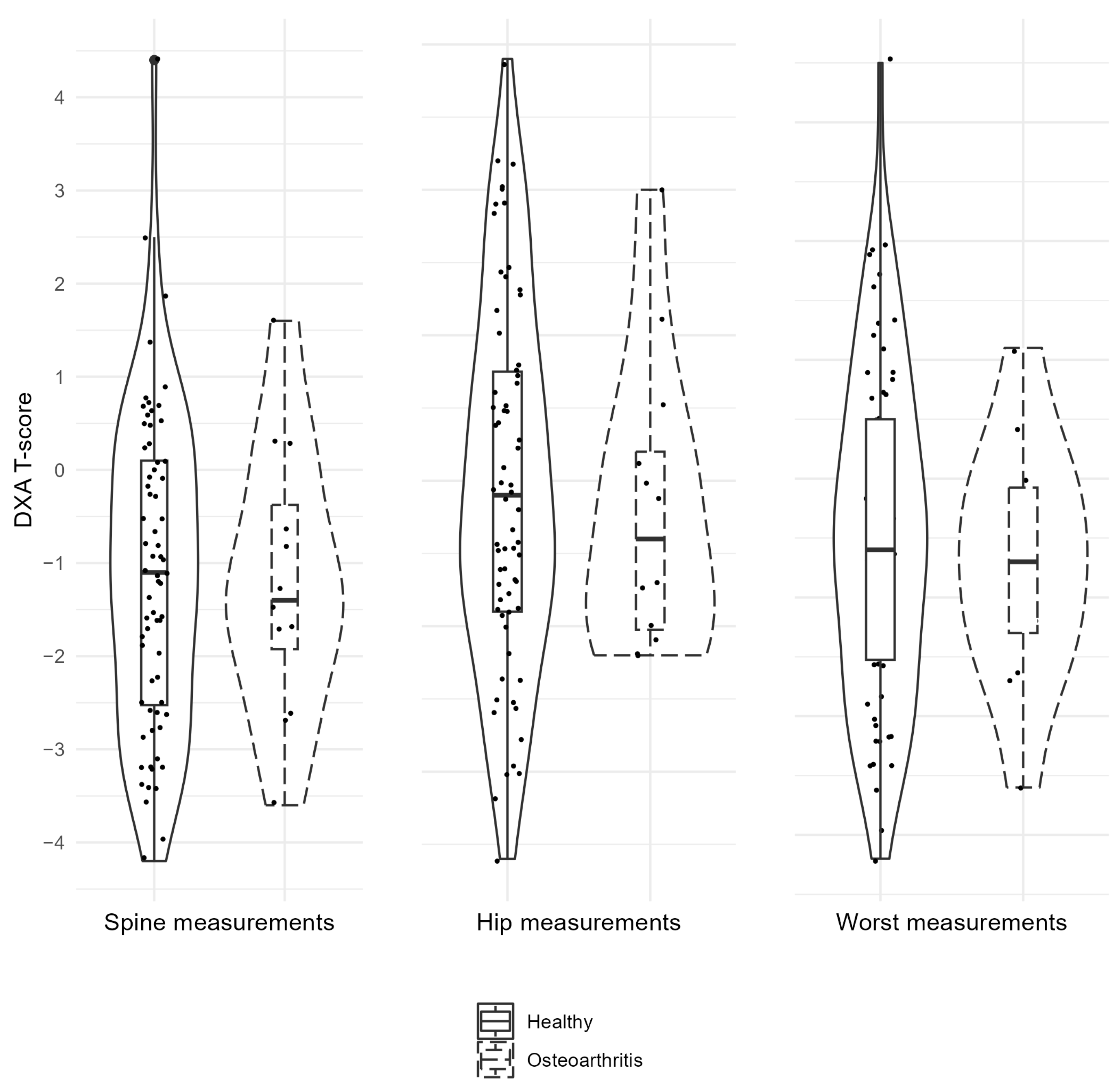
Figure 1. Violin plots for T-scores (DXA) of spine, hips and worst for healthy individuals and individuals with radiological signs of osteoarthritis. DXA—dual-energy X-ray absorptiometry.
Dentistry 12 00238 g001
Table 1. Number of radiographic bony alterations in TMJ.
Overall
N (%)
OA
N (%)
NonOA
N (%)
Indeterminant signs of OA
Flattening38 (47.5%)6 (7.5%)32 (40%)
Subcortical sclerosis33 (41.3%)6 (7.5%)17 (21.3%)
Signs of OA
Subcortical cyst8 (10%)8 (10%)0
Erosion4 (5%)4 (5%)0
Osteophytes000
N—number of findings, OA—radiological signs of osteoarthritis, NonOA—no radiological signs of osteoarthritis.
Table 2. Characteristics between groups observing and not observing signs of OA and age, DXA results.
Factor (N)DescriptivesDXA SpineDXA HipDXA WorstAge
OA (12)Mean (SD)−1.2 (1.5)−1.2 (1.0)−1.7 (1.0)72.3 (10.1)
NonOA (68)Mean (SD)−1.1 (1.7)−1.0 (1.2)−1.5 (1.4)72.0 (8.7)
CI, 95%−1.0; 1.1−0.6; 0.9−0.7; 1.0−5.9; 5.2
p-Value0.4580.2770.1700.856
N—number of findings, OA—radiological signs of osteoarthritis, NonOA—no radiological signs of osteoarthritis, DXA—dual-energy X-ray absorptiometry, CI—confidence interval.
Table 3. Characteristics between radiological findings of bony alterations and age, DXA scores.
FactorPositive/Negative Finding (N)DescriptivesDXA SpineDXA HipDXA WorstAge
FlatteningPositive (38)Mean (SD)−1.1 (1.5)−1.0 (1.3)−1.5 (1.2)73.1 (8.1)
Negative (42)Mean (SD)−1.1 (1.7)−1.1 (1.1)−1.6 (1.3)71.1 (9.5)
CI, 95%−0.8; 0.7−0.7; 0.4−0.7; 0.5−6.0; 1.9
p-Value0.7210.3020.8190.447
Subcortical sclerosisPositive (33)Mean (SD)−1.4 (1.5)−1.1 (1.1)−1.7 (1.4)70.8 (9.3)
Negative (47)Mean (SD)−0.9 (1.7)−1.1 (1.3)−1.5 (1.3)72.9 (8.5)
CI, 95%−0.2; 1.3−0.5; 0.5−0.4; 0.8−1.8; 6.2
p-Value0.6850.3470.9370.555
Subcortical cystPositive (8)Mean (SD)−1.4 (1.0)−0.8 (1.1)−1.5 (0.9)71.8 (11.2)
Negative (72) Mean (SD)−1.1 (1.7)−1.1 (1.2)−1.6 (1.4)72.1 (8.6)
CI, 95%−0.9; 1.5−1.1; 0.7−1.1; 0.9−6.3; 6.9
p-Value0.1040.4280.1990.582
ErosionPositive (4)Mean (SD)−0.8 (2.3)−1.8 (0.6)−2.2 (1.1)73.5 (8.9)
Negative (76)Mean (SD)−1.1 (1.6)−1.0 (1.2)−1.5 (1.3)72.0 (8.9)
CI, 95%−2.0; 1.3−0.4; 2.0−0.6; 2.1−10.6; 7.5
p-Value0.3640.0950.3420.681
N—number of findings, SD—standard deviation, DXA—dual-energy X-ray absorptiometry, CI—confidence interval.
Disclaimer/Publisher’s Note: The statements, opinions and data contained in all publications are solely those of the individual author(s) and contributor(s) and not of MDPI and/or the editor(s). MDPI and/or the editor(s) disclaim responsibility for any injury to people or property resulting from any ideas, methods, instructions or products referred to in the content.