
A Review of White Spot Lesions: Development and Treatment with Resin Infiltration


Abstract
1. Introduction
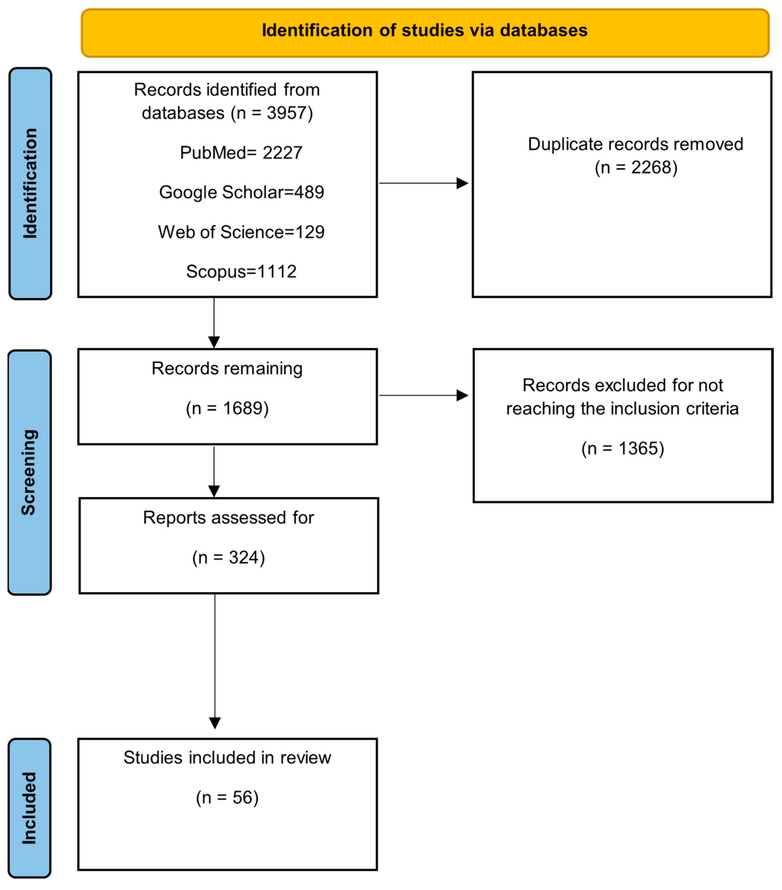
2. Materials and Methods
3. Results
3.1. Development of White Spot Lesions
3.2. Refractive Index
3.3. Treatment with Resin
3.4. Infiltration Technique
3.5. Clinical Studies Results

| Researcher | Study Design | Treatment | Results | References |
|---|---|---|---|---|
| Xi Gu et al. (2019) | Split-mouth, randomized clinical trial | RIT and micro-abrasion | RIT had better immediate and long-term results. It decreased the lesion size and maintained aesthetics for a 12-month period. | [30] |
| Giray et al. (2018) | Randomized Clinical Trial | RIT and FV | Using DIAGNOdent, it was demonstrated that RIT had a superior outcome than the FV. The carious lesion was better assessed by the RIT, and FV penetrated only the superficial layer of the WSLs. | [31] |
| Kannan et al. (2019) | Randomized controlled trial | RIT and light-cured FV | RIT had a better immediate outcome. After 6 months, the results were reversed. | [32] |
| Simon et al. (2022) | Randomized controlled trial | RIT and CPP-ACP | RIT had a better immediate outcome. CPP-ACP had significant results after one month and was stable through 12 months. | [33] |
| Gholami et al. (2023) | Clinical trial | RIT | Through spectrophotometry and digital photography, it was shown that RIT improved aesthetics and was durable for the duration of the study. | [28] |
| Knaup et al. (2023) | Clinical trial | RIT | RIT improved the appearance of WSLs and was stable for a 12-month period. | [29] |
| Knösel et al. (2019) | Second follow-up of split-mouth randomized controlled study | RIT | Some colour and lightness changes occurred between 24 and 45 months but are unlikely to be visible to the naked eye. | [36] |
4. Discussion
5. Conclusions
Author Contributions
Funding
Conflicts of Interest
References
- Perdigão, J. Resin infiltration of enamel white spot lesions: An ultramorphological analysis. J. Esthet. Restor. Dent. 2020, 32, 317–324. [Google Scholar] [CrossRef] [PubMed]
- Pitts, N.B.; Zero, D.T.; Marsh, P.D.; Ekstrand, K.; Weintraub, J.A.; Ramos-Gomez, F.; Tagami, J.; Twetman, S.; Tsakos, G.; Ismail, A. Dental caries. Nat. Rev. Dis. Primers 2017, 3, 17030. [Google Scholar] [CrossRef] [PubMed]
- Puleio, F.; Fiorillo, L.; Gorassini, F.; Iandolo, A.; Meto, A.; D’amico, C.; Cervino, G.; Pinizzotto, M.; Bruno, G.; Portelli, M.; et al. Systematic review on white spot lesions treatments. Eur. J. Dent. 2022, 16, 41–48. [Google Scholar] [CrossRef]
- Shimada, Y.; Sato, T.; Inoue, G.; Nakagawa, H.; Tabata, T.; Zhou, Y.; Hiraishi, N.; Gondo, T.; Takano, S.; Ushijima, K.; et al. Evaluation of incipient enamel caries at smooth tooth surfaces using SSOCT. Materials 2022, 15, 5947. [Google Scholar] [CrossRef]
- Solomon, R.V.; Priya, S.; Wahed, M.A.; Gopishetty, P.; Sathyanvesh, M. Minimalistic Intervention of White Spot Lesions and Dental Fluorosis with Resin Infiltration Technique—A Report of Two Cases. J. Clin. Diagn. Res. 2022, 16, ZD01–ZD04. [Google Scholar] [CrossRef]
- Borges, A.B.; Caneppele, T.M.F.; Masterson, D.; Maia, L. Is resin infiltration an effective esthetic treatment for enamel development defects and white spot lesions? A systematic review. J. Dent. 2017, 56, 11–18. [Google Scholar] [CrossRef]
- Al-Saeed, E.J.; AlMarhoon, Z.W.; Al-Eid, Z.A.A.; AlAhmari, T.A.; AlJamed, S.H.; AlSarhan, R.; AlShehri, A.; Al-Debasi, Y.T.; Badaoud, O.M.; AlHussain, B.S. Properties, Success, and Applications of Resin Infiltration for Minimal Invasive Restoration: A Scoping Review. Arch. Pharm. Pract. 2022, 13, 110–115. [Google Scholar] [CrossRef]
- Neuhaus, K.W.; Graf, M.; Lussi, A.; Katsaros, C. Late infiltration of post-orthodontic white spot lesions. J. Orofac. Orthop. 2010, 71, 442–447. [Google Scholar] [CrossRef] [PubMed]
- Struzycka, I. The oral microbiome in dental caries. Pol. J. Microbiol. 2014, 63, 127–135. [Google Scholar] [CrossRef]
- Jakubovics, N.S.; Goodman, S.D.; Mashburn-Warren, L.; Stafford, G.P.; Cieplik, F. The dental plaque biofilm matrix. Periodontol. 2000 2021, 86, 32–56. [Google Scholar] [CrossRef]
- Seneviratne, C.J.; Zhang, C.F.; Samaranayake, L.P. Dental plaque biofilm in oral health and disease. Chin. J. Dent. Res. 2011, 14, 87–94. [Google Scholar] [PubMed]
- Valm, A.M. The structure of dental plaque microbial communities in the transition from health to dentalncaries and periodontal disease. J. Mol. Biol. 2019, 431, 2957–2969. [Google Scholar] [CrossRef] [PubMed]
- Marsh, P.D.; Moter, A.; Devine, D.A. Dental plaque biofilms: Communities, conflict and control. Periodontol. 2000 2011, 55, 16–35. [Google Scholar] [CrossRef] [PubMed]
- Lamont, R.J.; Koo, H.; Hajishengallis, G. The oral microbiota: Dynamic communities and host interactions. Nat. Rev. Microbiol. 2018, 16, 745–759. [Google Scholar] [CrossRef]
- Khoroushi, M.; Kachuie, M. Prevention and treatment of white spot lesions in orthodontic patients. Contemp. Clin. Dent. 2017, 8, 11–19. [Google Scholar] [CrossRef]
- Sundararaj, D.; Venkatachalapathy, S.; Tandon, A.; Pereira, A. Critical evaluation of incidence and prevalence of white spot lesions during fixed orthodontic appliance treatment: A meta-analysis. J. Int. Soc. Prev. Community Dent. 2015, 5, 433–439. [Google Scholar] [CrossRef]
- Julien, K.C.; Buschang, P.H.; Campbell, P.M. Prevalence of white spot lesion formation during orthodontic treatment. Angle Orthod. 2013, 83, 641–647. [Google Scholar] [CrossRef]
- Lucchese, A.; Gherlone, E. Prevalence of white-spot lesions before and during orthodontic treatment with fixed appliances. Eur. J. Orthod. 2013, 35, 664–668. [Google Scholar] [CrossRef] [PubMed]
- Paris, S.; Schwendicke, F.; Keltsch, J.; Dörfer, C.; Meyer-Lueckel, H. Masking of white spot lesions by resin infiltration in vitro. J. Dent. 2013, 41, 28–34. [Google Scholar] [CrossRef]
- Chapman, J.A.; Roberts, W.E.; Eckert, G.J.; Kula, K.S.; González-Cabezas, C. Risk factors for incidence and severity of white spot lesions during treatment with fixed orthodontic appliances. Am. J. Orthod. Dentofac. Orthop. 2010, 138, 188–194. [Google Scholar] [CrossRef]
- Denis, M.; Atlan, A.; Vennat, E.; Tirlet, G.; Attal, J.-P. White defects on enamel: Diagnosis and anatomopathology: Two essential factors for proper treatment (part 1). Int. Orthod. 2013, 11, 139–165. [Google Scholar] [CrossRef] [PubMed]
- Meng, Z.; Yao, X.S.; Yao, H.; Liang, Y.; Liu, T.; Li, Y.; Wang, G.; Lan, S. Measurement of the refractive index of human teeth by optical coherence tomography. J. Biomed. Opt. 2009, 14, 034010. [Google Scholar] [CrossRef] [PubMed]
- Kim, S.; Kim, E.Y.; Jeong, T.S.; Kim, J.-W. The evaluation of resin infiltration for masking labial enamel white spot lesions. Int. J. Paediatr. Dent. 2011, 21, 241–248. [Google Scholar] [CrossRef] [PubMed]
- Oivanen, M.; Keulemans, F.; Garoushi, S.; Vallittu, P.K.; Lassila, L. The effect of refractive index of fillers and polymer matrix on translucency and color matching of dental resin composite. Biomater. Investig. Dent. 2021, 1, 48–53. [Google Scholar] [CrossRef] [PubMed]
- Neuhaus, K.W.; Schlafer, S.; Lussi, A.; Nyvad, B. Infiltration of natural caries lesions in relation to their activity status and acid pretreatment in vitro. Caries Res. 2013, 47, 203–210. [Google Scholar] [CrossRef]
- Manoharan, V.; Arun Kumar, S.; Arumugam, S.B.; Anand, V.; Krishnamoorthy, S.; Methippara, J.J. Is resin infiltration a microinvasive approach to white lesions of calcified tooth structures?: A systemic review. Int. J. Clin. Pediatr. Dent. 2019, 12, 53–58. [Google Scholar] [CrossRef]
- Lasfargues, J.J.; Bonte, E.; Guerrieri, A.; Fezzani, L. Minimal intervention dentistry: Part 6. Caries inhibition by resin infiltration. Br. Dent. J. 2013, 214, 53–59. [Google Scholar] [CrossRef]
- Gholami, S.; Boruziniat, A.; Talebi, A.; Yazdandoust, Y. Effect of extent of white spot lesions on the esthetic outcome after treatment by the resin infiltration technique: A clinical trial. Front. Dent. 2023, 22, 40. [Google Scholar] [CrossRef]
- Flynn, L.N.; Julien, K.; Noureldin, A.; Buschang, P.H. The efficacy of fluoride varnish vs a filled resin sealant for preventing white spot lesions during orthodontic treatment. Angle Orthod. 2022, 92, 204–212. [Google Scholar] [CrossRef]
- Gu, X.; Yang, L.; Yang, D.; Gao, Y.; Duan, X.; Zhu, X.; Yuan, H.; Li, J. Esthetic improvements of postorthodontic white-spot lesions treated with resin infiltration and microabrasion: A split-mouth, randomized clinical trial. Angle Orthod. 2019, 89, 372–377. [Google Scholar] [CrossRef]
- Giray, F.E.; Durhan, M.A.; Haznedaroglu, E.; Durmus, B.; Kalyoncu, O.I.; Tanboga, I. Resin infiltration technique and fluoride varnish on white spot lesions in children: Preliminary findings of a randomized clinical trial. Niger. J. Clin. Pract. 2018, 21, 1564–1569. [Google Scholar] [CrossRef] [PubMed]
- Kannan, A.; Padmanabhan, S. Comparative evaluation of Icon® resin infiltration and Clinpro™ XT varnish on colour and fluorescence changes of white spot lesions: A randomized controlled trial. Prog. Orthod. 2019, 20, 23. [Google Scholar] [CrossRef] [PubMed]
- Simon, L.S.; Dash, J.K.; U, D.; Philip, S.; Sarangi, S. Management of post orthodontic white spot lesions using resin infiltration and CPP-ACP materials—A clinical study. J. Clin. Pediatr. Dent. 2022, 46, 70–74. [Google Scholar] [CrossRef]
- Gugnani, N.; Pandit, I.K.; Gupta, M. Comparative evaluation of esthetic changes in nonpitted fluorosis stains when treated with resin infiltration, in-office bleaching, and combination therapies. J. Esthet. Restor. Dent. 2017, 29, 317–324. [Google Scholar] [CrossRef]
- Knaup, I.; Kobbe, C.; Ehrlich, E.E.; Esteves-Oliveira, M.; Abou-Ayash, B.; Meyer-Lueckel, H.; Wolf, M.; Wierichs, R.J. Correlation of quantitative light-induced fluorescence and qualitative visual rating in infiltrated post-orthodontic white spot lesions. Eur. J. Orthod. 2023, 45, 133–141. [Google Scholar] [CrossRef]
- Knösel, M.; Eckstein, A.; Helms, H.J. Long-term follow-up of camouflage effects following resin infiltration of post orthodontic white-spot lesions in vivo. Angle Orthod. 2019, 89, 33–39. [Google Scholar] [CrossRef]
- Karabekiroğlu, S.; Ünlü, N.; Küçükyilmaz, E.; Şener, S.; Botsali, M.S.; Malkoç, S. Treatment of post-orthodontic white spot lesions with CPPACP paste: A three year follow up study. Dent. Mater. J. 2017, 36, 791–797. [Google Scholar] [CrossRef] [PubMed]
- Singh, S.; Singh, S.P.; Goyal, A.; Utreja, A.K.; Jena, A.K. Effects of various remineralizing agents on the outcome of postorthodontic white spot lesions (WSLs): A clinical trial. Prog. Orthod. 2016, 17, 25. [Google Scholar] [CrossRef]
- Memarpour, M.; Fakhraei, E.; Dadaein, S.; Vossoughi, M. Efficacy of fluoride varnish and casein phosphopeptideamorphous calcium phosphate for remineralization of primary teeth: A randomized clinical trial. Med. Princ. Pract. 2015, 24, 231–237. [Google Scholar] [CrossRef]
- Hoffman, D.A.; Clark, A.E.; Rody, W.J., Jr.; McGorray, S.P.; Wheeler, T.T. A prospective randomized clinical trial into the capacity of a toothpaste containing NovaMin to prevent white spot lesions and gingivitis during orthodontic treatment. Prog. Orthod. 2015, 16, 25. [Google Scholar] [CrossRef]
- Atteya, S.M.; Amer, H.A.; Saleh, S.M.; Safwat, Y. Self-assembling peptide and nano-silver fluoride in remineralizing early enamel carious lesions: Randomized controlled clinical trial. BMC Oral Health. 2023, 23, 577. [Google Scholar] [CrossRef] [PubMed]
- Sedlakova Kondelova, P.; Mannaa, A.; Bommer, C.; Abdelaziz, M.; Daeniker, L.; di Bella, E.; Krejci, I. Efficacy of P11-4 for the treatment of initial buccal caries: A randomized clinical trial. Sci. Rep. 2020, 10, 20211. [Google Scholar] [CrossRef] [PubMed]
- Knösel, M.; Eckstein, A.; Helms, H.-J. Durability of esthetic improvement following Icon resin infiltration of multibracket-induced white spot lesions compared with no therapy over 6 months: A single-center, splitmouth, randomized clinical trial. Am. J. Orthod. Dentofac. Orthop. 2013, 144, 86–96. [Google Scholar] [CrossRef]
- Paris, S.; Hopfenmuller, W.; Meyer-Lueckel, H. Resin infiltration of caries lesions: An efficacy randomized trial. J. Dent. Res. 2010, 89, 823–826. [Google Scholar] [CrossRef]
- Yetkiner, E.; Wegehaupt, F.; Wiegand, A.; Attin, R.; Attin, T. Colour improvement and stability of white spot lesions following infiltration, micro-abrasion, or fluoride treatments in vitro. Eur. J. Orthod. 2014, 36, 595–602. [Google Scholar] [CrossRef]
- Paolone, G.; Pavan, F.; Mandurino, M.; Baldani, S.; Guglielmi, P.C.; Scotti, N.; Cantatore, G.; Vichi, A. Color stability of resin-based composites exposed to smoke. A systematic review. J. Esthet. Restor. Dent. 2023, 35, 309–321. [Google Scholar] [CrossRef]
- Leland, A.; Akyalcin, S.; English, J.D.; Tufekci, E.; Paravina, R. Evaluation of staining and color changes of a resin infiltration system. Angle Orthod. 2016, 86, 900–904. [Google Scholar] [CrossRef] [PubMed]
- Sabti, M.Y.; Alfarhan, I.Y.; Akbar, A.A.; Qudeimat, M.A. Evaluating color stability and enamel surface roughness following resin infiltration treatment. Clin. Exp. Dent. Res. 2024, 10, e2834. [Google Scholar] [CrossRef]
- Saccucci, M.; Corridore, D.; Di Carlo, G.; Bonucci, E.; Cicciù, M.; Vozza, I. Assessment of Enamel Color Stability of Resins Infiltration Treatment in Human Teeth: A Systematic Review. Int. J. Environ. Res. Public Health 2022, 19, 11269. [Google Scholar] [CrossRef]
- Skucha-Nowak, M. Attempt to assess the infiltration of enamel made with experimental preparation using a scanning electron microscope. Open Med. 2015, 10, 238–248. [Google Scholar] [CrossRef]
- Kantovitz, K.R.; Pascon, F.M.; Nobre-dos-Santos, M.; Puppin-Rontani, R.M. Review of the effects of infiltrants and sealers on non-cavitated enamel lesions. Oral Health Prev. Dent. 2010, 8, 295–305. [Google Scholar] [PubMed]
- Paris, S.; Schwendicke, F.; Seddig, S.; Müller, W.-D.; Dörfer, C.; Meyer-Lueckel, H. Micro-hardness and mineral loss of enamel lesions after infiltration with various resins: Influence of infiltrant composition and application frequency in vitro. J. Dent. 2013, 41, 543–548. [Google Scholar] [CrossRef] [PubMed]
- Tostes, M.A.; Santos, E., Jr.; Camargo, S.A., Jr. Effect of resin infiltration on the nanomechanical properties of demineralized bovine enamel. Indian J. Dent. 2014, 5, 116–122. [Google Scholar] [CrossRef] [PubMed] [PubMed Central]
- Chen, M.; Li, J.Z.; Zuo, Q.L.; Liu, C.; Jiang, H.; Du, M.-Q. Accelerated aging effects on color, microhardness and microstructure of ICON resin infiltration. Eur. Rev. Med. Pharmacol. Sci. 2019, 23, 7722–7731. [Google Scholar] [CrossRef] [PubMed]
- Paris, S.; Meyer-Lueckel, H.; Mueller, J.; Hummel, M.; Kielbassa, A.M. Progression of Sealed Initial Bovine Enamel Lesions under Demineralizing Conditions in vitro. Caries Res. 2006, 40, 124–129. [Google Scholar] [CrossRef]
- Stoica, A.M.; Stoica, O.E.; Dako, T.; Kovàcs-Ivàcson, A.-C.; Monea, M.; Bardocz-Veres, Z. Evaluation of the therapeutic performance of ICON infiltration resin in the treatment of white spot lesions in esthetic dental areas. Clinical Report. Acta Stomatol. Marisiensis J. 2023, 6, 25–32. [Google Scholar] [CrossRef]
| Level of Screening | Description of the Selection Process |
|---|---|
| First level of screening: database filters | Filters such as language, date of publication and article type were applied to the database search. |
| Second level of screening: title review | After the application of the filters above, the titles of the results were reviewed. Unclear sentencing or missing keywords in the title were reasons for exclusion. |
| Third level of screening: abstract review | Short, unspecific and vague abstracts were subject to dismissal. |
| Fourth level of screening: full article review | Only articles with a clear objective, valid methods and results were included in the research. |
| Researcher | Study Design | Treatment | Results | Reference |
|---|---|---|---|---|
| Karabekiroğlu et al. (2017) | Randomized controlled study | CPP-ACP | CPP-ACP was not more effective than 1450 ppm fluoridated toothpaste; the researcher suggested another way of treatment. | [37] |
| Singh et al. (2016) | Clinical trial | 1000 ppm fluoride toothpaste, FV, CPP-ACP | The fluoridated toothpaste had the better outcome. The use of varnish or CPP-ACP did not improve the outcome. | [38] |
| Hoffman et al. (2015) | Randomized controlled trial | Toothpaste containing NovaMin | No difference between NovaMin toothpaste and fluoride toothpaste. A trend of improvement was observed at the 3-month check-up, but no significant results. | [40] |
| Atteya et al. (2023) | Randomized controlled clinical trial | P11-4, NSF and NaF | P11-4 and NSF showed better management of the carious activity than the NaF. | [41] |
| Sedlakova Kondelova et al. (2020) | Randomized controlled, split-mouth study | P11-4, FV | P11-4 reduced the size of the WSLs; P11-4 and delayed FV showed improved aestethics. | [42] |
| Memarpour et al. (2015) | Randomized clinical trial | FV, CPP-ACP | FV and CPP-ACP showed improvement of WSLs in primary teeth. | [39] |
Disclaimer/Publisher’s Note: The statements, opinions and data contained in all publications are solely those of the individual author(s) and contributor(s) and not of MDPI and/or the editor(s). MDPI and/or the editor(s) disclaim responsibility for any injury to people or property resulting from any ideas, methods, instructions or products referred to in the content. |
© 2024 by the authors. Licensee MDPI, Basel, Switzerland. This article is an open access article distributed under the terms and conditions of the Creative Commons Attribution (CC BY) license (https://creativecommons.org/licenses/by/4.0/).
Share and Cite
Prada, A.M.; Potra Cicalău, G.I.; Ciavoi, G. A Review of White Spot Lesions: Development and Treatment with Resin Infiltration. Dent. J. 2024, 12, 375. https://doi.org/10.3390/dj12120375
Prada AM, Potra Cicalău GI, Ciavoi G. A Review of White Spot Lesions: Development and Treatment with Resin Infiltration. Dentistry Journal. 2024; 12(12):375. https://doi.org/10.3390/dj12120375
Chicago/Turabian StylePrada, Alexandra Maria, Georgiana Ioana Potra Cicalău, and Gabriela Ciavoi. 2024. "A Review of White Spot Lesions: Development and Treatment with Resin Infiltration" Dentistry Journal 12, no. 12: 375. https://doi.org/10.3390/dj12120375
APA StylePrada, A. M., Potra Cicalău, G. I., & Ciavoi, G. (2024). A Review of White Spot Lesions: Development and Treatment with Resin Infiltration. Dentistry Journal, 12(12), 375. https://doi.org/10.3390/dj12120375