
Maxillary Anterior Teeth Dimensions and Relative Width Proportions: A Narrative Literature Review


Abstract
:1. Introduction
2. Materials and Methods
2.1. Search Strategy
2.2. Study Selection, Inclusion and Exclusion Criteria
3. Results
4. Discussion
4.1. Anterior Teeth Dimensions
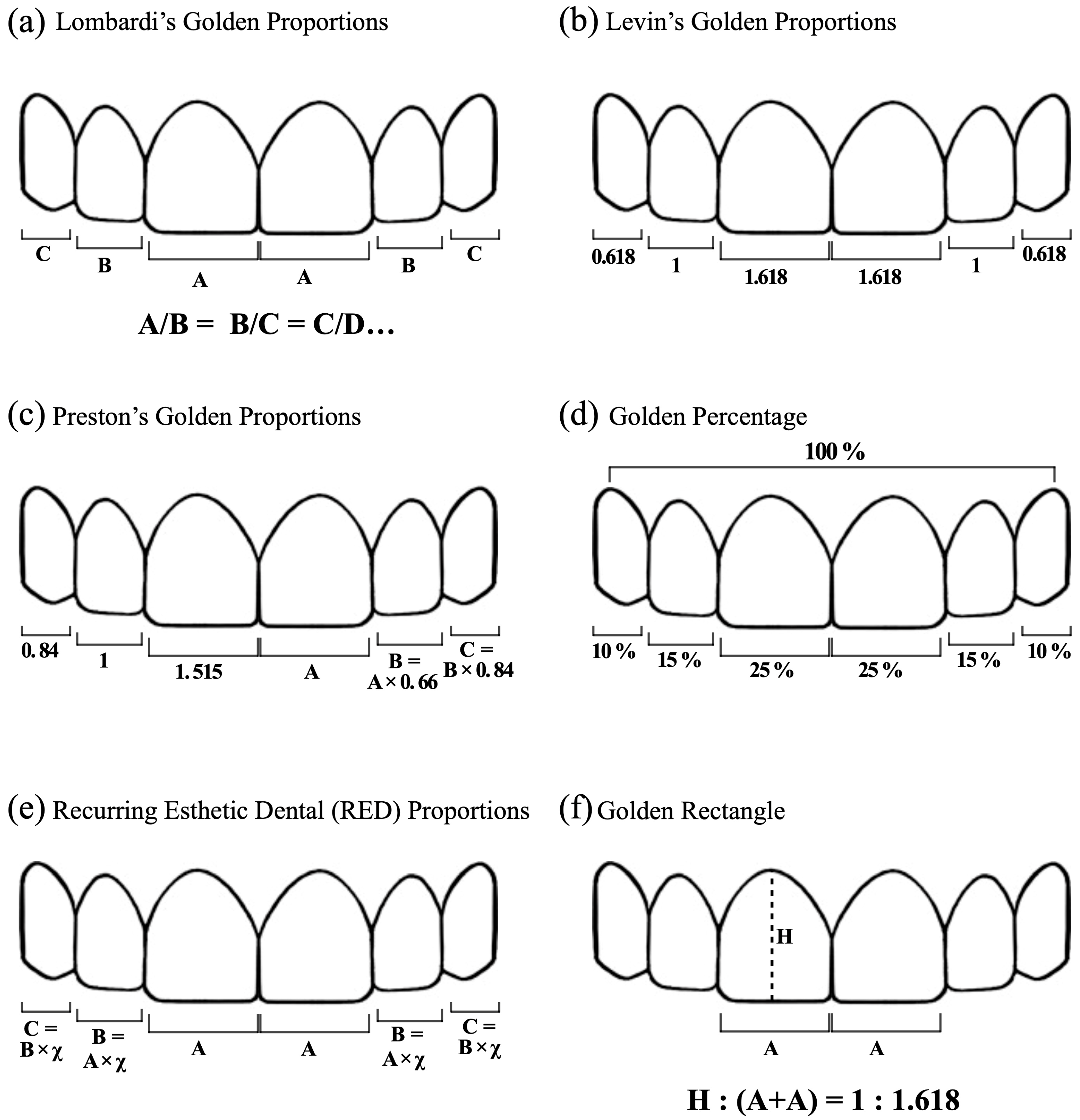
4.2. Golden Proportions, Golden Percentage, RED Proportion, and Golden Rectangle
5. Conclusions
- There are no standard tooth sizes. Size ranges can be taken as references, and they differ in the two sexes. Men show greater width and length; however, the width/length ratio is usually greater in women.
- Perfectly symmetrical contralateral elements are found in low percentages of subjects. In about 60% of cases, there are differences greater than 0.2 mm both in terms of length and width. In a more recent study, however, no asymmetries were found.
- The correlation between dental dimensions and facial parameters is not always present and is strongly influenced by ethnicity. Some studies propose formulas to determine dental dimensions starting from facial parameters. However, the authors underline how they are closely related to the ethnicity of the studied population.
- Regarding ethnic differences, Caucasians have greater width and W/L ratio in the central incisors than Asians, while the length is superimposable. The laterals and canines are longer in Asians, while their width is similar.
- Golden Proportions, Preston’s Golden Proportions, Golden Percentage, and RED Proportions are never fully matched. In a few cases, there are partial central–lateral and canine–lateral correlations. More recent studies propose a modified Golden Percentage with central–lateral–canine percentages of 22.5–15–12.5%. These values are much more representative of the Golden Percentage proposed by Snow and are more recommended as a principle of smile design. Golden Rectangle seems to be a suitable method to obtain central incisor dimensions.
Author Contributions
Funding
Conflicts of Interest
References
- Lombardi, R.E. The principles of visual perception and their clinical application to denture esthetics. J. Prosthet. Dent. 1973, 29, 358–382. [Google Scholar] [CrossRef] [PubMed]
- Levin, E.I. Dental esthetics and the golden proportion. J. Prosthet. Dent. 1978, 40, 244–252. [Google Scholar] [CrossRef] [PubMed]
- Preston, J.D. The Golden Proportion Revisited. J. Esthet. Restor. Dent. 1993, 5, 247–251. [Google Scholar] [CrossRef] [PubMed]
- Snow, S.R. Esthetic smile analysis of maxillary anterior tooth width: The golden percentage. J. Esthet. Dent. 1999, 11, 177–184. [Google Scholar] [CrossRef] [PubMed]
- Ward, D.H. Proportional smile design using the recurring esthetic dental (red) proportion. Dent. Clin. N. Am. 2001, 45, 143–154. [Google Scholar] [CrossRef]
- Marquardt, S.R. Marquardt on the Golden Decagon and human facial beauty. Interview by Dr. Gottlieb. J. Clin. Orthod. 2002, 36, 339–347. [Google Scholar]
- Magne, P.; Gallucci, G.O.; Belser, U.C. Anatomic crown width/length ratios of unworn and worn maxillary teeth in white subjects. J. Prosthet. Dent. 2003, 89, 453–461. [Google Scholar] [CrossRef]
- Orozco-Varo, A.; Arroyo-Cruz, G.; Martínez-de-Fuentes, R.; Jiménez-Castellanos, E. Biometric analysis of the clinical crown and the width/length ratio in the maxillary anterior region. J. Prosthet. Dent. 2015, 113, 565–570. [Google Scholar] [CrossRef]
- Saleem, B.; Mahmood, A.; Butt, A.M.; Najmi, N.; Aslam, S.; Shah, S.M.; Shams, S. Analysis of width, height and width/height ratio of crowns of maxillary anterior teeth. Pak. J. Med. Health Sci. 2022, 32, 46–49. [Google Scholar] [CrossRef]
- Zhao, Q.; Li, N.; Cao, J. Morphological features of maxillary anterior teeth in a sample of Chinese population. Homo 2015, 66, 448–454. [Google Scholar] [CrossRef]
- Mavroskoufis, F.; Ritchie, G.M. Variation in size and form between left and right maxillary central incisor teeth. J. Prosthet. Dent. 1980, 43, 254–257. [Google Scholar] [CrossRef] [PubMed]
- Vadavadagi, S.V.; Hombesh, M.N.; Choudhury, G.K.; Deshpande, S.; Anusha, C.V.; Murthy, D.K. Variation in Size and Form between Left and Right Maxillary Central Incisor Teeth. J. Int. Oral. Health 2015, 7, 33–36. [Google Scholar] [PubMed]
- Wang, Y.; Song, Y.; Zhong, Q.; Xu, C. Evaluation of influence factors on the width, length, and width to length ratio of the maxillary central incisor: A systematic review and meta-analysis. J. Esthet. Restor. Dent. 2021, 33, 351–363. [Google Scholar] [CrossRef] [PubMed]
- Sterrett, J.D.; Oliver, T.; Robinson, F.; Fortson, W.; Knaak, B.; Russell, C.M. Width/length ratios of normal clinical crowns of the maxillary anterior dentition in man. J. Clin. Periodontol. 1999, 26, 153–157. [Google Scholar] [CrossRef] [PubMed]
- Al Wazzan, K.A. The relationship between intercanthal dimension and the widths of maxillary anterior teeth. J. Prosthet. Dent. 2001, 86, 608–612. [Google Scholar] [CrossRef] [PubMed]
- Attokaran, G.; Shenoy, K. Correlation between Innercanthal Distance and Mesiodistal Width of Maxillary Anterior Teeth in a Thrissur, Kerala, India, Population. J. Contemp. Dent. Pract. 2016, 17, 382–387. [Google Scholar] [PubMed]
- Arun Kumar, K.V.; Gupta, S.H.; Sandhu, H.S. Determination of mesiodistal width of maxillary anterior teeth using inner canthal distance. Med. J. Armed. Forces India 2015, 71, S376–S381. [Google Scholar] [CrossRef] [PubMed]
- Ahmed, N.; Abbasi, M.; Khan, D.; Khalid, S.; Jawed, W.; Mahmood, M. Determination of the combined width of maxillary anterior teeth using innercanthal distance with respect to age gender and ethinicity. PAFMJ 2021, 71, S164–S169. [Google Scholar] [CrossRef]
- Hasanreisoglu, U.; Berksun, S.; Aras, K.; Arslan, I. An analysis of maxillary anterior teeth: Facial and dental proportions. J. Prosthet. Dent. 2005, 94, 530–538. [Google Scholar] [CrossRef]
- Barman, J.; Serin, S. Comparison of interpupillary distance and combined mesiodistal width of maxillary central incisor teeth in two ethnic groups of Northeast India: An in vivo study. Indian J. Dent. Res. 2018, 29, 155–160. [Google Scholar] [CrossRef]
- Kini, A.Y.; Angadi, G.S. Biometric ratio in estimating widths of maxillary anterior teeth derived after correlating anthropometric measurements with dental measurements. Gerodontology 2013, 30, 105–111. [Google Scholar] [CrossRef] [PubMed]
- Zlatarić, D.K.; Kristek, E.; Celebić, A. Analysis of width/length ratios of normal clinical crowns of the maxillary anterior dentition: Correlation between dental proportions and facial measurements. Int. J. Prosthodont. 2007, 20, 313–315. [Google Scholar] [PubMed]
- Radia, S.; Sherriff, M.; McDonald, F.; Naini, F.B. Relationship between maxillary central incisor proportions and facial proportions. J. Prosthet. Dent. 2016, 115, 741–748. [Google Scholar] [CrossRef] [PubMed]
- LaVere, A.M.; Marcroft, K.R.; Smith, R.C.; Sarka, R.J. Denture tooth selection: An analysis of the natural maxillary central incisor compared to the length and width of the face. Part I. J. Prosthet. Dent. 1992, 67, 661–663. [Google Scholar] [CrossRef] [PubMed]
- LaVere, A.M.; Marcroft, K.R.; Smith, R.C.; Sarka, R.J. Denture tooth selection: An analysis of the natural maxillary central incisor compared to the length and width of the face: Part II. J. Prosthet. Dent. 1992, 67, 810–812. [Google Scholar] [CrossRef] [PubMed]
- Isa, Z.M.; Tawfiq, O.F.; Noor, N.M.; Shamsudheen, M.I.; Rijal, O.M. Regression methods to investigate the relationship between facial measurements and widths of the maxillary anterior teeth. J. Prosthet. Dent. 2010, 103, 182–188. [Google Scholar] [CrossRef] [PubMed]
- Nalawade, S.S.; Shinde, S.K.; Pawar, R.L.; Gupta, A.; Kale, V.T.; Janrao, K.A. Estimation of dental and facial proportions using height as criteria. J. Int. Oral. Health 2014, 6, 25–28. [Google Scholar]
- Tsukiyama, T.; Marcushamer, E.; Griffin, T.J.; Arguello, E.; Magne, P.; Gallucci, G.O. Comparison of the anatomic crown width/length ratios of unworn and worn maxillary teeth in Asian and white subjects. J. Prosthet. Dent. 2012, 107, 11–16. [Google Scholar] [CrossRef]
- Parciak, E.C.; Dahiya, A.T.; AlRumaih, H.S.; Kattadiyil, M.T.; Baba, N.Z.; Goodacre, C.J. Comparison of maxillary anterior tooth width and facial dimensions of 3 ethnicities. J. Prosthet. Dent. 2017, 118, 504–510. [Google Scholar] [CrossRef]
- Mohammed, S.; Bakhsh, L. Evaluation of Relation between Bizygomatic Width and Mesiodistal Dimension of Maxillary Central Incisor in Saudi Population: An In-vivo Study. Study J. Clin. Diagn. Res. 2020, 14, ZC32–ZC36. [Google Scholar]
- Alqahtani, A.S.; Habib, S.R.; Ali, M.; Alshahrani, A.S.; Alotaibi, N.M.; Alahaidib, F.A. Maxillary anterior teeth dimension and relative width proportion in a Saudi subpopulation. J. Taibah. Univ. Med. Sci. 2021, 16, 209–216. [Google Scholar] [CrossRef]
- Marcuschamer, E.; Tsukiyama, T.; Griffin, T.J.; Arguello, E.; Magne, P.; Gallucci, G.O. Anatomical crown width/length ratios of worn and unworn maxillary teeth in Asian subjects. Int. J Periodontics Restor. Dent. 2011, 31, 495–503. [Google Scholar]
- Sah, S.K.; Zhang, H.D.; Chang, T.; Dhungana, M.; Acharya, L.; Chen, L.L.; Ding, Y.M. Maxillary Anterior Teeth Dimensions and Proportions in a Central Mainland Chinese Population. Chin. J. Dent. Res. 2014, 17, 117–124. [Google Scholar] [PubMed]
- Akl, M.A.; Mansour, D.E.; Mays, K.; Wee, A.G. Mathematical Tooth Proportions: A Systematic Review. J. Prosthodont. 2022, 31, 289–298. [Google Scholar] [CrossRef] [PubMed]
- Calçada, D.; Correia, A.; Araújo, F. Anthropometric analysis of anterior maxillary teeth with digital photography—A study in a Portuguese sample. Int. J. Esthet. Dent. 2014, 9, 370–380. [Google Scholar] [PubMed]
- Londono, J.; Ghasemi, S.; Lawand, G.; Dashti, M. Evaluation of the golden proportion in the natural dentition: A systematic review and meta-analysis. J. Prosthet. Dent. 2021, 129, 696–702. [Google Scholar] [CrossRef]
- Mahshid, M.; Khoshvaghti, A.; Varshosaz, M.; Vallaei, N. Evaluation of “Golden Proportion” in Individuals with an Esthetic Smile. J. Esthet. Restor. Dent. 2004, 16, 185–192. [Google Scholar] [CrossRef]
- Swelem, A.A.; Al-Rafah, E.M. Evaluation of "Golden Proportion" in Saudi individuals with natural smiles. Saudi. Dent. J. 2019, 31, 277–283. [Google Scholar] [CrossRef]
- Kalia, R. An analysis of the aesthetic proportions of anterior maxillary teeth in a UK population. Br. Dent. J. 2020, 228, 449–455. [Google Scholar] [CrossRef]
- Rodríguez-López, S.; Martínez, M.F.E.; Velasco, J.P.; Junquera, L.; García-Pola, M. Analysis of dental esthetic proportions in a Spanish population sample. J. Oral. Sci. 2021, 63, 257–262. [Google Scholar] [CrossRef]
- Melo, M.; Ata-Ali, F.; Huertas, J.; Cobo, T.; Shibli, J.A.; Galindo-Moreno, P.; Ata-Ali, J. Revisiting the Maxillary Teeth in 384 Subjects Reveals A Deviation from the Classical Aesthetic Dimensions. Sci. Rep. 2019, 9, 730. [Google Scholar] [CrossRef] [PubMed]
- Maharjan, A.; Joshi, S. Clinical Evaluation of Maxillary Anterior Teeth in Relation to Golden Proportion, Red Proportion and Golden Percentage. J. Nepal. Health Res. Counc. 2018, 16, 11–15. [Google Scholar] [CrossRef] [PubMed]
- Aldegheishem, A.; Azam, A.; Al-Madi, E.; Abu-Khalaf, L.; Bani Ali, B.; Anweigi, L. Golden proportion evaluation in maxillary anterior teeth amongst Saudi population in Riyadh. Saudi. Dent. J. 2019, 31, 322–329. [Google Scholar] [CrossRef] [PubMed]
- Kantrong, N.; Traiveat, K.; Wongkhantee, S. Natural upper anterior teeth display an increasing proportion in mesio-distal direction. J. Clin. Exp. Dent. 2019, 11, e890–e897. [Google Scholar] [CrossRef] [PubMed]
- Mahajan, V.; Nagpal, A.; Gupta, R.; Vaidya, S.; Jabeen, F.; Thakur, K. Comparative Evaluation of Golden Proportion, Recurring Esthetic Dental Proportion and Golden Percentage in Himachal Demographic. JAMMR 2019, 29, 1–7. [Google Scholar] [CrossRef]
- Özdemir, H.; Köseoğlu, M.; Bayindir, F. An investigation of the esthetic indicators of maxillary anterior teeth in young Turkish people. J. Prosthet. Dent. 2018, 120, 583–588. [Google Scholar] [CrossRef] [PubMed]
- Al-Kaisy, N.; Garib, B.T. Analysis of the golden proportion and width/height ratios of maxillary anterior teeth in Arab and Kurdish populations. J. Prosthet. Dent. 2018, 119, 981–986. [Google Scholar] [CrossRef]
- Sandeep, N.; Satwalekar, P.; Srinivas, S.; Reddy, C.S.; Reddy, G.R.; Reddy, B.A. An Analysis of Maxillary Anterior Teeth Dimensions for the Existence of Golden Proportion: Clinical Study. J. Int. Oral. Health 2015, 7, 18–21. [Google Scholar]
- Petričević, N.; Asja, C.; Ibrahimagić-Šeper, L. Appropriate proportions as guidelines in selection of anterior denture teeth. Med. Glas 2008, 5, 103–108. [Google Scholar]
- Agrawal, V.S.; Kapoor, S.; Bhesania, D.; Shah, C. Comparative photographic evaluation of various geometric and mathematical proportions of maxillary anterior teeth: A clinical study. Indian J. Dent. Res. 2016, 27, 32–36. [Google Scholar] [CrossRef]
- Ansari, A.; Jameel, A.; Charania, A. Evaluation of Golden Proportion Between Maxillary Anterior Teeth. Med. Forum 2015, 26, 45–48. [Google Scholar]
- Al-Marzok, M.I.; Majeed, K.R.; Ibrahim, I.K. Evaluation of maxillary anterior teeth and their relation to the golden proportion in Malaysian population. BMC Oral. Health 2013, 13, 9. [Google Scholar] [CrossRef] [PubMed]
- Wadud, M.A.; Kitisubkanchana, J.; Santiwong, P.; Srithavaj, T. Face Proportions, and Analysis of Maxillary Anterior Teeth and Facial Proportions in a Thai Population. Open Dent. J. 2021, 15, 398. [Google Scholar] [CrossRef]
- Fayyad, M.; Jamani, K.; Agrabawi, J. Geometric and Mathematical Proportions and their Relations to Maxillary Anterior Teeth. Contemp. Dent. Pract. 2006, 7, 62–70. [Google Scholar] [CrossRef]
- Condon, M.; Bready, M.; Quinn, F.; O’Connell, B.C.; Houston, F.J.; O’Sullivan, M. Maxillary anterior tooth dimensions and proportions in an Irish young adult population. J. Oral. Rehabil. 2011, 38, 501–508. [Google Scholar] [CrossRef]
- Rokaya, D.; Kitisubkanchana, J.; Wonglamsam, A.; Santiwong, P.; Srithavaj, T.; Humagain, M. Nepalese Esthetic Dental (NED) Proportion in Nepalese Population. Kathmandu Univ. Med. J. 2015, 13, 244–249. [Google Scholar] [CrossRef]
- Muhammad, S.; Shahid, R.; Siddiqui, M.I. Tooth morphology and aesthetics while smiling in accordance to golden proportion. Pak. J. Med. Health Sci. 2016, 10, 281–284. [Google Scholar]
- Forster, A.; Velez, R.; Antal, M.; Nagy, K. Width ratios in the anterior maxillary region in a Hungarian population: Addition to the golden proportion debate. J. Prosthet. Dent. 2013, 110, 211–215. [Google Scholar] [CrossRef]
- Ahmed, N.; Halim, M.S.; Khalid, S.; Ghani, Z.A.; Jamayet, N.B. Evaluation of golden percentage in natural maxillary anterior teeth width: A systematic review. J. Prosthet. Dent. 2021, 127, e1–e845. [Google Scholar] [CrossRef]
- Ahmed, N.; Halim, M.S.B.; Ghani, Z.A.; Khan, Z.A.; Abbasi, M.S.; Jamayet, N.B.; Alam, M.K. A 2D Photographic and 3D Digital Dental Model Analysis of Golden Percentage in Maxillary Anterior Teeth. Biomed. Res. Int. 2021, 2021, 6674400. [Google Scholar] [CrossRef]
- Shetty, S.; Pitti, V.; Satish Babu, C.; Surendra Kumar, G.; Jnanadev, K. To evaluate the validity of Recurring Esthetic Dental proportion in natural dentition. J. Conserv. Dent. 2011, 14, 314–317. [Google Scholar] [PubMed]
- Liao, P.; Fan, Y.; Nathanson, D. Evaluation of maxillary anterior teeth width: A systematic review. J. Prosthet. Dent. 2019, 122, 275–281.e7. [Google Scholar] [CrossRef] [PubMed]
- Ahmed, N.; Halim, M.S.; Ab-Ghani, Z.; Abbasi, M.S.; Aslam, A.; Safdar, J.; Das, G.; Ahmed, A.R.; Jamayet, N.B. The Analysis of Facio-Dental Proportions to Determine the Width of Maxillary Anterior Teeth: A Clinical Study. J. Clin. Med. 2022, 11, 7340. [Google Scholar] [CrossRef] [PubMed]
- Chaudhari, D.; Dange, D.; Khalikar, S. Golden Rectangle Ratio—How Precious Is It?: A Clinical Study. IOSR-JDMS 2014, 13, 1–6. [Google Scholar] [CrossRef]
- Singh, R.; Tripathi, A.; Singh, S.; Bhatnagar, A. A study on the practical applicability of the rule of golden rectangle in dental aesthetics. Eur. J. Prosthodont. Restor. Dent. 2011, 19, 85–89. [Google Scholar]
- Varghese, P.; Cherian, B.; Sukumaran, B.; Anu, S.; Jacob, B.M.; Raja, V.V. Analysis of Geometric Proportions on Maxillary Anterior Teeth for Esthetic Smile Design: An In vivo Study. J. Pharm. Bioallied. Sci. 2021, 13, s778–s782. [Google Scholar] [CrossRef]


| Database | PubMed, Google Scholar, Embase, Cochrane Library | |
|---|---|---|
| Publication date | Until June 2023 | |
| Keywords | Anterior teeth dimensions | “Anterior maxillary teeth”, “maxillary central incisor (MCI)”, “anatomic crown”, “width”, “length”, “width/length ratio”, “symmetry”, “ethnicity”, “gender”, “face”, “facial parameters”, “lips”, “inter-pupillary distance”, “alar distance”, “inter-canthal distance” |
| Golden Proportions, Golden Percentage, RED Proportions, and Golden Rectangle | “Golden Proportions”, “Golden Percentage”, “RED Proportions”, “Golden proportion revisited”, “Golden Rectangle”, “dental esthetic”, “esthetic dentistry”, “tooth proportions”, “facial esthetic”, “anterior maxillary teeth” | |
| Language | English | |
| Type of paper | In vitro studies, clinical studies, clinical technique articles, reviews, systematic reviews | |
| Inclusion criteria | Articles relating to main topics with similar materials and methods, with digital or analogic measurements on casts or photographs | |
| Exclusion criteria | Non-English language articles, books, other types of articles | |
| Journal category | All |
| Author | Year | Description | Study Findings |
|---|---|---|---|
| Magne et al. [7] | 2003 | In vitro study | Width is not influenced by incisal edge wear, so worn teeth show higher W/L ratio. Worn central incisors have the highest value, and unworn canines and lateral incisors the lowest. Sample (n): 146 extracted human teeth from Caucasian subjects |
| Orozco-Varo et al. [8] | 2015 | In vitro study | Different values for width, length, and W/L ratio are found for men and women. The results differ from other studies measurement. Sample (n): 412 subjects from an European origin population |
| Saleem et al. [9] | 2022 | In vitro study | Width, length, and W/L ratio of anterior maxillary teeth are different in men and women. Sample (n): 101 subjects from a Pakistani population |
| Zhao et al. [10] | 2015 | In vitro study | Chinese populations show square-like shape teeth because of high W/L ratio. Sample (n): 101 subjects from a Chinese population |
| Mavroskoufis and Ritchie [11] | 1980 | In vitro study | The percentage of identical central incisors is low. Women have more similar teeth than men. Sample (n): 70 subjects from an English population |
| Vadavadagi et al. [12] | 2015 | In vitro study | Men show larger values than women. Left central incisor in men is longer than right incisor. Sample (n): 70 subjects from an Indian population |
| Alqahtani et al. [13] | 2021 | In vitro study | Little asymmetry between left and right side, and W/L ratio is similar for men and women with a minor difference for the canines. The squarish form is like that of the Turkish population. Sample (n): 180 subjects from a Saudi population |
| Radia et al. [14] | 2016 | In vitro study | Little relationship between maxillary central incisor and face proportions. Proposal of ratios between maxillary central incisor height and face height. No asymmetry between left and right central incisors, and no sex influence. Men have larger teeth and face measurement, but W/L ratios are similar to women. Sample (n): 149 subjects from an English population |
| Wang et al. [15] | 2021 | Systematic review and meta-analysis | There are conflicting results in the literature. Clinically, contralateral central incisor can be used to create an aesthetic and symmetric restoration; face parameters, sex, and patient features have to be considered in an esthetic treatment. |
| Sterrett et al. [16] | 1999 | In vitro study | W/L ratios are influenced by genders. Study shows similar values for central incisor and lateral incisor W/L ratios between men and women. Canine ratio is significantly greater in women. Sample (n): 71 subjects from a Caucasian population |
| Kini and Angadi [17] | 2013 | In vitro study | Existence of a correlation between inter-commissural distance, inter-pupillary distance, and inter-canine distance in photographs and casts. However, there is an important ethnic influence. Sample (n): 70 subjects from an Indian population |
| Al Wazzan [18] | 2001 | In vitro study | Inter-canthal distance can be used as a preliminary method to find anterior maxillary teeth width in edentulous patients. Sample (n): 443 subjects from a Saudi population |
| Attokaran and Shenoy [19] | 2016 | In vitro study | Inter-canthal distance is correlated with mesiodistal width of six anterior teeth in women. Sample (n): 1200 subjects from an Indian population |
| Arun Kumar et al. [20] | 2014 | In vitro study | Inter-canthal distance can be used to find the combined mesiodistal width of maxillary anterior teeth if multiplied by 1.61. Sample (n): 800 subjects from four Indian ethnic groups |
| Ahmed et al. [21] | 2021 | In vitro study | A weak relationship exists between inner canthal distance and maxillary anterior teeth width. A multiplication ratio of 1.27 can be helpful to find combined mesiodistal width of maxillary anterior teeth. Sample (n): 100 subjects from a Pakistani population |
| Hasanreisoglu et al. [22] | 2005 | In vitro study | Maxillary central incisor and canine dimensions of men are greater than those of women in the Turkish population. Canines have the greatest gender variation. Absence of recurrent proportion for all anterior teeth. Correlation is found between bizygomatic width and central incisor width and alar width and inter-canine distance. Sample (n): 100 subjects from a Turkish population |
| Barman and Serin [23] | 2018 | In vitro study | Existence of a correlation between IPD and combined mesiodistal width of maxillary central incisors. Sample (n): 120 subjects from two ethnic Indian groups |
| Zlatarić et al. [24] | 2007 | In vitro study | Face parameters are influenced by gender, and dental W/L ratios are similar in men and women. The correlation is low: The selection of artificial teeth using face parameters is not accurate. Sample (n): 90 subjects from a Caucasian population |
| LaVere et al. [25] | 1992 | In vitro study | The selection of size of the anterior teeth depends on anatomic factors, sex, age, and patient’s desires. Facial length and facial width for anterior tooth selection may result in selection of teeth that are too large for the sample age group. Sample (n): 448 subjects from an American population |
| LaVere et al. (part II) [26] | 1992 | In vitro study | Trubyte Tooth Indicator leads to narrower and longer artificial teeth (in both sexes). The selected teeth remain within 1 mm of their natural size. Sample (n): 448 subjects from an American population |
| Isa et al. [27] | 2010 | In vitro study | Regression methods can be used to predict the widths of the anterior teeth within the population tested by a combination of inter-pupillary and alar width. Sample (n): 60 subjects from a Malaysian population |
| Nalawade et al. [28] | 2014 | In vitro study | Correlation is found between height, inter-canine distance, inter-commissural distance, inter-incisal distance, and lower facial height. Formulas are proposed for the backward calculation of the parameters. Sample (n): 144 subjects from an Indian population |
| Tsukiyama et al. [29] | 2012 | In vitro study | Anterior teeth appear to have a slenderer shape in the Asian population. Only central incisors are statistically wider in the White subjects. Sample (n): 157 extracted human teeth from Asian subjects and 142 from Caucasian subjects |
| Parciak et al. [30] | 2017 | In vitro study | Correlation is found between central incisor and bizygomatic width in the ethnicities. In the Asian women, inter-commissural width is correlated with central incisor width, the 2 central incisors width, the 4 incisors width, and the 6 anterior teeth width. Sample (n): 360 subjects (120 from an Asian population, 120 African American population, 120 from a Caucasian population) |
| Mohammed et al. [31] | 2020 | In vitro study | A weak correlation exists between bizygomatic distance and central incisor width. Sample (n): 200 subjects from a Saudi population |
| Marcuschamer et al. [32] | 2011 | In vitro study | Width is not influenced by incisal edge wear, so worn teeth show higher W/L ratio. Worn central incisors have the highest value, unworn canines and lateral incisors the lowest. Sample (n): 264 extracted human teeth from Asian subjects |
| Author | Year | Description | Study Findings |
|---|---|---|---|
| Lombardi [1] | 1973 | Review | Principles in esthetic dentistry can be free from subjectivity and make it possible to reach the perfect result. |
| Levin [2] | 1978 | Review | Golden Proportions are described as a method to predict dental esthetic. |
| Preston [3] | 1993 | In vitro study | Levin Golden Proportion is not found in the sample. Golden Proportion has to be revisited: Maxillary lateral incisor width is 66% of the central incisor, and canine is 84% of the lateral incisor (or 55% of the central incisor). Sample (n): 58 subjects from an American population |
| Snow [4] | 1999 | In vitro study | In the inter-canine distance, maxillary anterior teeth have the following percentages (from canine to central incisor) 10–15–25%. Golden Percentage is useful in diagnosing and developing symmetry. |
| Ward [5] | 2001 | Clinical study: case report | RED Proportion is a tool for smile design based on a constant distal reduction in anterior teeth width. The RED Proportion of 70% is preferred by the author. The higher is the percentage, the more square and shorter are the teeth. |
| Marquardt [6] | 2002 | Review | Interview with the Golden Rectangle author. |
| Sah et al. [33] | 2014 | In vitro study | Maxillary anterior teeth were greater for men than women with a small mean difference (<0.2 mm). The Golden Proportion, or any recurring anterior teeth proportions, was not found for the population. Sample (n): 140 subjects from a Chinese population |
| Akl et al. [34] | 2021 | Systematic review | Mathematical theories are not found in natural smiles. Golden Proportion exists in some cases only between central and lateral incisors or between lateral incisor and canine. Golden Percentage can be adjusted to be a starting point for an esthetic treatment of anterior teeth. |
| Calçada et al. [35] | 2014 | In vitro study | Golden Proportion, Preston’s proportions, and RED proportions are not found in the sample. Golden Percentage values could be adjusted and applicable to the population. Sample (n): 50 subjects from a Portuguese population |
| Londono et al. [36] | 2021 | Systematic review and meta-analysis | Golden Proportion is not found in the analyzed articles. It can be used as guidelines, modifying the percentages depending on the case and the patient features. |
| Mahshid et al. [37] | 2004 | In vitro study | Golden proportion does not exist in maxillary anterior teeth of the Iranian population. Sample (n): 157 subjects from an Iranian population |
| Swelem and Al-Rafah [38] | 2019 | In vitro study | Golden Proportion is not found in the sample. Males show larger teeth than women. There is no side-dependent factor for both genders. Sample (n): 360 subjects from a Saudi population |
| Kalia [39] | 2020 | In vitro study | Golden Proportions, Preston Golden Proportions, Golden Percentage, and RED Proportions are not found in the sample. Modified Golden Percentage values (22.5–15–12.5% from canine to central incisor) are vastly more represented and recommended as guidelines for an esthetic treatment plan. Sample (n): 509 subjects from an English population |
| Rodríguez-López et al. [40] | 2021 | In vitro study | Golden Proportion, Golden Percentage, and RED Proportions are not found in the sample. Modified Golden Percentage can be applied as guidelines for esthetic treatment of anterior teeth. Sample (n): 78 subjects from a Spanish population |
| Melo et al. [41] | 2019 | In vitro study | Golden Proportion, Golden Percentage, and RED Proportion are not found in the analyzed teeth. Sample (n): 384 subjects from a Spanish population |
| Maharjan and Joshi [42] | 2018 | In vitro study | Golden Percentage with modified values may serve as a guideline for the restoration of anterior tooth. RED proportion is applicable only in the Mongolian female population. Sample (n): 63 subjects from a Nepalese population |
| Aldegheishem et al. [43] | 2019 | In vitro study | Golden Proportion is not found in the Saudi population. Specific population characteristics and perception of an agreeable smile have to be taken into consideration in an esthetic treatment. Sample (n): 61 subjects from a Saudi population |
| Kantrong et al. [44] | 2019 | In vitro study | An increasing proportion of upper anterior teeth in the sample is found, with lateral-to-central incisor and canine-to-lateral incisor ratios measuring 0.72 and 0.80 on both sides. Sample (n): 140 subjects from a Thai population |
| Mahajan et al. [45] | 2019 | In vitro study | Only Golden Percentage can be used as a starting point for esthetic treatments in the sample population. Sample (n): 200 subjects from an Indian population |
| Özdemir et al. [46] | 2018 | In vitro study | Golden Proportion, RED Proportion, and the 50:40:30 rule are not found in the sample population. Sample (n): 150 subjects from a Turkish population |
| Al-Kaisy and Garib [47] | 2018 | In vitro study | Golden Proportion is found only for central and lateral incisors in both populations, in men and women. Ethnicity has to be taken into consideration in the valuation of dental proportions. Sample (n): 100 subjects from a Kurdish and Arab population |
| Sandeep et al. [48] | 2014 | In vitro study | Golden Proportion is not found in the sample. W/L ratio is 75–80%, and gender does not influence maxillary anterior teeth proportions. Sample (n): 240 subjects from an Indian population |
| Petričević et al. [49] | 2008 | In vitro study | Golden Proportion is not a suitable method to determine anterior teeth width. Sample (n): 80 subjects from a Croatian population |
| Agrawal et al. [50] | 2016 | In vitro study | Golden and Red Proportions are not found in the population sample. Golden Percentage is not found but average percentages in frontal view can be used to predict mesiodistal width. Sample (n): 80 subjects from an Indian population |
| Ansari et al. [51] | 2015 | In vitro study | Golden Proportion is not found in the Pakistani population. Sample (n): 500 subjects from a Pakistani population |
| Al-Marzok et al. [52] | 2013 | In vitro study | Golden Proportion is not found in Malaysian population. Sample (n): 49 subjects from a Malaysian population |
| Wadud et al. [53] | 2021 | In vitro study | Golden Proportion is not found in the Thai population. Sample (n): 200 subjects from a Thai population |
| Fayyad et al. [54] | 2006 | In vitro study | Golden and RED Proportions are not found in the population. Golden Percentage values adjusted could be applicable to determine anterior teeth width. Sample (n): 376 subjects from an Arabic population |
| Condon et al. [55] | 2011 | In vitro study | Golden Proportions exist only between lateral and central incisor in the Irish population. Sample (n): 109 subjects from an Irish population |
| Rokaya et al. [56] | 2015 | In vitro study | Golden Proportion is not found in the Nepalese population. Sample (n): 150 subjects from a Nepalese population |
| Muhammad et al. [57] | 2016 | In vitro study | Golden Proportion is not found in the sample population. Sample (n): 70 subjects from a Pakistani population |
| Forster et al. [58] | 2013 | In vitro study | Golden Proportion does not exist in the Hungarian population. Sample (n): 109 subjects from a Hungarian population |
| Ahmed et al. [59] | 2021 | Systematic review | Golden Percentage is not found in the population analyzed, so it cannot be used as a guideline for anterior teeth restoration. |
| Ahmed et al. [60] | 2021 | In vitro study | Golden Percentage values are not found in the population sample, and there is no correlation with gender. Golden Percentage cannot be used as a guideline for anterior teeth restoration. Sample (n): 190 subjects from a Pakistani population |
| Shetty et al. [61] | 2011 | In vitro study | RED Proportion is not found in the sample. Sample (n): 90 subjects from an Indian population |
| Liao et al. [62] | 2019 | Systematic review | RED proportions (70%) with alar distance can be used as an accurate method for predicting the combined width of central incisors. Other correlations between facial parameters and dental proportions are not found. |
| Ahmed et al. [63] | 2022 | In vitro study | Maxillary anterior teeth width can be obtained by modifying the inner inter-canthal distance with Golden Percentage and interpupillary distance with Golden Proportion. Sample (n): 230 subjects from a Pakistani population |
| Chaudhari et al. [64] | 2014 | In vitro study | Golden Rectangle concept is found with low variations from 1.618, both in men and women, so it can be applied in obtaining esthetically pleasing central incisors. Sample (n): 200 subjects from an Indian population |
| Singh et al. [65] | 2011 | In vitro study | Golden Rectangle concept is present in 80% of subjects within a 2 standard deviation, and no gender influence is observed. Sample (n): 70 subjects from an Indian population |
| Varghese [66] | 2021 | In vitro study | Golden Rectangle concept is found with a 1.59 ratio in men and a 1.6 ratio in women, so it can be used in determining central incisor dimensions. Sample (n): 150 subjects from an Indian population |
| Centrals Unworn | Centrals Worn | Laterals Unworn | Laterals Worn | Canines Unworn | Canines Worn | Premolars | |
|---|---|---|---|---|---|---|---|
| Length (mm) | 11.69 (0.70) | 10.67 (1.13) | 9.75 (0.83) | 9.34 (0.80) | 10.83 (0.77) | 9.90 (0.84) | 9.33 (0.94) |
| Width (mm) | 9.10 (0.62) | 9.24 (0.66) | 7.07 (0.76) | 7.38 (0.52) | 7.90 (0.64) | 8.06 (0.74) | 7.84 (0.73) |
| W/L Ratio | 0.78 (0.03) | 0.87 (0.08) | 0.73 (0.07) | 0.79 (0.06) | 0.73 (0.06) | 0.81 (0.06) | 0.84 (0.06) |
| Left Canine | Left Lateral | Left Central | Right Central | Right Lateral | Right Canine | ||
|---|---|---|---|---|---|---|---|
| Length (mm) | Female | 9.63 (0.7637) | 8.43 (0.7474) | 10.06 (0.7101) | 10.08 (0.7202) | 8.55 (0.8096) | 9.67 (0.8283) |
| Male | 10.31 (0.8935) | 8.70 (0.7719) | 10.47 (0.8113) | 10.47 (0.8271) | 8.78 (0.8409) | 10.43 (0.9665) | |
| Width (mm) | Female | 7.71 (0.4494) | 6.65 (0.5573) | 8.60 (0.5200) | 8.61 (0.5212) | 6.69 (0.5668) | 7.66 (0.4141) |
| Male | 8.02 (0.4317) | 6.87 (0.5509) | 8.87 (0.5114) | 8.87 (0.4972) | 6.90 (0.5404) | 7.96 (0.4511) | |
| W/L Ratio | Female | 0.80 (0.0619) | 0.79 (0.0753) | 0.85 (0.0602) | 0.85 (0.0609) | 0.78 (0.0856) | 0.79 (0.0623) |
| Male | 0.78 (0.0711) | 0.79 (0.0789) | 0.85 (0.0663) | 0.85 (0.0708) | 0.79 (0.0802) | 0.76 (0.0761) |
| Tooth | Model | r |
|---|---|---|
| Central incisor | Y2 = 4.22 + 0.07 (IPD) | 0.99 |
| Lateral incisor | Y3 = 2.24 + 0.07 (IPD) + 0.02 (IA) | 0.99 |
| Canine | Y6 = 4.16 + 0.05 (IPD) + 0.02 (IA) | 0.94 |
| Variable | Regression Formula |
|---|---|
| Inter-incisal distance | =0.73 + 0.012 × height in cm |
| Inter-canine distance | =1.16 + 0.014 × height in cm |
| Inter-commissural distance | =0.4 + 0.04 × height in cm |
| Lower facial height | =−5.25 + 0.067 × height in cm |
| Dental Dimensions | W/L Ratio | Symmetry | Face Parameters Correlation | |
|---|---|---|---|---|
| Males | >Width and length | Not influenced by gender (conflicting results) | Less symmetrical teeth | ICD–MDW of all anterior groups; IPD–inter-commissural width–MDW of all anterior groups; Facial height–CIH (conflicting results, ethnicity influence) |
| Females | <Width and length | Not influenced by gender (conflicting results) | More symmetrical teeth | Inter-canine distance–IAD; IPD–inter-commissural width–MDW of all anterior groups; BZW–CIW; Facial height–CIH (conflicting results, ethnicity influence) |
| Afro-American | Turkish | Arabic | Caucasian | Asian | |
|---|---|---|---|---|---|
| Afro-American | - | - | - | Afro-Americans have bigger dental dimensions | Afro-Americans have bigger dental dimensions |
| Turkish | - | - | Similar dental dimensions | - | - |
| Arabic | - | Similar dental dimensions | - | Different dental dimensions | Different dental dimensions |
| Caucasian | Afro-Americans have bigger dental dimensions | - | Different dental dimensions | - | Caucasians have bigger CIW and W/L ratio and lower LI and canine length; Similar correlation CIW–BZW |
| Asian | Afro-Americans have bigger dental dimensions | - | Different dental dimensions | Caucasians have bigger CIW and W/L ratios and lower LI and canine length; Similar correlation CIW–BZW | Inter-commissural width–single central incisor width–two central incisors width–four incisor width–MDW of all anterior group correlation in women |
Disclaimer/Publisher’s Note: The statements, opinions and data contained in all publications are solely those of the individual author(s) and contributor(s) and not of MDPI and/or the editor(s). MDPI and/or the editor(s) disclaim responsibility for any injury to people or property resulting from any ideas, methods, instructions or products referred to in the content. |
© 2023 by the authors. Licensee MDPI, Basel, Switzerland. This article is an open access article distributed under the terms and conditions of the Creative Commons Attribution (CC BY) license (https://creativecommons.org/licenses/by/4.0/).
Share and Cite
Cinelli, F.; Piva, F.; Bertini, F.; Russo, D.S.; Giachetti, L. Maxillary Anterior Teeth Dimensions and Relative Width Proportions: A Narrative Literature Review. Dent. J. 2024, 12, 3. https://doi.org/10.3390/dj12010003
Cinelli F, Piva F, Bertini F, Russo DS, Giachetti L. Maxillary Anterior Teeth Dimensions and Relative Width Proportions: A Narrative Literature Review. Dentistry Journal. 2024; 12(1):3. https://doi.org/10.3390/dj12010003
Chicago/Turabian StyleCinelli, Francesca, Francesco Piva, Fabio Bertini, Daniele Scaminaci Russo, and Luca Giachetti. 2024. "Maxillary Anterior Teeth Dimensions and Relative Width Proportions: A Narrative Literature Review" Dentistry Journal 12, no. 1: 3. https://doi.org/10.3390/dj12010003
APA StyleCinelli, F., Piva, F., Bertini, F., Russo, D. S., & Giachetti, L. (2024). Maxillary Anterior Teeth Dimensions and Relative Width Proportions: A Narrative Literature Review. Dentistry Journal, 12(1), 3. https://doi.org/10.3390/dj12010003