
Cantilevers: Multi-Tool in Orthodontic Treatment


Abstract
1. Introduction
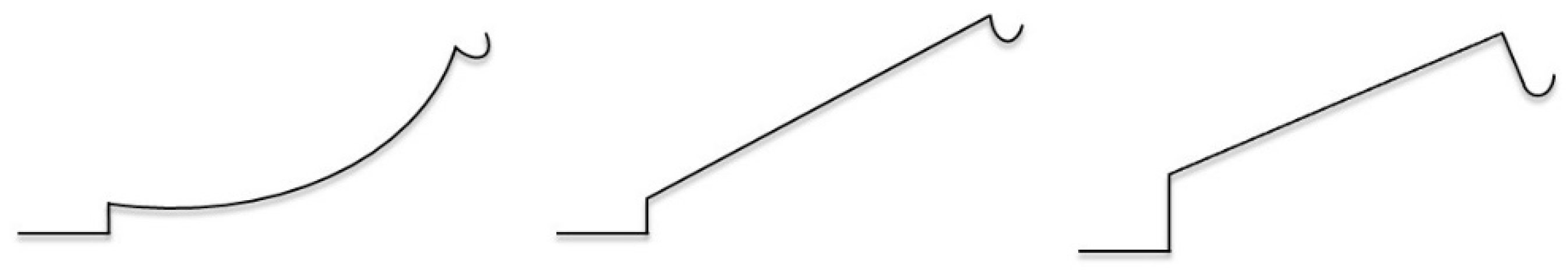
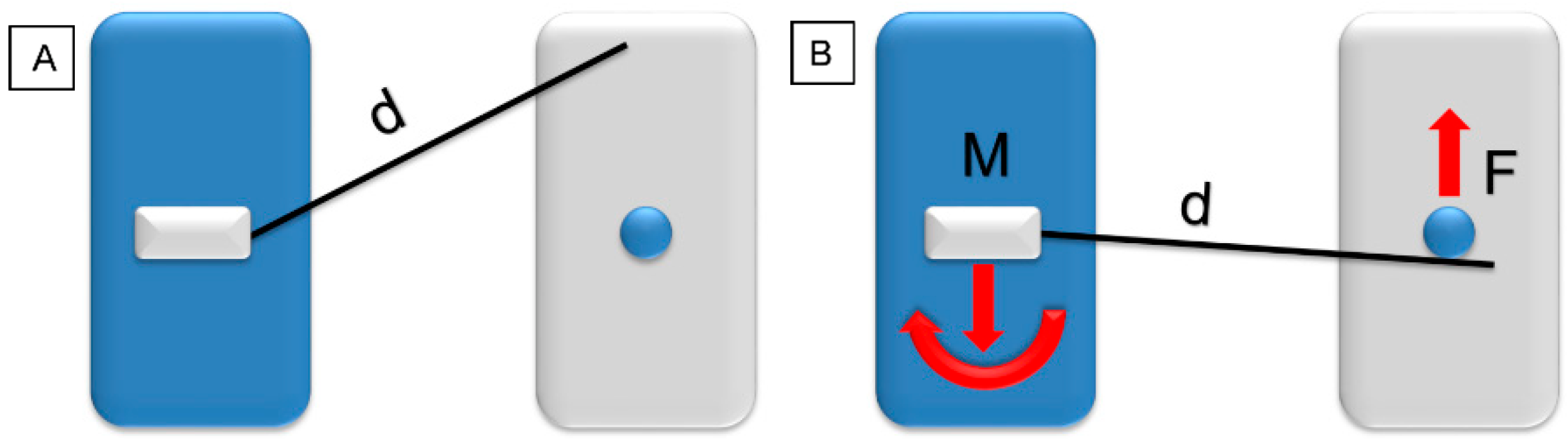
2. Anchorage Considerations
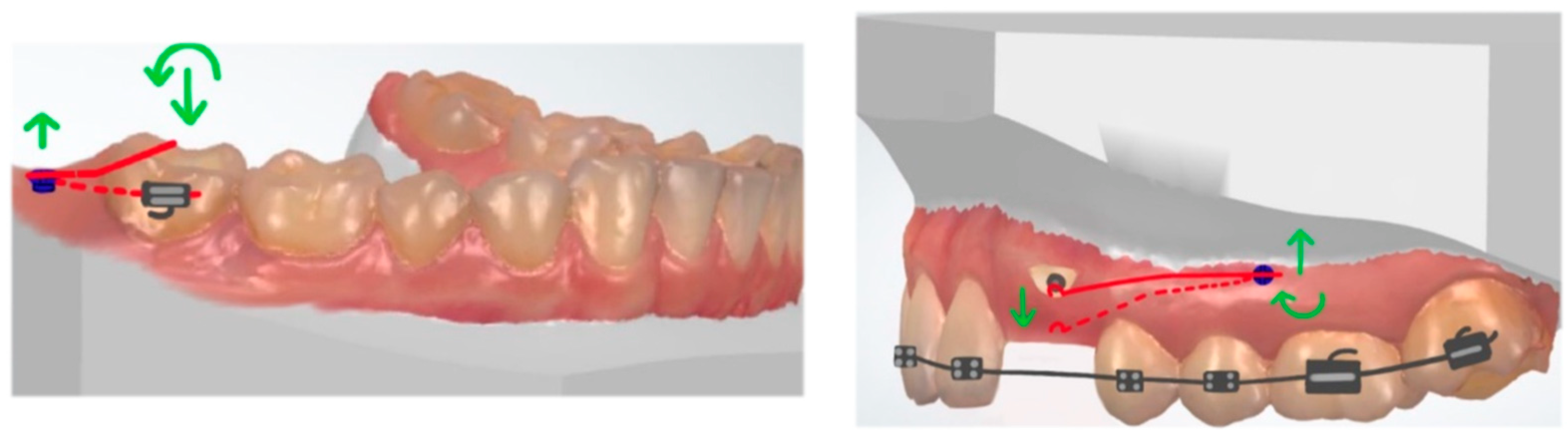
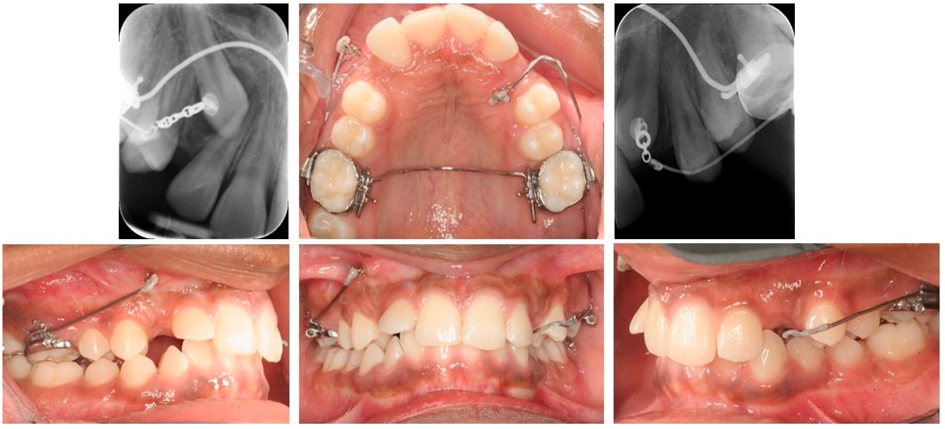
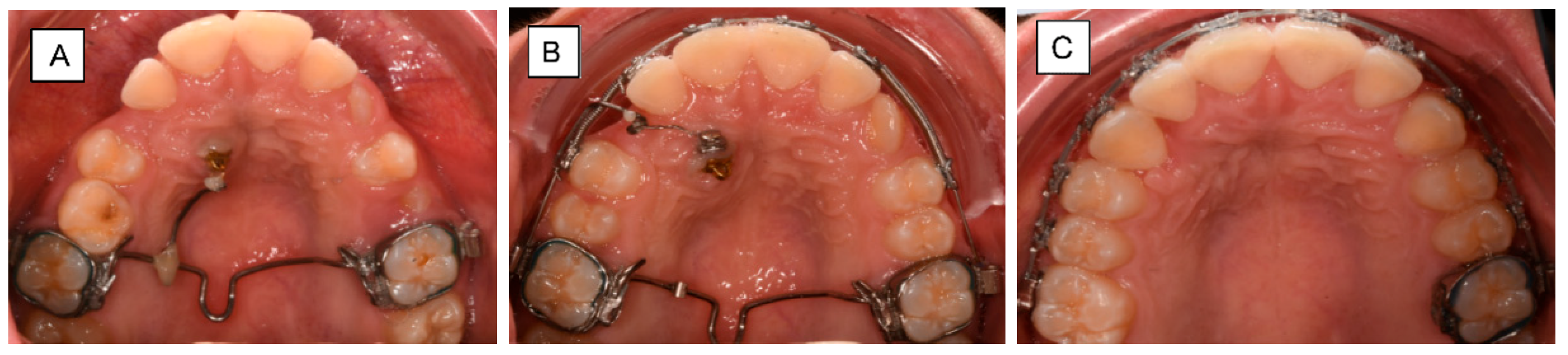
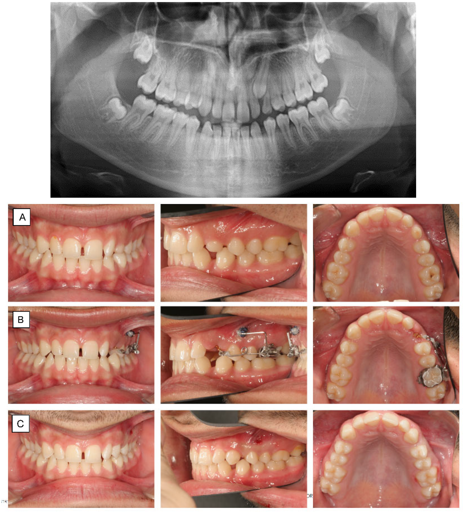
3. Impacted Teeth
3.1. Buccal Impaction
3.2. Palatal Impaction
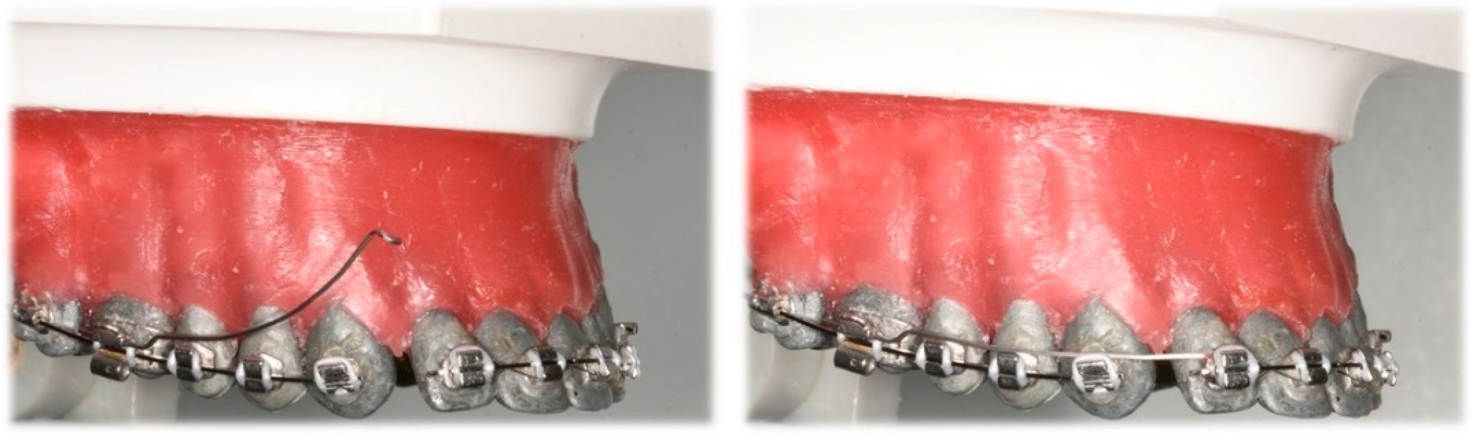
4. Deep Bite Correction
5. Open Bite
6. Intrusion
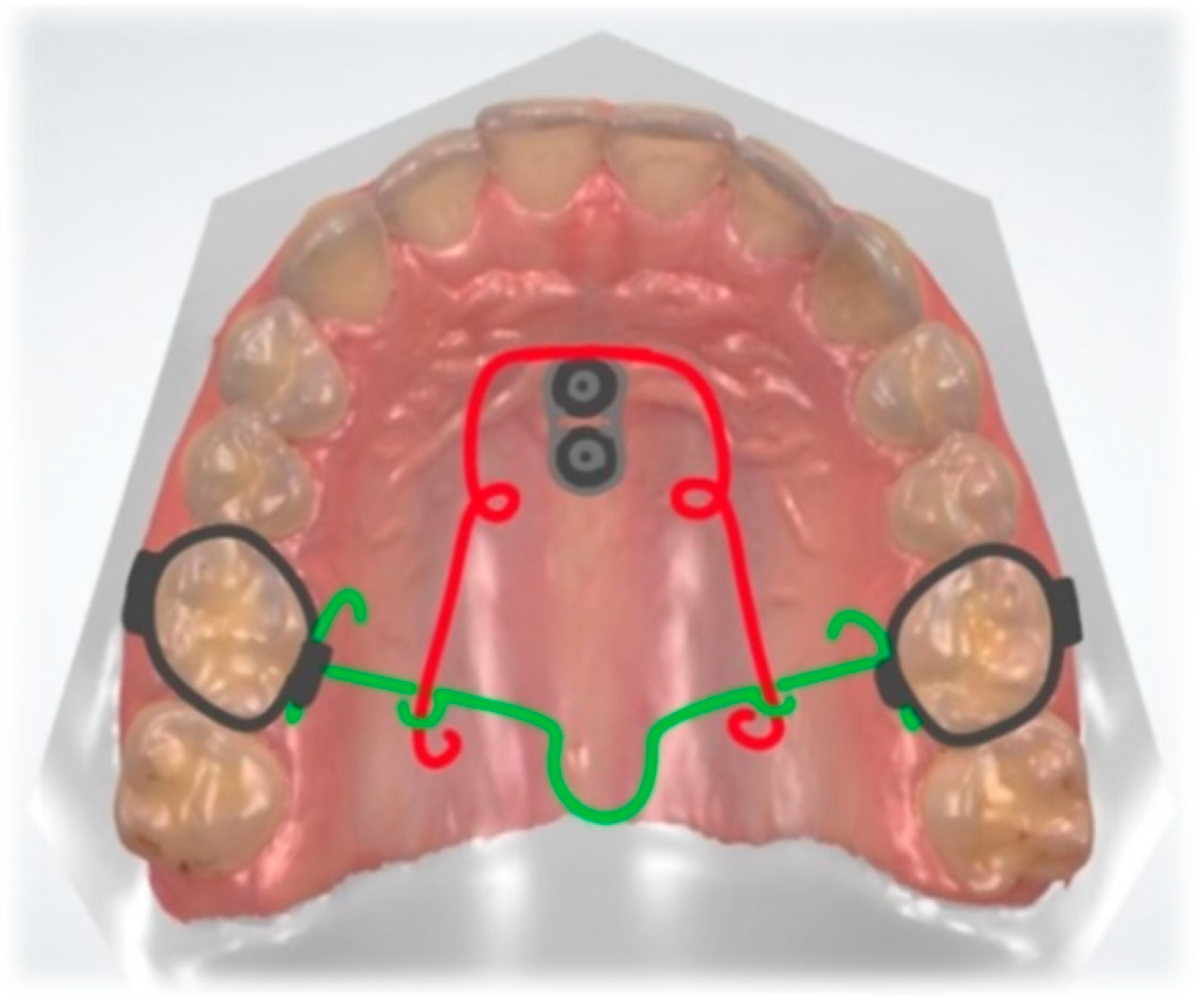
7. Space Closure
8. Occlusal Cant
9. Asymmetry, Midline Correction
10. Molar Uprighting
11. Dental Transposition
12. Single Tooth Extrusion
13. Molar Distalization
14. Discussion
15. Conclusions
Author Contributions
Funding
Institutional Review Board Statement
Informed Consent Statement
Data Availability Statement
Conflicts of Interest
References
- Burstone, C.J. Rationale of the segmented arch. Am. J. Orthod. 1962, 48, 805–822. [Google Scholar] [CrossRef]
- Laino, A.; Cacciafesta, V.; Martina, R. Treatment of tooth impaction and transposition with a segmented-arch technique. J. Clin. Orthod. 2001, 35, 79–86. [Google Scholar] [PubMed]
- Caballero, G.M.; Carvalho Filho, O.A.; Hargreaves, B.O.; Brito, H.H.; Magalhaes Junior, P.A.; Oliveira, D.D. Mandibular canine intrusion with the segmented arch technique: A finite element method study. Am. J. Orthod. Dentofac. Orthop. 2015, 147, 691–697. [Google Scholar] [CrossRef] [PubMed]
- Fischer, T.J.; Ziegler, F.; Lundberg, C. Cantilever mechanics for treatment of impacted canines. J. Clin. Orthod. 2000, 34, 647–650. [Google Scholar]
- Gurgel, J.A.; Pinzan-Vercelino, C.R.; Powers, J.M. Mechanical properties of beta-titanium wires. Angle Orthod. 2011, 81, 478–483. [Google Scholar] [CrossRef] [PubMed]
- Dalstra, M.; Melsen, B. Force systems developed by six different cantilever configurations. Clin. Orthod. Res. 1999, 2, 3–9. [Google Scholar] [CrossRef] [PubMed]
- Bilinska, M.; Dalstra, M. The Effect of Symmetric and Asymmetric Loading of Frontal Segment with Two Curved Cantilevers: An In Vitro Study. Dent. J. 2022, 10, 52. [Google Scholar] [CrossRef]
- Jacob, H.B.; Gonzaga, A.S.; Trinh, B.; Le, E.T.; English, J.D. Effects of stress relaxation in beta-titanium cantilevers used in orthodontic mechanics. Dent. Press J. Orthod. 2021, 26, e212069. [Google Scholar] [CrossRef] [PubMed]
- van Steenbergen, E.; Burstone, C.J.; Prahl-Andersen, B.; Aartman, I.H. The influence of force magnitude on intrusion of the maxillary segment. Angle Orthod. 2005, 75, 723–729. [Google Scholar] [CrossRef]
- Heravi, F.; Shafaee, H.; Forouzanfar, A.; Zarch, S.H.; Merati, M. The effect of canine disimpaction performed with temporary anchorage devices (TADs) before comprehensive orthodontic treatment to avoid root resorption of adjacent teeth. Dent. Press J. Orthod. 2016, 21, 65–72. [Google Scholar] [CrossRef]
- Thebault, B.; Dutertre, E. Disimpaction of maxillary canines using temporary bone anchorage and cantilever springs. Int. Orthod. 2015, 13, 61–80. [Google Scholar] [CrossRef]
- Chandhoke, T.K.; Nanda, R.; Uribe, F.A. Clinical applications of predictable force systems, part 2: Miniscrew anchorage. J. Clin. Orthod. 2015, 49, 229–239. [Google Scholar] [PubMed]
- Sukh, R.; Singh, G.P.; Tandon, P. Interdisciplinary approach for the management of bilaterally impacted maxillary canines. Contemp. Clin. Dent. 2014, 5, 539–544. [Google Scholar] [CrossRef] [PubMed]
- Yadav, S.; Upadhyay, M.; Uribe, F.; Nanda, R. Mechanics for treatment of impacted and ectopically erupted maxillary canines. J. Clin. Orthod. 2013, 47, 305–313. [Google Scholar] [PubMed]
- Potrubacz, M.I.; Chimenti, C.; Marchione, L.; Tepedino, M. Retrospective evaluation of treatment time and efficiency of a predictable cantilever system for orthodontic extrusion of impacted maxillary canines. Am. J. Orthod. Dentofac. Orthop. 2018, 154, 55–64. [Google Scholar] [CrossRef] [PubMed]
- Tepedino, M.; Iancu-Potrubacz, M.; Grippaudo, C.; Chimenti, C.; Lagana, G. Does muscular activity related to vertical facial divergence influence the time needed for orthodontic extrusion of palatally impacted maxillary canines? A retrospective study. J. Clin. Exp. Dent. 2018, 10, e869–e875. [Google Scholar] [CrossRef]
- Vijayashree, U.H.; Pai, V. Canine extrusion with a vertical tube supported cantilever spring. Apos Trends Orthod. 2017, 7, 49–51. [Google Scholar] [CrossRef]
- Zeno, K.G.; El-Mohtar, S.J.; Mustapha, S.; Ghafari, J.G. Finite element analysis of stresses on adjacent teeth during the traction of palatally impacted canines. Angle Orthod. 2019, 89, 418–425. [Google Scholar] [CrossRef]
- Fleming, P.S.; Sharma, P.K.; DiBiase, A.T. How to...mechanically erupt a palatal canine. J. Orthod. 2010, 37, 262–271. [Google Scholar] [CrossRef]
- Katiyar, R.; Singh, G.P.; Tandon, P. A cantilever spring for alignment of buccally impacted canines. J. Clin. Orthod. 2012, 46, 354–355. [Google Scholar]
- Patel, S.; Cacciafesta, V.; Bosch, C. Alignment of impacted canines with cantilevers and box loops. J. Clin. Orthod. 1999, 33, 82–85. [Google Scholar] [PubMed]
- Paduano, S.; Spagnuolo, G.; Franzese, G.; Pellegrino, G.; Valletta, R.; Cioffi, I. Use of cantilever mechanics for impacted teeth: Case series. Open Dent. J. 2013, 30, 186–197. [Google Scholar] [CrossRef] [PubMed][Green Version]
- Paduano, S.; Cioffi, I.; Iodice, G.; d’Anto, V.; Riccitiello, F.; Pellegrino, G.; Valletta, R. Correction of multiple canine impactions by mixed straightwire and cantilever mechanics: A case report. Case Rep. Dent. 2014, 2014, 925019. [Google Scholar] [CrossRef]
- Nakandakari, C.; Goncalves, J.R.; Cassano, D.S.; Raveli, T.B.; Bianchi, J.; Raveli, D.B. Orthodontic Traction of Impacted Canine Using Cantilever. Case Rep. Dent. 2016, 2016, 4386464. [Google Scholar] [CrossRef] [PubMed]
- Tepedino, M.; Chimenti, C.; Masedu, F.; Potrubacz, M.I. Predictable method to deliver physiologic force for extrusion of palatally impacted maxillary canines. Am. J. Orthod. Dentofac. Orthop. 2018, 153, 195–203. [Google Scholar] [CrossRef]
- Annarumma, F.; D’Emidio, M.; Rodi, G.; Battista, G.; Papi, G.; Migliorati, M. The effectiveness of miniscrews in the three-dimensional control of a palatal impacted canine: “Canine Only” approach. Case report. Int. Orthod. 2021, 19, 716–725. [Google Scholar] [CrossRef]
- Gandini, L.G., Jr.; Gandini, M.R.; Amaral, R.M. Continuous torque system with control of the reaction unit. Am. J. Orthod. Dentofac. Orthop. 2010, 137, 393–395. [Google Scholar] [CrossRef]
- Burstone, C. Biomechanics of Deep Overbite Correction. Semin. Orthod. 2001, 7, 26–33. [Google Scholar] [CrossRef]
- Melsen, B.; Konstantellos, V.; Lagoudakis, M.; Planert, J. Combined intrusion and retraction generated by cantilevers with helical coils. J. Orofac. Orthop. 1997, 58, 232–241. [Google Scholar] [CrossRef]
- Shroff, B.; Lindauer, S.J.; Burstone, C.J.; Leiss, J.B. Segmented approach to simultaneous intrusion and space closure: Biomechanics of the three-piece base arch appliance. Am. J. Orthod. Dentofac. Orthop. 1995, 107, 136–143. [Google Scholar] [CrossRef]
- Albelasy, N.F.; Montasser, M.A.; Hafez, A.M.; Abdelnaby, Y.L. Effects on root axes and resorption of simultaneous intrusion and retraction of maxillary central and lateral incisors using mini-implant supported three-piece burstone base arch: A prospective observational study. Int. Orthod. 2022, 20, 100595. [Google Scholar] [CrossRef] [PubMed]
- Kuhlberg, A. Cantilever Springs: Force System and Clinical Applications. Semin. Orthod. 2001, 7, 150–159. [Google Scholar] [CrossRef]
- Wilmes, B.; Vasudavan, S.; Stocker, B.; Willmann, J.H.; Drescher, D. Closure of an open bite using the ‘Mousetrap’ appliance: A 3-year follow-up. Aust. Orthod. J. 2015, 31, 208–215. [Google Scholar] [CrossRef] [PubMed]
- Flieger, S.; Ziebura, T.; Kleinheinz, J.; Wiechmann, D. A simplified approach to true molar intrusion. Head Face Med. 2012, 8, 30. [Google Scholar] [CrossRef]
- Nojima, L.I.; Barreto, B.C.T.; Vargas, E.O.A.; Starling, C.R.; Nojima, M.D.C.G. A clinical approach to managing open-bite malocclusion associated with severe crowding. Am. J. Orthod. Dentofac. Orthop. 2022, 162, 122–134. [Google Scholar] [CrossRef]
- El-Dawlatly, M.M.; Fayed, M.M.; Mostafa, Y.A. Deep overbite malocclusion: Analysis of the underlying components. Am. J. Orthod. Dentofac. Orthop. 2012, 142, 473–480. [Google Scholar] [CrossRef]
- Burstone, C.R. Deep overbite correction by intrusion. Am. J. Orthod. 1977, 72, 1–22. [Google Scholar] [CrossRef]
- Thote, A.M.; Sharma, K.; Uddanwadiker, R.V.; Shrivastava, S. Optimum pure intrusion of a mandibular canine with the segmented arch in lingual orthodontics. Biomed. Mater. Eng. 2017, 28, 247–256. [Google Scholar] [CrossRef]
- Vu, B.T.; Soroushian, S. Single-tooth intrusion with a cross tube and a cantilever spring. J. Clin. Orthod. 2013, 47, 427. [Google Scholar]
- Choy, K.; Pae, E.K.; Kim, K.H.; Park, Y.C.; Burstone, C.J. Controlled space closure with a statically determinate retraction system. Angle Orthod. 2002, 72, 191–198. [Google Scholar] [CrossRef]
- Deluke, M.; Uribe, F.; Nanda, R. Correction of a canted lower incisal plane. J. Clin. Orthod. 2006, 40, 555–559. [Google Scholar] [PubMed]
- Musilli, M.; Grampone, F.; Melsen, B. A new auxiliary spring for correction of a canted incisal plane. J. Clin. Orthod. 2014, 48, 500–504. [Google Scholar] [PubMed]
- van Steenbergen, E.; Nanda, R. Biomechanics of orthodontic correction of dental asymmetries. Am. J. Orthod. Dentofac. Orthop. 1995, 107, 618–624. [Google Scholar] [CrossRef]
- Fiorelli, G.; Melsen, B.; Modica, C. Differentiated orthodontic mechanics for dental midline correction. J. Clin. Orthod. 2001, 35, 239–244. [Google Scholar]
- Nanda, R.; Margolis, M.J. Treatment strategies for midline discrepancies. Semin. Orthod. 1996, 2, 84–89. [Google Scholar] [CrossRef]
- Mittal, T.; Singh, H.; Kapoor, P.; Sharma, P. Dental midline correction using a cantilever spring: A novel approach. Int. J. Orthod. Rehabil. 2020, 11, 145–149. [Google Scholar] [CrossRef]
- Musilli, M.; Marsico, M.; Romanucci, A.; Grampone, F. Molar uprighting with mini screws: Comparison among different systems and relative biomechanical analysis. Prog. Orthod. 2010, 11, 166–173. [Google Scholar] [CrossRef]
- Khouw, F.E.; Norton, L.A. The mechanism of fixed molar uprighting appliances. J. Prosthet. Dent. 1972, 27, 381–389. [Google Scholar] [CrossRef]
- Kojima, Y.; Mizuno, T.; Fukui, H. A numerical simulation of tooth movement produced by molar uprighting spring. Am. J. Orthod. Dentofac. Orthop. 2007, 132, 630–638. [Google Scholar] [CrossRef]
- Ma, Z.; Yang, C.; Zhang, S.; Xie, Q.; Shen, Y.; Shen, P. Orthodontic extrusion of horizontally impacted mandibular molars. Int. J. Clin. Exp. Med. 2014, 7, 3320–3326. [Google Scholar]
- Tay, A.B.; Go, W.S. Effect of exposed inferior alveolar neurovascular bundle during surgical removal of impacted lower third molars. J. Oral. Maxillofac. Surg. 2004, 62, 592–600. [Google Scholar] [CrossRef]
- Bonetti, G.A.; Bendandi, M.; Laino, L.; Checchi, V.; Checchi, L. Orthodontic extraction: Riskless extraction of impacted lower third molars close to the mandibular canal. J. Oral. Maxillofac. Surg. 2007, 65, 2580–2586. [Google Scholar] [CrossRef]
- Barros, S.E.; Janson, G.; Chiqueto, K.; Ferreira, E.; Rosing, C. Expanding torque possibilities: A skeletally anchored torqued cantilever for uprighting “kissing molars”. Am. J. Orthod. Dentofac. Orthop. 2018, 153, 588–598. [Google Scholar] [CrossRef] [PubMed]
- Sawicka, M.; Racka-Pilszak, B.; Rosnowska-Mazurkiewicz, A. Uprighting partially impacted permanent second molars. Angle Orthod. 2007, 77, 148–154. [Google Scholar] [CrossRef] [PubMed]
- Morita, Y.; Koga, Y.; Nguyen, T.A.; Yoshida, N. Biomechanical considerations for uprighting impacted mandibular molars. Korean J. Orthod. 2020, 50, 268–277. [Google Scholar] [CrossRef]
- Allen, W.A. Bilateral transposition of teeth in two brothers. Br. Dent. J. 1967, 123, 439–440. [Google Scholar]
- Filho, L.C.; Mde, A.C.; An, T.L.; Bertoz, F.A. Maxillary canine—First premolar transposition. Angle Orthod. 2007, 77, 167–175. [Google Scholar] [CrossRef]
- Lorente, C.; Lorente, P.; Perez-Vela, M.; Esquinas, C.; Lorente, T. Orthodontic management of a complete and an incomplete maxillary canine-first premolar transposition. Angle Orthod. 2020, 90, 457–466. [Google Scholar] [CrossRef] [PubMed]
- Gebert, T.J.; Palma, V.C.; Borges, A.H.; Volpato, L.E. Dental transposition of canine and lateral incisor and impacted central incisor treatment: A case report. Dent. Press J. Orthod. 2014, 19, 106–112. [Google Scholar] [CrossRef] [PubMed]
- Fu, P.S.; Wang, J.C.; Wu, Y.M.; Huang, T.K.; Chen, W.C.; Tseng, Y.C.; Tseng, C.H.; Hung, C.C. Unilaterally impacted maxillary central incisor and canine with ipsilateral transposed canine-lateral incisor. Angle Orthod. 2013, 83, 920–926. [Google Scholar] [CrossRef]
- Noh, H.K.; Park, H.S. An efficient and noncompliant method for forced eruption with microimplants that is bracket free, and its long-term stability. J. Am. Dent. Assoc. 2019, 150, 369–377. [Google Scholar] [CrossRef] [PubMed]
- Kumar, G.; Verma, N.; Parashar, S. Management of Subgingival Root Fracture with Decoronation and Orthodontic Extrusion in Mandibular Dentition: A Report of Two Cases. Contemp. Clin. Dent. 2019, 10, 554–557. [Google Scholar] [CrossRef] [PubMed]
- Vilanova, L.; Henriques, J.F.C.; Patel, M.P.; Da Costa Grec, R.H.; Aliaga-Del Castillo, A. The Miniscrew-Anchored Cantilever: A Simple Molar Distalizer. J. Clin. Orthod. 2020, 54, 773–774. [Google Scholar]
- Adel, S.; Zaher, A.; El Harouni, N.; Venugopal, A.; Premjani, P.; Vaid, N. Robotic Applications in Orthodontics: Changing the Face of Contemporary Clinical Care. Biomed. Res. Int. 2021, 2021, 9954615. [Google Scholar] [CrossRef] [PubMed]
- Liu, R.; Wan, W.; Isomura, E.T.; Harada, K. Metal wire manipulation planning for 3D curving—How a low payload robot can use a bending machine to bend stiff metal wire. arXiv 2022, arXiv:2203.04024. [Google Scholar] [CrossRef]
- van der Meer, W.J.; Vissink, A.; Ren, Y. Full 3-dimensional digital workflow for multicomponent dental appliances: A proof of concept. J. Am. Dent. Assoc. 2016, 147, 288–291. [Google Scholar] [CrossRef]
- Pellizzari, M.; Jam, A.; Tschon, M.; Fini, M.; Lora, C.; Benedetti, M. A 3D-Printed Ultra-Low Young’s Modulus β-Ti Alloy for Biomedical Applications. Materials 2020, 13, 2792. [Google Scholar] [CrossRef]
- Arefin, A.M.E.; Khatri, N.R.; Kulkarni, N.; Egan, P.F. Polymer 3D Printing Review: Materials, Process, and Design Strategies for Medical Applications. Polymers 2021, 13, 1499. [Google Scholar] [CrossRef]
- Thurzo, A.; Urbanová, W.; Novák, B.; Waczulíková, I.; Varga, I. Utilization of a 3D Printed Orthodontic Distalizer for Tooth-Borne Hybrid Treatment in Class II Unilateral Malocclusions. Materials 2022, 15, 1740. [Google Scholar] [CrossRef]
- Guerrero-Gironés, J.; López-García, S.; Pecci-Lloret, M.R.; Pecci-Lloret, M.P.; Lozano, F.J.R.; García-Bernal, D. In vitro biocompatibility testing of 3D printing and conventional resins for occlusal devices: Biocompatibility of 3D printing and conventional resins. J. Dent. 2022, 123, 104163. [Google Scholar] [CrossRef]


Publisher’s Note: MDPI stays neutral with regard to jurisdictional claims in published maps and institutional affiliations. |
© 2022 by the authors. Licensee MDPI, Basel, Switzerland. This article is an open access article distributed under the terms and conditions of the Creative Commons Attribution (CC BY) license (https://creativecommons.org/licenses/by/4.0/).
Share and Cite
Bilinska, M.; Kristensen, K.D.; Dalstra, M. Cantilevers: Multi-Tool in Orthodontic Treatment. Dent. J. 2022, 10, 135. https://doi.org/10.3390/dj10070135
Bilinska M, Kristensen KD, Dalstra M. Cantilevers: Multi-Tool in Orthodontic Treatment. Dentistry Journal. 2022; 10(7):135. https://doi.org/10.3390/dj10070135
Chicago/Turabian StyleBilinska, Malgorzata, Kasper Dahl Kristensen, and Michel Dalstra. 2022. "Cantilevers: Multi-Tool in Orthodontic Treatment" Dentistry Journal 10, no. 7: 135. https://doi.org/10.3390/dj10070135
APA StyleBilinska, M., Kristensen, K. D., & Dalstra, M. (2022). Cantilevers: Multi-Tool in Orthodontic Treatment. Dentistry Journal, 10(7), 135. https://doi.org/10.3390/dj10070135