Review

A Review of Synthesis, Characterization, Properties, and Applications of Double Perovskite Oxides

by
Pablo V. Tuza
1,* and
Mariana M. V. M. Souza
2
1
Facultad de Ciencia e Ingeniería en Alimentos y Biotecnología, Universidad Técnica de Ambato, Ambato 180216, Ecuador
2
Escola de Química, Universidade Federal do Rio de Janeiro (UFRJ), Rio de Janeiro 21941-909, RJ, Brazil
*
Author to whom correspondence should be addressed.
Inorganics 2025, 13(11), 372; https://doi.org/10.3390/inorganics13110372
Submission received: 14 September 2025 / Revised: 23 October 2025 / Accepted: 4 November 2025 / Published: 7 November 2025
(This article belongs to the Special Issue Recent Progress in Perovskites)

Abstract

Double perovskites are represented by the formula A2BB’O6 and AA’BB’O6. These materials have been synthesized using the solid-state reaction, sol–gel, Pechini, and hydrothermal methods. X-ray fluorescence, X-ray diffraction, magnetic measurements, transmission electron microscopy, X-ray photoelectron spectroscopy, temperature-programmed reduction, synchrotron X-ray diffraction, neutron powder diffraction, extended X-ray absorption fine structure, and Raman spectroscopy have been used for the characterization of double perovskites. X-ray diffraction, synchrotron X-ray diffraction, and neutron powder diffraction coupled with the Rietveld method determine the crystal structure of a sample. These materials present various properties and applications. The present review aims (i) to report a process to determine the symmetry, apparent size, and apparent strain using the Rietveld method; (ii) show how experimental characterization techniques complement each other in the investigation of double perovskites; (iii) describe how the synthesis method can help in the uncovering of double perovskites with improved properties; and (iv) exemplify some of the main applications of double perovskites.

1. Introduction

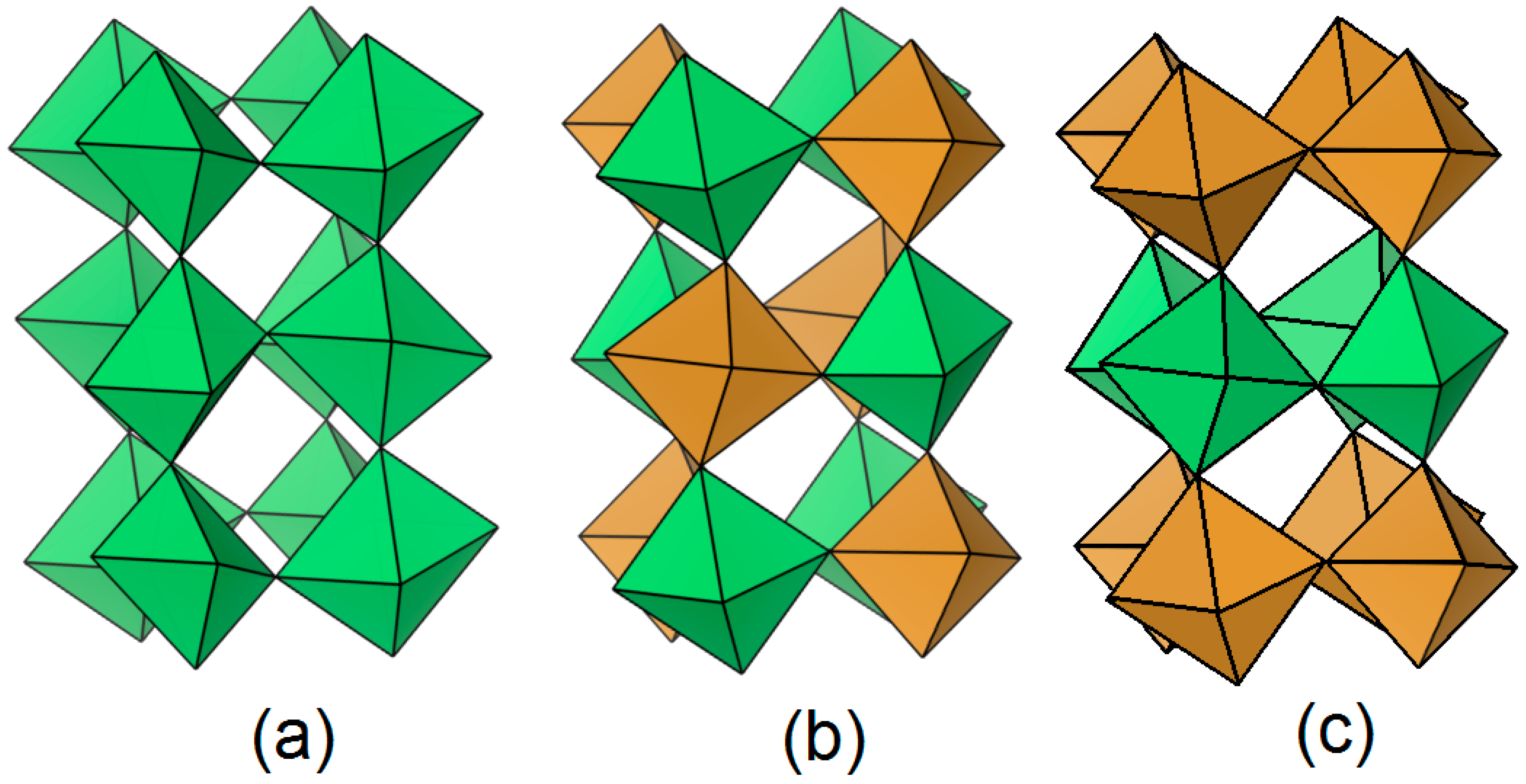
Perovskites are mixed oxides represented by the formula ABO3, where A is an alkali earth or rare earth metal, and B can be a transition metal [1]. The crystalline structure of this material can be represented using octahedra sharing corners. The oxygen atoms are positioned at the octahedra vertices, the octahedra centers are occupied by B cations, and the A cations are placed in the cavities between the octahedra, as depicted in Figure 1, which was drawn using the Vesta software (version 3.90.0a) [2].
Double perovskites (DPs) can be represented by the formula A2BB’O6 or AA’BB’O6 [3], where the apostrophe symbol indicates a DP containing distinct metal ions with the same molar composition. There are three types of B-cation arrangement for DPs: random, rock salt, and layered [3], which are indicated in Figure 2. For the case of the random arrangement, the octahedra are randomly distributed in the structure. In the rock salt ordering case, the octahedra are ordered as the sodium and chloride ions in the NaCl structure. As indicated in its name, in the layered DP, BO6, and B’O6 octahedra layers are intercalated between them. It is worth mentioning that ABO3 perovskites with B cations with a molar ratio different from 0.5 mol can be represented using the DP random arrangement.
Surface exchange and diffusion of oxygen ions, conductivity enhancement [4], and improvement of catalytic properties are advantages of DPs over ABO3 materials. For example, La2NiTiO6, which contains LaNiO3 and LaTiO3, shows stable catalytic activity for the steam reforming of methane compared to the low-stable Ni or the inactive Ti perovskite, as the DP presents enhanced metal–support interaction compared to the conventional perovskites [5].
B and B’ cation arrangement in DPs mostly depends on B and B’ valence differences [6], but it also may be influenced by B and B’ ionic radius differences, as occurs for the Bi2AlGaO6 and Bi2AlInO6 [7]. Furthermore, the calcination time may influence the arrangement, as La-Ni-Ti-containing DPs with random ordering changed to a rock-salt arrangement by modifying the calcination step from 17 h at 800 °C to 135 h at 1000 °C [8].

2. Synthesis

DPs can contain B cations with mixed valences, and this allows for the design of several mixed oxides in accord with the octet rule and the preparation. Four preparation methods are mostly used to prepare these mixed oxides: the solid-state reaction, the sol–gel, the modified Pechini, and the hydrothermal methods.

2.1. Solid-State Reaction Method

The solid-state reaction is the most employed method to synthesize DPs. It consists of mixing adequate amounts of oxide [9] or carbonate [10] precursors of DPs. Then, the mixture is calcined at high temperatures, like 1400 °C [11], for diffusion and chemical reaction of the starting materials. It is worth noting that oxide diffusion is so low at 500 °C [12]. Similarly, higher pressures employed during the synthesis process ensure high diffusion rates [13], and this warrants the formation of the DP phase.
The ball-milling period of the precursors prior to their heating influences the morphology. Zouridi et al. [14] established that the mechanochemical step influences the particle size and produces a spherical nanometric single-phase La0.5Sr0.5Ti0.5Mn0.5O3−δ. Moreover, the perovskite phase can form during the ball-milling process, as observed for the La1−xSrxTi1−yMnyO3±δ [15]. Furthermore, the microstrain depends on the ball-milling time, as described for the SrTiO3 [16].
An oxygenless atmosphere can influence the formation of DPs. Wlodarczyk et al. [13] established that the Ar atmosphere inhibits cation oxidation during the preparation of the Ba2CeWO6. Moreover, the final calcination step for the formation of the single-phase Sr2FeMoO6 was 1500 °C for 12 h in 2% H2/Ar, followed by 1500 °C for 6 h [17], whereas it was 1074 °C for 4 h in 3% H2/Ar [18]. It is worth noting that the last sample contained about 2 wt% impurities of Fe and SrMoO4.
The sintering temperature affects the coherent length [16], but this condition also affects other properties. Nadig et al. [19] indicated that the La0.67Ca0.33MnO3 calcined from 900 °C to 1200 °C presented the optimal oxygen content for the sample heated to 1100 °C, and this oxygen amount affects its cooling power.

2.2. Sol–Gel Method

The sol–gel method, also known as the citrate sol–gel method [20], is widely used for the preparation of DPs. In this method, nitrates [21], oxides, organometallic compounds [22], and metals [23] of DPs are used as starting materials. As can be found in work by Valdés et al. [24], all precursors are mixed, including the citric acid, in pH-adjusted water using NH4OH at 80 °C. The resulting mixture was transformed into a gel at 90 °C for 24 h after evaporation at 80 °C. Finally, this last solid was calcined and then reduced.
The sol–gel method can be used to obtain DPs at temperatures lower than those employed in the solid-state reaction method. To illustrate this, Valdés et al. [24] used the sol–gel method for the synthesis of Sr2FeMoO6 and employed a reducing atmosphere. The maximum temperature used in this work was 1200 °C, and this is less than that used in the solid-state synthesis for the same material (1500 °C, [17]).
Although the sol–gel method followed by ball milling has not been reported in the literature for DPs, this additional step may improve the mixed oxide properties. For instance, Bartoletti et al. [25] reported that the sol–gel synthesis of CaCu3Ti4O12, before a ball-milling process, increased its number of defects compared to the same material without the mechanochemical step, and this additional step in the synthesis method improved the CaCu3Ti4O12 catalytic activity.
Some technological differences in the sol–gel method that influence the DP properties have been reported in the literature. For instance, Xu et al. [26] reported the effect of polyvinyl alcohol (PVA) on the synthesis of La2CoMnO6. The final calcination temperature was lower compared to that reported without PVA (700 °C, Xu et al. [26] vs. 900 °C, Yousif et al. [27]). Martinez-Rodriguez et al. [28] prepared the Pr1−xBaxMnO3−δ using EDTA and ethylene glycol (EG) in the preparation method. These authors reported that the sol–gel EDTA synthesis produced a sample with the highest proportion of cubic phase compared to the sol–gel EG preparation for x = 0.5, as the first method produces a better thermally stable sample.
Environmentally friendly fuels, such as lactose, fructose, maltose, sugar, and an organic compound-based powder called liquorice, as well as other types of fuels like oxalic acid, maleic acid, green coffee powder, and pomegranate paste, were employed for the preparation of Tb2ZnMnO6 [29] and Tb2FeMnO6 [30], respectively. These authors claimed homogeneous and smaller particle sizes for the case of maltose fuel used to synthesize the Zn-containing DP, while the best results in size and morphology corresponded to the maleic acid addition to obtain the Fe-containing DP. Additionally, the grape juice used as fuel can modulate the particle size, as reported for the Y2CrMnO6 [31].

2.3. Pechini Method

The Pechini method, also known as Pechini sol–gel [32], is characterized by producing a metal-containing polymer. This method is usually carried out in four stages: (i) metal solution preparation, (ii) chelate production through attachment of solution-containing cations and an organic acid like citric acid, (iii) resin production through chelate polymerization, by employing an alcohol such as the EG, and (iv) synthesis of DPs after multiple thermal treatments.
In the first step, precursor solubilization is required. Some low-cost precursors, such as titanium species produced through titanium isopropoxide evaporation, are not water-soluble. Nitric acid can be employed to help solubilize this titanium precursor, like 3.4 mol/L nitric acid [33].
Moreover, the addition of ammonia hydroxide can be carried out in the first step or to adjust the pH after the third step [34]. The addition of ammonia promotes the production of a foam-like structure. For example, Córdova-Calderón et al. [8] reported the formation of this structure after calcining the resin at 450 °C during La2NiTiO6 preparation, and this was not claimed by Pérez-Flores et al. [35] in the same sample produced from the calcination of an NH4OH-free resin.
Some technical differences in the method include ball-milling of the powder from resin calcination [36] or the use of ethylenediaminetetraacetic acid with citric acid as complexing agents [37]. Related to the CoFe2O4 preparation, Cheraghi et al. [38] used lemon juice, whereas Al-Wasidi and Abdelrahman [32] employed tartaric acid as a source of complexing agents. This last acid may provide small crystallite sizes, as in the synthesis of MgAl2O4 [39].
Moreover, the EG may be replaced by another alcohol. For instance, dos Santos et al. [40] employed glycerol instead of EG in the synthesis of SiO2. These authors claimed a higher surface area for the higher weight alcohol, as a result of the polymeric matrix influence in the product. Cheraghi et al. [38] reported a mixture of sucrose and EG, while Al-Wasidi and Abdelrahman [32] used 1,2-propanediol.
The resin calcination can be performed using a heating rate of 2 °C/min, which may help maintain low gas production from this polyester decomposition. Bataliotti et al. [41] described that the polymer containing Te ions presented three steps of degradation, and these processes accounted for about 90% weight loss and gas production.

2.4. Hydrothermal Method

The hydrothermal method is carried out by employing nitrates and ammonium salts [42], chlorides [43], and organometallic compounds [44] of double-perovskite-containing metals. To begin with, a solution is obtained from the precursors, and then it is placed in a Teflon-lined stainless-steel autoclave for thermal treatment. Finally, the products are washed and dried.
The hydrothermal method has influenced the structure of DPs. Yi et al. [45] claimed that the dielectric behavior of La2FeCrO6 is related to its oxygen vacancies. Irfan et al. [46] reported that the Ce2NiCrO6, Pr2NiCrO6, and Nd2NiCrO6 presented agglomerated particles related to the synthesis method. Valdés et al. [24] reported lower particle sizes of Sr2FeMoO6 compared to those from the microwave sol–gel method by providing uniform heating in the particle production. Zhang et al. [47] reported that the hydrothermal method promotes the stabilization of the NiO6 and MnO6 structures, which are influenced by the size of R3+ ions in the R2NiMnO6.
The materials containing DPs prepared using the hydrothermal method have shown better catalytic properties than the conventional catalysts. McGuire et al. [48] used Y2CoMnO6 and Y2NiMnO6 as supports for the Pt catalyst in performing methanol oxidation. Pt/Y2CoMnO6 provided a better performance compared to the commercial Pt/C and the Pt/Y2NiMnO6, as it has a stronger metal-to-support interaction. These authors also stated that the Y2CoMnO6 was more active and stable than the Y2NiMnO6 and the commercial IrO2 for oxygen evolution reaction (OER). Additionally, Y2CoMnO6 is a bifunctional catalyst, as it is also active for the oxygen reduction reaction.
The hydrothermal method has been used to prepare DP-containing composites. Sharma et al. [49] observed that the reduced graphite oxide Sr2TiMnO6 (Sr2TiMnO6-rGO), prepared by mixing graphite oxide and the DP and then aging the blend using the hydrothermal method, presented activity for the Rhodamine B dye due to its absorbance range and bandgap value. The Sr2TiMnO6 was prepared following the solid-state reaction method. Moreover, Khan et al. [50] indicated that the LSTN@NiMn-LDH, where LSTN and LDH are LaSrTiNiO6 and lamellar double hydroxides, were prepared by the hydrothermal method and used as a catalyst for the OER and hydrogen evolution reaction (HER). The authors reported that OER occurs because of the interaction between oxygen species and the catalyst, whereas the HER activity is due to a synergistic effect between LSTN and NiMn-LDH.

3. Characterization

3.1. X-Ray Fluorescence

The chemical composition of DPs can be determined through X-ray fluorescence (XRF) using a spectrometer. The results can be expressed on an oxide or a metal basis. Popov et al. [51] reported that the experimental chemical composition of the Ba2CoNbO6 on a metallic basis was 2(0.03):1(0.03):0.998(0.03) for Ba:Nb:Co; hence, the composition was within the interval formed by the mean value of the corresponding experimental chemical composition and the standard deviation. The chemical composition and the presence of impurities were established using the X-ray fluorescence method. A value of 5.4 for oxygen mols in the Sr2CoNbO6−δ and impurities in the sample was determined using XRF and confirmed by magnetization measurements [52]. Jin et al. [53] indicated that the Al:Fe experimental molar ratio for the La2FeAlO6 prepared using glucose and citric acid was about 1, and these values were obtained using X-ray fluorescence. Also, the chemical composition quantified using X-ray fluorescence indicates mixed valences of Mn in the Sr2MnNbO6−δ [54].

3.2. X-Ray Diffraction (XRD)

XRD can be used to determine the crystal structure, the crystallite size, and the phase composition of DPs. XRD is performed in a diffractometer, and its operating parameters are the 2θ range, step size, and count time. The intensity of the main reflection of the XRD pattern should be at least 10,000 a.u., as reported for the XRD of NdCaFeSnO6 [55].
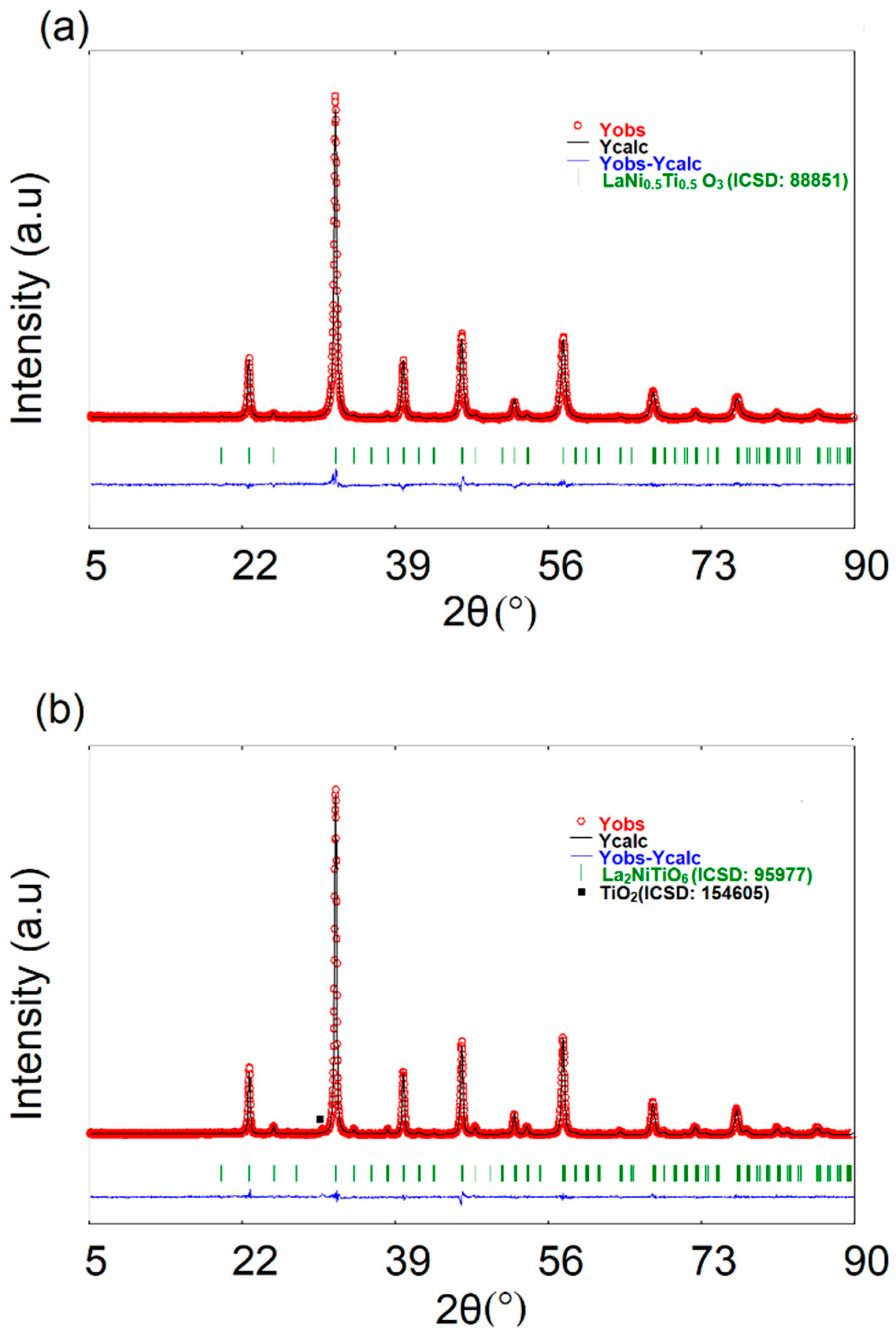
This characterization technique is largely employed to determine the crystal structure. Fabrelli et al. [56] reported that the La2−xBaxCoMnO6 presented an evolution from orthorhombic (Pnma) to rhombohedral (R 3 ¯ c), and then to hexagonal (P63/mmc) as Ba content increases (Figure 3). Zaraq et al. [57] established that the Sr2Co0.75Fe0.25TeO6, Sr2Co0.5Fe0.5TeO6, and Sr2Co0.25Fe0.75TeO6 present monoclinic symmetry and space group I2/m at room temperature. The symmetry was maintained, but the space group changed to P21/n at temperatures below 100 K, whereas the symmetry and the space group changed to tetragonal (I4/m) and then to cubic (Fm 3 ¯ m) at high temperatures up to 1100 K. Córdova-Calderón et al. [8] indicated two different symmetries for the La-Ni-Ti DP as a function of the calcination temperature (Figure 4), fostering the synthesis of DPs with desired crystal structures.

3.2.1. Crystal Structure Determination

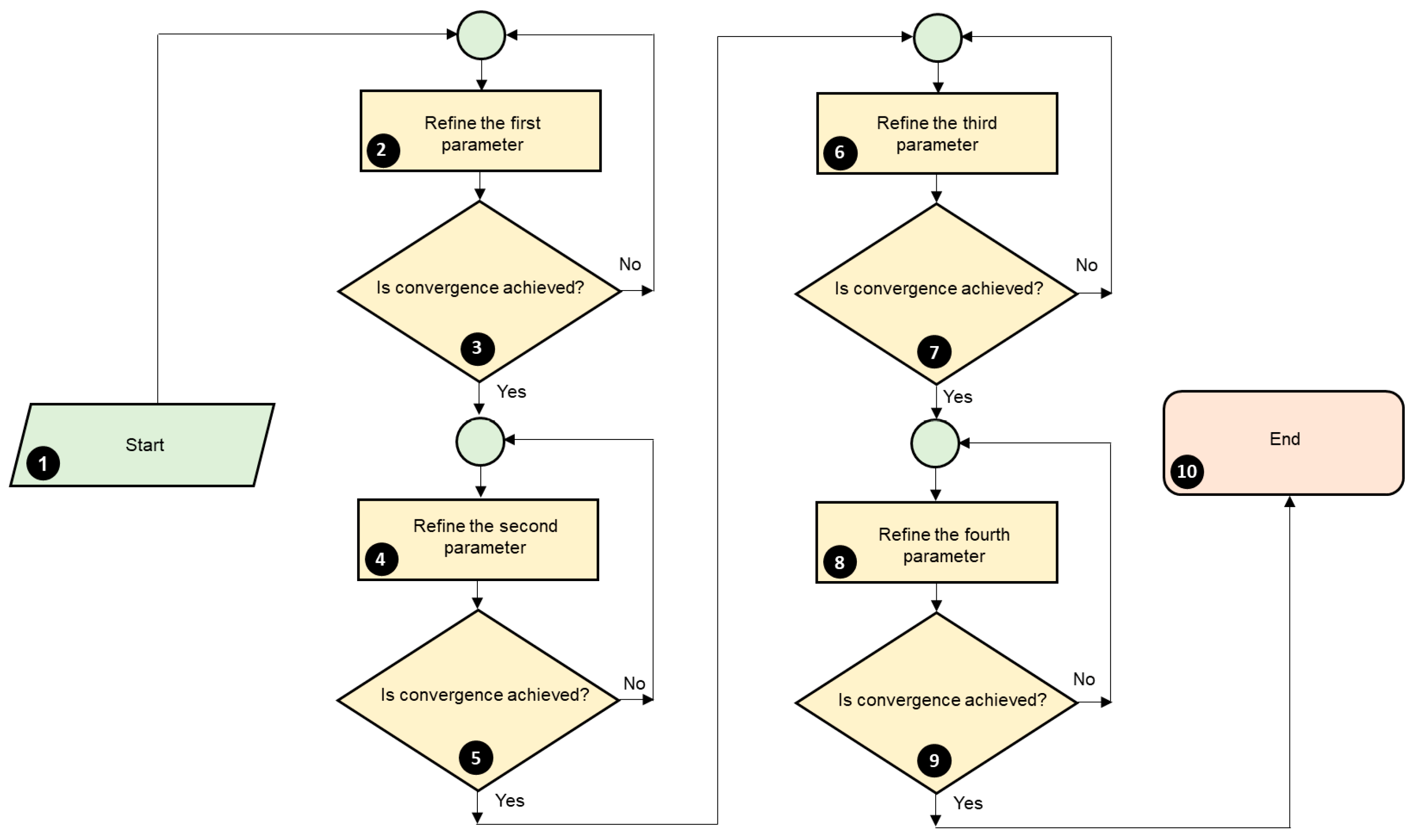
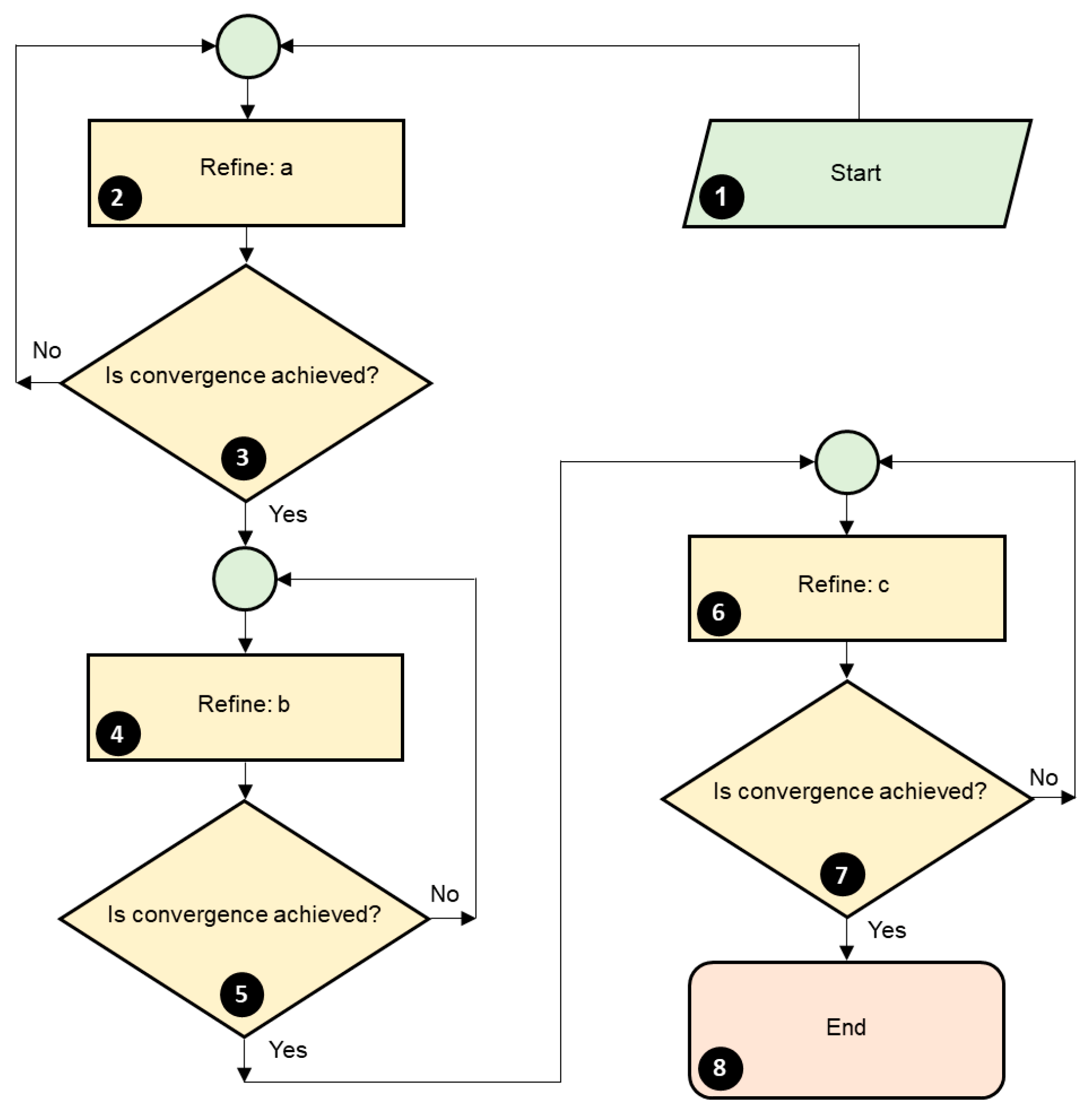
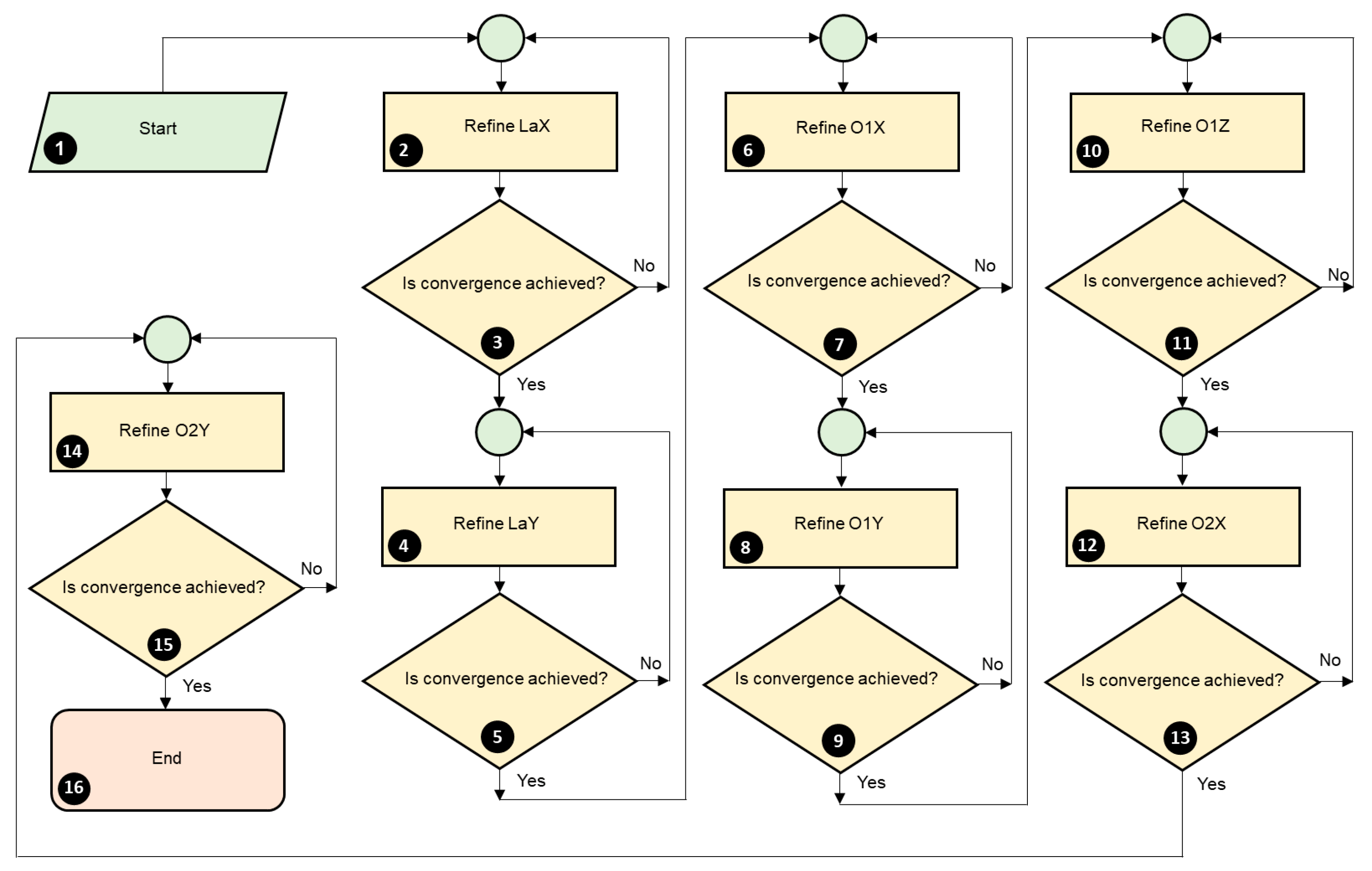
Crystal structures are determined using the Rietveld method, which is implemented in various programs, such as Fullprof [58]. This free and reliable package has been extensively used to refine crystal structures through cyclic steps where the convergence must be achieved.
Free databases like the Crystallographic Open Database share crystallographic information of materials through CIF files. The operator obtains a crystal structure template in a PCR file through the default crystal structure simulation option by opening the CIF file with the PCR tool of Winplotr [59] and then saving it. The PCR file will be saved in the same CIF file folder. Steps 2 and 3 of Figure 5 depict this procedure, whereas the default and written parameters in a PCR file showing hypothetical values are presented in Table S1. It is important to note that the written parameters in Table S1 should be immediately placed after their prior default row. For instance, row 3 must be added to the PCR file after row 2. In addition, two rows of zeros in rows 8 and 12–17 of Table S1 represent values (top row) and codewords (bottom row) for each parameter, where the codeword enables the parameter refinement. Finally, for the case of the refinement of various parameters in each row, if not specified in the refinement process, the refinement proceeds from left to right.
The Rietveld refinement process using the PCR file may be described as five steps: (i) parameter setting, (ii) background, (iii) unit cell parameters, (iv) peak shape parameters, and (v) atomic coordinates refinement.
In the parameter-setting step, an operator changes from simulation to refinement by altering the Job parameter value in row 1 of Table S1 from 2 to 0. Then, the peak shape function is selected and added below the Npr parameter from rows 1 and 9 of Table S1. There are seven different peak functions in Fullprof (version 3.0), and the pseudo-Voigt function is chosen and presented in Table S1. However, other authors prefer the Thompson–Cox–Hastings pseudo-Voigt convoluted with the axial divergence asymmetry function [60]. The suitable function provides better values for the criteria’s parameters to judge the quality of refinement.
Next, the researcher specifies the number of excluded 2θ ranges by writing a value for the Nex parameter in row 1 of Table S1 through adding code, depicted in row 6 of Table S1. For instance, if the operator performs the instructions in step 5 of Figure 5, the order-ascending range-containing values, i.e., the lowest and the highest values, replace each row of zeros in row 6 of Table S1.
Moreover, the format of the experimental data, the cut-off of peak profile tails, the monochromator polarization correction, the maximum number of iterations per refinement, the minimum, and the maximum 2θ values are specified through the Ins, Wdt, Cthm, NCY, Thmin, and Thmax parameters in rows 2, 4, and 5 of Table S1.
Furthermore, the operator should add the number of atoms, the atoms, and their valences describing the researched material, as depicted in steps 6 to 8 of Figure 5. The number of atoms (Nat) parameter comprises row 9 of Table S1, whereas the atoms, their types, and valences should be written one above another at row 12 of Table S1. Similarly, the researcher should write literature-based values to isotropic displacement parameters (Biso) for investigations where these values are not refined, as presented by our group [61], and indicated in step 9 of Figure 5. The Biso parameter is contained in row 12 of Table S1.
Additionally, the operator should write 1 for the More parameter located in row 9 of Table S1 and write the code to calculate bond distances and angles, as indicated in row 10 of Table S1. This procedure is shown in step 10 of Figure 5.
Also, the Sycos parameter that accounts for the largest systematic aberration in the Bragg–Brentano geometry [62] and Scale parameters located in rows 7 and 13 of Table S1 are refined, as presented in steps 11 to 14 of Figure 5. In step 11, the operator should select the experimental data saved in the DAT file previously placed in the PCR file folder. Silva et al. [63] refined the Zero parameter in row 7 of Table S1 instead of Sycos. It is worth noting that if the goniometer of the diffractometer is correctly aligned, the Zero parameter should not be refined.
In the background refinement step, four parameters describing the background located in row 8 of Table S1 are refined to ensure convergence is achieved in the program, as depicted in Figure 6.
In the refinement of the unit cell parameters, the operator should refine the unit cell parameters in row 15 of Table S1, as presented in Figure 7, for an orthorhombic symmetry. If the symmetry enables an angle to be refined, like the β angle in the symmetry monoclinic, this angle should be refined after the c parameter (Figure 7), and followed by a convergence verification step, similar to step 7 in Figure 7.
In the refinement step of the peak shape parameters, the operator should refine U, V, W, and X parameters for the peak shape function that are depicted in row 14 of Table S1. It should be observed whether peak shape models enable the refinement of other parameters, like the so-called Shape1; these parameters should be refined right after the X parameter. The procedure of peak shape parameter refinement is presented in Figure 8.
The last refinement step involves the atomic coordinates: X, Y, and Z parameters shown in row 14 of Table S1 are refined, as depicted in Figure 9 for a material containing La, Ni, Ti, and O, with orthorhombic symmetry. After the operator has refined all of the atomic coordinates, other parameters, such as Biso and Occ parameters, should be refined.
Furthermore, if the sample powder is packed on a depression of the sample holder prior to the experimental data collection, as recommended by the diffractometer manufacturer, and the diffraction peaks are symmetric, the preferred orientation (Pref1 and Pref2) and asymmetry parameters (Asy1 to Asy4) in row 16 of Table S1 should not be refined. Otherwise, each parameter should be refined after step 16 of Figure 9, following the order of refinement shown in this figure. For this last case, the last 2θ value should be placed under AsyLim located in row 4 of Table S1, to enable the refinement of asymmetry parameters. It is worth mentioning that preferred orientation is a source of error, as it modifies the diffraction peak intensities [64], and it may occur in compressed samples [65].
Finally, other authors have refined most of the parameters presented in this work. Silva et al. [63] refined the scale factor, background coefficients, zero-point error, asymmetry, atomic coordinates, Biso, Occ parameters, and unit cell parameters, although they did not describe the order of refinement.

3.2.2. Graph of the Observed and Calculated XRD Pattern

A graph for the observed, calculated, and difference in XRD patterns is presented after finishing the Rietveld refinement method. Bhuyan et al. [66] reported that this graph showed no secondary phase for the Gd2FeCrO6 sample. Hosen et al. [20] established the absence of long-range ordering of B cations through the refinement of the crystal structure of Gd2CoCrO6 nanoparticles as a result of a peak absence around 20°. However, the graph showed background noise in this sample, which is characteristic of particle sizes at the nanoscale. Guo et al. [67] claimed that the La2NiRuO6-calculated pattern corresponds to its experimental profile, as observed in the difference between the data, which is represented below these profiles in the graph. Zaraq et al. [57] reported that the monoclinic Sr2Co0.5Fe0.5TeO6 presents (111) and (11 1 ¯ ) reflections in the 2θ range 23.5° to 25.5° and (311) ( 3 ¯ 11) (131) ( 1 ¯ 31) reflections in the 2θ range 51° to 53° characterizing the P21/n at 100 K; hence, they ensured that the Sr-Co-Fe-Te DP cannot be assigned the space group I2/m.
Steps 1 to 5 of Figure 10a present a graph generation procedure for the observed and calculated XRD patterns through the Winplotr tool of the Fullprof package. Step 6 of Figure 10a depicts the edition step, where the operator adds a format to the patterns. Some usual procedures are indicated in Table S2. Step 7 of Figure 10a indicates the export file procedure. This graph is in a BMP file that can be edited in other image-editing programs like Paint.

3.2.3. Criteria for Judging the Quality of the Refinement

Several factors, like Rp, Rwp, Rexp, and χ2, have been used to quantify the quality of a crystal structure refinement. However, there is no consensus on a range of parameter-accepted values. For example, Rp and Rwp values of 21.4 and 26.8 were reported by our group in the refinement of La, Ni, Ti, and Co perovskites [61]. El Hachmi and Sadoune [68] reported values of 4.61 and 5.99 for these same parameters of the crystal structure of the SrLa2NiFeNbO9.
The R factors should be presented along with bond distances to address the quality of the refinement. For instance, Córdova-Calderón et al. [8] reported Ni-O and Ti-O distances from the structure refinement of LaNi0.5Ti0.5O3 and La2NiTiO6 and compared them with previously reported values in the literature to assess the quality of the refinement.
Moreover, the R factors and χ2 help assign the symmetry and space group for a sample with the same chemical composition but different B cation arrangements. Our group verified that the symmetry and space group for LaNi0.5Ti0.5O3 provided higher R-factor and χ2 values when used to describe the La2NiTiO6 [8].

3.2.4. CIF File Generation

CIF files contain crystallographic sample information that other researchers can use to simulate the XRD pattern using the Fullprof program and then refine the crystal structures of their own samples, as shown in Figure 5, Figure 6, Figure 7, Figure 8, Figure 9 and Figure 10a. Additionally, they can even draw an image of the crystal structure of the sample using other packages, such as the Vesta software (version 3.90.0a). If the operator has completed the procedures outlined in Figure 5, Figure 6, Figure 7, Figure 8, Figure 9 and Figure 10a and then enters a codeword of −1 for the Rpa [69] parameter in row 2 of Table S1, the Fullprof program will generate the CIF file for the sample.

3.2.5. Apparent Crystallite Size and Apparent Strain

The apparent crystallite size and apparent strain can be determined through a micro-structural analysis in the Fullprof program. Both parameters depend on the full width at half maximum, but the apparent strain is also derived from the Stokes–Wilson apparent strain. Figure 10b presents a summary of the procedure in determining the apparent crystallite size and apparent strain for an orthorhombic structure and space group Pbnm. The analysis is performed using the Fullprof program and the default Thompson–Cox–Hastings pseudo-Voigt convoluted with axial divergence asymmetry peak function [70], i.e., Npr of 7, located in rows 1 and 9 of Table S1. The IRF file is derived from a refinement of a standard, such as the CeO2, and its name and extension are included in the PCR file, as depicted in row 3 of Table S1. A value of 4 for the Res parameter (row 1, Table S1) can describe the information format for this resolution file [62].
Moreover, parameters located from row 12 to 15 of Table S1 should be unchanged by writing zero for their codeword, except for the Scale and the GauSiz. Furthermore, the symmetry and space group should be specified in the Size-Model parameter. For instance, the orthorhombic symmetry with space group Pbnm corresponds to a value of 18 [70]. Additionally, the operator should write the crystallite size and strain parameters particular to a crystal structure and space group, as described in steps 7 to 20 of Figure 10b. For example, the parameters shown in row 17 of Table S1 were manually written, and these parameters describe an orthorhombic symmetry, with space group Pbnm. As a matter of fact, each symmetry presents its own set of size and strain parameters [70], and these parameters should be refined following the method presented in Figure 10b. On the other hand, it is worth noting that row 17 should replace row 18 in the PCR, and this last row will appear automatically at step 9 of Figure 10b. An MIC file containing the apparent size and apparent strain will be created and saved in the PCR file folder.

3.3. Magnetic Measurements

Magnetic measurements, such as temperature-dependent magnetization in zero-field-cooled (ZFC) and field-cooled (FC) modes at various magnetic fields, can be conducted using a magnetometer. DPs can show various magnetic behaviors. Almadhi et al. [71] demonstrated that (Ca0.5Mn0.5)2MnTeO6 is a spin glass, whereas Shibuya et al. [72] synthesized Ba2(Cd1−xCax)ReO6 and observed that canted antiferromagnetic (AFM) order and quadrupolar order can be observed for 0 ≤ x ≤ 0.6, AFM order and quadrupolar order correspond to 0.6 ≤ x ≤ 0.9, and AFM order can be verified for 0.9 ≤ x ≤ 1. Agarwal et al. [73] demonstrated different valences and ionic radius mismatches among Co, Cr, and Mn in La2Co(1−x)Cr(x)MnO6 by increasing the Cr amount through magnetic measurements. This influence reduced the Curie temperature, coercive field, and remanence.
Macchiutti et al. [74] showed the magnetization vs. temperature (M(T)) curves in ZFC and FC modes, the inverse of the magnetic susceptibility vs. temperature ((T)), and the Curie–Weiss law fit for the La1.5Sr0.5CoMnO6 calcined at various temperatures. The authors observed that a decrease in the M(T) values occurs due to the increase in boundaries, as the number of antiphase domains increases with the grain size [74].
Zhang et al. [75] depicted the ZFC and FC magnetization curves, the first derivative of ZFC curves, the variation in the Curie temperature (TC), and the glass transition temperature (Tg) as a function of the Ga content for the La1.5Sr0.5Co1−xGaxMnO6. It could be observed that the Tg is practically imperceptible for x ≥ 0.3 due to the dilution effect of the Ga ions in the sample [75].
An important interfacial effect is the Exchange Bias (EB) [76], and it depends essentially on magnetic interactions in the sample. Qiu et al. [77] claimed that an EB field of 1200 Oe at 10 K for the Sr2FeReO6-LaFeO3 may be attributed to the B cations arrangement and ferrimagnetic (FiM) nature of the DP interfacing the G-type AFM FeO6-containing perovskite. Pal et al. [78] contended that the EB results from atoms not occupying their sites and competition of ferromagnetic (FM), AFM, and spin glass interactions in the Sr2FeRuO6. Deng et al. [79] described that the EB depends on the spin–orbit coupling on Ir and AFM behavior of the B and B’ cations for the Y2NiIrO6. Kush and Srivastava [80] established that the EB depends on surface spins in interfacial frozen glassy states at the FiM particle’s interface. Datta et al. [81] indicated that the EB phenomenon results from the competing FiM and AFM interactions for the NdSrCoIrO6. Moreover, while Mahalle et al. [82] reported that the EB is influenced by the applied field for the Gd2CoRuO6, Li et al. [83] stated that the EB depends on the FM layer thickness in the Bi0.5Sr0.5Fe0.5Cr0.5O3.
The FM, AFM, FiM, and paramagnetic nature are determined using magnetic scans. While these characteristics are typical of DPs, the AFM behavior is related to B cation ordering. For instance, Córdova-Calderón et al. [8] reported NiO6 and TiO6 random ordering in LaNi0.5Ti0.5O3 and a Néel temperature (TN) of 15 K. Moreover, these authors stated the LaNi0.5Ti0.5O3 Curie–Weiss temperature and the effective magnetic moment by fitting the inverse susceptibility to the Curie–Weiss law. Furthermore, a TN of 80 K from magnetic measurements is due to the NiO6 and MoO6 AFM ordering in the Sr2NiMoO6, whereas a TN of 60 K is related to MoO6 substitution by the WO6 [84]. Additionally, a magnetization vs. magnetic field curve justifies FM and AFM domains [20] and indicates multiferroic [85] and ferromagnetic properties [86]. Moreover, the FiM nature in the Ba2FeMnO6 depends on the double-exchange interaction of the B and B’ cations through the oxygen [87].
Magnetic susceptibility values derived from magnetization values also present magnetic transitions, as shown by Zaraq et al. [88] for Sr2FeTeO6, Sr2Fe0.75Ni0.25TeO6, Sr2Fe0.5Ni0.5TeO6, Sr2Fe0.25Ni0.75TeO6, and Sr2NiTeO6. The Curie temperature showed magnetic transitions, and its change with x in Sr2−xCexFe1+x/2Mo1−x/2O6 was identified using magnetic susceptibility measurements [89].
Some structural studies can be performed through magnetic measurements. The progressive Pr substitution in Sr2−xPrxFeTiO6−δ from x = 0.2 to x = 0.8 promoted a transition from AFM to FM [90]. A glassy phase is produced by adding W in Sr2NiMo1−xWxO6 (0 ≤ x ≤ 1), as indicated in the ZFC and FC curves at low temperatures [84].

3.4. Transmission Electron Microscopy

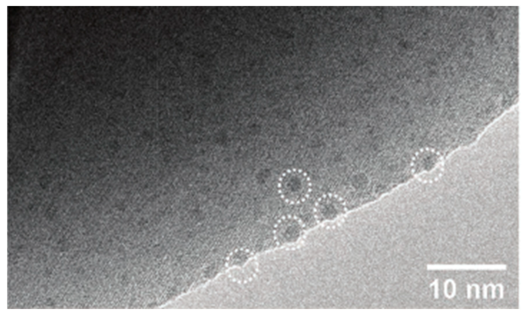
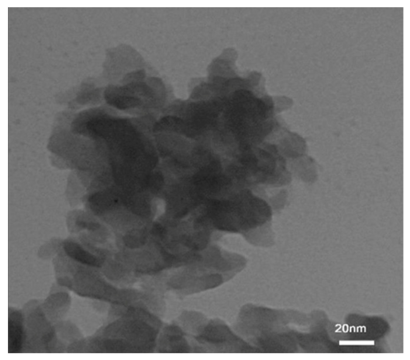
Transmission electron microscopy (TEM) provides sample images at the nanometric scale, containing information about the sample morphology and particle size. Guo et al. [67] employed the ImageJ [91] package in the exsolution process of Ni-Ru particles from La1.85NiRuO6 reduced using 3% H2O-wetted 5% H2/Ar at 350 °C, finding Ni-Ru particle sizes of about 2.2 nm (Figure 11). Also, these authors measured the interplanar spacing and angles using fringe patterns through the DigitalMicrograph and the ImageJ program. Moreover, these authors manually selected the particles for their size determination through ImageJ, using a time-consuming procedure. Dara et al. [92] reported that Tb2CoMnO6 presented agglomerated particles at the nanoscale, as depicted in Figure 12.
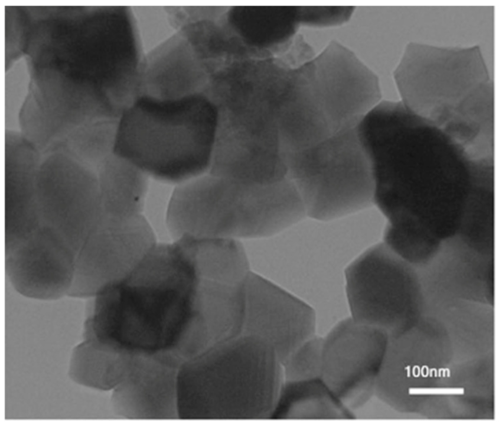
The diameter of a spherical particle describes its size. For the case of an elongated particle, its size is the mean of the ellipse axis simulating its shape [93]. Figure 10c presents these axes for a hypothetical particle. Additionally, particle sizes should be equal to or higher than the crystallite size obtained using Size and Strain Analysis from Fullprof, as a particle can be constituted of one or many crystallites. For instance, the Lu2FeMnO6’s TEM’s particle size was 55 nm, whereas its crystallite size was 25.2 nm (Figure 13) [94].

3.5. X-Ray Photoelectron Spectroscopy

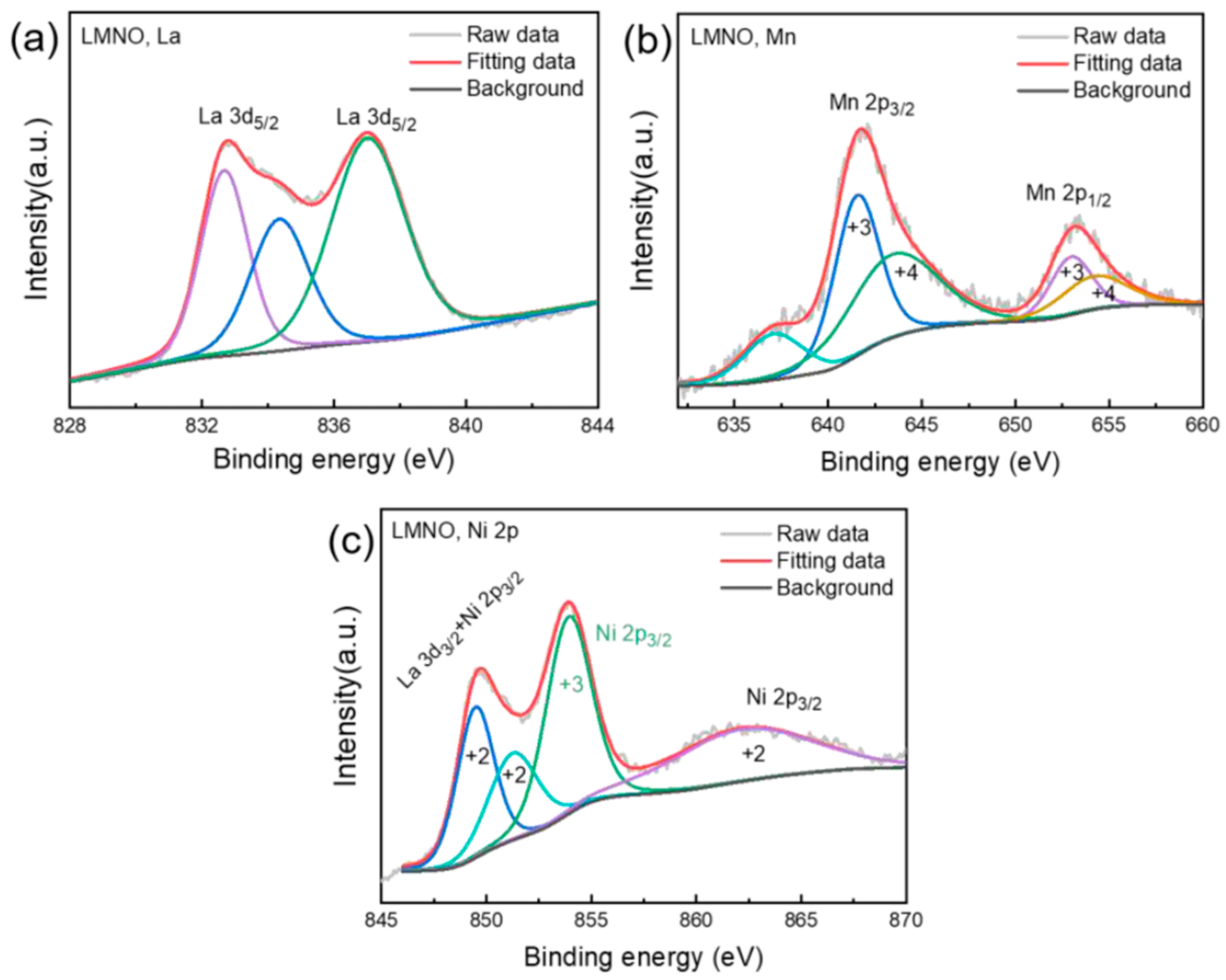
X-ray photoelectron spectroscopy (XPS) provides structural information from the surface of the analyzed material, approximately 10 nm deep [95], and indicates the oxidation states of each ion [96]. For example, Ali et al. [97] reported surface Mn3+O6 and Fe3+O6 in La2MnFeO6 and surface Mn3+O6 and Co3+O6 in La2MnCoO6 using XPS, thereby verifying the structure of a DP in their samples. Sharmili et al. [42] claimed that La2NiMnO6 presents a uniform distribution of its constituents. Hosen et al. [20] determined mixed valences for Co and Cr cations in the Gd2CoCrO6 through XPS; thus, this DP presents short-range ordering of B cations. Kush and Srivastava [80] established defects in the La2FeCoO6 through the analysis of their B cations by using the XPS technique. Bhattacharjee et al. [98] indicated that some Fe2+-O-Fe2+ and Fe2+-O-Fe3+ interactions for the Ba2FeVO6 and Ca2FeVO6 were identified through XPS. Zhang et al. [11] reported the La 3d, Mn 2p, and Ni 2p spectra for the La2MnNiO6 (Figure 14) that resemble the perovskite characteristic through XPS.
Furthermore, XPS provides the surface chemical composition of a sample. For instance, Raji et al. [99] reported that the experimental surface molar ratios for Ba, Co, W, and O in Ba2CoWO6 measured through XPS were 1.9, 0.9, 0.85, and 5.8.
Additionally, XPS states the presence of atoms with different valences. For instance, Tang and Zhu [100] reported mixed valences for Fe and Re and two types of oxygen in La2FeReO6+δ: lattice and adsorbed oxygen. Also, this characterization technique was used to report oxygen vacancies, as in the NdBaFeTiO6 [101]. Additionally, Ghorbani and Ehsani [102] established that the mean oxygen valence of the Ba2FeMoO6 was −1.59. Kumar et al. [103] reported an increase in the adsorbed oxygen with a decrease in the Sr content for Sr2−xFeCoO6−δ, as determined by core-level XPS, due to an increase in the number of oxygen vacancies. Furthermore, Ghorbani and Ehsani [104] claimed the generation of mixed valences in Ba2−xAgxFeMoO6 for x of 0.025, prepared using the sol–gel method. Yi et al. [105] indicated Cr3+, Cr4+, Ni2+, and Ni3+, lattice and adsorbed oxygen in the La2CrNiO6. Moreover, Solanki et al. [106] reported that the ratio of Co2+/Co3+ depends on x up to 0.2 for the La2CoTi1−xNixO6.

3.6. Temperature-Programmed Reduction

The temperature-programmed reduction (TPR) can be carried out in conventional equipment with a TCD or mass detector. Initially, the sample is pretreated to eliminate adsorbed water by heating it under a flow of an inert gas like Ar. Subsequently, the sample is reduced under a flowing reduction mixture like H2/Ar.
TPR is used to determine surface oxygen species and reduction processes in the sample. La2AlFeO6, prepared using glucose and citric acid, demonstrated two peaks: one peak stating the reduction of Fe3+ to Fe2+ and another peak at high temperatures describing the Fe2+ reduction to Fe0. More Fe3+ species are present in the material prepared using glucose, and this was determined through the TPR peak area. Moreover, the mixed oxide prepared using glucose was reduced at lower temperatures than the same material prepared using citric acid [53].
Roozbahania et al. [107] stated that the La2NiMnO6 presented two peaks in the TPR profile: the first peak at 378 °C represents the reduction of (i) surface oxygen species, (ii) Mn4+ to Mn3+, and (iii) Ni3+ to Ni2+. The second peak (at 466 °C) depicts the reduction of (i) lattice oxygen and (ii) Ni2+ reduction to Ni0. These authors also claimed that for La2CoMnO6, there was one peak (at 409 °C) showing the reduction of (i) Mn4+ to Mn3+ and (ii) Co3+ to Co2+.
Guamán-Ayala et al. [33] reported that LaNi0.5Ti0.5O3 DP presented two TPR peaks. One peak at low temperatures described the reduction of surface oxygen and Ni3+ to Ni2+, whereas the peak at high temperatures indicated the reduction of Ni2+ to Ni0. The peak at high temperatures was confirmed through quantitative phase analysis using the Rietveld method. There is a more delayed H2 consumption in the La2NiTiO6 compared to the LaNi0.5Ti0.5O3 [108].

3.7. Synchrotron X-Ray Diffraction (SXRD)

Synchrotron X-ray powder diffraction measurements can be collected through Debye–Scherrer cameras [109]. Before the data collection, the sample is packed into a capillary, and the measurements can be collected at low temperatures using a cryostat and a blower [110].
These scans can be used to determine the symmetry and space group of a sample. Zhao et al. [111] reported that Pb2NiMoO6 presented a monoclinic symmetry with space group Pc using synchrotron X-ray powder diffraction dataset. Liang et al. [112] claimed a triclinic crystal structure for Ca2CuWO6 with space group P 1 ¯ among 100 to 1000 K.
Moreover, these patterns have been used to verify crystal structure stabilities with temperature. da Cruz Pinha Barbosa et al. [113] contended that Ba2ZnReO6 presents a transition from cubic to tetragonal at 23 K by using synchrotron radiation. Silva et al. [114] claimed that SmSrCoFeO6 and EuSrCoFeO6 maintain the orthorhombic crystal structure and Pnma space group from 9 to 300 K, as stated using a synchrotron X-ray powder diffraction dataset. Morimura and Yamada [109] argued that the CoO6 and RuO6 octahedra arrangement in La2CoRuO6 changed from rock-salt to random ordering by pressing the sample using 8 GPa and 1373 K, as confirmed by SXRD.
The refinement of the crystal structure can be performed using the Fullprof program, and most of the parameters proposed in this work for the Rietveld method using XRD patterns are fitted in the Rietveld refinement coupled to the SXRD dataset, but following a different refinement strategy. For instance, the unit cell, overall isotropic displacement (Bov, row 13 Table S1), scale, peak shape of the pseudo-Voigt, atomic coordinates, asymmetry, and the background parameters were refined for the Ce1–x(Nd0.74Tm0.26)xO2–x/2, where several background points were previously selected, A and B cations were fixed, and the order of refinement is not reported [115].

3.8. Neutron Powder Diffraction (NPD)

The symmetry, space group, and B cation arrangements at low temperatures have been determined using NPD measurements. Ni and Ir moments present ordering below 74 K, as confirmed through 1.5 to 90 K neutron thermodiffractogram [116]. Naveen et al. [117] reported that the Ca2−xLaxFeRuO6 presents orthorhombic symmetry and space group Pbnm; hence, these DPs present random ordering of FeO6 and RuO6 at temperatures lower than 3 K. The crystal structure and space group at 150 K for CaMnCoWO6 were determined, making use of a neutron-derived fit [118]. Almadhi et al. [71] reported no apparent long arrangement of spins in (Ca0.5Mn0.5)2MnTeO6 at 1.6 K. Sr2MnNbO6−δ presents the Pbnm space group from 4 to 300 K as established via the NPD method [54].
Magnetic properties can also be studied through neutron diffraction data. Short-range spin orders in CaMnFeTaO6 were verified by the absence of magnetic diffraction peaks in the neutron diffraction patterns [119]. A ferromagnetic order between Mn and Cr in CaMnCrSbO6 was observed through neutron diffraction [120]. Moreover, neutron diffraction patterns of the CaMnCoWO6 revealed magnetic peaks that disappeared through heating from 15 to 20 K [118]. The absence of a magnetic peak at 1.5 K using neutron diffraction was observed for the CaMnMnWO6, and this was related to a spin glass transition at 8 K [121]. A magnetic phase separation for Sr2CuTe0.3W0.7O6 and Sr2CuTe0.2W0.8O6 was observed through neutron diffraction measurements [122].
The investigation of the structure has been carried out using neutron diffraction profiles. The amount of Er in La2−xErxNiMnO6 preserved the monoclinic symmetry, as verified through Rietveld refinement of neutron diffraction data [123]. Pal et al. [124] stated that the Pr1.5Sr0.5CoMnO6 presented a CoO6 and MnO6 random arrangement composed of two symmetries, monoclinic (P21/n) and orthorhombic (Pbnm). Almadhi et al. [71] claimed accurate cation Occ parameters using the contrast in neutron-scattering lengths. Copper sites were partially substituted by iron in CaCuFeReO6, as indicated by the refinement of the mixed oxide crystal structure [125]. Moreover, these authors reported magnetic diffraction peaks in the neutron diffraction patterns collected from 5 to 500 K. To research oxygen positions in Sr2Ga1−xMnxSbO6, the Rietveld refinement of NPD data was employed [126]. Oxygen diffusion paths in the La2Ge1−xCrxMgO6−δ structure are described through neutron diffraction measurements [127].
The refinement can be performed using the Fullprof program. López et al. [127] reported that in the refinement of the crystal structure of La2Ge1−xCrxMgO6−δ using NPD, the pseudo-Voigt function was selected, and the scale, background, zero, peak shape, asymmetry, unit cell, and isotropic displacement parameters were refined. The authors also reported the use of scattering lengths in the refinement. The scattering length of oxygen is detectable through NPD [128], compared to XRD, where oxygen cannot be well detected [126]. Similarly, the Ni and Ti in the La2NiTiO6 are more detectable in NPD than in XRD, as their scattering lengths are different [108].

3.9. Extended X-Ray Absorption Fine Structure (EXAFS)

EXAFS provides information about the symmetry and the structure. Jeevanandham et al. [129] observed defects in the La2Ni0.2Cu0.8MnO6. Majumder et al. [130] reported the concentration and distribution of antisite disorders in Sm2NiMnO6. Kavaliuk et al. [131] established that the bond length difference between values obtained from EXAFS and XRD are of 0.2 Å in the BaLn8Co2O6−δ, BaLn10Co2O6−δ, BaLn12Co2O6−δ, and BaLn14Co2O6−δ, where Ln can be La, Ce, Pr, Nd, Sm, Eu, Gd, Tb, Dy, Ho, Er, Tm, Yb, and Lu, as XRD provides an average value for this parameter.

3.10. Raman Spectroscopy

The crystal structure of DPs has been investigated using Raman spectroscopy. Murthy et al. [132] synthesized the SrLa1−xMnxLiTeO6 and claimed the same symmetry for all samples and the Mn inclusion in the perovskite structure. The Cr-O and Fe-O bond lengths in Gd2FeCrO6 were calculated using the Raman spectrum and compared to those calculated through XRD, and the difference was 0.04 Å [66]. Chahla et al. [55] indicated that the NdCaFeSnO6 showed bands in the Raman spectrum related to the bending and stretching modes of SnO6 and FeO6. Moreover, they reported that the unit cell expansion with the temperature was observed through a peak displacement around 350 cm−1 in the Raman spectrum.
Khan et al. [133] established that A-site ordering and anti-site disorder in Gd-doped Y2CoMnO6 influence the Raman spectra. Meena et al. [134] stated that La2−xCaxFeMnO6 presents improper ordering of FeO6 and MnO6 by Raman scattering spectroscopy. Boudad et al. [10] indicated five band shifts in the LaBaFeTiO6 Raman spectrum compared to that of EuBaFeTiO6. Wlodarczyk et al. [13] reported that Ba2CeWO6 decomposed in air above 573 K, whereas Silva et al. [135] contended that La2CoMnO6 showed phonon energy behaviors associated with short-range Co and Mn ferromagnetic clusters through Raman spectroscopy measurements.

4. Properties and Applications

DPs present several properties like FM [136], magnetoresistance effect [137], ferroelectric [138], AFM [139], FiM [140], multiferroic [141], metal–insulator transition [142], magnetocaloric effect [143], luminescent [144], magnetic [53], optical, electrical, and mechanical [145], semiconducting [146], and half-metallic FM [147].
Some material properties are related to magnetic properties. BaBiFeTiO6 presents ferroelectric properties with remanent polarization, as depicted by the polarization vs. electric field hysteresis loop [138]. The FiM behavior of Sr2CrHfO6 was observed at 2 K [140]. Gd2CrFeO6 exhibits magnetic entropy changes and refrigerant capacity similar to those of other materials with magnetocaloric effects [143]. The magnetic moments of La2BB’O6 (BB’ = CrCo, CrNi, ScNi, VNi, VSc) confirm their properties [147].
Other properties are related to their bandgap values. Ba2GdBiO6 can be used in ultraviolet and infrared absorption due to its bandgap [145]. The bandgap of the semiconducting Sr2FeMnO6 is 1.38 eV [146], whereas the direct bandgap of the semiconducting Gd2CoCrO6 justifies its optical property [20]. Other DPs for optoelectronic applications are LaBaFeTiO6 and EuBaFeTiO6, which present high bandgap values [10], whereas the bandgaps of Sr2InSbO6 and Ba2InSbO6 are 2.55 eV and 1.75 eV [148].
DPs can be used in memory devices [138], sensors [141], cooling [143], LED [144], energy storage and photonics [149], catalytic [53], biomedical [150], photocatalytic [20], electronic, spintronic, optoelectronic, and magnetic [147], solar cell, thermoelectric, and transport [148] applications.
Several characteristics make DPs appealing for their applications. Transport parameters determine the Sr2PrSnO6 applications in spintronics [136]. The electronic, magnetic, optical, and mechanical properties foster the photovoltaic applications of the Ca2TaNiO6, Sr2TaNiO6, and Ba2TaNiO6 [139]. The strain-influenced properties promote the LaSrNiReO6 and LaSr0.5Ca0.5NiReO6 for memory devices [142]. Sm-doped Sr2LaSbO6 presents excitation at 406 nm, suitable for LED applications [144]. La2CoMnO6 exhibits appealing absorption behavior and superior supercapacitor performance, making it suitable for applications in photonics and energy storage [149]. Fe2+ from the reduction of surface Fe3+ in the La2FeAlO6 through artificial light is active for OH production from H2O2 in the Fenton reaction [53], a catalytic application of DPs. The Seebeck coefficient and UV response in the Sr2NdNbO6 appeal for its biomedical applications [150]. The magnetoresistance effect, along with the mixed oxide AFM nature, fosters Mn2CoReO6 spintronic applications [137]. The FM arrangement, insulating behavior, and ferroelectricity promote the Dy2MnCoO6-sensing application [141]. The Ce-doped Ba2TiMoO6 degrades methylene blue, which may be related to its capacity to absorb UV radiation that can be used for photocatalytic reactions [151]. The Sr2FeMoO6 prepared using the sol–gel method at a pH of 8 and irradiated for 35 min converted methylene blue completely, which can be related to its absorption of UV and VIS radiation and a higher amount of surface oxygen [152]. The Lu2CrMnO6 can be used for hydrogen storage, as it presents nanostructures with appealing hydrogen sorption and oxidation-reduction processes [153].

5. Summary

Double perovskites (DP), represented by the formula A2BB’O6 and AA’BB’O6, present three arrangements (random, rock salt, and layered). They can be synthesized using the solid-state reaction, sol–gel, Pechini, and hydrothermal methods, and characterized using XRF, XRD, magnetic measurements, TEM, XPS, TPR, SXRD, NPD, EXAFS, and Raman spectroscopy.
The Rietveld method was coupled to XRD, SXRD, and NPD and implemented in the Fullprof program. Several options are available in the Winplotr tool of the Fullprof package to draw the Rietveld refinement results. A CIF file can be generated from the sample after refinement of the crystal structure. The results of the Rietveld method can be used to determine the apparent size and apparent strain.
The results from the characterization techniques complement those obtained from the Rietveld method. For the case of La2NiTiO6, XRF verifies the occupation number of La, Ni, and Ti in the crystalline structure determined from the Rietveld method coupled to XRD, SXRD, or NPD. Magnetic measurements can verify the Ni and Ti arrangement and symmetry. The apparent crystallite size of the La2NiTiO6 can be contrasted with TEM information. The adsorbed oxygen on the surface can be verified by the XPS and the TPR. The Ni-O and Ti-O bond length distances from EXAFS and Raman spectroscopy can complement the results of the Rietveld refinement.
The synthesis method can have an important role in investigating DPs with enhanced properties. Green chemicals in the sol–gel method have been reported to provide DPs with an appealing particle size. The ball-milling step can introduce defects in the sample. NH4OH can produce a structure-like foam after the resin calcination using the Pechini method. DPs-containing composites have been prepared using the hydrothermal method.
The combination of magnetic, electronic, optical, and catalytic properties makes DPs highly versatile for applications in spintronics, photovoltaics, memory devices, sensors, LEDs, cooling systems, energy storage and photonics, catalysis, photocatalysis, biomedicine, thermoelectric, and hydrogen storage.

6. Outlook and Challenges

Several DPs have been reported in the literature; however, more research on this type of mixed oxides is required. First of all, additional studies should be carried out on cheap ion-containing DPs like Fe, Co, or Cu, as these metals present catalytic properties to produce ammonia, hydrocarbons, or hydrogen. These systems may be designed using active metals with non-active ions to improve the metal–support interaction, as demonstrated for the La2NiTiO6. Furthermore, cheap, active, and stable supports like core shell structures should be studied to favor the cost–benefit ratio to implement DPs materials in catalytic applications.
The calcination temperature may influence the oxygen content, as observed in the solid-state reaction method, or the symmetry, as stated for the Pechini method. The partial substitution of B cations may produce vacancies (Pechini method) or mixed valences in the structure (sol–gel). In the sol–gel synthesis, the inclusion of a milling step may modulate the properties of DPs. In the Pechini method, alternative carboxylic acids or alcohols to the citric acid and ethylene glycol may influence the morphology of the samples.
Moreover, more fundamental knowledge about cost-effective systems, such as materials containing DPs and activated carbon, is required to develop technologies for separating, purifying, and storing hydrogen.
Finally, biomedical items may be a new extensive application of DPs, as a result of DP properties, and this new horizon may be fostered by meeting customer needs with starting materials through extensive DP characterization. Therefore, additional research is required on DPs, especially on cost-effective materials and preparation methods, promoting catalytic processes, biomedical applications, and renewable energies.

Supplementary Materials

The following supporting information can be downloaded at https://www.mdpi.com/article/10.3390/inorganics13110372/s1: Table S1: Default (black) and written (blue) parameters in a portion of a PCR file using the Fullprof program with hypothetical values; Table S2: Procedures in the edition step for the observed and calculated XRD pattern.

Author Contributions

Conceptualization, P.V.T. and M.M.V.M.S.; methodology, P.V.T. and M.M.V.M.S.; software, P.V.T. and M.M.V.M.S.; validation, P.V.T. and M.M.V.M.S.; formal analysis, P.V.T. and M.M.V.M.S.; investigation, P.V.T. and M.M.V.M.S.; resources, P.V.T. and M.M.V.M.S.; data curation, P.V.T. and M.M.V.M.S.; writing—original draft preparation, P.V.T. and M.M.V.M.S.; writing—review and editing, P.V.T. and M.M.V.M.S.; visualization, P.V.T. and M.M.V.M.S.; supervision, P.V.T. and M.M.V.M.S.; project administration, P.V.T. and M.M.V.M.S.; funding acquisition, P.V.T. All authors have read and agreed to the published version of the manuscript.

Funding

Mariana M. V. M. Souza thanks Brazilian funding agencies, FAPERJ and CNPq, for the financial support granted to carry out this work.

Data Availability Statement

No new data were created or analyzed in this study. Data sharing is not applicable to this article.

Conflicts of Interest

The authors declare no conflicts of interest.

Abbreviations

The following abbreviations are used in this manuscript:
AFMAntiferromagnetic
DPDouble perovskite
BMPFile containing a figure of a crystal structure
CIFCrystallographic information file
DATFile containing observed X-ray diffraction data
EBExchange Bias
EGEthylene glycol
EXAFSExtended X-ray absorption fine structure
FCField-cooled mode
FiMFerrimagnetic
FMFerromagnetic
HERHydrogen evolution reaction
IRFFile for an instrumental resolution function containing information of a standard like CeO2
LEDLight-Emitting Diode
MMagnetization
MICFile containing the apparent size and apparent strain
OEROxygen evolution reaction
PCRA tool of the Fullprof program that generates editing files used during the refinement of a crystal structure, called PCR files
PVAPolyvinyl alcohol
NPDNeutron Powder Diffraction
SXRDSynchrotron X-ray diffraction
TTemperature
TCCurie Temperature
TEMTransmission electron microscopy
TgGlass transition temperature
TNNéel temperature
TPRTemperature-programmed reduction
XPSX-ray photoelectron spectroscopy
XRDX-ray Diffraction
XRFX-ray Fluorescence
ZFCZero-field cooled mode
χMagnetic susceptibility

References

  1. Bibi, N.; Usman, M.; Noreen, S. Predictions on new Cu-based ABO3 (A=Cu and B=Lu, Y) oxide-perovskite for energy storage and optoelectronic applications: A DFT study. Mater. Sci. Semicond. Process. 2025, 185, 109001. [Google Scholar] [CrossRef]
  2. Momma, K.; Izumi, F. VESTA 3 for three-dimensional visualization of crystal, volumetric and morphology data. J. Appl. Crystallogr. 2011, 44, 1272–1276. [Google Scholar] [CrossRef]
  3. Malik, S.; Dutta, A. Examining the effects of sintering temperature on double perovskite La2NiTiO6: Analysis of structural, optical, electrical properties, and leakage current characteristics. J. Phys. Chem. Solids 2024, 190, 112027. [Google Scholar] [CrossRef]
  4. Banerjee, A.; Awasthi, M.K.; Maji, P.; Pal, M.; Aziz, S.T.; Lahiri, G.K.; Dutta, A. Double Perovskite Oxides Bringing a Revelation in Oxygen Evolution Reaction Electrocatalyst Design. ChemElectroChem 2023, 10, e202201098. [Google Scholar] [CrossRef]
  5. Tuza, P.V.; Souza, M.M.V.M. Steam Reforming of Methane over Catalyst Derived from Ordered Double Perovskite: Effect of Crystalline Phase Transformation. Catal. Lett. 2016, 146, 47–53. [Google Scholar] [CrossRef]
  6. Luo, H.; Li, Y.; Li, T.; Wang, Z.; Wang, L.; Liu, Y.-Q. Double perovskite type catalysts with improved anti-coking and sulfur-resisting performance for diesel reforming. Int. J. Hydrogen Energy 2023, 48, 9929–9944. [Google Scholar] [CrossRef]
  7. Kaczkowski, J.; Pugaczowa-Michalska, M.; Płowaś-Korus, I. Isovalent cation ordering in Bi-based double perovskites: A density functional analysis. J. Magn. Magn. Mater. 2022, 548, 168984. [Google Scholar] [CrossRef]
  8. Córdova-Calderón, J.; Tuza, P.V.; Souza, M.M.V.M. Synthesis and Characterization of LaNi0.5Ti0.5O3 and La2NiTiO6 Double Perovskite Nanoparticles. Materials 2022, 15, 2411. [Google Scholar] [CrossRef]
  9. Mohandas, M.; Ugendar, K.; Harsita, M.; Rao, T.D.; Kanakaraju, P.; Mondal, R.; Sattibabu, B. Structural, magnetic and dielectric properties in double perovskite La2-xScxCoMnO6 (x = 0.0, 0.1, and 0.2). Mater. Today Commun. 2024, 41, 110917. [Google Scholar] [CrossRef]
  10. Boudad, L.; Taibi, M.; Belayachi, A.; Abd-Lefdil, M. Structural, morphological, dielectric and optical properties of double perovskites RBaFeTiO6(R = La, Eu). RSC Adv. 2021, 11, 40205–40215. [Google Scholar] [CrossRef]
  11. Zhang, C.; Zhang, Y.; Nie, Z.; Wu, C.; Gao, T.; Yang, N.; Yu, Y.; Cui, Y.; Gao, Y.; Liu, W. Double Perovskite La2MnNiO6 as a High-Performance Anode for Lithium-Ion Batteries. Adv. Sci. 2023, 10, 2300506. [Google Scholar] [CrossRef]
  12. Levitas, B.; Piligian, S.; Ireland, T.; Gopalan, S. Elucidating the influence of molten salt chemistries on the synthesis and stability of perovskites oxides. RSC Adv. 2021, 11, 29156–29163. [Google Scholar] [CrossRef]
  13. Wlodarczyk, D.; Amilusik, M.; Kosyl, K.M.; Chrunik, M.; Lawniczak-Jablonska, K.; Strankowski, M.; Zajac, M.; Tsiumra, V.; Grochot, A.; Reszka, A.; et al. Synthesis Attempt and Structural Studies of Novel A2CeWO6 Double Perovskites (A2+ = Ba, Ca) in and outside of Ambient Conditions. ACS Omega 2022, 7, 18382–18408. [Google Scholar] [CrossRef] [PubMed]
  14. Zouridi, L.; Vourros, A.; Garagounis, I.; Marnellos, G.E.; Binas, V. Solvent-free direct salt precursor mechanochemical synthesis of La0.5Sr0.5Ti0.5Mn0.5O3-δ oxide perovskite and its electrocatalytic behavior as oxygen electrode for solid oxide cells. J. Solid State Chem. 2023, 328, 124293. [Google Scholar] [CrossRef]
  15. Zouridi, L.; Totnios, D.; Papoutsakis, L.; Daskalos, E.; Karagiannakis, G.; Marnellos, G.E.; Binas, V. Direct Salt Precursor Mechanochemical Synthesis for La1– xSrxTi1– yMnyO3±δ Perovskite Nanomaterials as Solid Oxide Oxygen Electrodes. ACS Appl. Nano Mater. 2025, 8, 3389–3401. [Google Scholar] [CrossRef]
  16. Siemek, K.; Olejniczak, A.; Konieczny, P.; Perzanowski, M. Defect engineering in Ba-doped SrTiO3 ball-milled particles. Appl. Surf. Sci. 2025, 710, 163843. [Google Scholar] [CrossRef]
  17. Phuyal, D.; Mukherjee, S.; Panda, S.K.; Man, G.J.; Simonov, K.; Simonelli, L.; Butorin, S.M.; Rensmo, H.; Karis, O. Nonlocal Interactions in the Double Perovskite Sr2FeMoO6 from Core-Level X-ray Spectroscopy. J. Phys. Chem. C 2021, 125, 11249–11256. [Google Scholar] [CrossRef]
  18. Alvarado-Flores, J.J.; Mondragón-Sánchez, R.; Ávalos-Rodríguez, M.L.; Alcaraz-Vera, J.V.; Rutiaga-Quiñones, J.G.; Guevara-Martínez, S.J. Synthesis, characterization and kinetic study of the Sr2FeMoO6-δ double perovskite: New findings on the calcination of one of its precursors. Int. J. Hydrogen Energy 2021, 46, 26185–26196. [Google Scholar] [CrossRef]
  19. Nadig, P.R.; Murari, M.S.; Daivajna, M.D. Influence of heat sintering on the physical properties of bulk La0.67Ca0.33 MnO3 perovskite manganite: Role of oxygen in tuning the magnetocaloric response. Phys. Chem. Chem. Phys. 2024, 26, 5237–5252. [Google Scholar] [CrossRef]
  20. Hosen, M.J.; Basith, M.A.; Syed, I.M. Structural, magnetic and optical properties of disordered double perovskite Gd2CoCrO6 nanoparticles. RSC Adv. 2023, 13, 17545–17555. [Google Scholar] [CrossRef]
  21. Islam, M.A.; Sato, T.; Ara, F.; Basith, M.A. Sol-gel based synthesis to explore structure, magnetic and optical properties of double perovskite Y2FeCrO6 nanoparticles. J. Alloys Compd. 2023, 94, 169066. [Google Scholar] [CrossRef]
  22. Boudad, L.; Taibi, M.; Belayachi, A.; Abd-Lefdil, M. Sol-gel synthesis and characterization of novel double perovskites RBaFeTiO6 (R= Pr, Nd). Ceram. Int. 2022, 48, 6087–6096. [Google Scholar] [CrossRef]
  23. Zhao, X.; Ge, W.; Gu, J.; Tang, Q.; Wu, Z.; Yi, K.; Zhu, X. Effects of synthesis methods on the structural and magnetic properties of double perovskite Sr2CrReO6 oxide powders. J. Alloys Compd. 2023, 952, 170004. [Google Scholar] [CrossRef]
  24. Valdés, J.; Reséndiz, D.; Cuán, Á.; Nava, R.; Aguilar, B.; Cortés-Romero, C.M.; Navarro, O. Sol-Gel Synthesis of the Double Perovskite Sr2FeMoO6 by Microwave Technique. Materials 2021, 14, 3876. [Google Scholar] [CrossRef]
  25. Bartoletti, A.; Gondolini, A.; Sangiorgi, N.; Aramini, M.; Ardit, M.; Rancan, M.; Armelao, L.; Kondrat, S.A.; Sanson, A. Identification of structural changes in CaCu3Ti4O12 on high energy ball milling and their effect on photocatalytic performance. Catal. Sci. Technol. 2023, 13, 1041–1058. [Google Scholar] [CrossRef]
  26. Xu, Z.; Feng, Z.; Xu, Y. Preparation and characterization of R2CoMnO6 (R=La, Nd) via PVA sol-gel route. J. Asian Ceram. Soc. 2021, 9, 119–127. [Google Scholar] [CrossRef]
  27. Yousif, N.M.; Makram, N.; Wahab, L.A. Structural, dielectric, and magnetic properties of LaCo0.2Mn0.8O3 and La2CoMnO6 perovskite materials. J. Sol-Gel Sci. Technol. 2021, 98, 238–251. [Google Scholar] [CrossRef]
  28. Martinez-Rodriguez, H.A.; Jurado, J.F.; Herrera-Pérez, G.; Espinoza-Magana, F.; Reyes-Rojas, A. Enhancing Pr1-xBaxMnO3-δ perovskite charge-transport by electronic structure modulation. J. Mater. Sci. 2021, 56, 16510–16523. [Google Scholar] [CrossRef]
  29. Dara, M.; Hassanpour, M.; Alshamsi, H.A.; Baladi, M.; Salavati-Niasari, M. Green sol-gel auto combustion synthesis and characterization of double perovskite Tb2ZnMnO6 nanoparticles and a brief study of photocatalytic activity. RSC Adv. 2021, 11, 8228–8238. [Google Scholar] [CrossRef]
  30. Dara, M.; Hassanpour, M.; Amiri, O.; Baladi, M.; Salavati-Niasari, M. Sol–gel auto combustion synthesis, characterization, and application of Tb2FeMnO6 nanostructures as an effective photocatalyst for the discoloration of organic dye contaminants in wastewater. RSC Adv. 2021, 11, 26844–26854. [Google Scholar] [CrossRef]
  31. Oroumi, G.; Monsef, R.; Dawi, E.A.; Aljeboree, A.M.; Alubiady, M.H.S.; Al-Ani, A.M.; Salavati-Niasari, M. Achieving new insights on rational design and application of double perovskite Y2CrMnO6 nanostructures as potential materials for electrochemical hydrogen storage performance. J. Energy Storage 2024, 85, 111161. [Google Scholar] [CrossRef]
  32. Al-Wasidi, A.S.; Abdelrahman, E.A. Simple Synthesis and Characterization of Cobalt Ferrite Nanoparticles for the Successful Adsorption of Indigo Carmine Dye from Aqueous Media. Inorganics 2023, 11, 453. [Google Scholar] [CrossRef]
  33. Guamán-Ayala, M.; Tuza, P.V.; Souza, M.M.V.M. Cation reducibility of LaNi0.5Ti0.5O3, LaNi0.5Ti0.45Co0.05O3, and LaNi0.45Co0.05Ti0.5O3 perovskites from X-ray powder diffraction data using the Rietveld method. Powder Diffr. 2022, 37, 84–90. [Google Scholar] [CrossRef]
  34. Swadchaipong, N.; Tongnan, V.; Maneesard, P.; Hartley, M.; Li, K.; Ampairojanawong, R.; Makdee, A.; Hartley, U.W.; Sereewatthanawut, I. Study of nitrous oxide utilization for syngas production via partial oxidation of methane using Ni-doped perovskite catalysts. RSC Adv. 2025, 15, 3080–3088. [Google Scholar] [CrossRef] [PubMed]
  35. Pérez-Flores, J.C.; Castro-García, M.; Crespo-Muñoz, V.; Valera-Jiménez, J.F.; García-Alvarado, F.; Canales-Vázquez, J. Analysis of performance losses and degradation mechanism in porous La2−xNiTiO6−δ:YSZ electrodes. Materials 2021, 14, 2819. [Google Scholar] [CrossRef]
  36. Zhao, Z.; Zou, M.; Huang, H.; Zhai, X.; Wofford, H.; Tong, J. Insight of BaCe0.5Fe0.5O3−δ twin perovskite oxide composite for solid oxide electrochemical cells. J. Am. Ceram. Soc. 2023, 106, 186–200. [Google Scholar] [CrossRef]
  37. Zhai, X.; Ding, F.; Zhao, Z.; Santomauro, A.; Luo, F.; Tong, J. Predicting the formation of fractionally doped perovskite oxides by a function-confined machine learning method. Commun. Mater. 2022, 3, 42. [Google Scholar] [CrossRef]
  38. Cheraghi, A.; Davar, F.; Homayoonfal, M.; Hojjati-Najafabadi, A. Effect of lemon juice on microstructure, phase changes, and magnetic performance of CoFe2O4 nanoparticles and their use on release of anti-cancer drugs. Ceram. Int. 2021, 47, 20210–20219. [Google Scholar] [CrossRef]
  39. Al-Farraj, E.S.; Khairy, M.; Saad, F.A.; Shah, R.K.; Abdelrahman, E.A. Efficient Photocatalytic Decomposition of Acid Blue 25 Dye using Facilely Synthesized Magnesium Aluminate Nanoparticles. Water Conserv. Sci. Eng. 2024, 9, 3. [Google Scholar] [CrossRef]
  40. dos Santos, T.V.; Pryston, D.B.A.; Assis, G.C.; Meneghetti, M.R.; Meneghetti, S.M.P. Tin, Niobium and Tin-Niobium oxides obtained by the Pechini method using glycerol as a polyol: Synthesis, characterization and use as a catalyst in fructose conversion. Catal. Today 2021, 379, 62–69. [Google Scholar] [CrossRef]
  41. Bataliotti, M.D.; Costa, F.B.; Minussi, F.B.; Araújo, E.B.; de Lima, N.B.; Moraes, J.C.S. Characterization of tellurium dioxide thin films obtained through the Pechini method. J. Sol-Gel Sci. Technol. 2022, 103, 378–385. [Google Scholar] [CrossRef]
  42. Sharmili, T.; Preethi, A.J.; Ragam, M. Comprehensive study on double perovskite La2NiMnO6 structures synthesized via hydrothermal and sol-gel methods for energy storage applications. Mater. Today Commun. 2025, 48, 113315. [Google Scholar] [CrossRef]
  43. Wu, Z.; Lv, C.; Fang, J.; Wang, Y.; Jin, X.; Ge, J. Cobalt-doping-controlled synthesis of CoMnO3 as a novel electrode material for supercapacitors. Inorg. Chem. Commun. 2025, 180, 115005. [Google Scholar] [CrossRef]
  44. Priya, L.S.; Dhanemozhi, A.C.; Mayandi, J.; Pratha Govindaraj, B.S.; Somu, A.R. Structural, optical, morphological and electrochemical properties of dual-shaped SrTiO3 nanostructures with enhanced photocatalytic and supercapacitor performances. J. Indian Chem. Soc. 2025, 102, 101819. [Google Scholar] [CrossRef]
  45. Yi, K.; Wu, Z.; Tang, Q.; Gu, J.; Ding, J.; Chen, L.; Zhu, X. Microstructural Characterization and Magnetic, Dielectric, and Transport Properties of Hydrothermal La2FeCrO6 Double Perovskites. Nanomaterials 2023, 13, 3132. [Google Scholar] [CrossRef]
  46. Irfan, M.; Murtaza, G.; Muhammad, N.; Tahir, S.; Raza, H.H.; Sabir, B.; Iftikhar, M.; Sharif, S. Experimental and theoretical studies of structural, electronic and magnetic properties of RE2NiCrO6 (RE = Ce, Pr and Nd) double perovskites. Phys. E 2023, 148, 115635. [Google Scholar] [CrossRef]
  47. Zhang, C.; Wang, Z.; Yuan, L.; Ti, R.; Wu, H.; Yuan, H. Double perovskites R2NiMnO6 with small R3+ cations: Magnetic interactions tuned by R3+ ionic radius and the role of orbital ordering. J. Alloys Compd. 2025, 1039, 182997. [Google Scholar] [CrossRef]
  48. McGuire, S.C.; Wesley, W.; Sasaki, K.; Tong, X.; Wong, S.S. Yttrium-based Double Perovskite Nanorods for Electrocatalysis. ACS Appl. Mater. Interfaces 2022, 14, 30914–30926. [Google Scholar] [CrossRef]
  49. Sharma, A.; Bhardwaj, U.; Kushwaha, H.S. Efficacious visible-light photocatalytic degradation of toxics by using Sr2TiMnO6-rGO composite for the wastewater treatment. Clean. Eng. Technol. 2021, 2, 100087. [Google Scholar] [CrossRef]
  50. Khan, R.; Mehran, M.T.; Baig, M.M.; Sarfraz, B.; Naqvi, S.R.; K. Niazi, M.B.; Khan, M.Z.; Khoja, A.H. 3D hierarchical heterostructured LSTN@NiMn-layered double hydroxide as a bifunctional water splitting electrocatalyst for hydrogen production. Fuel 2021, 285, 119174. [Google Scholar] [CrossRef]
  51. Popov, D.V.; Batulin, R.G.; Cherosov, M.A.; Yatsyk, I.V.; Chupakhina, T.I.; Deeva, Y.A.; Makarchenko, A.S.; Fazlizhanova, D.I.; Shustov, V.A.; Eremina, R.M.; et al. Intermediate-spin state of Co ions in magnetic and thermoelectric properties of double perovskite Ba2CoNbO6. J. Alloys Compd. 2024, 1009, 176900. [Google Scholar] [CrossRef]
  52. Popov, D.V.; Batulin, R.G.; Cherosov, M.A.; Yatsyk, I.V.; Chupakhina, T.I.; Deeva, Y.A.; Eremina, R.M.; Maiti, T. Magnetic properties of the double perovskite Sr2CoNbO6-δ. Magn. Reson. Solids 2023, 25, 23301. [Google Scholar] [CrossRef]
  53. Jin, Z.; Xu, C.; Zhou, T.; Hu, J.; Hu, R.; Meng, H.; Shen, J.; Yang, M.; Motkuri, R.K. High surface area magnetic double perovskite La2AlFeO6 as an efficient and stable photo-Fenton catalyst under a wide pH range. Appl. Surf. Sci. 2022, 574, 151554. [Google Scholar] [CrossRef]
  54. Popov, D.V.; Batulin, R.G.; Cherosov, M.A.; Chupakhina, T.I.; Deeva, Y.A.; Shustov, V.A.; Agafonov, S.S.; Kolkov, M.I.; Kurbakov, A.I.; Eremina, R.M.; et al. Magnetic phase separation in double perovskite Sr2MnNbO6-δ. Mater. Chem. Phys. 2025, 345, 131139. [Google Scholar] [CrossRef]
  55. Chahla, S.; Gagou, Y.; El Marssi, M.; Chaker, H.; Ben Hassen, R. An analysis of the crystal structure, vibrational properties, and optoelectronic behavior of NdCaFeSnO6. RSC Adv. 2025, 15, 22843–22862. [Google Scholar] [CrossRef]
  56. Fabrelli, H.; Silva, A.G.; Boldrin, M.; Bufaiçal, L.; Bittar, E.M. Structural transitions and spontaneous exchange bias in La2−xBaxCoMnO6 series. J. Solid State Chem. 2023, 322, 123944. [Google Scholar] [CrossRef]
  57. Zaraq, A.; Orayech, B.; Igartua, J.M.; El Bouari, A.; Gregory, D.H.; Gesing, T.M. Crystallography at non-ambient conditions and physical properties of the synthesized double perovskites, Sr2(Co1−xFex)TeO6. Dalton Trans. 2023, 52, 4086–4102. [Google Scholar] [CrossRef] [PubMed]
  58. Rodríguez-Carvajal, J. Recent advances in magnetic structure determination by neutron powder diffraction. Phys. B 1993, 192, 55–69. [Google Scholar] [CrossRef]
  59. Roisnel, T.; Rodríquez-Carvajal, J. WinPLOTR: A Windows Tool for Powder Diffraction Pattern Analysis. In Proceedings of the Seventh European Powder Diffraction Conference (EPDIC 7), Barcelona, Spain, 20–23 May 2020. [Google Scholar]
  60. Graversen, L.G.; Juelsholt, M.; Aalling-Frederiksen, O.; Friis-Jensen, U.; Pittkowski, R.K.; Thomsen, M.S.; Kirsch, A.; Magnard, N.P.L.; Jensen, K.M.Ø. Mechanistic insights into solvent-guided growth and structure of MoO2 nanoparticles in solvothermal synthesis. Chem. Sci. 2025, 16, 14350–14365. [Google Scholar] [CrossRef]
  61. Souza, M.M.V.M.; Maza, A.; Tuza, P.V. X-ray powder diffraction data of LaNi0.5Ti0.45Co0.05O3, LaNi0.45Co0.05Ti0.5O3, and LaNi0.5Ti0.5O3 perovskites. Powder Diffr. 2021, 36, 29–34. [Google Scholar] [CrossRef]
  62. Rodríguez-Carvajal, J. An Introduction to the Program Fullprof 2000 (Version July 2001); Laboratoire Léon Brillouin (CEA-CNRS): Saclay, France, 2001. [Google Scholar]
  63. Silva, R.S.; Gainza, J.; dos Santos, C.; Rodrigues, J.E.F.S.; Dejoie, C.; Huttel, Y.; Biskup, N.; Nemes, N.M.; Martínez, J.L.; Ferreira, N.S.; et al. Magnetoelastic Coupling and Cryogenic Magnetocaloric Effect in Two-Site Disordered GdSrCoFeO6 Double Perovskite. Chem. Mater. 2023, 35, 2439–2455. [Google Scholar]
  64. Binns, J.; Darmanin, C.; Kewish, C.M.; Pathirannahalge, S.K.; Berntsen, P.; Adams, P.L.R.; Paporakis, S.; Wells, D.; Roque, F.G.; Abbey, B.; et al. Preferred orientation and its effects on intensity-correlation measurements. IUCrJ 2022, 9, 231–242. [Google Scholar] [CrossRef]
  65. Yotsumoto, Y.; Nakajima, Y.; Takamoto, R.; Takeichi, Y.; Ono, K. Autonomous robotic experimentation system for powder X-ray diffraction. Digit. Discov. 2024, 3, 2523–2532. [Google Scholar] [CrossRef]
  66. Bhuyan, M.D.I.; Das, S.; Basith, M.A. Sol-gel synthesized double perovskite Gd2FeCrO6 nanoparticles: Structural, magnetic and optical properties. J. Alloys Compd. 2021, 878, 160389. [Google Scholar] [CrossRef]
  67. Guo, J.; Cai, R.; Cali, E.; Wilson, G.E.; Kerherve, G.; Haigh, S.J.; Skinner, S.J. Low-Temperature Exsolution of Ni–Ru Bimetallic Nanoparticles from A-Site Deficient Double Perovskites. Small 2022, 18, 2107020. [Google Scholar] [CrossRef]
  68. El Hachmi, A.; Sadoune, Z. Synthesis and structural characterization of new perovskite phases, Ba2Bi0.572TeO6±δ and SrLa2NiFeNbO9. Powder Diffr. 2024, 39, 235–244. [Google Scholar] [CrossRef]
  69. Fullprof Suite. Fullprof News—2002. 2002. Available online: https://www.ill.eu/sites/fullprof/Old_FP/php/Fullprof_News_2002.htm (accessed on 8 September 2025).
  70. Rodríguez-Carvajal, J. Rietveld Refinement of Complex Inorganic Materials Using FullProf; Laboratoire Léon Brillouin (CEA-CNRS): Saclay, France, 2003; Available online: http://ccp14.cryst.bbk.ac.uk/projects/ecm21-durban2003/juan/ecm21_fullprof_juan.pdf (accessed on 9 September 2025).
  71. Almadhi, A.; Ji, K.; Injac, S.D.; Ritter, C.; Attfield, J.P. (Ca0.5Mn0.5)2MnTeO6—An Anomalously Stable High-Pressure Double Perovskite. Chem Asian J. 2024, 19, e202400280. [Google Scholar] [CrossRef]
  72. Shibuya, K.; Hirai, D.; Takenaka, K. Chemical pressure tuning of multipolar and magnetic orders in Ba2(Cd1-xCax)ReO6 double perovskites. Phys. Rev. Mater. 2025, 9, 034406. [Google Scholar] [CrossRef]
  73. Agarwal, C.; Verma, J.K.; Bano, T.; Kumawat, S.; Gora, M.K.; Kumar, A.; Chandra, S.; Gautam, Y.K.; Kumar, S. Effect of Cr Substitution for Co on the Structural, Optical, and Magnetic Properties of La2CoMnO6 Double Perovskite Nanomaterials: A Facile Auto-Combustion Sol-Gel Approach. ECS J. Solid State Sci. Technol. 2025, 14, 043012. [Google Scholar] [CrossRef]
  74. Macchiutti, C.; Jesus, J.R.; Carneiro, F.B.; Bufaiçal, L.; Klein, R.A.; Zhang, Q.; Kirkham, M.; Brown, C.M.; dos Reis, R.D.; Perez, G.; et al. Tuning the spontaneous exchange bias effect in La1.5Sr0.5CoMnO6 with sintering temperature. Phys. Rev. Mater. 2024, 8, 044408. [Google Scholar] [CrossRef]
  75. Zhang, H.; Chen, W.; Xie, L.; Zhao, H.; Li, Q. Tunable exchange bias in La1.5Sr0.5CoMnO6 double perovskite doped with nonmagnetic Ga ions. Curr. Appl. Phys. 2022, 35, 58–66. [Google Scholar] [CrossRef]
  76. Fourmont, P.; Cho, E.; Cloutier, S.G.; Ross, C.A. Exchange Bias in La0.67Sr0.33MnO3/YFeO3 Ferromagnet/Antiferromagnet Multilayer Heterostructures. Small 2025, 21, 2501644. [Google Scholar] [CrossRef]
  77. Qiu, X.; Wang, Z.; Chen, H.; Liang, Y.; Jiang, X.; Zhang, Y.; Ma, J.; Zhu, F.; Nan, T.; Chen, Z.; et al. Enhanced exchange bias in all-oxide heterostructures with cation-ordered ferrimagnetic double-perovskite. Npj Spintron. 2024, 2, 44. [Google Scholar] [CrossRef]
  78. Pal, K.; Mandal, S.; Dey, S.; Pradhan, K.; Das, I. Unraveling the origins of exchange bias and magnetic anomalies in Sr2FeRuO6: Experimental and theoretical insights. Mater. Today Commun. 2025, 46, 112814. [Google Scholar] [CrossRef]
  79. Deng, Z.; Wang, X.; Wang, M.; Shen, F.; Zhang, J.; Chen, Y.; Feng, H.L.; Xu, J.; Peng, Y.; Li, W.; et al. Giant Exchange-Bias-Like Effect at Low Cooling Fields Induced by Pinned Magnetic Domains in Y2NiIrO6 Double Perovskite. Adv. Mater. 2023, 35, 2370120. [Google Scholar] [CrossRef]
  80. Kush, L.; Srivastava, S. Observation of Giant Zero-Field Cooled Spontaneous Exchange Bias Effect in La2FeCoO6 Double Perovskite Ceramic. J. Supercond. Nov. Magn. 2025, 38, 82. [Google Scholar] [CrossRef]
  81. Datta, R.; Mondal, S.; Mondal, S.; Kalyan Pradhan, S.; Majumdar, S.; Kumar De, S. Evolution of structural, magnetic and transport properties of 3d-5d based double perovskites Nd2–xSrxCoIrO6. J. Phys. Condens. Matter 2025, 37, 065801. [Google Scholar] [CrossRef]
  82. Mahalle, P.; Kumar, A.; Cuello, G.J.; da Silva, I.; Krzystyniak, M.; Yusuf, S.M. Investigating the genesis of negative magnetization, exchange bias, and electrical properties in Gd2CuRuO6. Phys. Rev. Mater. 2025, 9, 104407. [Google Scholar] [CrossRef]
  83. Li, S.Z.; Rahman, A.; Ma, C.L.; Zhao, X.; Sun, Z.Y.; Liu, M.F.; Wang, X.Z.; Xu, X.F.; Liu, J.M. Exchange bias effect in polycrystalline Bi0.5Sr0.5Fe0.5Cr0.5O3 bulk. Sci. Rep. 2023, 13, 6333. [Google Scholar] [CrossRef]
  84. Patel, R.K.; Chikara, K.S.; Hossain, S.M.; Majumder, M.; Nath, C.; Yusuf, S.M.; Saravanan, M.P.; Bera, A.K.; Pramanik, A.K. Crystal structure, magnetic, and magnetostructural correlations in double perovskite antiferromagnets Sr2NiMo1−xWxO6 (0≤x≤1). Phys. Rev. Mater. 2025, 9, 044406. [Google Scholar] [CrossRef]
  85. Mohanty, S.; Satapathy, S.; Nayak, M.; Rai, S.; Singh, R.; Behera, S. Effect of low Ni- substitution on optical, dielectric and magnetic properties of double perovskite Mg2FeNbO6. Inorg. Chem. Commun. 2024, 165, 112513. [Google Scholar] [CrossRef]
  86. Sahoo, S.; Sahoo, L.; Nayak, N.C.; Parida, B.N.; Parida, R.K. Investigation of the structural, dielectric, magnetic properties and NTC-thermistor response of CaBiFeMnO6 double perovskites. Mater. Adv. 2024, 5, 5442–5457. [Google Scholar] [CrossRef]
  87. Wu, Z.; Yi, K.; Tang, Q.; Gu, J.; Ding, J.; Chen, L.; Zhu, X. Microstructures and physical properties of sol–gel derived Ba2FeMnO6 double perovskite. J. Am. Ceram. Soc. 2023, 106, 3663–3675. [Google Scholar] [CrossRef]
  88. Zaraq, A.; Gregory, D.H.; Orayech, B.; Igartua, J.M.; El Bouari, A.; Eales, J.D.; Bingham, P.A.; Gesing, T.M. Effects of iron substitution and anti-site disorder on crystal structures, vibrational, optical and magnetic properties of double perovskites Sr2(Fe1−xNix)TeO6. Dalton Trans. 2022, 51, 17368–17380. [Google Scholar] [CrossRef] [PubMed]
  89. García-Ruíz, D.L.; Aguilar, B.; Navarro, O.; de la Torre Medina, J.; Soto, T.E.; Cervantes-Solano, M. Effects of the electron-doping on the structural and magnetic properties of the Sr2−xCexFe1+x/2Mo1−x/2O6 double perovskite. Solid State Commun. 2025, 404, 116046. [Google Scholar] [CrossRef]
  90. Punitha, J.S.; Raji, R.K.; Ramachandran, T.; Kumar, K.S.; Dhilip, M.; Hamed, F.; Nataraj, A. Influence of Pr3+ substitution on the structural, optical, magnetic, and dielectric properties of Sr2FeTiO6−δ double perovskites. Solid State Sci. 2025, 160, 107825. [Google Scholar] [CrossRef]
  91. Rasband, W.S. ImageJ: Image Processing and Analysis and Java. Available online: https://imagej.net/ij/ (accessed on 22 August 2025).
  92. Dara, M.; Hassanpour, M.; Amiri, O.; Baladi, M.; Salavati-Niasari, M. Tb2CoMnO6 double perovskites nanoparticles as photocatalyst for the degradation of organic dyes: Synthesis and characterization. Arab. J. Chem. 2021, 14, 103349. [Google Scholar] [CrossRef]
  93. Nartova, A.V.; Mashukov, M.Y.; Astakhov, R.R.; Kudinov, V.Y.; Matveev, A.V.; Okunev, A.G. Particle Recognition on Transmission Electron Microscopy Images Using Computer Vision and Deep Learning for Catalytic Applications. Catalysts 2022, 12, 135. [Google Scholar] [CrossRef]
  94. Rahimkhoei, V.; Salavati-Niasari, M.; Alsultany, F.H.; Aljeboree, A.M.; Hamadanian, M. Exploration of electrochemical energy storage potential of MWCNT scaffolds functionalized with Lu2FeMnO6 synthesized via a facile sol–gel Pechini chemical method. Appl. Water Sci. 2025, 15, 126. [Google Scholar] [CrossRef]
  95. Sha, Z.; Shen, Z.; Calì, E.; Kilner, J.A.; Skinner, S.J. Understanding surface chemical processes in perovskite oxide electrodes. J. Mater. Chem. A 2023, 11, 5645–5659. [Google Scholar] [CrossRef]
  96. Wlodarczyk, D.; Amilusik, M.; Kosyl, K.M.; Chrunik, M.; Lawniczak-Jablonska, K.; Przybylinska, H.; Kosmela, P.; Strankowski, M.; Bulyk, L.-I.; Tsiumra, V.; et al. Synthesis and Properties of the Ba2PrWO6 Double Perovskite. Inorg. Chem. 2024, 63, 10194–10206. [Google Scholar] [CrossRef] [PubMed]
  97. Ali, A.; Gondal, M.A.; Khan, J.A.; Mustaqeem, M.; Almessiere, M.A.; Baykal, A. Optimizing La2MnXO6 Double Perovskite for Superior Electrochemical Efficiency in Supercapacitors. Energy Storage 2025, 7, e70123. [Google Scholar] [CrossRef]
  98. Bhattacharjee, S.; Parida, R.K.; Parida, B.N. Investigation of multifunction features of double perovskite oxide A2FeVO6 (where A = Ba, Ca). Phys. B 2023, 659, 414849. [Google Scholar] [CrossRef]
  99. Raji, R.K.; Ramachandran, T.; Dhilip, M.; Aravindan, V.; Punitha, J.S.; Hamed, F. Integrating Experimental and Computational Insights: A Dual Approach to Ba2CoWO6 Double Perovskites. Ceramics 2024, 7, 2006–2023. [Google Scholar] [CrossRef]
  100. Tang, Q.; Zhu, X. Structural Characterization and Physical Properties of Double Perovskite La2FeReO6+δ Powders. Nanomaterials 2022, 12, 244. [Google Scholar] [CrossRef]
  101. Islam, Z.U.; Want, B. Structural, optical, and dielectric properties of double perovskite NdBaFeTiO6. Ceram. Int. 2025, 51, 1585–1594. [Google Scholar] [CrossRef]
  102. Ghorbani, Z.; Ehsani, M.H. Synthesis and magnetic properties of Ba2-xSmxFeMoO6 (0.0 ≤ x ≤ 0.1) Double perovskite. Ceram. Int. 2023, 49, 27362–27372. [Google Scholar] [CrossRef]
  103. Kumar, P.; Tripathi, A.; Verma, H.; Mittal, A.; Bhattacharya, B.; Upadhyay, S. Cation deficiency driven enhancement in electrochemical performance of Sr2-xFeCoO6-δ double perovskite for supercapacitor electrodes. Electrochim. Acta 2025, 538, 146992. [Google Scholar] [CrossRef]
  104. Ghorbani, Z.; Ehsani, M.H. Double perovskite oxides Ba2-xAgxFeMoO6 (x = 0.0, 0.025, 0.05). Phys. B 2024, 682, 415867. [Google Scholar] [CrossRef]
  105. Yi, K.; Tang, Q.; Wu, Z.; Gu, J.; Zhu, X. Structural, magnetic, and electrical transport properties of half-metallic double perovskite La2CrNiO6 oxides. J. Alloys Compd. 2023, 933, 167742. [Google Scholar] [CrossRef]
  106. Solanki, N.; Choudhary, R.J.; Kaurav, N. Qualitative study of structural phase transition in nickel doped La2CoTi(1−x)NixO6 double perovskite. J. Alloys Compd. 2023, 943, 169126. [Google Scholar] [CrossRef]
  107. Roozbahania, H.; Maghsoodia, S.; Raeia, B.; Kootenaeia, A.S.; Azizi, Z. MgO support mediated enhancement of La2BMnO6 (B = Co, Ni) perovskite oxide in catalytic combustion of propane. Iran. J. Catal. 2023, 2, 211–222. [Google Scholar]
  108. Winterstein, T.F.; Malleier, C.; Mohammadi, A.; Krüger, H.; Kahlenberg, V.; Venter, A.M.; Bekheet, M.F.; Müller, J.T.; Gurlo, A.; Heggen, M.; et al. Lanthanum nickel titanate perovskites as model systems for Ni-perovskite interfacial engineering in methane dry reforming. Mater. Today Chem. 2025, 45, 102620. [Google Scholar] [CrossRef]
  109. Morimura, A.; Yamada, I. Pressure-Induced Order-Disorder Transition in the Double Perovskite Oxide La2CoRuO6. Mater. Trans. 2023, 64, 2093–2096. [Google Scholar] [CrossRef]
  110. Rodrigues, J.E.F.S.; Gainza, J.; Serrano-Sánchez, F.; Silva, R.S.; Dejoie, C.; Nemes, N.M.; Dura, O.J.; Martínez, J.L.; Alonso, J.A. Thermal Expansion and Rattling Behavior of Gd-Filled Co4Sb12 Skutterudite Determined by High-Resolution Synchrotron X-ray Diffraction. Materials 2022, 16, 370. [Google Scholar] [CrossRef] [PubMed]
  111. Zhao, J.; Wang, X.; Shen, X.; Sahle, C.J.; Dong, C.; Hojo, H.; Sakai, Y.; Zhang, J.; Li, W.; Duan, L.; et al. Magnetic Ordering and Structural Transition in the Ordered Double-Perovskite Pb2NiMoO6. Chem. Mater. 2022, 34, 97–106. [Google Scholar] [CrossRef]
  112. Liang, X.; Yamaura, K.; Tsirlin, A.A.; Belik, A.A. Ca2CuWO6: A triclinically distorted double perovskite with low-dimensional magnetic behavior. Phys. Rev. B 2025, 111, 094432. [Google Scholar] [CrossRef]
  113. da Cruz Pinha Barbosa, V.; Maharaj, D.D.; Cronkright, Z.W.; Wang, Y.; Cong, R.; Garcia, E.; Reyes, A.P.; Yan, J.; Ritter, C.; Mitrović, V.F.; et al. Exploring the Links between Structural Distortions, Orbital Ordering, and Multipolar Magnetic Ordering in Double Perovskites Containing Re(VI) and Os(VII). Chem. Mater. 2024, 36, 11478–11489. [Google Scholar] [CrossRef]
  114. Silva, R.S.; Rodrigues, J.E.; Gainza, J.; Serrano-Sánchez, F.; Martínez, L.; Huttel, Y.; Martínez, J.L.; Alonso, J.A. Magnetoelastic Coupling Evidence by Anisotropic Crossed Thermal Expansion in Magnetocaloric RSrCoFeO6 (R = Sm, Eu) Double Perovskites. Inorg. Chem. 2024, 63, 7007–7018. [Google Scholar] [CrossRef]
  115. Artini, C.; Massardo, S.; Carnasciali, M.M.; Joseph, B.; Pani, M. Evaluation of the Defect Cluster Content in Singly and Doubly Doped Ceria through In Situ High-Pressure X-ray Diffraction. Inorg. Chem. 2021, 60, 7306–7314. [Google Scholar] [CrossRef]
  116. Sharma, S.; Ritter, C.; Adroja, D.T.; Stenning, G.B.; Sundaresan, A.; Langridge, S. Magnetic structure of the double perovskite La2NiIrO6 investigated using neutron diffraction. Phys. Rev. Mater. 2022, 6, 014407. [Google Scholar] [CrossRef]
  117. Naveen, K.; Rom, T.; Islam, S.S.; Reehuis, M.; Adler, P.; Felser, C.; Hoser, A.; Nath, R.C.; Yadav, A.K.; Jha, S.N.; et al. Evolution of transition metal charge states in correlation with the structural and magnetic properties in disordered double perovskites Ca2− xLaxFeRuO6 (0.5 ≤ x ≤ 2). Phys. Chem. Chem. Phys. 2021, 23, 21769–21783. [Google Scholar] [CrossRef]
  118. Ji, K.; Chen, R.; Manuel, P.; Attfield, J.P. Coexisting commensurate and incommensurate magnetic orders in the double double perovskite CaMnCoWO6. Z. Anorg. Allg. Chem. 2023, 649, e202200084. [Google Scholar] [CrossRef]
  119. Kearins, P.; Solana-Madruga, E.; Ji, K.; Ritter, C.; Attfield, J.P. Cluster Spin Glass Formation in the Double Double Perovskite CaMnFeTaO6. J. Phys. Chem. C 2021, 125, 9550–9555. [Google Scholar] [CrossRef]
  120. Dhawan, R.; Balasubramanian, P.; Nautiyal, T. Origins of multi-sublattice magnetism and superexchange interactions in double–double perovskite CaMnCrSbO6. J. Phys.:Condens. Matter 2024, 36, 305801. [Google Scholar] [CrossRef]
  121. Ji, K.; Alharbi, K.N.; Solana-Madruga, E.; Moyo, G.T.; Ritter, C.; Attfield, J.P. Double Double to Double Perovskite Transformations in Quaternary Manganese Oxides. Angew. Chem. Int. Ed. 2021, 60, 22248–22252. [Google Scholar] [CrossRef]
  122. Mustonen, O.H.J.; Fogh, E.; Paddison, J.A.M.; Mangin-Thro, L.; Hansen, T.; Playford, H.Y.; Diaz-Lopez, M.; Babkevich, P.; Vasala, S.; Karppinen, M.; et al. Structure, Spin Correlations, and Magnetism of the S = 1/2 Square-Lattice Antiferromagnet Sr2CuTe1– xWx O6 (0 ≤ x ≤ 1). Chem. Mater. 2024, 36, 501–513. [Google Scholar] [CrossRef] [PubMed]
  123. Harsita, M.; Kodam, U.; Mishra, S.K.; Rao, T.D.; Borole, S.; Rayaprol, S.; Krishna, P.S.R.; Sattibabu, B. Combined structural refinement using x-ray and neutron diffraction and magnetic properties of double perovskites La2−xErxNiMnO6. Mater. Lett. 2025, 391, 138481. [Google Scholar] [CrossRef]
  124. Pal, A.; Anand, K.; Patel, N.; Das, A.; Ghosh, S.; Yen, P.T.-W.; Huang, S.-M.; Singh, R.K.; Yang, H.D.; Ghosh, A.K.; et al. Interplay of spin, phonon, and lattice degrees in a hole-doped double perovskite: Observation of spin–phonon coupling and magnetostriction effect. J. Appl. Phys. 2022, 132, 223906. [Google Scholar] [CrossRef]
  125. Solana-Madruga, E.; Kearins, P.S.; Ritter, C.; Arévalo-López, Á.M.; Attfield, J.P. 1:1 Ca2+:Cu2+ A-site Order in a Ferrimagnetic Double Double Perovskite. Angew. Chem. 2022, 134, e202209497. [Google Scholar] [CrossRef]
  126. Jin, L.; Ni, D.; Gui, X.; Straus, D.B.; Zhang, Q.; Cava, R.J. Ferromagnetic Double Perovskite Semiconductors with Tunable Properties. Adv. Sci. 2022, 9, 2104319. [Google Scholar] [CrossRef]
  127. López, C.A.; Singh, P.; Martínez-Coronado, R.; Alonso, J.A. Structural peculiarities of La2Ge1-xCrxMgO6-δ (0< x ≤0.5): A superior oxide-ion electrolyte for low-temperature solid-oxide fuel cells. Int. J. Hydrogen Energy 2023, 48, 12485–12492. [Google Scholar]
  128. Yatoo, M.A.; Seymour, I.D.; Skinner, S.J. Neutron diffraction and DFT studies of oxygen defect and transport in higher-order Ruddlesden–Popper phase materials. RSC Adv. 2023, 13, 13786–13797. [Google Scholar] [CrossRef] [PubMed]
  129. Jeevanandham, K.; Annadata, H.; Vinod, K.; Sathyanarayana, A.T.; Pandian, R.; Chakraborty, S.; Goutam, U.K.; Ghosh, B. Investigations of structural, magnetic, and magnetocaloric properties of La2Ni1−xCuxMnO6 double perovskites. Phys. Rev. B 2025, 111, 214402. [Google Scholar] [CrossRef]
  130. Majumder, S.; Tripathi, M.; Raghunathan, R.; Rajput, P.; Jha, S.N.; de Souza, D.O.; Olivi, L.; Chowdhury, S.; Choudhary, R.J.; Phase, D.M. Mapping the magnetic state as a function of antisite disorder in Sm2NiMnO6 double perovskite thin films. Phys. Rev. B 2022, 105, 024408. [Google Scholar] [CrossRef]
  131. Kavaliuk, H.; Mielewczyk-Gryń, A.; Gazda, M.; Miruszewski, T.; Gajewska, M.; Wachowski, S.L. High entropy oxides: How many lanthanides can be introduced into Co-based double perovskite oxides? J. Eur. Ceram. Soc. 2025, 45, 117617. [Google Scholar] [CrossRef]
  132. Murthy, P.S.R.; Salkar, K.; Xec, S.; Zambaulikar, S. Unraveling the role of Mn doping in transforming SrLaLiTeO6 perovskites: Structural, optical, and dielectric insights. J. Mater. Sci. Mater. Electron. 2025, 36, 369. [Google Scholar] [CrossRef]
  133. Khan, A.; Banerjee, D.; Rawat, D.; Nath, T.K.; Soni, A.; Chatterjee, S.; Taraphder, A. Emergence of spin-phonon coupling in Gd-doped Y2CoMnO6 double perovskite oxide: A combined experimental and ab-initio study. Phys. Chem. Chem. Phys. 2025, 27, 18005–18014. [Google Scholar] [CrossRef]
  134. Meena, B.R.; Chatterjee, S.; Ghosh, A.K. Insight into charge conduction and relaxation in La2-xCaxFeMnO6 solid solution double perovskites. J. Alloys Compd. 2025, 1037, 182341. [Google Scholar] [CrossRef]
  135. Silva, A.V.S.; Silva, R.X.; Paschoal, C.W.A.; Paschoal, A.R.; Nonato, A. Dynamics of magnetic inhomogeneity in La2CoMnO6 films probed by Raman spectroscopy. Spectrochim. Acta Part A 2025, 325, 125112. [Google Scholar] [CrossRef]
  136. Apu, R.S.; Hasan, N.; Haque, R.I.; Kabir, A.; Rashid, M.H. Ferromagnetic double Sr2PrSnO6 perovskite: DFT analysis of physical, optoelectronic, and transport properties for advanced opto-spintronics. AIP Adv. 2025, 15, 035043. [Google Scholar] [CrossRef]
  137. Wang, X.; Zhang, J.; Pan, Z.; Lu, D.; Pi, M.; Ye, X.; Dong, C.; Chen, J.; Chen, K.; Radu, F.; et al. X-ray Absorption Spectroscopic Study of the Transition-Metal-Only Double Perovskite Oxide Mn2CoReO6. J. Phys. Chem. C 2024, 128, 15668–15675. [Google Scholar] [CrossRef]
  138. Sahoo, L.; Parida, B.N.; Parida, R.K.; Padhee, R.; Mahapatra, A.K. Structural, optical dielectric and ferroelectric properties of double perovskite BaBiFeTiO6. Inorg. Chem. Commun. 2022, 146, 110102. [Google Scholar] [CrossRef]
  139. Ali, H.S.; Murtaza, G.; Ayyaz, A.; Ismail, K.; Touqir, M.; Rehman, H.U.; Alanazi, Y.M. First-principles predictions of structure, half-metallic antiferromagnetism, optoelectronic, and elastic properties of double perovskites A2TaNiO6 (A = Ca, Sr, and Ba) for energy harvesting. Mater. Sci. Semicond. Process. 2024, 181, 108638. [Google Scholar] [CrossRef]
  140. Tang, Q.; Zhu, X. Microstructure and physical properties of Sr2CrHfO6 ferrimagnetic double perovskite oxides. J. Am. Ceram. Soc. 2024, 107, 968–978. [Google Scholar] [CrossRef]
  141. Chatterjee, S.; Das, I. Structural, magnetic, magnetocaloric behavior and magneto-transport, electrical polarization study in 3d based bulk and nano-crystalline multiferroic double perovskite Dy2MnCoO6. J. Phys.: Condens. Matter 2024, 36, 385802. [Google Scholar] [CrossRef]
  142. Faiza-Rubab, S.; Nazir, S. Interplay between spin–orbital coupling and electron-correlation: Induction of phase transitions and giant magnetic anisotropy in strained LaSr1− xCaxNiReO6. Phys. Chem. Chem. Phys. 2022, 24, 17174–17184. [Google Scholar] [CrossRef]
  143. Hao, W.; Ji, R.; Zhu, L.; Huang, S.; Zhang, Y. Structural, magnetic and cryogenic magnetocaloric properties in Gd2CrFeO6 ceramic oxide. Solid State Commun. 2025, 399, 115876. [Google Scholar] [CrossRef]
  144. Verma, S.; Chaurasia, L.; Kumari, S.; Prasad, A.; Rao, A.S. Reddish-Orange emitting thermally stable Sm3+ doped Sr2LaSbO6 phosphor for applications in w-LEDs. J. Photochem. Photobiol. A 2025, 467, 116405. [Google Scholar] [CrossRef]
  145. Ali, M.L.; Hasan, Z.; Akter, M.S.; Khan, M. Pressure induced physical properties of lead-free double perovskite Ba2GdBiO6 for optoelectronic applications. Comput. Condens. Matter 2025, 44, e01078. [Google Scholar] [CrossRef]
  146. Rather, M.R.; Bilal, F.; Mushtaq, A.; Parvaiz, A.; Hassan, T.; Ghosh, S.; Sultan, K. Investigating the Multifunctional Role of Sr2FeMnO6 Double Perovskite in Spintronic and Thermoelectric Properties. Phys. Status Solidi B 2025, 262, 2400562. [Google Scholar] [CrossRef]
  147. Arif, S.; Ali, Y. The electronic, magnetic, and optical properties of double perovskite La2BB′O6 (B = Cr, V and B′ = Co, Ni and Sc). RSC Adv. 2022, 12, 35279–35289. [Google Scholar] [CrossRef]
  148. Amin, M.A.; Nazir, G.; Mahmood, Q.; Alzahrani, J.; Kattan, N.A.; Mera, A.; Mirza, H.; Mezni, A.; Refat, M.S.; Gobouri, A.A.; et al. Study of double perovskites X2InSbO6 (X = Sr, Ba) for renewable energy; alternative of organic-inorganic perovskites. J. Mater. Res. Technol. 2022, 18, 4403–4412. [Google Scholar] [CrossRef]
  149. Shereef, A.; Aleena, P.A.; Kunjumon, J.; Jose, A.K.; Thomas, S.A.; Tomy, M.; Xavier, T.S.; Hussain, S.; Sajan, D. Third order nonlinear optical properties and electrochemical performance of La2CoMnO6 double perovskite. Mater. Sci. Eng. B 2023, 289, 116262. [Google Scholar] [CrossRef]
  150. Alheibshy, F.; Belhachi, S.; Qureshi, M.T.; Hussein, W.; Alqarni, S.; Younes, K.M.; Abdel-Hameed, R.; Iqbal, M.W.; Sillanpää, M. Biomedical applications of double perovskites Sr2TmNbO6 and Sr2NdNbO6: A pathway to innovative healthcare solutions. Talanta 2026, 296, 128440. [Google Scholar] [CrossRef] [PubMed]
  151. Pattanaik, R.; Pradhan, D.; Dash, S.K. A Brief Review on Solar Light Assisted Photocatalytic Degradation of Dyes using Double/Layered Perovskites. Curr. Nanosci. 2024, 21, 201–217. [Google Scholar] [CrossRef]
  152. Roudgar-Amoli, M.; Abedini, E.; Alizadeh, A.; Shariatinia, Z. Understanding double perovskite oxides capabilities to improve photocatalytic contaminants degradation performances in water treatment processes: A review. J. Ind. Eng. Chem. 2024, 129, 579–619. [Google Scholar] [CrossRef]
  153. Mahdi, M.A.; Oroumi, G.; Samimi, F.; Dawi, E.A.; Abed, M.J.; Alzaidy, A.H.; Jasim, L.S.; Salavati-Niasari, M. Tailoring the innovative Lu2CrMnO6 double perovskite nanostructure as an efficient electrode materials for electrochemical hydrogen storage application. J. Energy Storage 2024, 88, 111660. [Google Scholar] [CrossRef]
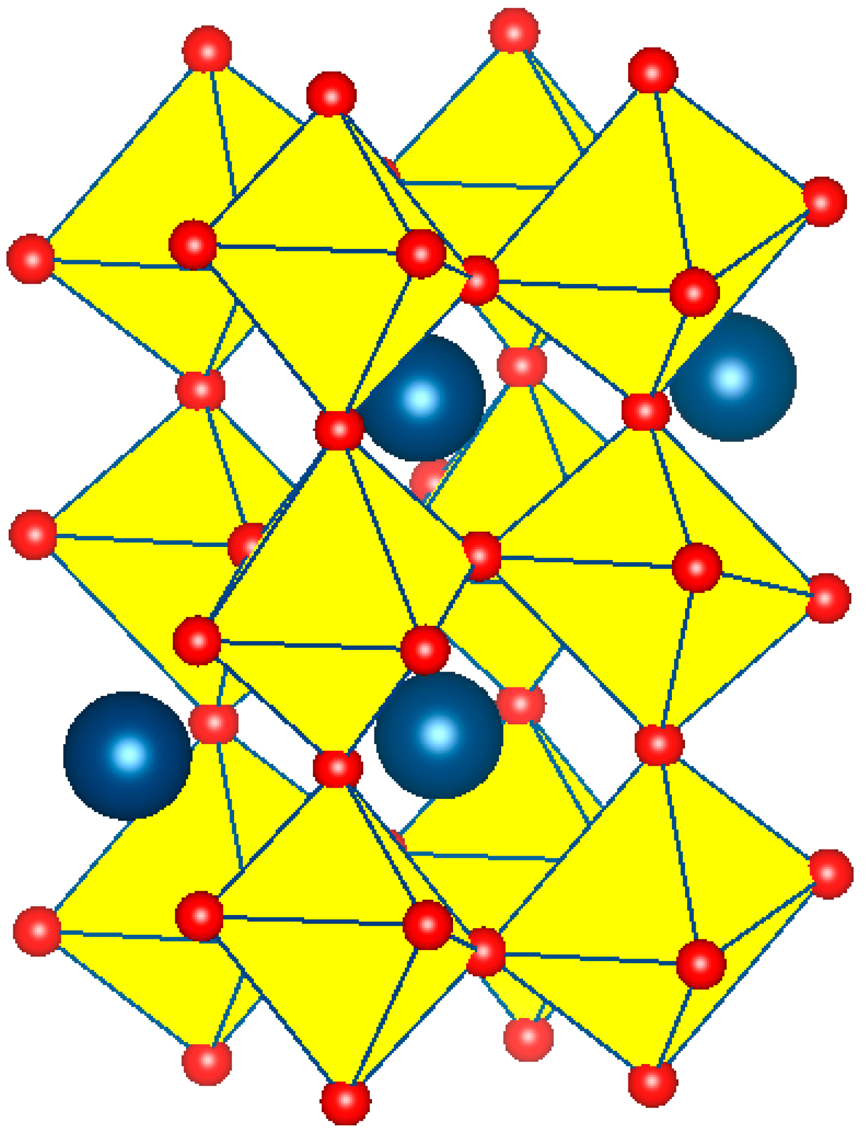
Figure 1. Perovskite crystal structure, where the A cation and oxygen ions are dark-blue and red spheres, and the B cation is in the octahedral center.
Inorganics 13 00372 g001
Figure 2. (a) Random, (b) rock salt, and (c) layered arrangements for DPs. For the random arrangement, both BO6 and B’O6 octahedra are depicted in green. In contrast, for the rock-salt and layered orderings, BO6 octahedra are shown in green, while B’O6 octahedra are depicted in light brown.
Inorganics 13 00372 g002
Figure 3. Observed, calculated, and difference XRD patterns of the La2−xBaxCoMnO6 for x of (a) 0.25, (b) 0.5, (c) 0.75, and (d) 1. Reprinted with permission from Ref. [56].Copyright 2025, Elsevier.
Inorganics 13 00372 g003
Figure 4. Observed, calculated, and difference XRD patterns of (a) LaNi0.5Ti0.5O3 and (b) La2NiTiO6. Reprinted from Ref. [8].
Inorganics 13 00372 g004
Figure 5. Initial procedure for refining the structure of a DP.
Inorganics 13 00372 g005
Figure 6. Background refinement through a trial-and-error method.
Inorganics 13 00372 g006
Figure 7. Refinement of unit cell parameters.
Inorganics 13 00372 g007
Figure 8. Order of refinement of U, V, W, and X parameters.
Inorganics 13 00372 g008
Figure 9. Order of refinement of atomic coordinates for a material containing La, Ni, Ti, and O and an orthorhombic symmetry.
Inorganics 13 00372 g009
Figure 10. (a) Graph generation for the observed and calculated XRD pattern using Fullprof (version 3.0). (b) Crystallite size and strain procedure in a PCR file for an orthorhombic symmetry with space group Pbnm. (c) The a- and b-axes of the orange-dashed ellipse correspond to the axes of the elongated hypothetical particle.
Inorganics 13 00372 g010
Figure 11. TEM micrograph of reduced La1.85NiRuO6. Reprinted from Ref. [67]. The white-dashed circles indicate Ni-Ru nanoparticles.
Inorganics 13 00372 g011
Figure 12. TEM image of Tb2CoMnO6. Reprinted from Ref. [92].
Inorganics 13 00372 g012
Figure 13. TEM micrograph of the Lu2FeMnO6. Reprinted from Ref. [94].
Inorganics 13 00372 g013
Figure 14. XPS survey scan of the La2MnNiO6. XPS spectra of (a) La 3d, (b) Mn 2p, and (c) Ni 2p. Reprinted/adapted with permission from Ref. [11].
Inorganics 13 00372 g014
Disclaimer/Publisher’s Note: The statements, opinions and data contained in all publications are solely those of the individual author(s) and contributor(s) and not of MDPI and/or the editor(s). MDPI and/or the editor(s) disclaim responsibility for any injury to people or property resulting from any ideas, methods, instructions or products referred to in the content.