Generation of Uniform X-ray Illumination and Its Application to X-ray Diffraction Microscopy


Abstract
1. Introduction
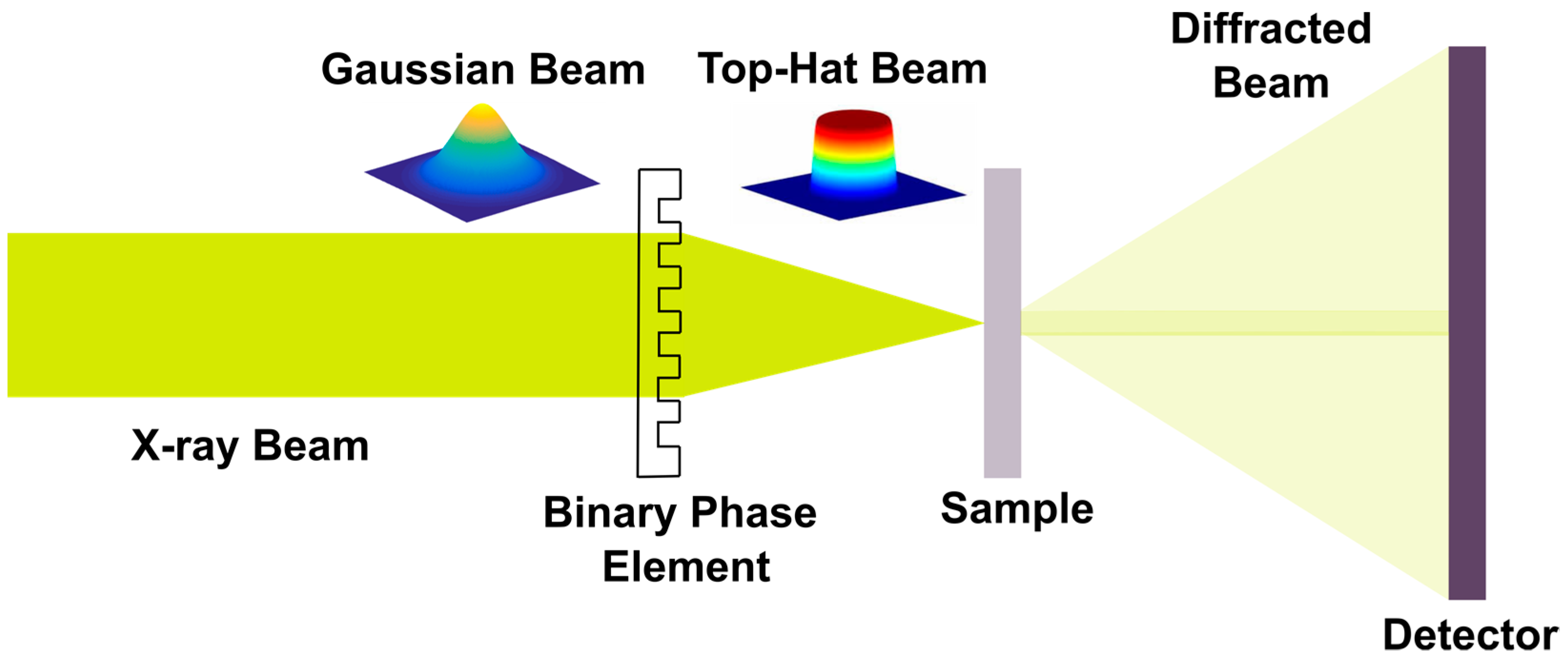
2. Experimental Setup and Design of the Phase Grating
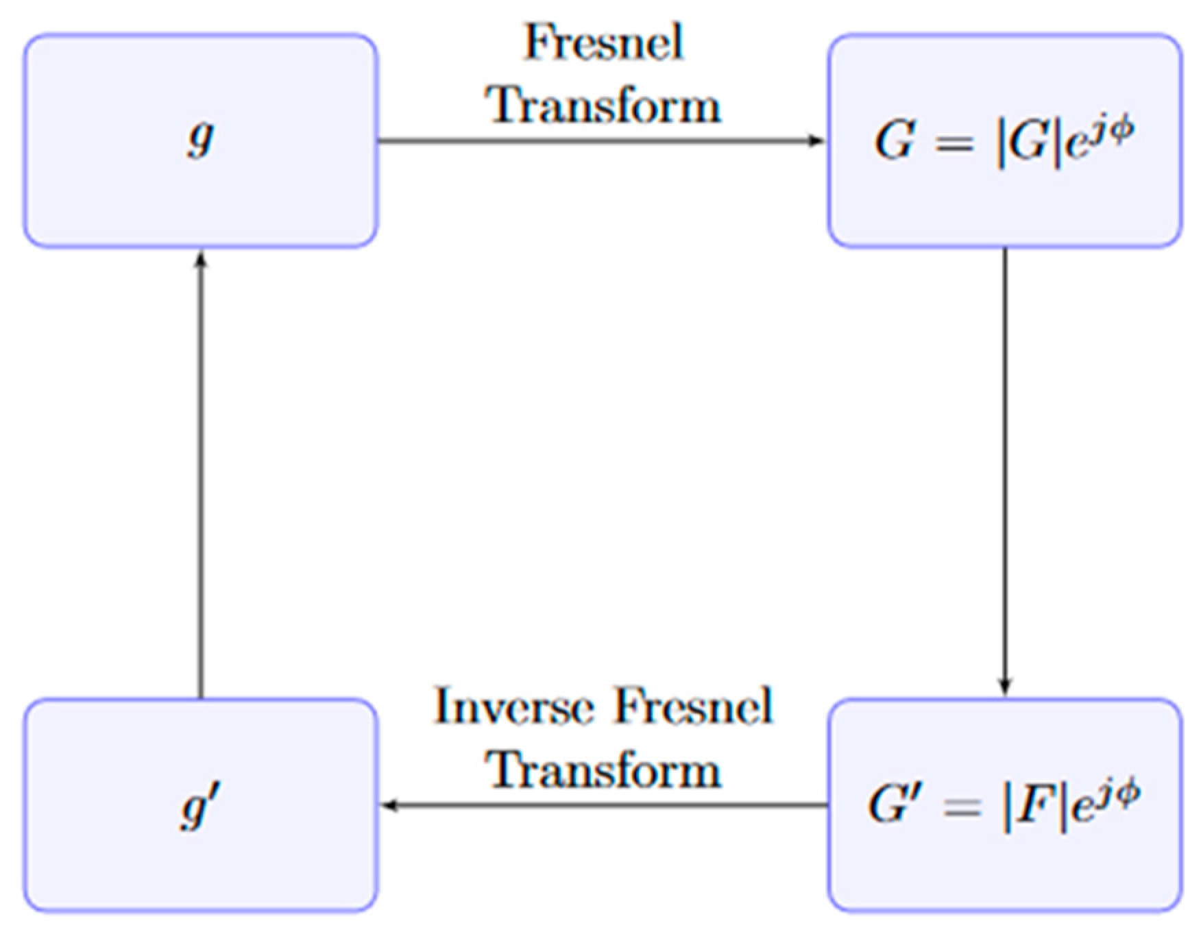
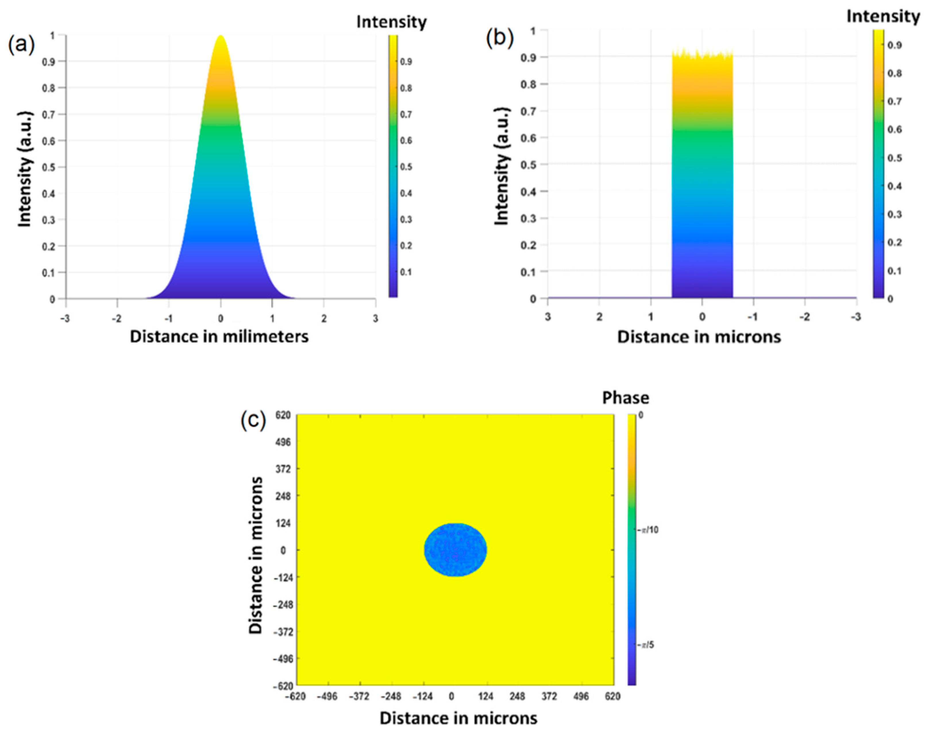
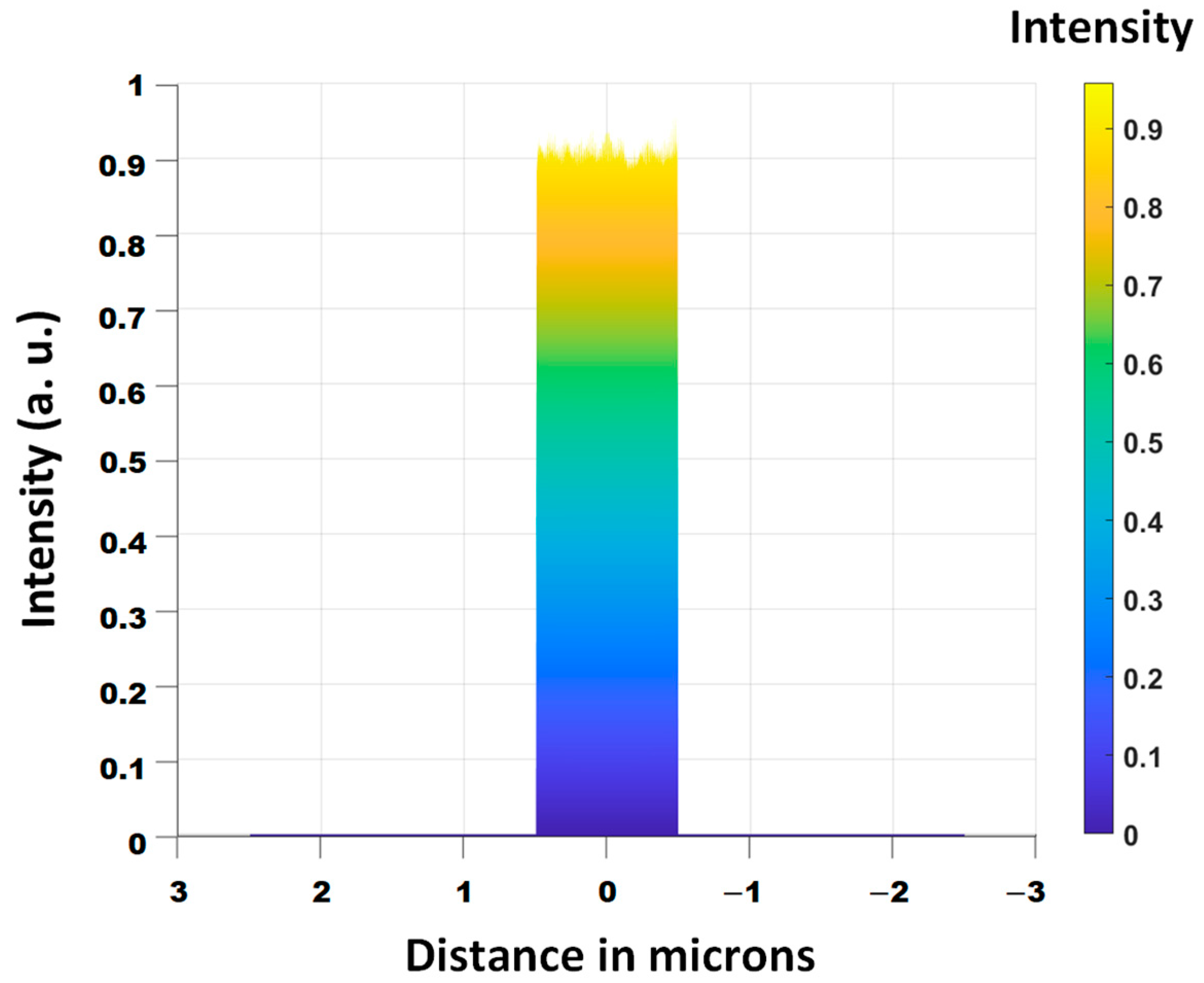
2.1. Design of the Optical Element
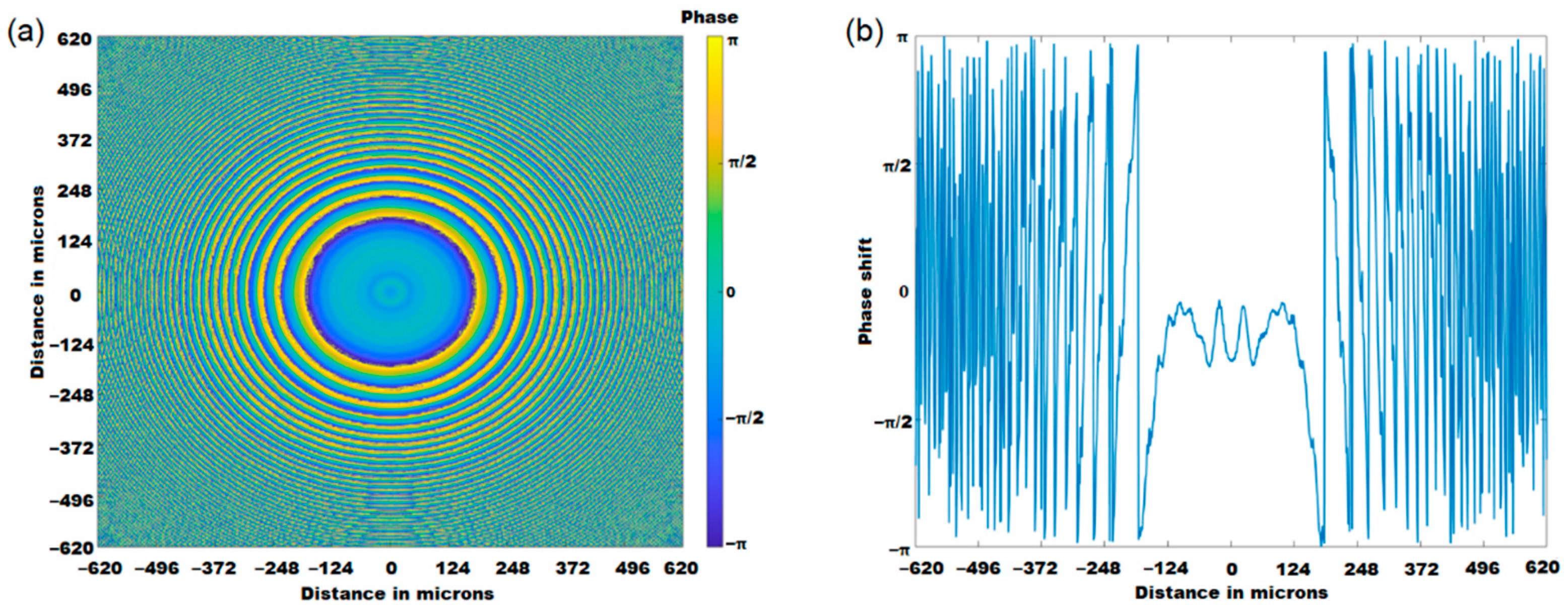
2.2. Description of the Optical Grating
3. Coherent Diffraction Imaging of Extended Objects
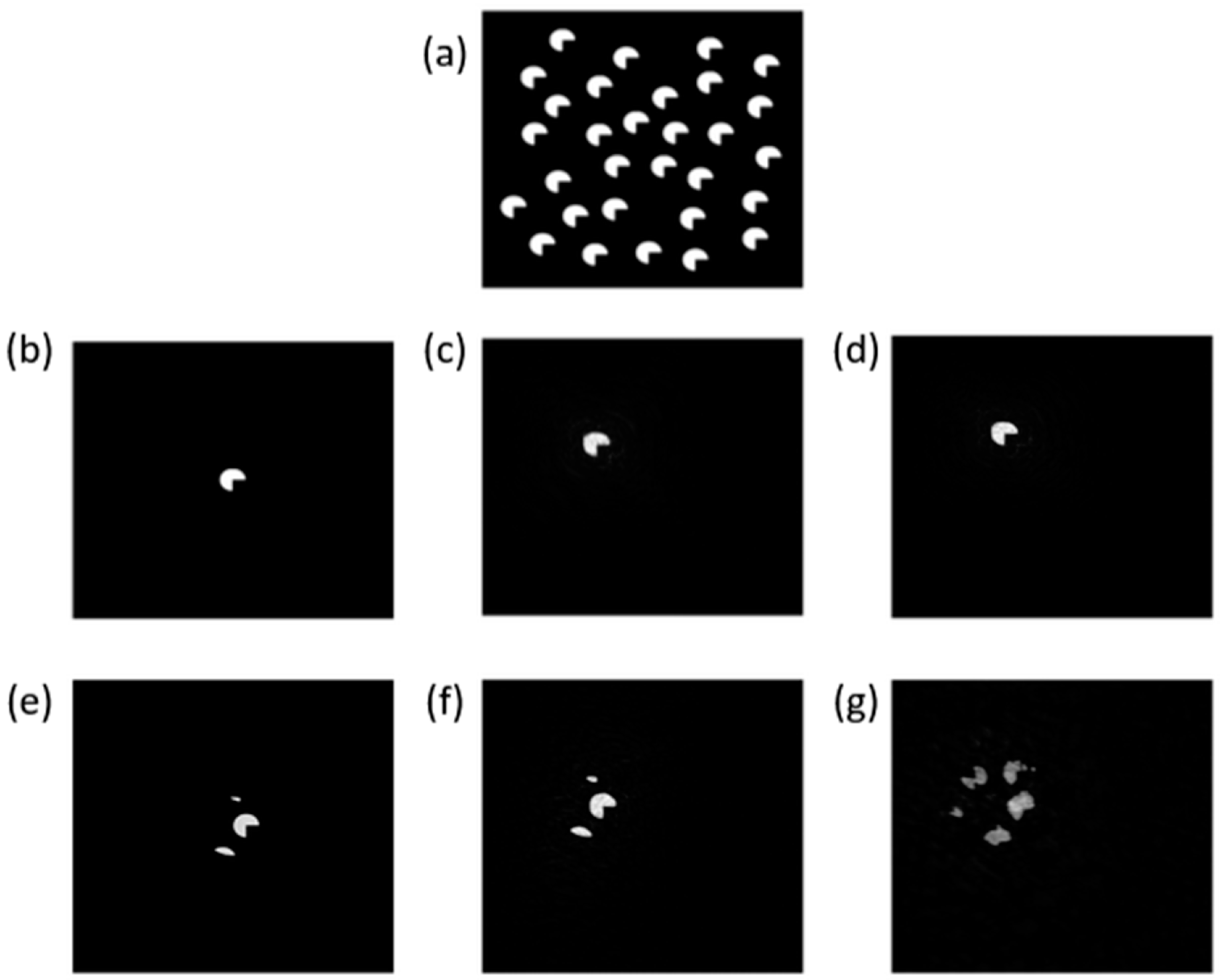
4. A Study of the Hit-Rate Improvement with the Top-Hat Beam
5. Conclusions
Author Contributions
Funding
Institutional Review Board Statement
Informed Consent Statement
Data Availability Statement
Conflicts of Interest
References
- Miao, J.; Charalambous, P.; Kirz, J.; Sayre, D. Extending the methodology of X-ray crystallography to allow imaging of micrometre-sized non-crystalline specimens. Nature 1999, 400, 342–344. [Google Scholar] [CrossRef]
- Miao, J.; Hodgson, K.O.; Ishikawa, T.; Larabell, C.A.; LeGros, M.A.; Nishino, Y. Imaging whole Escherichia coli bacteria by using single-particle X-ray diffraction. Proc. Natl. Acad. Sci. USA 2003, 100, 110–112. [Google Scholar] [CrossRef] [PubMed]
- Nishino, Y.; Takahashi, Y.; Imamoto, N.; Ishikawa, T.; Maeshima, K. Three-dimensional visualization of a human chromosome using coherent X-ray diffraction. Phys. Rev. Lett. 2009, 102, 018101. [Google Scholar] [CrossRef] [PubMed]
- Seibert, M.M.; Ekeberg, T.; Maia, F.R.N.C.; Svenda, M.; Andreasson, J.; Jönsson, O.; Odić, D.; Iwan, B.; Rocker, A.; Westphal, D.; et al. Single mimivirus particles intercepted and imaged with an X-ray laser. Nature 2011, 470, 78–81. [Google Scholar] [CrossRef]
- Ekeberg, T.; Svenda, M.; Abergel, C.; Maia, F.R.; Seltzer, V.; Claverie, J.M.; Hantke, M.; Jönsson, O.; Nettelblad, C.; van der Schot, G.; et al. Three-dimensional reconstruction of the giant mimivirus particle with an X-ray free-electron laser. Phys. Rev. Lett. 2015, 114, 098102. [Google Scholar] [CrossRef]
- Zuo, J.M.; Vartanyants, I.; Gao, M.; Zhang, R.; Nagahara, L.A. Atomic resolution imaging of a carbon nanotube from diffraction intensities. Science 2003, 300, 1419–1421. [Google Scholar] [CrossRef]
- Latychevskaia, T.; Longchamp, J.-N.; Fink, H.-W. When holography meets coherent diffraction imaging. Opt. Express 2012, 20, 28871–28892. [Google Scholar] [CrossRef]
- Marathe, S.; Kim, S.S.; Kim, S.N.; Kim, C.; Kang, H.C.; Nickles, P.V.; Noh, D.Y. Coherent diffraction surface imaging in reflection geometry. Opt. Express 2010, 18, 7253–7262. [Google Scholar] [CrossRef]
- Khakurel, K.P.; Kimura, T.; Joti, Y.; Matsuyama, S.; Yamauchi, K.; Nishino, Y. Coherent diffraction imaging of non-isolated object with apodized illumination. Opt. Express 2015, 23, 28182–28190. [Google Scholar] [CrossRef]
- Neutze, R.; Wouts, R.; van der Spoel, D.; Weckert, E.; Hajdu, J. Potential for biomolecular imaging with femtosecond X-ray pulses. Nature 2000, 406, 752–757. [Google Scholar] [CrossRef]
- Fienup, J.R. Phase retrieval algorithms: A comparison. Appl. Opt. 1982, 21, 2758–2769. [Google Scholar] [CrossRef] [PubMed]
- Marchesini, S.; He, H.; Chapman, H.N.; Hau-Riege, S.P.; Noy, A.; Howells, M.R.; Weierstall, U.; Spence, J.C.H. X-ray image reconstruction from a diffraction pattern alone. Phys. Rev. B 2003, 68, 140101. [Google Scholar] [CrossRef]
- Luke, D.R. Relaxed averaged alternating reflections for diffraction imaging. Inverse Probl. 2005, 21, 37–50. [Google Scholar] [CrossRef]
- Rodenburg, J.M.; Hurst, A.C.; Cullis, A.G.; Dobson, B.R.; Pfeiffer, F.; Bunk, O.; David, C.; Jefimovs, K.; Johnson, I. Hard-X-Ray Lensless Imaging of Extended Objects. Phys. Rev. Lett. 2007, 98, 034801. [Google Scholar] [CrossRef] [PubMed]
- Thibault, P.; Dierolf, M.; Menzel, A.; Bunk, O.; David, C.; Pfeiffer, F. High-resolution scanning X-ray diffraction microscopy. Science 2008, 321, 379–382. [Google Scholar] [CrossRef]
- Pfeiffer, F. X-ray ptychography. Nat. Photonics 2018, 12, 9–17. [Google Scholar] [CrossRef]
- Baksh, P.D.; Ostrčil, M.; Miszczak, M.; Pooley, C.; Chapman, R.T.; Wyatt, A.S.; Springate, E.; Chad, J.E.; Deinhardt, K.; Frey, J.G.; et al. Quantitative and correlative extreme ultraviolet coherent imaging of mouse hippocampal neurons at high resolution. Sci. Adv. 2020, 6, eaaz3025. [Google Scholar] [CrossRef]
- Tanksalvala, M.; Porter, C.L.; Esashi, Y.; Wang, B.; Jenkins, N.W.; Zhang, Z.; Miley, G.P.; Knobloch, J.L.; McBennett, B.; Horiguchi, N.; et al. Nondestructive, high-resolution, chemically specific 3D nanostructure characterization using phase-sensitive EUV imaging reflectometry. Sci. Adv. 2021, 7, eabd9667. [Google Scholar] [CrossRef]
- Eschen, W.; Loetgering, L.; Schuster, V.; Klas, R.; Kirsche, A.; Berthold, L.; Steinert, M.; Pertsch, T.; Gross, H.; Krause, M.; et al. Material-specific high-resolution table-top extreme ultraviolet microscopy. Light Sci. Appl. 2022, 11, 117. [Google Scholar] [CrossRef]
- Brooks, N.J.; Wang, B.; Binnie, I.; Tanksalvala, M.; Esashi, Y.; Knobloch, J.L.; Nguyen, Q.L.; McBennett, B.; Jenkins, N.W.; Gui, G.; et al. Temporal and spectral multiplexing for EUV multibeam ptychography with a high harmonic light source. Opt. Exp. 2022, 30, 30331–30346. [Google Scholar] [CrossRef]
- Abbey, B.; Nugent, K.; Williams, G.J.; Clark, J.N.; Peele, A.G.; Pfeifer, M.A.; De Jonge, M.; McNulty, I. Keyhole coherent diffractive imaging. Nat. Phys. 2008, 4, 394–398. [Google Scholar] [CrossRef]
- Khakurel, K.P.; Kimura, T.; Nakamori, H.; Goto, T.; Matsuyama, S.; Sasaki, T.; Takei, M.; Kohmura, Y.; Ishikawa, T.; Yamauchi, K.; et al. Generation of apodized X-ray illumination and its application to scanning and diffraction microscopy. J. Synchrotron Radiat. 2017, 24, 142–149. [Google Scholar] [CrossRef]
- Zhang, F.; Chen, B.; Morrison, G.R.; Vila-Comamala, J.; Guizar-Sicairos, M.; Robinson, I.K. Phase retrieval by coherent modulation imaging. Nat. Commun. 2016, 7, 13367. [Google Scholar] [CrossRef] [PubMed]
- Levitan, A.L.; Keskinbora, K.; Sanli, U.T.; Weigand, M.; Comin, R. Single-frame far-field diffractive imaging with randomized illumination. Opt. Express 2020, 28, 37103–37117. [Google Scholar] [CrossRef]
- Jefimovs, K.; Vila-Comamala, J.; Stampanoni, M.; Kaulich, B.; David, C. Beam-shaping condenser lenses for full-field transmission X-ray microscopy. J. Synchrotron Radiat. 2008, 15, 106–108. [Google Scholar] [CrossRef]
- Vogt, U.; Lindblom, M.; Charalambous, P.; Kaulich, B.; Wilhein, T. Condenser for Koehler-like illumination in transmission X-ray microscopes at undulator sources. Opt. Lett. 2006, 31, 1465–1467. [Google Scholar] [CrossRef][Green Version]
- Reddy, A.N.K.; Pal, V.; Mahler, S.; Friesem, A.A.; Davidson, N. Flat-top laser beams over an extended range. J. Phys. Conf. Ser. 2019, 1410, 012126. [Google Scholar] [CrossRef]
- Mimura, H.; Handa, S.; Kimura, T.; Yumoto, H.; Yamakawa, D.; Yokoyama, H.; Matsuyama, S.; Inagaki, K.; Yamamura, K.; Sano, Y.; et al. Breaking the 10 nm barrier in hard-X-ray focusing. Nat. Phys. 2010, 6, 122–125. [Google Scholar] [CrossRef]
- Fienup, J.R. Reconstruction of an object from the modulus of its Fourier transform. Opt. Lett. 1978, 3, 27–29. [Google Scholar] [CrossRef]
- Liu, H.; Spence, J.C.H. XFEL data analysis for structural biology. Quant. Biol. 2016, 4, 159–176. [Google Scholar] [CrossRef]
- Maiden, A.M.; Rodenburg, J.M.; Humphry, M.J. Optical ptychography: A practical implementation with useful resolution. Opt. Lett. 2010, 35, 2585–2587. [Google Scholar] [CrossRef] [PubMed]


| Parameter | Value |
|---|---|
| X-ray wavelength | 1.24 Å |
| Propagation distance | 10−2 m |
| Input beam size HWHM | 1 mm |
| Top-hat beam size (diameter) | 10−6 m |
Publisher’s Note: MDPI stays neutral with regard to jurisdictional claims in published maps and institutional affiliations. |
© 2022 by the authors. Licensee MDPI, Basel, Switzerland. This article is an open access article distributed under the terms and conditions of the Creative Commons Attribution (CC BY) license (https://creativecommons.org/licenses/by/4.0/).
Share and Cite
Kunio, K.; Espinoza, S.; Khakurel, K.P. Generation of Uniform X-ray Illumination and Its Application to X-ray Diffraction Microscopy. Photonics 2022, 9, 934. https://doi.org/10.3390/photonics9120934
Kunio K, Espinoza S, Khakurel KP. Generation of Uniform X-ray Illumination and Its Application to X-ray Diffraction Microscopy. Photonics. 2022; 9(12):934. https://doi.org/10.3390/photonics9120934
Chicago/Turabian StyleKunio, Katarzyna, Shirly Espinoza, and Krishna P. Khakurel. 2022. "Generation of Uniform X-ray Illumination and Its Application to X-ray Diffraction Microscopy" Photonics 9, no. 12: 934. https://doi.org/10.3390/photonics9120934
APA StyleKunio, K., Espinoza, S., & Khakurel, K. P. (2022). Generation of Uniform X-ray Illumination and Its Application to X-ray Diffraction Microscopy. Photonics, 9(12), 934. https://doi.org/10.3390/photonics9120934