Article

Computational Design of Highly-Sensitive Graphene-Based Multilayer SPR Biosensor

by
Seyyed Mohammad Ghasem Mousavi-Kiasari
1,
Kamyar Rashidi
1,
Davood Fathi
1,*,
Hussein Taleb
1,
Seyed Mohammad Mirjalili
2 and
Vahid Faramarzi
1
1
Department of Electrical and Computer Engineering, Tarbiat Modares University (TMU), Tehran P.O. Box 14115-194, Iran
2
Department of Engineering Physics, Polytechnique Montréal, Montreal, QC H3C 3A7, Canada
*
Author to whom correspondence should be addressed.
Photonics 2022, 9(10), 688; https://doi.org/10.3390/photonics9100688
Submission received: 9 August 2022 / Revised: 7 September 2022 / Accepted: 13 September 2022 / Published: 24 September 2022
(This article belongs to the Section Biophotonics and Biomedical Optics)

Abstract

:
In this paper, we present a set of optimal graphene-based multilayer surface plasmon resonance (SPR) biosensors for highly sensitive detection of biomolecules. To optimize the biosensor structure, we employed a multi-objective gray wolf optimizer (MOGWO) to maximize the sensitivity and minimize the structure full width at half maximum (FWHM). The main advantages of the optimized structures are high sensitivity, low FWHM, as well as easy implementation. We developed an algorithm that enables us to achieve nine different optimized structures. The best sensitivity, FWHM and FOM are obtained equal to 264.6°/RIU (for the structure #5), 1.905° and 56.6/RIU (for the structure #8), respectively. The results of this paper pave the way for the development of highly-sensitive SPR biosensors.

1. Introduction

A surface plasmon (SP) is a collective oscillation of conductive electrons propagating along the surface of a metal-dielectric interface. When at a specific wavelength the wave vector of the incident transverse magnetic (TM) polarized light couples with the propagation constant of SPs, the wave vector matching condition is satisfied and the result is the surface plasmon resonance(SPR) phenomena [1]. SPR biosensors are a type of optical sensors that due to high reliability and efficiency have a high potential to be employed in various applications such as the label-free detection of biomolecules, environmental monitoring, and security, etc. [2,3,4,5] Compared to other biosensing mechanisms, SPR biosensors take the advantages of high reliability and real-time sensing with a high sensitivity [6].
Increasing the sensitivity as well as improving the figure of merit (FOM)—defined as the ratio of sensitivity over full width at half maximum (FWHM)—have been the center of attention for scientists and researchers in the field of biosensors.
A typical SPR biosensor is composed of a metallic thin film coated on a prism, and the sensing medium is located on the surface of the metallic layer. The metallic layer is usually made of gold or silver. Since gold exhibits a relatively high resistance to oxidation and corrosion, it is more commonly used as the metallic layer [7]. However, due to poor adhesion of biomolecules on the gold surface, the conventional SPR biosensors are not very sensitive to the presence of biomolecules. To overcome this challenge, graphene has been proposed as a biomolecular recognition element (BRE) for enhancing the surface adhesion and consequently improving the sensitivity of SPR biosensor [5]. Because of the π-stacking interaction between the graphene hexagonal cells and biomolecules with the carbon-based ring structure, adhesion of biomolecules on the surface of graphene would be stronger and more stable [8,9,10,11,12,13].
Wu et al. reported a highly sensitive graphene-based SPR biosensor and demonstrated that by adding ten layers of graphene to the gold thin film the sensitivity can be enhanced by 25% [7]. Choi et al. explored a graphene-on-silver substrate for sensitive SPR imaging biosensor [14]. Gupta et al. demonstrated an SPR biosensor by employing graphene and silicon to enhance the sensitivity [15]. Zenget et al. proposed an SPR biosensor using a graphene-MoS2 nanostructured to enhance the sensitivity of the sensor [16]. In another work, Leiming et al. introduced a highly sensitive SPR biosensor based on a MoS2/graphene-aluminum hybrid structure with a maximum sensitivity of 190.83°/RIU [1]. However, FWHM of this structure is as high as 19° that results in an increase in the SPR measurement error. Furthermore, due to the use of six layers of MoS2, this structure will be subjected to stress and impurities which degrades the properties of the MoS2 layers. Moreover, the realization of a uniform air gap is a technological challenge. To improve the sensing performance of plasmonic devices, the sensor based on a Metal-Insulator-Metal waveguide has been investigated with a maximum sensitivity of 700 nm/RIU and FOM of 21.9. The sensor consists of two simple resonant cavities having a square and circular shape, with the side coupled to an MIM bus waveguide [17]. Also, plasmonic biosensor platform based on a hyperbolic metamaterial has been proposed using a grating-coupling technique and sweeping the wavelength from visible to near infrared by fixing incident angle, which presented different extreme sensitivity modes with a maximum of 30,000 nm/RIU and a record FOM of 590. It has been shown that use of graphene and other layered materials for functionalization can broaden the range of metals which can be used for plasmonic biosensing and increases the sensitivity by several orders of magnitude [18]. Furthermore, hybrid heterostructures can be utilized for SPR applications that include both thin layers of oxides and 2D layered materials. Vasyl, et al. proposed a SPR-sensing hybrid heterostructures based on noble-metal/dielectric/graphene heterostructures to achieve significant amplitude and phase sensitivity improvement of SPR sensing to refractive index of environment. Using this approach, a sensitivity of 30,000 nm/RIU and a large FOM of ≈400–690 can be obtained by sweeping the wavelength at a fixed angle [19].
In this work we propose an optimization framework to optimize the physical structure of an SPR graphene-based biosensor with the aim of improving the sensitivity and FOM of the biosensor. This framework enables us to obtain a set of optimal structures for biosensing applications with different requirements.

2. Theoretical Background

The investigated structure of SPR biosensor is shown in Figure 1. This structure is composed of a prism, metal layer (gold or silver), interlayer (IL), and a graphene layer. Compared to conventional SPR biosensors, this structure has an IL embedded between gold and graphene layers. It will be seen later that adding an appropriate IL with the suitable refractive index and the optimized thickness can improve the sensitivity of the SPR biosensor. The sensing medium is water with a refractive index of 1.330. At the presence of biomolecules on the graphene surface, the refractive index on the graphene (biomolecule layer) experiences a local increase of 0.005 for a thickness of 100 nm, as depicted in Figure 1. As shown in this figure, the SPR biosensor is illuminated by a TM-polarized light with the incident angle of θ and the wavelength of 633 nm.
According to the Drude model, the refractive index of gold can be expressed as [16]
n = ( 1 λ 2 λ c λ p 2 ( λ c + i λ ) ) 1 / 2
where λp = 0.16826 µm and λc = 8.9342 µm are the plasma and collision wavelengths, respectively, and λ is the wavelength of the incident light in µm. In the visible range, the refractive index of graphene can be approximately expressed as n = 3 + iC1λ/3 where C1 ≅ 5.446/µm [20]. The optical constants ε = (n, k) of silver are set (0.0562, 4.2776), at λ = 633 nm [21].
The angle of the minimum reflection in the SPR reflectance spectrum is called the resonance angle (θres) that is dependent on the refractive index of the sensing medium. After adhesion of the biomolecules on the surface of the graphene layer, the refractive index undergoes a small change (∆ns), and consequently the resonance angle experiences a small change (∆θres). The sensitivity of the SPR biosensor and the related FOM are expressed as S = ∆θres/∆ns and FOM = S/FWHM.
We performed full-wave optical simulations using the finite element method (FEM) to investigate the reflectance (far-field response) of proposed structures, and radio frequency module with periodic boundary condition is used. The periodic structures which have repeated in x direction is applied to the unit cell on the respective x direction to simulate the real structure with an array of infinite unit cells. Mesh numbers for prism is (13 × 20), for gold or silver layer is (13 × 15), for inter layer is (13 × 15), for biomolecule layer is (13 × 15), and for top of biomolecule layer (50 × 13). Non-uniform mesh sizes are used to decrease the simulation time and memory during the simulation. Each simulation run takes approximately 20 min to complete. The minimum mesh size of 0.1 nm is assigned to the graphene boundaries and refined mesh size is used in other regions. Mesh sizes increase gradually outside the graphene with a mesh growth rate of 1.3. The accuracy of the calculations in the mesh generation process has been assured by performing simulations until convergence is achieved. In the simulations, the maximum mesh element size is 105 nm and a light source illuminates the structure from the top port. Scattering parameters of the proposed device in this work are calculated by using the finite element method. The scattering parameters not only can be used to derive the effective refractive index of the plasmonic sensors but also are employed to determine optical responses (i.e., transmission, reflection, or absorption spectra). The transmission and reflection coefficient spectra can be achieved by solving Maxwell equations and defined as Ti = |S21|2 and Ri = |S11|2, respectively. Therefore, the absorption spectrum can be obtained as Ai = 1 − Ti − Ri. In our numerical simulations, the graphene layer is modeled as a thin film with a thickness of 0.34 nm. A periodic port is chosen in the y-direction in order to illuminate the structure.

3. Multi-Objective Optimization Framework

Due to a large number of structural parameters, employing an optimization technique is beneficial for designing an SPR biosensor with the desired characteristics. There are two goals to be optimized and some restrictions should be taken into account in the optimization process. To find the optimal structures for the biosensor, the multi-objective gray wolf optimizer (MOGWO) method [22,23] is employed to maximize the sensitivity of the structure (S) and minimize the FWHM. It is worth mentioning that if the reflection spectrum of the biosensor is too broad, the error in the SPR measurement will be large [24]. Moreover, the multi-objective problems with more than one objective don’t have unique solutions, and several merit factors can be considered in order to achieve a group of optimal structures known as the Pareto-optimal solutions [25].
Our framework is composed of three main modules known as the parameter module, the constraint module, and the optimizer module. The structural parameters including the thickness of gold (tAu) or silver (tAg) layer the thickness of the Interlayer (tIL), the type of the prism (nprism), the material of the Interlayer (IL), and the number of graphene layers (NGr) are defined in the parameter module. nprism is determined by the type of prism material chosen as BK7, SF10 or 2S2G. At the operating wavelength of λ = 633 nm, the refractive index of BK7, SF10, and 2S2G are 1.5151, 1.723, and 2.358 respectively [1]. Ta2O5 and TiO2 and SiO2 with the refractive indices of 2.0945 + 0.0071732i and 2.39 and 1.46 + i1.66 × 10−3 are selected as the materials of ILs [26,27], which are defined in the parameter module. The limitations and range of device structural parameters are defined in the constraint module as 0 < Min(R) < 0.35, 20 nm < tAu and tAg < 80 nm, 1 < NGr < 20, 5 nm < tIL < 100 nm, S > 40°/RIU and FWHM < 25°. The optimizer and optimization objectives are defined in the optimizer module.
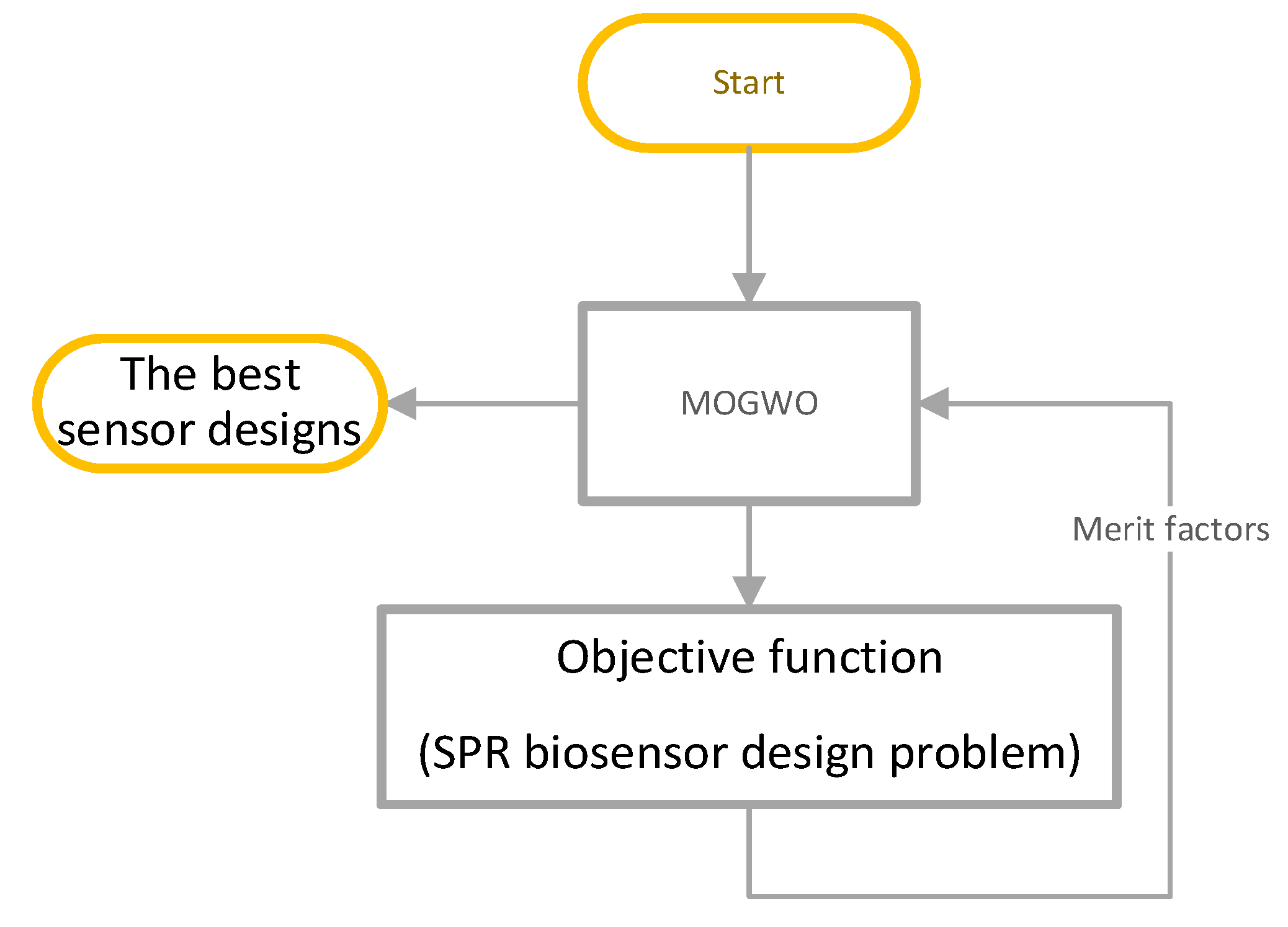
In order to implement the optimization module, we should use a powerful optimization method since there are a large number of structural parameters and the search space probably contains many local maximums. Also, the relationships between the structural parameters and two independent output merit factors (S, FWHM) are unknown. Therefore, we should deal with this optimization problem in a way that the SPR biosensor design problem is considered as a black box (the objective function). Thus, we choose MOGWO as the optimizer to solve the problem. It is worth mentioning here that the gradient descent optimization algorithm is not useful for this problem since this algorithm is prone to trapped in local optimums. The MOGWO simulates the leadership and collaboration mechanism of gray wolves for hunting prey [24,28,29] Also, MOGWO has a mechanism to escape from trapping in local optimums and has proven its excellent performance in various fields of engineering [30]. Therefore, by moving the MOGWO search agents toward the optimal solutions a set of optimal answers will be obtained. The overall fellow chart of how the MOGWO solves the problem of designing SPR biosensors is shown in Figure 2.
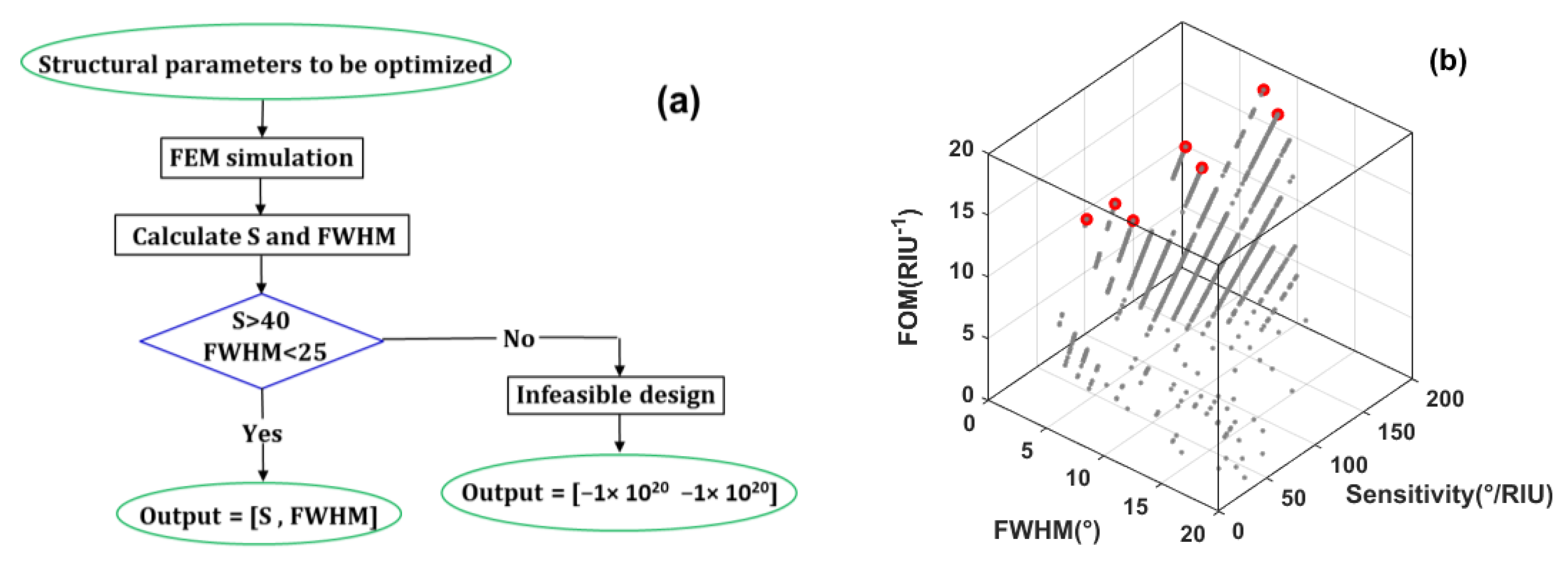
We used MOGWO with 60 search agents and maximum iteration of 200. The internal flowchart of the objective function is shown in Figure 3a. As seen in this figure, the numerical simulation is performed using the FEM for the candidate design, and the merit factors of the structure are evaluated. The candidate designs in which the constraints are not satisfied are removed during the optimization process by assigning a large negative number to their objectives.
When the metal layer is Au the optimization search history with 12,000 simulations is illustrated in Figure 3b, where 15 selected designs with optimum performance are marked with red color markers. Since some optimal designs have very close similarities, we have removed these similar designs and finally chosen four designs, the properties of which are given in Table 1. Also, we repeated these computations for Ag and finally 5 optimized structures have been selected.

4. Results and Discussion

The optimizer selects the optimal designs based on the best merit factors. A set of optimal structures is given in Table 1. In all these optimal structures NGr = 1. In other words, SPR biosensors with more than one graphene layers are not as optimal as the SPR with one layer. Our simulation results show that the sensitivity of the structure can be increased by increasing NGr. However, due to the increased FWHM of the structure, the SPR measurement error increases and consequently the optimal performance cannot be achieved. It is worth mentioning that all these optimal designs are desirable and each of them has a superior merit factor compared to other designs. These structures not only have high sensitivity to the presence of biomolecules in the sensing medium but also show narrow FWHM. Also, a Comparison between our proposed SPR biosensor with previously reported 2D materials based SPR sensors has been shown in Table 2.
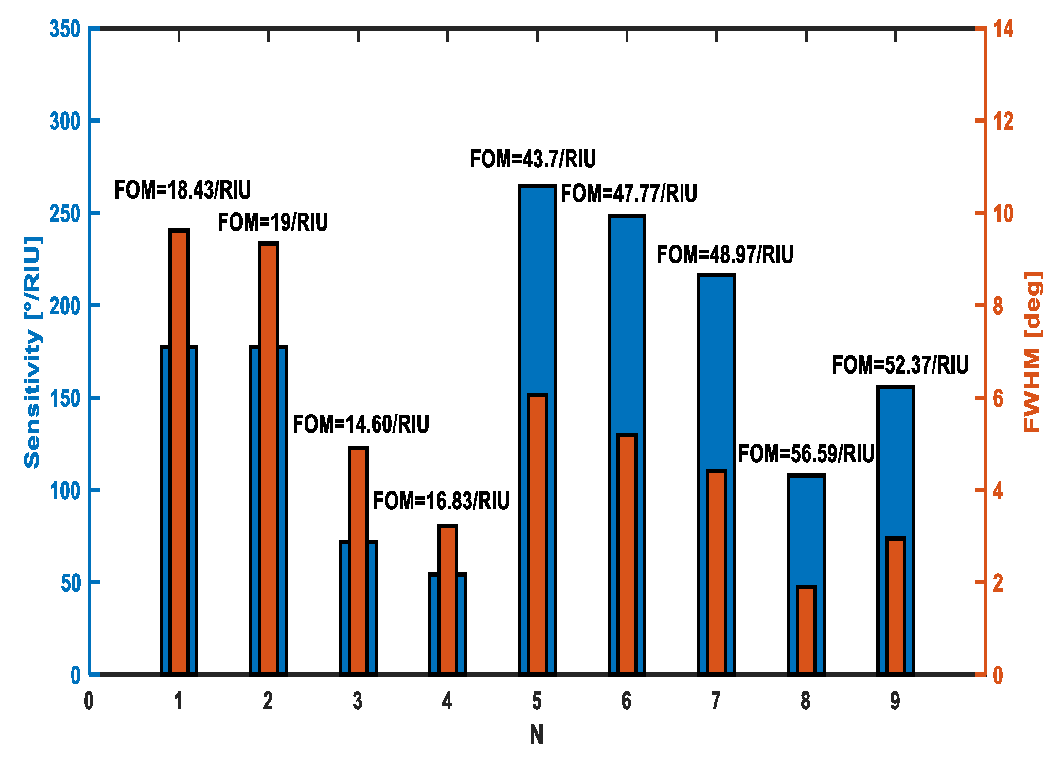
As can be seen in Table 1, structures #1 to #4 are optimal designs for gold metal layer. Structures #1 and #2 have more sensitivity than the other optimal designs. Structure #4 has the lowest FWHM compared to other optimal structures. Structure #3 has reasonable sensitivity and moderate FWHM. These results demonstrate that there is a trade-off between the sensitivity and FWHM of the SPR biosensors. A comparison between the sensitivity, FWHM, and FOM values of different optimal structures is illustrated in Figure 4. As seen in this figure, structure #2 has the highest FOM that demonstrates a good compromise between S and FWHM.
The increased FOM of structure #2 compared to #1 is a result of the increased thickness of the gold layer. With increasing the thickness of the gold layer, sufficient energy is absorbed into the structure which promotes a strong SPR excitation [16]. On the other hand, with a further increase in the thickness of the gold layer in structures #3 and #4, the sensitivity of the structure decreases due to the over-absorption of the gold layer [16]. In other words, the effect of the SPR enhancement is destroyed by the increased electron energy loss in the system. Furthermore, we found that the FWHM of the structure will be widened due to the increased loss in the gold layer.
The same behavior is obtained for silver. In structure 5 up to 8, as the thickness of silver increases, the FOM of the structure increases, and thru additional increase of silver layer thickness in structure 9, the FOM decreases due to the over-absorption of this layer.
One important point implied in Table 1 is that the refractive index of the prism has a huge impact on the sensitivity and FWHM of the SPR biosensor. The higher refractive index of SF10 compared to BK7 has improved the FWHM and degraded the sensitivity of the structure. This result is in agreement with what reported in the literature [1,34].
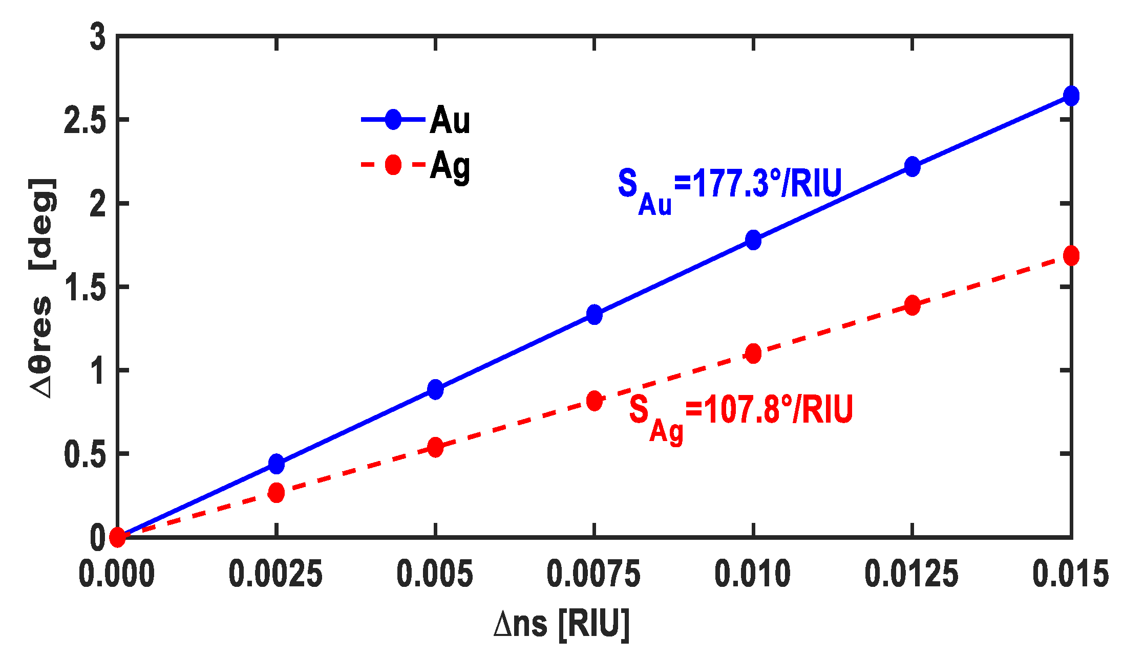
The linear behavior of the SPR biosensor is a very important factor for practical applications. The magnitude of the changes in the refractive index of the sensing medium (∆ns) caused by the adsorption of the biomolecules on the graphene surface is a function of the biomolecule density. Therefore, the linear variation of ∆θres versus ∆ns is a crucial factor for the SPR biosensors, as shown in Figure 5 for both Au and Ag layers. As seen in this figure, by introducing a small change of ∆ns, the resonance angle, θres, changes with a constant slope. The reason behind this behavior is that by changing the refractive index of the sensing medium the surface plasmon wavenumber (ksp) changes according to the excitation condition expressed as [16]
k sp = R e [ k 0 ( ε g i g ε s ε g i g + ε s ) 1 / 2 ]
where k0 is the free space wavenumber; εgig and εs are the relative permittivities of gold/Interlayer/graphene and the sensing medium, respectively. With the adhesion of biomolecules on the graphene surface, εs increases, and consequently ksp will be increased. Increased ksp requires a larger SPR angle according to [16]
k sp = k x   ,   k x = k 0   n p r i s m   s i n   ( θ r e s ) .
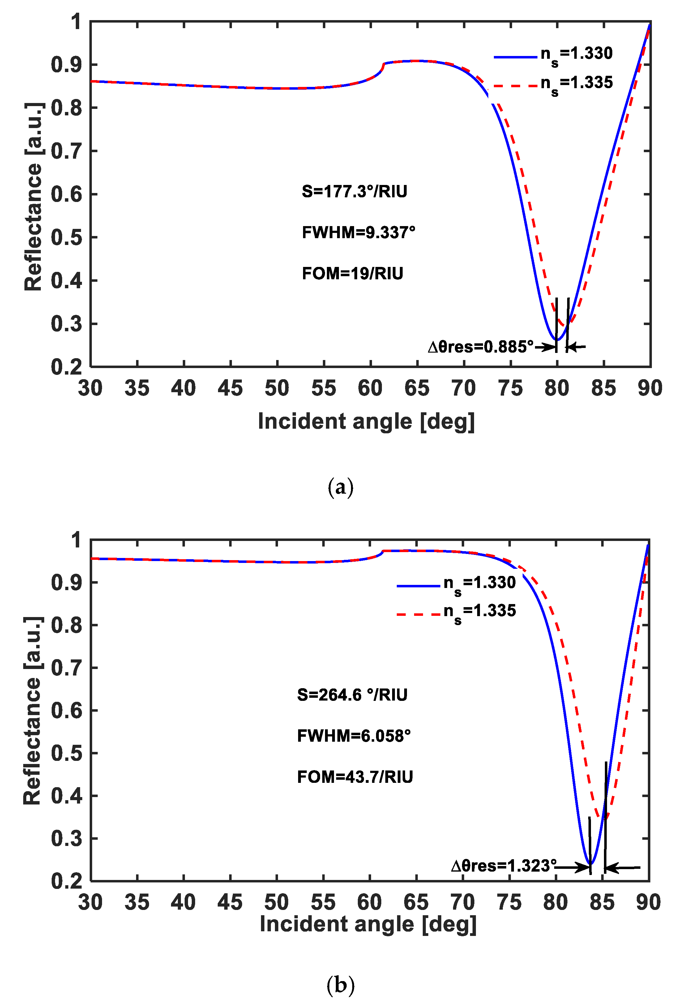
Figure 6 demonstrate the reflectance variation versus the incident angle, corresponding to the best sensitivity obtained in Table 1, for gold and silver layers.
Figure 5 demonstrate the reflectance variation versus the incident angle, corresponding to the best sensitivity obtained in Table 1, for gold and silver layers.

5. Conclusions

In summary, we employed a multi-objective optimization algorithm for designing SPR biosensors with high sensitivity and low FWHM. Our simulation results demonstrated that the proposed multi-objective algorithm can effectively optimize the performance of an SPR biosensor. In this way, nine optimal structures were achieved using the best figure of merits that can be selected based on the desired applications. The optimization algorithm does not need any initial design. This methodology enabled us to achieve a sensitivity as high as 264.6°/RIU and an FWHM as low as 1.905°. The proposed algorithm facilitates the development of high-performance SPR biosensors.

Author Contributions

Writing—original draft preparation and investigation, S.M.G.M.-K.; methodology, K.R.; writing—review and validation, H.T.; software, S.M.M.; visualization, V.F.; supervision, D.F. All authors have read and agreed to the published version of the manuscript.

Funding

This research received no external funding.

Institutional Review Board Statement

Not applicable.

Informed Consent Statement

Not applicable.

Data Availability Statement

All codes developed in this work will be made available from the corresponding author upon reasonable request.

Conflicts of Interest

The authors declare no conflict of interest.

References

  1. Wu, L.; Jia, Y.; Jiang, L.; Guo, J.; Dai, X.; Xiang, Y.; Fan, D. Sensitivity improved SPR biosensor based on the MoS2/graphene–aluminum hybrid structure. J. Lightwave Technol. 2017, 35, 82–87. [Google Scholar] [CrossRef]
  2. Huang, Y.H.; Ho, H.P.; Wu, S.Y.; Kong, S.K.; Wong, W.W.; Shum, P. Phase sensitive SPR sensor for wide dynamic range detection. Opt. Lett. 2011, 36, 4092–4094. [Google Scholar] [CrossRef] [PubMed]
  3. Liu, Y.; Liu, Q.; Chen, S.; Cheng, F.; Wang, H.; Peng, W. Surface Plasmon Resonance Biosensor Based on Smart Phone Platforms. Sci. Rep. 2015, 5, 12864. [Google Scholar] [CrossRef] [PubMed]
  4. Xu, Y.; Hsieh, C.-Y.; Wu, L.; Ang, L. Two-dimensional transition metal dichalcogenides mediated long range surface plasmon resonance biosensors. J. Phys. D Appl. Phys. 2018, 52, 065101. [Google Scholar] [CrossRef]
  5. Ouyang, Q.; Zeng, S.; Jiang, L.; Qu, J.; Dinh, X.Q.; Qian, J.; He, S.; Coquet, P.; Yong, K.T. Two-dimensional transition metal dichalcogenide enhanced phase-sensitive plasmonic biosensors: Theoretical insight. J. Phys. Chem. C 2017, 121, 6282–6289. [Google Scholar] [CrossRef]
  6. Johansen, K.; Arwin, H.; Lundström, I.; Liedberg, B. Imaging surface plasmon resonance sensor based on multiple wavelengths: Sensitivity considerations. Rev. Sci. Instrum. 2000, 71, 3530–3538. [Google Scholar] [CrossRef]
  7. Wu, L.; Chu, H.S.; Koh, W.S.; Li, E.P. Highly sensitive graphene biosensors based on surface plasmon resonance. Opt. Express 2010, 18, 14395–14400. [Google Scholar] [CrossRef] [PubMed]
  8. Song, B.; Li, D.; Qi, W.; Elstner, M.; Fan, C.; Fang, H. Graphene on Au(111): A Highly Conductive Material with Excellent Adsorption Properties for High-Resolution Bio/Nanodetection and Identification. ChemPhysChem 2010, 11, 585–589. [Google Scholar] [CrossRef] [PubMed]
  9. McGaughey, G.B.; Gagné, M.; Rappé, A.K. pi-Stacking interactions. Alive and well in proteins. J. Biol. Chem. 1998, 273, 15458–15463. [Google Scholar] [CrossRef] [PubMed]
  10. Tang, Z.; Wu, H.; Cort, J.R.; Buchko, G.W.; Zhang, Y.; Shao, Y.; Aksay, I.A.; Liu, J.; Lin, Y. Constraint of DNA on functionalized graphene improves its biostability and specificity. Small 2010, 6, 1205–1209. [Google Scholar] [CrossRef]
  11. Faramarzi, V.; Ahmadi, V.; Hwang, M.T.; Snapp, P. Highly sensitive crumpled 2D material-based plasmonic biosensors. Biomed. Opt. Express 2021, 12, 4544–4559. [Google Scholar] [CrossRef]
  12. Faramarzi, V.; Ahmadi, V.; Golmohamadi, F.G.; Fotouhi, B. A biosensor based on plasmonic wave excitation with diffractive grating structure. Sci. Iran. 2017, 24, 3441–3447. [Google Scholar] [CrossRef]
  13. Lei, Z.-L.; Guo, B. 2D material-based optical biosensor: Status and prospect. Adv. Sci. 2022, 9, 2102924. [Google Scholar] [CrossRef] [PubMed]
  14. Choi, S.H.; Kim, Y.L.; Byun, K.M. Graphene-on-silver substrates for sensitive surface plasmon resonance imaging biosensors. Opt. Express 2011, 19, 458–466. [Google Scholar] [CrossRef] [PubMed]
  15. Verma, R.; Gupta, B.D.; Jha, R. Sensitivity enhancement of a surface plasmon resonance based biomolecules sensor using graphene and silicon layers. Sens. Actuators B Chem. 2011, 160, 623–631. [Google Scholar] [CrossRef]
  16. Zeng, S.; Hu, S.; Xia, J.; Anderson, T.; Dinh, X.Q.; Meng, X.M.; Coquet, P.; Yong, K.T. Graphene-MoS2 hybrid nanostructures enhanced surface plasmon resonance biosensors. Sens. Actuators B 2015, 207, 801–810. [Google Scholar] [CrossRef]
  17. Kazanskiy, N.L.; Khonina, S.N.; Butt, M.A.; Kaźmierczak, A.; Piramidowicz, R. A numerical investigation of a plasmonic sensor based on a metal-insulator-metal waveguide for simultaneous detection of biological analytes and ambient temperature. Nanomaterials 2021, 11, 2551. [Google Scholar] [CrossRef] [PubMed]
  18. Wu, F.; Thomas, P.A.; Kravets, V.G.; Arola, H.O.; Soikkeli, M.; Iljin, K.; Kim, G.; Kim, M.; Shin, H.S.; Andreeva, D.V.; et al. Layered material platform for surface plasmon resonance biosensing. Sci. Rep. 2019, 9, 20286. [Google Scholar] [CrossRef] [PubMed]
  19. Kravets, V.G.; Wu, F.; Yu, T.; Grigorenko, A.N. Metal-dielectric-graphene hybrid heterostructures with enhanced surface plasmon resonance sensitivity based on amplitude and phase measurements. Plasmonics 2022, 17, 973–987. [Google Scholar] [CrossRef]
  20. Bruna, M.; Borini, S. Optical constants of graphene layers in the visible range. Appl. Phys. Lett. 2009, 94, 031901. [Google Scholar] [CrossRef]
  21. Johnson, P.B.; Christy, R.W. Optical Constants of the Noble Metals. Phys. Rev. B 1972, 6, 4370–4379. [Google Scholar] [CrossRef]
  22. Rashidi, K.; Mirjalili, S.M.; Taleb, H.; Fathi, D. Optimal Design of Large Mode Area Photonic Crystal Fibers Using a Multiobjective Gray Wolf Optimization Technique. J. Lightwave Technol. 2018, 36, 5626–5632. [Google Scholar] [CrossRef]
  23. Mirjalili, S.; Mirjalili, S.M.; Lewis, A. Grey wolf optimizer. Adv. Eng. Softw. 2014, 69, 46–61. [Google Scholar] [CrossRef]
  24. Maharana, P.K.; Jha, R. Chalcogenide prism and graphene multilayer based surface plasmon resonance affinity biosensor for high performance. Sens. Actuators B Chem. 2012, 169, 161–166. [Google Scholar] [CrossRef]
  25. Safdari, M.; Mirjalili, S.; Bianucci, P.; Zhang, X. Multi-objective optimization framework for designing photonic crystal sensors. Appl. Opt. 2018, 57, 1950. [Google Scholar] [CrossRef] [PubMed]
  26. Siefke, T.; Kroker, S.; Pfeiffer, K.; Puffky, O.; Dietrich, K.; Franta, D.; Ohlídal, I.; Szeghalmi, A.; Kley, E.-B.; Tünnermann, A. Materials Pushing the Application Limits of Wire Grid Polarizers further into the Deep Ultraviolet Spectral Range. Adv. Opt. Mater. 2016, 4, 1780–1786. [Google Scholar] [CrossRef]
  27. Marcos, L.V.R.; Larruquert, J.I.; Méndez, J.A.; Aznárez, J.A. Self-consistent optical constants of SiO2 and Ta2O5 films. Opt. Mater. Express 2016, 6, 3622–3637. [Google Scholar] [CrossRef]
  28. Mirjalili, S.; Saremi, S.; Mirjalili, S.M.; Coelho, L.d.S. Multi-objective grey wolf optimizer: A novel algorithm for multi-criterion optimization. Expert Syst. Appl. 2016, 47, 106–119. [Google Scholar] [CrossRef]
  29. Saremi, S.; Mirjalili, S.Z.; Mirjalili, S.M. Evolutionary population dynamics and grey wolf optimizer. Neural Comput. Appl. 2015, 26, 1257–1263. [Google Scholar] [CrossRef]
  30. Faris, H.; Aljarah, I.; Al-Betar, M.A.; Mirjalili, S. Grey wolf optimizer: A review of recent variants and applications. Neural Comput. Appl. 2018, 30, 413–435. [Google Scholar] [CrossRef]
  31. Sun, P.; Wang, M.; Liu, L.; Jiao, L.; Du, W.; Xia, F.; Liu, M.; Kong, W.; Dong, L.; Yun, M. Sensitivity enhancement of surface plasmon resonance biosensor based on graphene and barium titanate layers. Appl. Surf. Sci. 2019, 475, 342–347. [Google Scholar] [CrossRef]
  32. Mishra, A.K.; Mishra, S.K. Gas sensing in Kretschmann configuration utilizing bi-metallic layer of Rhodium-Silver in visible region. Sens. Actuators B Chem. 2016, 237, 969–973. [Google Scholar] [CrossRef]
  33. Ouyang, Q.; Zeng, S.; Jiang, L.; Hong, L.; Xu, G.; Dinh, X.-Q.; Qian, J.; He, S.; Qu, J.; Coquet, P.; et al. Sensitivity enhancement of transition metal dichalcogenides/silicon nanostructure-based surface plasmon resonance biosensor. Sci. Rep. 2016, 6, 28190. [Google Scholar] [CrossRef]
  34. Gupta, G.; Kondoh, J. Tuning and sensitivity enhancement of surface plasmon resonance sensor. Sens. Actuators B Chem. 2007, 122, 381–388. [Google Scholar] [CrossRef]
Figure 1. Schematic of the investigated SPR biosensor based on the metal layer/Interlayer/graphene layer structure and the Kretschmann configuration. The thickness of the biomolecule layer is assumed Z0 = 100 nm.
Photonics 09 00688 g001
Figure 2. Overall fellow chart of how the MOGWO solves the optimization problems.
Photonics 09 00688 g002
Figure 3. (a) objective function flowchart, (b) search history for Au.
Photonics 09 00688 g003
Figure 4. The sensitivity, FWHM and FOM for different optimized structures for Au (#1–#4) and Ag (#5–#9).
Photonics 09 00688 g004
Figure 5. Linear Variation of ∆θres with respect to ∆ns for the best FOM obtained in Table 2 for gold and silver layers.
Photonics 09 00688 g005
Figure 6. The reflectance characteristics versus the incident angle, corresponding to the best sensitivity obtained in Table 1, for (a) the gold layer and (b) the silver layer.
Photonics 09 00688 g006
Table 1. Structural parameters of optimal designs.
#Tmetal
(nm)
ILtIL
(nm)
PrismS
(°/RIU)
FWHM (°)FOM
1Au = 53TiO25BK7177.39.62218.43
2Au = 56TiO25BK7177.39.33719
3Au = 63TiO25SF1071.694.90914.60
4Au = 62SiO26SF1054.273.22516.83
5Ag = 52Ta2O516BK7264.66.05843.7
6Ag = 55Ta2O516BK7248.45.247.77
7Ag = 56Ta2O515BK7216.24.41548.97
8Ag = 58Ta2O58BK7107.81.90556.6
9Ag = 59Ta2O513BK7155.82.9552.37
Table 2. Comparison between our proposed SPR biosensor with previously reported 2D materials based SPR sensors.
StructureWavelength (nm)S
(°/RIU)
FWHM
(°)
FOM
(1/RIU)
REF
Ag (45 nm)-BaTiO3(10 nm)-graphene (1 L)6332575.70545.05[31]
RH (10 nm)-
Ag (45 nm)-
Si (14 nm)-graphene (1 L)
63222010.20421.56[32]
Au (35 nm)-Si (7 nm)-WS2 (1 L)600155.6817.4648.914[33]
Au (40 nm)-Si (5 nm)-WS2 (13 L)785127.9015.5238.239[33]
Au (40 nm)-Si (7 nm)-graphene (2 L)633134.6017.9757.488[15]
Air (35 nm)-MoS2(6 L)-AL (35 nm)-
Graphene (1 L)
633190.831910.05[1]
This work633107.81.90556.59
This work633216.24.41548.97
This work633264.66.05843.7
Publisher’s Note: MDPI stays neutral with regard to jurisdictional claims in published maps and institutional affiliations.