Surface Enhanced Raman Spectroscopy: Applications in Agriculture and Food Safety


Abstract
1. Introduction
2. Raman Spectroscopy and Surface Enhanced Raman Scattering (SERS)
2.1. Localized Surface Plasmon Resonance (LSPR)—Electromagnetic Enhancement
2.2. Chemical Enhancement
2.3. Enhancement Factor (EF)
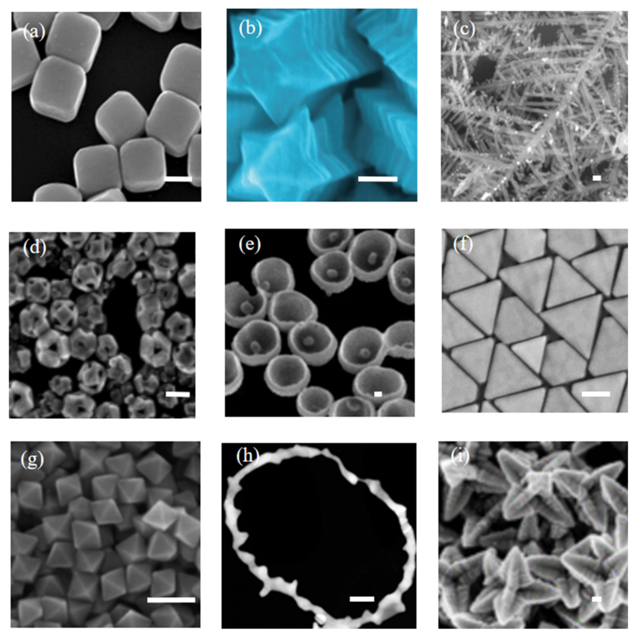
3. Fabrication of SERS-Active Substrates
3.1. Bottom-Up Assembly
3.2. Top-Down Synthesis
4. Chemical Functionalization of SERS Substrates
5. Application of SERS in Agri-Food
5.1. Detection of Pesticide Residues

| Organophosphates (Matrix) | SERS Substrate | LOD (Reported) | LOD (Normalised) | Excitation Wavelength | |
|---|---|---|---|---|---|
| Phosmet | Ooling tea [204] | Ag NPs | 0.1 mg/kg | 0.1 ppm | 633 nm |
| fruit [205] | multi-walled carbon nanotubes | 0.5 mg/kg | 0.5 ppm | 785 nm | |
| paddy water [206] | Au nanorods | 0.25 mg/L | 0.25 ppm | Portable Raman spectrometer, 785 nm | |
| fruit skin [207] | polyurethane-Ag NPs | 0.6 µg/mL | 0.6 ppb | 785 nm | |
| Plant Surfaces [208] | polyurethane micelle/Ag NP | 0.08 g/mL | 80 ppm | / | |
| Parathion-methyl | fruit or vegetable peels [209] | Snowflake-like Au NPs | 0.026 ng/cm | / | 638 nm |
| solvent [210] | Ag NP decorated ZnO-nanorods | 10−8 M | 2.63 ppb | 532 nm | |
| solvent [211] | nanoporous structure | 12 ppb | 12 ppb | 785 nm | |
| Malathion | solvent [212] | nanostructured Ag | 10 nM | 3.30 ppb | 632.8 nm |
| Chlorpyrifos | tomato surface [183] | Ag colloid | 10−9 mol/L | 0.35 ppb | 638 nm |
| soil [213] | Au NP | 10 ppm | 10 ppm | 785 nm | |
| fruits [214] | Ag NP | 10 ng/mL | 10 ppb | 633 nm | |
5.2. Detection of Chemical Additives
5.2.1. Dye Molecules
5.2.2. Melamine
6. Conclusions and Perspective
Author Contributions
Funding
Institutional Review Board Statement
Informed Consent Statement
Conflicts of Interest
References
- Food Agriculture Organization. The Future of Food and Agriculture—Trends and Challenges; United Nations: Rome, Italy, 2017. [Google Scholar]
- Henry, M.; Beguin, M.; Requier, F.; Rollin, O.; Odoux, J.-F.; Aupinel, P.; Aptel, J.; Tchamitchian, S.; Decourtye, A. A common pesticide decreases foraging success and survival in honey bees. Science 2012, 336, 348–350. [Google Scholar] [CrossRef]
- Gong, T.X.; Huang, Y.F.; Wei, Z.J.; Huang, W.; Wei, X.B.; Zhang, X.S. Magnetic assembled 3D SERS substrate for sensitive detection of pesticide residue in soil. Nanotechnology 2020, 31, 205501. [Google Scholar] [CrossRef]
- Guselnikova, O.; Postnikov, P.; Elashnikov, R.; Miliutina, E.; Svorcik, V.; Lyutakov, O. Metal-organic framework (MOF-5) coated SERS active gold gratings: A platform for the selective detection of organic contaminants in soil. Anal. Chim. Acta 2019, 1068, 70–79. [Google Scholar] [CrossRef]
- Krafft, B.; Tycova, A.; Urban, R.D.; Dusny, C.; Belder, D. Microfluidic device for concentration and SERS-based detection of bacteria in drinking water. Electrophoresis 2021, 42, 86–94. [Google Scholar] [CrossRef] [PubMed]
- Zhu, C.H.; Zhao, Q.S.; Meng, G.W.; Wang, X.J.; Hu, X.Y.; Han, F.M.; Lei, Y. Silver nanoparticle-assembled micro-bowl arrays for sensitive SERS detection of pesticide residue. Nanotechnology 2020, 31, 126300. [Google Scholar] [CrossRef] [PubMed]
- Wang, K.; Sun, D.-W.; Pu, H.; Wei, Q. Polymer multilayers enabled stable and flexible Au@Ag nanoparticle array for nondestructive SERS detection of pesticide residues. Talanta 2021, 223, 205303. [Google Scholar] [CrossRef] [PubMed]
- Grubisha, D.S.; Lipert, R.J.; Park, H.-Y.; Driskell, J.; Porter, M.D. Femtomolar Detection of Prostate-Specific Antigen: An Immunoassay Based on Surface-Enhanced Raman Scattering and Immunogold Labels. Anal. Chem. 2003, 75, 5936–5943. [Google Scholar] [CrossRef] [PubMed]
- Fan, M.; Andrade, G.F.S.; Brolo, A.G. A review on the fabrication of substrates for surface enhanced Raman spectroscopy and their applications in analytical chemistry. Anal. Chim. Acta 2011, 693, 7–25. [Google Scholar] [CrossRef] [PubMed]
- Justin, L.A.; Jeremy, D.D.; Ralph, A.T.; Yiping, Z. Current Progress on Surface-Enhanced Raman Scattering Chemical/Biological Sensing. In Functional Nanoparticles for Bioanalysis, Nanomedicine, and Bioelectronic Devices Volume 2; American Chemical Society: Washington, DC, USA, 2012; Volume 1113, pp. 235–272. [Google Scholar]
- Xie, W.; Schlucker, S. Medical applications of surface-enhanced Raman scattering. Phys. Chem. Chem. Phys. 2013, 15, 5329–5344. [Google Scholar] [CrossRef]
- Willets, K.A. Probing nanoscale interfaces with electrochemical surface-enhanced Raman scattering. Curr. Opin. Electrochem. 2019, 13, 18–24. [Google Scholar] [CrossRef]
- Craig, A.P.; Franca, A.S.; Irudayaraj, J. Surface-Enhanced Raman Spectroscopy Applied to Food Safety. Annu. Rev. Food Sci. Technol. 2013, 4, 369–380. [Google Scholar] [CrossRef]
- Fan, C.; Hu, Z.Q.; Mustapha, A.; Lin, M.S. Rapid detection of food- and waterborne bacteria using surface-enhanced Raman spectroscopy coupled with silver nanosubstrates. Appl. Microbiol. Biotechnol. 2011, 92, 1053–1061. [Google Scholar] [CrossRef]
- Zhang, Y.; Huang, Y.; Zhai, F.; Du, R.; Liu, Y.; Lai, K. Analyses of enrofloxacin, furazolidone and malachite green in fish products with surface-enhanced Raman spectroscopy. Food Chem. 2012, 135, 845–850. [Google Scholar] [CrossRef] [PubMed]
- Cialla, D.; Marz, A.; Bohme, R.; Theil, F.; Weber, K.; Schmitt, M.; Popp, J. Surface-enhanced Raman spectroscopy (SERS): Progress and trends. Anal. Bioanal. Chem. 2012, 403, 27–54. [Google Scholar] [CrossRef]
- Granger, J.H.; Schlotter, N.E.; Crawford, A.C.; Porter, M.D. Prospects for point-of-care pathogen diagnostics using surface-enhanced Raman scattering (SERS). Chem. Soc. Rev. 2016, 45, 3865–3882. [Google Scholar] [CrossRef] [PubMed]
- Jarvis, R.M.; Goodacre, R. Characterisation and identification of bacteria using SERS. Chem. Soc. Rev. 2008, 37, 931–936. [Google Scholar] [CrossRef] [PubMed]
- Laserna, J. Modern Techniques in Raman Spectroscopy; John Wiley and Sons: New York, NY, USA, 1996. [Google Scholar]
- Wen, P.; Yang, F.; Ge, C.; Li, S.; Xu, Y.; Chen, L. Self-assembled nano-Ag/Au@Au film composite SERS substrates show high uniformity and high enhancement factor for creatinine detection. Nanotechnology 2021, 32, 395502. [Google Scholar] [CrossRef]
- Kalantar-zadeh, K.; Fry, B. Nanotechnology-Enabled Sensors; Springer Science & Business Media: Berlin/Heidelberg, Germany, 2007. [Google Scholar]
- Settle, F.A. Handbook of Instrumental Techniques for Analytical Chemistry; Prentice Hall PTR: Hoboken, NJ, USA, 1997. [Google Scholar]
- Gardiner, D.J. Practical Raman Spectroscopy; Spinger: Berlin/Heidelberg, Germany, 1989. [Google Scholar]
- Jablonski, A. Efficiency of anti-Stokes fluorescence in dyes. Nature 1933, 131, 839–840. [Google Scholar] [CrossRef]
- Fleischmann, M.; Hendra, P.J.; McQuillan, A.J. Raman spectra of pyridine adsorbed at a silver electrode. Chem. Phys. Lett. 1974, 26, 163–166. [Google Scholar] [CrossRef]
- Gersten, J.I. The effect of surface roughness on surface enhanced Raman scattering. J. Chem. Phys. 1980, 72, 5779–5780. [Google Scholar] [CrossRef]
- Gersten, J.; Nitzan, A. Electromagnetic theory of enhanced Raman scattering by molecules adsorbed on rough surfaces. J. Chem. Phys. 1980, 73, 3023–3037. [Google Scholar] [CrossRef]
- Moskovits, M. Surface-enhanced spectroscopy. Rev. Mod. Phys. 1985, 57, 783–826. [Google Scholar] [CrossRef]
- Otto, A.; Mrozek, I.; Grabhorn, H.; Akemann, W. Surface-enhanced Raman scattering. J. Phys. Condens. Matter 1992, 4, 1143. [Google Scholar] [CrossRef]
- Stiles, P.L.; Dieringer, J.A.; Shah, N.C.; Duyne, R.P.V. Surface-Enhanced Raman Spectroscopy. Annu. Rev. Anal. Chem. 2008, 1, 601–626. [Google Scholar] [CrossRef]
- Hutter, E.; Fendler, J.H. Exploitation of Localized Surface Plasmon Resonance. Adv. Mater. 2004, 16, 1685–1706. [Google Scholar] [CrossRef]
- Camden, J.P.; Dieringer, J.A.; Wang, Y.; Masiello, D.J.; Marks, L.D.; Schatz, G.C.; Van Duyne, R.P. Probing the Structure of Single-Molecule Surface-Enhanced Raman Scattering Hot Spots. J. Am. Chem. Soc. 2008, 130, 12616–12617. [Google Scholar] [CrossRef]
- Sun, M.; Wan, S.; Liu, Y.; Jia, Y.; Xu, H. Chemical mechanism of surface-enhanced resonance Raman scattering via charge transfer in pyridine–Ag2 complex. J. Raman Spectrosc. 2008, 39, 402–408. [Google Scholar] [CrossRef]
- Adrian, F.J. Charge transfer effects in surface-enhanced Raman scatteringa. J. Chem. Phys. 1982, 77, 5302–5314. [Google Scholar] [CrossRef]
- Schatz, G.C. Theoretical studies of surface enhanced Raman scattering. Acc. Chem. Res. 1984, 17, 370–376. [Google Scholar] [CrossRef]
- Campion, A.; Ivanecky, J., III; Child, C.; Foster, M. On the mechanism of chemical enhancement in surface-enhanced Raman scattering. J. Am. Chem. Soc. 1995, 117, 11807–11808. [Google Scholar] [CrossRef]
- Le Ru, E.C.; Blackie, E.; Meyer, M.; Etchegoin, P.G. Surface Enhanced Raman Scattering Enhancement Factors: A Comprehensive Study. J. Phys. Chem. C 2007, 111, 13794–13803. [Google Scholar] [CrossRef]
- Weaver, M.J.; Zou, S.; Chan, H.Y.H. Peer Reviewed: The New Interfacial Ubiquity of Surface-Enhanced Raman Spectroscopy. Anal. Chem. 2000, 72, 38A–47A. [Google Scholar] [CrossRef]
- Etchegoin, P.G.; Le Ru, E.C. A perspective on single molecule SERS: Current status and future challenges. Phys. Chem. Chem. Phys. 2008, 10, 6079–6089. [Google Scholar] [CrossRef] [PubMed]
- Park, W.H.; Kim, Z.H. Charge Transfer Enhancement in the SERS of a Single Molecule. Nano Lett. 2010, 10, 4040–4048. [Google Scholar] [CrossRef]
- Bell, S.E.; McCourt, M.R. SERS enhancement by aggregated Au colloids: Effect of particle size. Phys. Chem. Chem. Phys. 2009, 11, 7455–7462. [Google Scholar] [CrossRef]
- Lee, S.J.; Guan, Z.; Xu, H.; Moskovits, M. Surface-Enhanced Raman Spectroscopy and Nanogeometry: The Plasmonic Origin of SERS. J. Phys. Chem. C 2007, 111, 17985–17988. [Google Scholar] [CrossRef]
- Lancaster, C.A.; Scholl, W.E.; Ticknor, M.A.; Shumaker-Parry, J.S. Uniting Top-Down and Bottom-Up Strategies Using Fabricated Nanostructures as Hosts for Synthesis of Nanomites. J. Phys. Chem. C 2020, 124, 6822–6829. [Google Scholar] [CrossRef]
- Banholzer, M.J.; Millstone, J.E.; Qin, L.; Mirkin, C.A. Rationally designed nanostructures for surface-enhanced Raman spectroscopy. Chem. Soc. Rev. 2008, 37, 885–897. [Google Scholar] [CrossRef]
- Pan, X.-T.; Liu, Y.-Y.; Qian, S.-Q.; Yang, J.-M.; Li, Y.; Gao, J.; Liu, C.-G.; Wang, K.; Xia, X.-H. Free-Standing Single Ag Nanowires for Multifunctional Optical Probes. ACS Appl. Mater. Interfaces 2021, 13, 19023–19030. [Google Scholar] [CrossRef] [PubMed]
- Xu, K.-X.; Chen, X.; Huang, Z.; Chen, Z.-N.; Chen, J.; Sun, J.-J.; Fang, Y.; Li, J.-F. Ligand-Free Fabrication of Ag Nanoassemblies for Highly Sensitive and Reproducible Surface-Enhanced Raman Scattering Sensing of Antibiotics. ACS Appl. Mater. Interfaces 2021, 13, 1766–1772. [Google Scholar] [CrossRef]
- Choi, S.; Jeong, H.; Choi, K.H.; Song, J.Y.; Kim, J. Electrodeposition of triangular Pd rod nanostructures and their electrocatalytic and SERS activities. ACS Appl. Mater. Interfaces 2014, 6, 3002–3007. [Google Scholar] [CrossRef]
- Ge, D.; Wei, J.; Ding, J.; Zhang, J.; Ma, C.; Wang, M.; Zhang, L.; Zhu, S. Silver Nano-Dendrite-Plated Porous Silicon Substrates Formed by Single-Step Electrochemical Synthesis for Surface-Enhanced Raman Scattering. ACS Appl. Nano Mater. 2020, 3, 3011–3018. [Google Scholar] [CrossRef]
- Jeong, H.; Kim, J. Electrodeposition of nanoflake Pd structures: Structure-dependent wettability and SERS activity. ACS Appl. Mater. Interfaces 2015, 7, 7129–7135. [Google Scholar] [CrossRef]
- Raveendran, J.; Stamplecoskie, K.G.; Docoslis, A. Tunable Fractal Nanostructures for Surface-Enhanced Raman Scattering via Templated Electrodeposition of Silver on Low-Energy Surfaces. ACS Appl. Nano Mater. 2020, 3, 2665–2679. [Google Scholar] [CrossRef]
- Longoni, M.; Zalaffi, M.S.; de Ferri, L.; Stortini, A.M.; Pojana, G.; Ugo, P. Surface Enhanced Raman Spectroscopy With Electrodeposited Copper Ultramicro-Wires With/Without Silver Nanostars Decoration. Nanomaterials 2021, 11, 518. [Google Scholar] [CrossRef]
- Grzelczak, M.; Vermant, J.; Furst, E.M.; Liz-Marzán, L.M. Directed Self-Assembly of Nanoparticles. ACS Nano 2010, 4, 3591–3605. [Google Scholar] [CrossRef] [PubMed]
- Zhu, S.; Fan, C.; Wang, J.; He, J.; Liang, E. Self-assembled Ag nanoparticles for surface enhanced Raman scattering. Opt. Rev. 2013, 20, 361–366. [Google Scholar] [CrossRef]
- Gao, T.; Wang, Y.; Wang, K.; Zhang, X.; Dui, J.; Li, G.; Lou, S.; Zhou, S. Controlled synthesis of homogeneous Ag nanosheet-assembled film for effective SERS substrate. ACS Appl. Mater. Interfaces 2013, 5, 7308–7314. [Google Scholar] [CrossRef]
- Zhang, L. Self-assembly Ag nanoparticle monolayer film as SERS Substrate for pesticide detection. Appl. Surf. Sci. 2013, 270, 292–294. [Google Scholar] [CrossRef]
- Wu, Y.; Dang, H.; Park, S.-G.; Chen, L.; Choo, J. SERS-PCR assays of SARS-CoV-2 target genes using Au nanoparticles-internalized Au nanodimple substrates. Biosens. Bioelectron. 2022, 197, 113736. [Google Scholar] [CrossRef] [PubMed]
- Lee, S.J.; Morrill, A.R.; Moskovits, M. Hot Spots in Silver Nanowire Bundles for Surface-Enhanced Raman Spectroscopy. J. Am. Chem. Soc. 2006, 128, 2200–2201. [Google Scholar] [CrossRef]
- Zheng, G.; Patolsky, F.; Cui, Y.; Wang, W.U.; Lieber, C.M. Multiplexed electrical detection of cancer markers with nanowire sensor arrays. Nat. Biotechnol. 2005, 23, 1294–1301. [Google Scholar] [CrossRef] [PubMed]
- Tang, J.; Zhao, Q.; Zhang, N.; Man, S.-Q. Facile fabrication of large-area and uniform silica nanospheres monolayer for efficient surface-enhanced Raman scattering. Appl. Surf. Sci. 2014, 308, 247–252. [Google Scholar] [CrossRef]
- Li, W.; Camargo, P.H.C.; Lu, X.; Xia, Y. Dimers of Silver Nanospheres: Facile Synthesis and Their Use as Hot Spots for Surface-Enhanced Raman Scattering. Nano Lett. 2009, 9, 485–490. [Google Scholar] [CrossRef] [PubMed]
- Martín, A.; Pescaglini, A.; Schopf, C.; Scardaci, V.; Coull, R.; Byrne, L.; Iacopino, D. Surface-Enhanced Raman Scattering of 4-Aminobenzenethiol on Au Nanorod Ordered Arrays. J. Phys. Chem. C 2014, 118, 13260–13267. [Google Scholar] [CrossRef]
- Martin, A.; Wang, J.J.; Iacopino, D. Flexible SERS active substrates from ordered vertical Au nanorod arrays. RSC Adv. 2014, 4, 20038–20043. [Google Scholar] [CrossRef]
- Xie, Z.; Tao, J.; Lu, Y.; Lin, K.; Yan, J.; Wang, P.; Ming, H. Polymer optical fiber SERS sensor with gold nanorods. Opt. Commun. 2009, 282, 439–442. [Google Scholar] [CrossRef]
- Oh, M.K.; Shin, Y.S.; Lee, C.L.; De, R.; Kang, H.; Yu, N.E.; Kim, B.H.; Kim, J.H.; Yang, J.K. Morphological and SERS Properties of Silver Nanorod Array Films Fabricated by Oblique Thermal Evaporation at Various Substrate Temperatures. Nanoscale Res. Lett. 2015, 10, 962. [Google Scholar] [CrossRef]
- Lovera, P.; Creedon, N.; Alatawi, H.; Mitchell, M.; Burke, M.; Quinn, A.J.; O’Riordan, A. Low-cost silver capped polystyrene nanotube arrays as super-hydrophobic substrates for SERS applications. Nanotechnology 2014, 25, 175502. [Google Scholar] [CrossRef]
- Lovera, P.; Creedon, N.; Alatawi, H.; O’Riordan, A. Metal Capped Polystyrene Nanotubes Arrays as Super-Hydrophobic Substrates for SERS Applications; SPIE: Washington, DC, USA, 2014; Volume 9129. [Google Scholar]
- Walker, D.A.; Browne, K.P.; Kowalczyk, B.; Grzybowski, B.A. Self-Assembly of Nanotriangle Superlattices Facilitated by Repulsive Electrostatic Interactions. Angew. Chem. Int. Ed. 2010, 49, 6760–6763. [Google Scholar] [CrossRef]
- Kumar-Krishnan, S.; Esparza, R.; Pal, U. Controlled Fabrication of Flower-Shaped Au-Cu Nanostructures Using a Deep Eutectic Solvent and Their Performance in Surface-Enhanced Raman Scattering-Based Molecular Sensing. ACS Omega 2020, 5, 3699–3708. [Google Scholar] [CrossRef]
- Liu, Z.; Yang, Z.; Peng, B.; Cao, C.; Zhang, C.; You, H.; Xiong, Q.; Li, Z.; Fang, J. Highly Sensitive, Uniform, and Reproducible Surface-Enhanced Raman Spectroscopy from Hollow Au-Ag Alloy Nanourchins. Adv. Mater. 2014, 26, 2431–2439. [Google Scholar] [CrossRef]
- Yang, M.; Alvarez-Puebla, R.; Kim, H.-S.; Aldeanueva-Potel, P.; Liz-Marzán, L.M.; Kotov, N.A. SERS-Active Gold Lace Nanoshells with Built-in Hotspots. Nano Lett. 2010, 10, 4013–4019. [Google Scholar] [CrossRef]
- Barbillon, G. Applications of Shell-Isolated Nanoparticle-Enhanced Raman Spectroscopy. Photonics 2021, 8, 46. [Google Scholar] [CrossRef]
- Barbillon, G.; Ivanov, A.; Sarychev, A.K. Hybrid Au/Si Disk-Shaped Nanoresonators on Gold Film for Amplified SERS Chemical Sensing. Nanomaterials 2019, 9, 1588. [Google Scholar] [CrossRef]
- Vigderman, L.; Zubarev, E.R. Starfruit-Shaped Gold Nanorods and Nanowires: Synthesis and SERS Characterization. Langmuir 2012, 28, 9034–9040. [Google Scholar] [CrossRef] [PubMed]
- Li, H.; Wang, Y.; Li, Y.; Zhang, J.; Qiao, Y.; Wang, Q.; Che, G. Fabrication of pollutant-resistance SERS imprinted sensors based on SiO2@TiO2@Ag composites for selective detection of pyrethroids in water. J. Phys. Chem. Solids 2020, 138, 109254. [Google Scholar] [CrossRef]
- Krishnan, S.K.; Chipatecua Godoy, Y. Deep Eutectic Solvent-Assisted Synthesis of Au Nanostars Supported on Graphene Oxide as an Efficient Substrate for SERS-Based Molecular Sensing. ACS Omega 2020, 5, 1384–1393. [Google Scholar] [CrossRef] [PubMed]
- Zhu, C.; Meng, G.; Huang, Q.; Huang, Z.; Chu, Z. Au Hierarchical Micro/Nanotower Arrays and Their Improved SERS Effect by Ag Nanoparticle Decoration. Cryst. Growth Des. 2011, 11, 748–752. [Google Scholar] [CrossRef]
- Subramanian, B.; Theriault, G.; Robichaud, J.; Tchoukanova, N.; Djaoued, Y. Large-area crack-free Au-SiO2 2D inverse opal composite films: Fabrication and SERS applications. Mater. Chem. Phys. 2020, 244, 122630. [Google Scholar] [CrossRef]
- Barbillon, G. Latest Novelties on Plasmonic and Non-Plasmonic Nanomaterials for SERS Sensing. Nanomaterials 2020, 10, 1200. [Google Scholar] [CrossRef]
- Sun, Y.; Xia, Y. Shape-controlled synthesis of gold and silver nanoparticles. Science 2002, 298, 2176–2179. [Google Scholar] [CrossRef]
- Chen, J.; McLellan, J.M.; Siekkinen, A.; Xiong, Y.; Li, Z.-Y.; Xia, Y. Facile Synthesis of Gold-Silver Nanocages with Controllable Pores on the Surface. J. Am. Chem. Soc. 2006, 128, 14776–14777. [Google Scholar] [CrossRef] [PubMed]
- Ye, J.; Van Dorpe, P.; Van Roy, W.; Borghs, G.; Maes, G. Fabrication, Characterization, and Optical Properties of Gold Nanobowl Submonolayer Structures. Langmuir 2009, 25, 1822–1827. [Google Scholar] [CrossRef] [PubMed]
- Liebig, F.; Sarhan, R.M.; Sander, M.; Koopman, W.; Schuetz, R.; Bargheer, M.; Koetz, J. Deposition of Gold Nanotriangles in Large Scale Close-Packed Monolayers for X-ray-Based Temperature Calibration and SERS Monitoring of Plasmon-Driven Catalytic Reactions. ACS Appl. Mater. Interfaces 2017, 9, 20247–20253. [Google Scholar] [CrossRef]
- Li, C.; Shuford, K.L.; Chen, M.; Lee, E.J.; Cho, S.O. A Facile Polyol Route to Uniform Gold Octahedra with Tailorable Size and Their Optical Properties. ACS Nano 2008, 2, 1760–1769. [Google Scholar] [CrossRef]
- Nguyen, P.-D.; Zhang, X.; Su, J. One-Step Controlled Synthesis of Size-Tunable Toroidal Gold Particles for Biochemical Sensing. ACS Appl. Nano Mater. 2019, 2, 7839–7847. [Google Scholar] [CrossRef]
- Qiu, Y.H.; Ding, S.J.; Lin, Y.J.; Chen, K.; Yang, D.J.; Ma, S.; Li, X.; Lin, H.Q.; Wang, J.; Wang, Q.Q. Growth of Au Hollow Stars and Harmonic Excitation Energy Transfer. ACS Nano 2020, 14, 736–745. [Google Scholar] [CrossRef] [PubMed]
- Maier, S.A.; Brongersma, M.L.; Kik, P.G.; Meltzer, S.; Requicha, A.A.; Atwater, H.A. Plasmonics—A route to nanoscale optical devices. Adv. Mater. 2001, 13, 1501–1505. [Google Scholar] [CrossRef]
- Alvarez-Puebla, R.; Liz-Marzán, L.M.; García de Abajo, F.J. Light Concentration at the Nanometer Scale. J. Phys. Chem. Lett. 2010, 1, 2428–2434. [Google Scholar] [CrossRef]
- Aroca, R.; Alvarez-Puebla, R.; Pieczonka, N.; Sanchez-Cortez, S.; Garcia-Ramos, J. Surface-enhanced Raman scattering on colloidal nanostructures. Adv. Colloid Interface Sci. 2005, 116, 45–61. [Google Scholar] [CrossRef]
- Lee, P.C.; Meisel, D. Adsorption and surface-enhanced Raman of dyes on silver and gold sols. J. Phys. Chem. 1982, 86, 3391–3395. [Google Scholar] [CrossRef]
- Rivas, L.; Sanchez-Cortes, S.; García-Ramos, J.V.; Morcillo, G. Mixed Silver/Gold Colloids: A Study of Their Formation, Morphology, and Surface-Enhanced Raman Activity. Langmuir 2000, 16, 9722–9728. [Google Scholar] [CrossRef]
- Faulds, K.; Littleford, R.E.; Graham, D.; Dent, G.; Smith, W.E. Comparison of Surface-Enhanced Resonance Raman Scattering from Unaggregated and Aggregated Nanoparticles. Anal. Chem. 2004, 76, 592–598. [Google Scholar] [CrossRef] [PubMed]
- Schopf, C.; Martín, A.; Burke, M.; Jones, D.; Pescaglini, A.; O’Riordan, A.; Quinn, A.J.; Iacopino, D. Au nanorod plasmonic superstructures obtained by a combined droplet evaporation and stamping method. J. Mater. Chem. C 2014, 2, 3536–3541. [Google Scholar] [CrossRef]
- Yang, J.K.; Kang, H.; Lee, H.; Jo, A.; Jeong, S.; Jeon, S.J.; Kim, H.I.; Lee, H.Y.; Jeong, D.H.; Kim, J.H.; et al. Single-Step and Rapid Growth of Silver Nanoshells as SERS-Active Nanostructures for Label-Free Detection of Pesticides. ACS Appl. Mater. Interfaces 2014, 6, 12541–12549. [Google Scholar] [CrossRef] [PubMed]
- Aroca, R.F.; Goulet, P.J.G.; dos Santos, D.S.; Alvarez-Puebla, R.A.; Oliveira, O.N. Silver Nanowire Layer-by-Layer Films as Substrates for Surface-Enhanced Raman Scattering. Anal. Chem. 2005, 77, 378–382. [Google Scholar] [CrossRef]
- Freeman, R.G.; Grabar, K.C.; Allison, K.J.; Bright, R.M.; Davis, J.A.; Guthrie, A.P.; Hommer, M.B.; Jackson, M.A.; Smith, P.C.; Walter, D.G.; et al. Self-assembled metal colloid monolayers: An approach to SERS substrates. Science 1995, 267, 1629–1632. [Google Scholar] [CrossRef] [PubMed]
- Wang, Z.; Pan, S.; Krauss, T.D.; Du, H.; Rothberg, L.J. The structural basis for giant enhancement enabling single-molecule Raman scattering. Proc. Natl. Acad. Sci. USA 2003, 100, 8638–8643. [Google Scholar] [CrossRef]
- Fan, M.; Brolo, A.G. Silver nanoparticles self assembly as SERS substrates with near single molecule detection limit. Phys. Chem. Chem. Phys. 2009, 11, 7381–7389. [Google Scholar] [CrossRef]
- Yu, W.W.; White, I.M. A simple filter-based approach to surface enhanced Raman spectroscopy for trace chemical detection. Analyst 2012, 137, 1168–1173. [Google Scholar] [CrossRef] [PubMed]
- Shiohara, A.; Langer, J.; Polavarapu, L.; Liz-Marzan, L.M. Solution processed polydimethylsiloxane/gold nanostar flexible substrates for plasmonic sensing. Nanoscale 2014, 6, 9817–9823. [Google Scholar] [CrossRef]
- Polavarapu, L.; La Porta, A.; Novikov, S.M.; Coronado-Puchau, M.; Liz-Marzan, L.M. Pen-on-Paper Approach Toward the Design of Universal Surface Enhanced Raman Scattering Substrates. Small 2014, 10, 3065–3071. [Google Scholar] [CrossRef] [PubMed]
- Lee, C.H.; Tian, L.; Singamaneni, S. Paper-based SERS swab for rapid trace detection on real-world surfaces. ACS Appl. Mater. Interfaces 2010, 2, 3429–3435. [Google Scholar] [CrossRef] [PubMed]
- Chen, J.; Huang, Y.; Kannan, P.; Zhang, L.; Lin, Z.; Zhang, J.; Chen, T.; Guo, L. Flexible and Adhesive Surface Enhance Raman Scattering Active Tape for Rapid Detection of Pesticide Residues in Fruits and Vegetables. Anal. Chem. 2016, 88, 2149–2155. [Google Scholar] [CrossRef]
- Kahl, M.; Voges, E.; Kostrewa, S.; Viets, C.; Hill, W. Periodically structured metallic substrates for SERS. Sens. Actuators B Chem. 1998, 51, 285–291. [Google Scholar] [CrossRef]
- Yu, Q.; Guan, P.; Qin, D.; Golden, G.; Wallace, P.M. Inverted Size-Dependence of Surface-Enhanced Raman Scattering on Gold Nanohole and Nanodisk Arrays. Nano Lett. 2008, 8, 1923–1928. [Google Scholar] [CrossRef]
- Wu, T.; Lin, Y.-W. Surface-enhanced Raman scattering active gold nanoparticle/nanohole arrays fabricated through electron beam lithography. Appl. Surf. Sci. 2018, 435, 1143–1149. [Google Scholar] [CrossRef]
- Krishnamoorthy, S.; Krishnan, S.; Thoniyot, P.; Low, H.Y. Inherently Reproducible Fabrication of Plasmonic Nanoparticle Arrays for SERS by Combining Nanoimprint and Copolymer Lithography. ACS Appl. Mater. Interfaces 2011, 3, 1033–1040. [Google Scholar] [CrossRef]
- Robinson, C.; Justice, J.; Petäjä, J.; Karppinen, M.; Corbett, B.; O’Riordan, A.; Lovera, P. Nanoimprint Lithography–Based Fabrication of Plasmonic Array of Elliptical Nanoholes for Dual-Wavelength, Dual-Polarisation Refractive Index Sensing. Plasmonics 2019, 14, 951–959. [Google Scholar] [CrossRef]
- Yang, J.; Li, J.; Du, Z.; Teng, J.; Hong, M. Laser Hybrid Micro/nano-structuring of Si Surfaces in Air and its Applications for SERS Detection. Sci. Rep. 2014, 4, 6657. [Google Scholar] [CrossRef] [PubMed]
- Sun, W.; Hong, R.; Liu, Q.; Li, Z.; Shi, J.; Tao, C.; Zhang, D. SERS-active Ag–Al alloy nanoparticles with tunable surface plasmon resonance induced by laser ablation. Opt. Mater. 2019, 96, 109298. [Google Scholar] [CrossRef]
- Bian, X.; Xu, J.; Yang, J.; Chiu, K.-L.; Jiang, S. Flexible Ag SERS substrate for non-destructive and rapid detection of toxic materials on irregular surface. Surf. Interfaces 2021, 23, 100995. [Google Scholar] [CrossRef]
- Zhou, Q.; Thokchom, A.K.; Kim, D.-J.; Kim, T. Inkjet-printed Ag micro-/nanostructure clusters on Cu substrates for in-situ pre-concentration and surface-enhanced Raman scattering. Sens. Actuators B Chem. 2017, 243, 176–183. [Google Scholar] [CrossRef]
- Huebner, U.; Boucher, R.; Schneidewind, H.; Cialla, D.; Popp, J. Microfabricated SERS-arrays with sharp-edged metallic nanostructures. Microelectron. Eng. 2008, 85, 1792–1794. [Google Scholar] [CrossRef]
- Yang, J.-Y.; Park, S.-G.; Jung, S.; Byeon, E.-Y.; Kim, D.-G.; Jung, H.S.; Kim, H.J.; Lee, S. SERS substrates based on self-organized dimple nanostructures on polyethylene naphthalate films produced via oxygen ion beam sputtering. Appl. Surf. Sci. 2022, 572, 151452. [Google Scholar] [CrossRef]
- Muhammad, M.; Shao, C.-S.; Huang, Q. Aptamer-functionalized Au nanoparticles array as the effective SERS biosensor for label-free detection of interleukin-6 in serum. Sens. Actuators B Chem. 2021, 334, 129607. [Google Scholar] [CrossRef]
- Zhang, C.; Yi, P.; Peng, L.; Lai, X.; Chen, J.; Huang, M.; Ni, J. Continuous fabrication of nanostructure arrays for flexible surface enhanced Raman scattering substrate. Sci. Rep. 2017, 7, 39814. [Google Scholar] [CrossRef] [PubMed]
- Shi, G.; Wang, M.; Zhu, Y.; Yan, X.; Pan, S.; Zhang, A. Nanoflower-like Ag/AAO SERS platform with quasi-photonic crystal nanostructure for efficient detection of goat serum. Curr. Appl. Phys. 2019, 19, 1276–1285. [Google Scholar] [CrossRef]
- Li, M.; Wu, J.; Wang, C.; Fang, J. The cascade structure of periodic micro/nanoscale Au nano-islands @ Ag-frustum arrays as effective SERS substrates. Vacuum 2020, 175, 109265. [Google Scholar] [CrossRef]
- Lin, B.; Kannan, P.; Qiu, B.; Lin, Z.; Guo, L. On-spot surface enhanced Raman scattering detection of Aflatoxin B1 in peanut extracts using gold nanobipyramids evenly trapped into the AAO nanoholes. Food Chem. 2020, 307, 125528. [Google Scholar] [CrossRef]
- Hu, M.; Ou, F.S.; Wu, W.; Naumov, I.; Li, X.; Bratkovsky, A.M.; Williams, R.S.; Li, Z. Gold Nanofingers for Molecule Trapping and Detection. J. Am. Chem. Soc. 2010, 132, 12820–12822. [Google Scholar] [CrossRef]
- Diebold, E.D.; Mack, N.H.; Doorn, S.K.; Mazur, E. Femtosecond Laser-Nanostructured Substrates for Surface-Enhanced Raman Scattering. Langmuir 2009, 25, 1790–1794. [Google Scholar] [CrossRef]
- Aleknavičienė, I.; Pabrėža, E.; Talaikis, M.; Jankunec, M.; Račiukaitis, G. Low-cost SERS substratefeaturing laser-ablated amorphous nanostructure. Appl. Surf. Sci. 2022, 571, 151248. [Google Scholar] [CrossRef]
- Restaino, S.M.; White, I.M. A critical review of flexible and porous SERS sensors for analytical chemistry at the point-of-sample. Anal. Chim. Acta 2019, 1060, 17–29. [Google Scholar] [CrossRef]
- Li, L.; Yang, S.Y.; Duan, J.L.; Huang, L.; Xiao, G.N. Fabrication and SERS performance of silver nanoarrays by inkjet printing silver nanoparticles ink on the gratings of compact disc recordable. Spectrochim. Acta A—Mol. Biomol. Spectrosc. 2020, 225, 117598. [Google Scholar] [CrossRef]
- Wu, J.; Zhang, L.; Huang, F.; Ji, X.; Dai, H.; Wu, W. Surface enhanced Raman scattering substrate for the detection of explosives: Construction strategy and dimensional effect. J. Hazard. Mater. 2020, 387, 121714. [Google Scholar] [CrossRef] [PubMed]
- Yu, B.; Ge, M.; Li, P.; Xie, Q.; Yang, L. Development of surface-enhanced Raman spectroscopy application for determination of illicit drugs: Towards a practical sensor. Talanta 2019, 191, 1–10. [Google Scholar] [PubMed]
- D’Apuzzo, F.; Sengupta, R.N.; Overbay, M.; Aronoff, J.S.; Rogacs, A.; Barcelo, S.J. A Generalizable Single-Chip Calibration Method for Highly Quantitative SERS via Inkjet Dispense. Anal. Chem. 2020, 92, 1372–1378. [Google Scholar] [PubMed]
- Joshi, P.; Santhanam, V. Inkjet-Based Fabrication Process with Control over the Morphology of SERS-Active Silver Nanostructures. Ind. Eng. Chem. Res. 2018, 57, 5250–5258. [Google Scholar] [CrossRef]
- Kumar, A.; Santhanam, V. Paper swab based SERS detection of non-permitted colourants from dals and vegetables using a portable spectrometer. Anal. Chim. Acta 2019, 1090, 106–113. [Google Scholar] [CrossRef]
- Lan, L.L.; Hou, X.Y.; Gao, Y.M.; Fan, X.C.; Qiu, T. Inkjet-printed paper-based semiconducting substrates for surface-enhanced Raman spectroscopy. Nanotechnology 2020, 31, 055502. [Google Scholar] [CrossRef] [PubMed]
- Li, L.; Xiao, G.N. Research Progress of Preparing Surface-Enhanced Raman Scattering Active Substrates by Printing Technologies. Spectrosc. Spectr. Anal. 2019, 39, 3326–3332. [Google Scholar]
- Micciche, C.; Arrabito, G.; Amato, F.; Buscarino, G.; Agnello, S.; Pignataro, B. Inkjet printing Ag nanoparticles for SERS hot spots. Anal. Methods 2018, 10, 3215–3223. [Google Scholar] [CrossRef]
- Oravec, M.; Sasinkova, V.; Tomanova, K.; Gal, L.; Parciova, S.; Huck, C.W. In-situ surface-enhanced Raman scattering and FT-Raman spectroscopy of black prints. Vib. Spectrosc. 2018, 94, 16–21. [Google Scholar] [CrossRef]
- Weng, G.J.; Yang, Y.; Zhao, J.; Zhu, J.; Li, J.J.; Zhao, J.W. Preparation and SERS performance of Au NP/paper strips based on inkjet printing and seed mediated growth: The effect of silver ions. Solid State Commun. 2018, 272, 67–73. [Google Scholar] [CrossRef]
- Yu, W.W.; White, I.M. Inkjet-printed paper-based SERS dipsticks and swabs for trace chemical detection. Analyst 2013, 138, 1020–1025. [Google Scholar] [CrossRef]
- Shen, S.; Zhao, B.; Wang, H.; Li, Z.; Qu, G.; Guo, Z.; Zhou, T.; Song, W.; Wang, X.; Ruan, W. CdTe quantum dots modified polystyrene spheres with Ag nanoparticle caps: Applications both in fluorescence and in SERS. Colloids Surf. A Physicochem. Eng. Asp. 2014, 443, 467–472. [Google Scholar] [CrossRef]
- Sammi, H.; Nair, R.V.; Sardana, N. Recent advances in nanoporous AAO based substrates for surface-enhanced raman scattering. Mater. Today Proc. 2021, 41, 843–850. [Google Scholar] [CrossRef]
- Huang, Z.; Meng, G.; Huang, Q.; Chen, B.; Zhu, C.; Zhang, Z. Large-area Ag nanorod array substrates for SERS: AAO template-assisted fabrication, functionalization, and application in detection PCBs. J. Raman Spectrosc. 2013, 44, 240–246. [Google Scholar] [CrossRef]
- Ruan, C.; Eres, G.; Wang, W.; Zhang, Z.; Gu, B. Controlled Fabrication of Nanopillar Arrays as Active Substrates for Surface-Enhanced Raman Spectroscopy. Langmuir 2007, 23, 5757–5760. [Google Scholar] [CrossRef]
- Batista, E.A.; dos Santos, D.P.; Andrade, G.F.S.; Sant’Ana, A.C.; Brolo, A.G.; Temperini, M.L.A. Using Polycarbonate Membranes as Templates for the Preparation of Au Nanostructures for Surface-Enhanced Raman Scattering. J. Nanosci. Nanotechnol. 2009, 9, 3233–3238. [Google Scholar] [CrossRef] [PubMed]
- Penn, M.A.; Drake, D.M.; Driskell, J.D. Accelerated Surface-Enhanced Raman Spectroscopy (SERS)-Based Immunoassay on a Gold-Plated Membrane. Anal. Chem. 2013, 85, 8609–8617. [Google Scholar] [CrossRef]
- Wigginton, K.R.; Vikesland, P.J. Gold-coated polycarbonate membrane filter for pathogen concentration and SERS-based detection. Analyst 2010, 135, 1320–1326. [Google Scholar] [CrossRef] [PubMed]
- Piao, L.; Park, S.; Lee, H.B.; Kim, K.; Kim, J.; Chung, T.D. Single Gold Microshell Tailored to Sensitive Surface Enhanced Raman Scattering Probe. Anal. Chem. 2010, 82, 447–451. [Google Scholar] [CrossRef]
- Wang, J.J.; Zhou, F.; Duan, G.T.; Li, Y.; Liu, G.Q.; Su, F.H.; Cai, W.P. A controlled Ag-Au bimetallic nanoshelled microsphere array and its improved surface-enhanced Raman scattering effect. RSC Adv. 2014, 4, 8758–8763. [Google Scholar] [CrossRef]
- Zhao, Y.H.; Luo, W.Q.; Kanda, P.; Cheng, H.W.; Chen, Y.Y.; Wang, S.P.; Huan, S.Y. Silver deposited polystyrene (PS) microspheres for surface-enhanced Raman spectroscopic-encoding and rapid label-free detection of melamine in milk powder. Talanta 2013, 113, 7–13. [Google Scholar] [CrossRef]
- Charconnet, M.; Kuttner, C.; Plou, J.; García-Pomar, J.L.; Mihi, A.; Liz-Marzán, L.M.; Seifert, A. Mechanically Tunable Lattice-Plasmon Resonances by Templated Self-Assembled Superlattices for Multi-Wavelength Surface-Enhanced Raman Spectroscopy. Small Methods 2021, 5, 2100453. [Google Scholar] [CrossRef]
- Wu, L.; Wang, W.; Zhang, W.; Su, H.; Liu, Q.; Gu, J.; Deng, T.; Zhang, D. Highly sensitive, reproducible and uniform SERS substrates with a high density of three-dimensionally distributed hotspots: Gyroid-structured Au periodic metallic materials. NPG Asia Mater. 2018, 10, e462. [Google Scholar] [CrossRef]
- Klutse, C.K.; Mayer, A.; Wittkamper, J.; Cullum, B.M. Applications of Self-Assembled Monolayers in Surface-Enhanced Raman Scattering. J. Nanotechnol. 2012, 2012, 319038. [Google Scholar] [CrossRef]
- Tielens, F.; Santos, E. AuS and SH Bond Formation/Breaking during the Formation of Alkanethiol SAMs on Au(111): A Theoretical Study. J. Phys. Chem. C 2010, 114, 9444–9452. [Google Scholar] [CrossRef]
- Cohen-Atiya, M.; Mandler, D. Studying thiol adsorption on Au, Ag and Hg surfaces by potentiometric measurements. J. Electroanal. Chem. 2003, 550–551, 267–276. [Google Scholar] [CrossRef]
- Xue, Y.; Li, X.; Li, H.; Zhang, W. Quantifying thiol–gold interactions towards the efficient strength control. Nat. Commun. 2014, 5, 4348. [Google Scholar] [CrossRef]
- Inkpen, M.S.; Liu, Z.-F.; Li, H.; Campos, L.M.; Neaton, J.B.; Venkataraman, L. Non-chemisorbed gold–sulfur binding prevails in self-assembled monolayers. Nat. Chem. 2019, 11, 351–358. [Google Scholar] [CrossRef]
- Grönbeck, H.; Curioni, A.; Andreoni, W. Thiols and Disulfides on the Au(111) Surface: The Headgroup-Gold Interaction. J. Am. Chem. Soc. 2000, 122, 3839–3842. [Google Scholar] [CrossRef]
- Levin, C.S.; Bishnoi, S.W.; Grady, N.K.; Halas, N.J. Determining the Conformation of Thiolated Poly(ethylene glycol) on Au Nanoshells by Surface-Enhanced Raman Scattering Spectroscopic Assay. Anal. Chem. 2006, 78, 3277–3281. [Google Scholar] [CrossRef]
- Yang, E.L.; Li, D.; Yin, P.K.; Xie, Q.Y.; Li, Y.; Lin, Q.Y.; Duan, Y.X. A novel surface-enhanced Raman scattering (SERS) strategy for ultrasensitive detection of bacteria based on three-dimensional (3D) DNA walker. Biosens. Bioelectron. 2021, 172, 112758. [Google Scholar] [CrossRef]
- Kamińska, A.; Winkler, K.; Kowalska, A.; Witkowska, E.; Szymborski, T.; Janeczek, A.; Waluk, J. SERS-based Immunoassay in a Microfluidic System for the Multiplexed Recognition of Interleukins from Blood Plasma: Towards Picogram Detection. Sci. Rep. 2017, 7, 10656. [Google Scholar] [CrossRef]
- Oss, C.J.V. Antibody-Antigen Intermolecular Forces. In Encyclopedia of Immunology, 2nd ed.; Elsevier: Amsterdam, The Netherlands, 1998; pp. 163–167. [Google Scholar]
- Kim, H.; Kang, H.; Kim, H.-N.; Kim, H.; Moon, J.; Guk, K.; Park, H.; Yong, D.; Bae, P.K.; Park, H.G.; et al. Development of 6E3 antibody-mediated SERS immunoassay for drug-resistant influenza virus. Biosens. Bioelectron. 2021, 187, 113324. [Google Scholar] [CrossRef]
- He, D.Y.; Wu, Z.Z.; Cui, B.; Xu, E.B. Aptamer and gold nanorod-based fumonisin B1 assay using both fluorometry and SERS. Microchim. Acta 2020, 187, 215. [Google Scholar] [CrossRef]
- He, X.; Zhou, X.; Liu, Y.; Wang, X.L. Ultrasensitive, recyclable and portable microfluidic surface-enhanced raman scattering (SERS) biosensor for uranyl ions detection. Sens. Actuators B Chem. 2020, 311, 127676. [Google Scholar] [CrossRef]
- Huang, D.D.; Chen, J.M.; Ding, L.; Guo, L.H.; Kannan, P.; Luo, F.; Qiu, B.; Lin, Z.Y. Core-satellite assemblies and exonuclease assisted double amplification strategy for ultrasensitive SERS detection of biotoxin. Anal. Chim. Acta 2020, 1110, 56–63. [Google Scholar] [CrossRef] [PubMed]
- Wang, J.R.; Xia, C.; Yang, L.; Li, Y.F.; Li, C.M.; Huang, C.Z. DNA Nanofirecrackers Assembled through Hybridization Chain Reaction for Ultrasensitive SERS Immunoassay of Prostate Specific Antigen. Anal. Chem. 2020, 92, 4046–4052. [Google Scholar] [CrossRef] [PubMed]
- Wang, Q.; Hu, Y.J.; Jiang, N.J.; Wang, J.J.; Yu, M.; Zhuang, X.M. Preparation of Aptamer Responsive DNA Functionalized Hydrogels for the Sensitive Detection of alpha-Fetoprotein Using SERS Method. Bioconjugate Chem. 2020, 31, 813–820. [Google Scholar] [CrossRef] [PubMed]
- Zhou, S.S.; Lu, C.; Li, Y.Z.; Xue, L.; Zhao, C.Y.; Tian, G.F.; Bao, Y.M.; Tang, L.H.; Lin, J.H.; Zheng, J.K. Gold Nanobones Enhanced Ultrasensitive Surface-Enhanced Raman Scattering Aptasensor for Detecting Escherichia coil O157:H7. ACS Sens. 2020, 5, 588–596. [Google Scholar] [CrossRef] [PubMed]
- Šimáková, P.; Gautier, J.; Procházka, M.; Hervé-Aubert, K.; Chourpa, I. Polyethylene-glycol-Stabilized Ag Nanoparticles for Surface-Enhanced Raman Scattering Spectroscopy: Ag Surface Accessibility Studied Using Metalation of Free-Base Porphyrins. J. Phys. Chem. C 2014, 118, 7690–7697. [Google Scholar] [CrossRef]
- Lin, D.; Hsieh, C.-L.; Hsu, K.-C.; Liao, P.-H.; Qiu, S.; Gong, T.; Yong, K.-T.; Feng, S.; Kong, K.V. Geometrically encoded SERS nanobarcodes for the logical detection of nasopharyngeal carcinoma-related progression biomarkers. Nat. Commun. 2021, 12, 3430. [Google Scholar] [CrossRef]
- Jeyaratnam, J. Acute pesticide poisoning: A major global health problem. World Health Stat. Q. 1990, 43, 139–144. [Google Scholar]
- Aktar, M.W.; Sengupta, D.; Chowdhury, A. Impact of pesticides use in agriculture: Their benefits and hazards. Interdiscip. Toxicol. 2009, 2, 1–12. [Google Scholar] [CrossRef]
- Food Agriculture Organization. International Code of Conduct on the Distribution and Use of Pesticides; United Nations: Rome, Italy, 2005. [Google Scholar]
- Han, L.J.; Sapozhnikova, Y. Semi-automated high-throughput method for residual analysis of 302 pesticides and environmental contaminants in catfish by fast low-pressure GC-MS/MS and UHPLC-MS/MS. Food Chem. 2020, 319, 126592. [Google Scholar] [CrossRef]
- Velkoska-Markovska, L.; Petanovska-Ilievska, B. Rapid Resolution Liquid Chromatography Method for Determination of Malathion in Pesticide Formulation. Acta Chromatogr. 2020, 32, 256–259. [Google Scholar] [CrossRef]
- Geto, A.; Noori, J.S.; Mortensen, J.; Svendsen, W.E.; Dimaki, M. Electrochemical determination of bentazone using simple screen-printed carbon electrodes. Environ. Int. 2019, 129, 400–407. [Google Scholar] [CrossRef]
- Santana, P.C.A.; Lima, J.B.S.; Santana, T.B.S.; Santos, L.F.S.; Matos, C.R.S.; da Costa, L.P.; Gimenez, I.F.; Sussuchi, E.M. Semiconductor Nanocrystals-Reduced Graphene Composites for the Electrochemical Detection of Carbendazim. J. Braz. Chem. Soc. 2019, 30, 1302–1308. [Google Scholar] [CrossRef]
- Alak, A.M.; Vo-Dinh, T. Surface-enhanced Raman spectrometry of organo phosphorus chemical agents. Anal. Chem. 1987, 59, 2149–2153. [Google Scholar] [CrossRef]
- European Food Safety Authority. The 2010 European Union Report on Pesticide Residues in Food. EFSA J. 2013, 11, 3130. [Google Scholar]
- Song, D.; Yang, R.; Long, F.; Zhu, A. Applications of magnetic nanoparticles in surface-enhanced Raman scattering (SERS) detection of environmental pollutants. J. Environ. Sci. 2019, 80, 14–34. [Google Scholar] [CrossRef] [PubMed]
- Fan, Y.X.; Lai, K.Q.; Rasco, B.A.; Huang, Y.Q. Analyses of phosmet residues in apples with surface-enhanced Raman spectroscopy. Food Control 2014, 37, 153–157. [Google Scholar] [CrossRef]
- Liu, B.; Zhou, P.; Liu, X.M.; Sun, X.; Li, H.; Lin, M.S. Detection of Pesticides in Fruits by Surface-Enhanced Raman Spectroscopy Coupled with Gold Nanostructures. Food Bioprocess Technol. 2013, 6, 710–718. [Google Scholar] [CrossRef]
- Yaseen, T.; Pu, H.B.; Sun, D.W. Rapid detection of multiple organophosphorus pesticides (triazophos and parathion-methyl) residues in peach by SERS based on core-shell bimetallic Au@Ag NPs. Food Addit. Contam. A—Chem. Anal. Control Expo. Risk Assess. 2019, 36, 762–778. [Google Scholar] [CrossRef]
- Benitta, T.A.; Kapoor, S.; Christy, R.S.; Raj, C.I.S.; Kumaran, J.T.T. Surface Enhanced Raman Spectra and Theoretical Study of an Organophosphate Malathion. Orient. J. Chem. 2017, 33, 760–767. [Google Scholar] [CrossRef]
- Nie, Y.H.; Teng, Y.J.; Li, P.; Liu, W.H.; Shi, Q.W.; Zhang, Y.C. Label-free aptamer-based sensor for specific detection of malathion residues by surface-enhanced Raman scattering. Spectrochim. Acta A—Mol. Biomol. Spectrosc. 2018, 191, 271–276. [Google Scholar] [CrossRef]
- Banks, K.E.; Hunter, D.H.; Wachal, D.J. Chlorpyrifos in surface waters before and after a federally mandated ban. Environ. Int. 2005, 31, 351–356. [Google Scholar] [CrossRef]
- Feng, S.L.; Hu, Y.X.; Ma, L.Y.; Lu, X.N. Development of molecularly imprinted polymers-surface-enhanced Raman spectroscopy/colorimetric dual sensor for determination of chlorpyrifos in apple juice. Sens. Actuators B Chem. 2017, 241, 750–757. [Google Scholar] [CrossRef]
- Ma, P.; Wang, L.Y.; Xu, L.; Li, J.Y.; Zhang, X.D.; Chen, H. Rapid quantitative determination of chlorpyrifos pesticide residues in tomatoes by surface-enhanced Raman spectroscopy. Eur. Food Res. Technol. 2020, 246, 239–251. [Google Scholar] [CrossRef]
- Hussain, A.; Sun, D.W.; Pu, H.B. Bimetallic core shelled nanoparticles (Au@AgNPs) for rapid detection of thiram and dicyandiamide contaminants in liquid milk using SERS. Food Chem. 2020, 317, 126429. [Google Scholar] [CrossRef]
- Hu, X.N.; Bian, X.Z.; Yu, S.Z.; Dan, K. Magnetic Fe3O4@SiO2@Ag@COOH NPs/Au Film with Hybrid Localized Surface Plasmon/Surface Plasmon Polariton Modes for Surface-Enhanced Raman Scattering Detection of Thiabendazole. J. Nanosci. Nanotechnol. 2020, 20, 2079–2086. [Google Scholar] [CrossRef]
- Wang, K.Q.; Sun, D.W.; Pu, H.B.; Wei, Q.Y. Two-dimensional Au@Ag nanodot array for sensing dual-fungicides in fruit juices with surface-enhanced Raman spectroscopy technique. Food Chem. 2020, 310, 125923. [Google Scholar] [CrossRef] [PubMed]
- Costa, J.C.S.; Ando, R.A.; Sant’Ana, A.C.; Rossi, L.M.; Santos, P.S.; Temperini, M.L.A.; Corio, P. High performance gold nanorods and silver nanocubes in surface-enhanced Raman spectroscopy of pesticides. Phys. Chem. Chem. Phys. 2009, 11, 7491–7498. [Google Scholar] [CrossRef] [PubMed]
- Dowgiallo, A.M. Trace level pesticide detection utilizing gold nanoparticles and surface enhanced Raman spectroscopy (SERS). In Synthesis and Photonics of Nanoscale Materials XVI; Kabashin, A.V., Dubowski, J.J., Geohegan, D.B., Eds.; SPIE: Washington, DC, USA, 2019; Volume 10907. [Google Scholar]
- Wang, Q.; Zhao, Y.; Bu, T.; Wang, X.; Xu, Z.; Zhangsun, H.; Wang, L. Semi-sacrificial template growth-assisted self-supporting MOF chip: A versatile and high-performance SERS sensor for food contaminants monitoring. Sens. Actuators B Chem. 2022, 352, 131025. [Google Scholar] [CrossRef]
- Canamares, M.V.; Feis, A. Surface-enhanced Raman spectra of the neonicotinoid pesticide thiacloprid. J. Raman Spectrosc. 2013, 44, 1126–1135. [Google Scholar] [CrossRef]
- Wu, J.; Xi, J.; Chen, H.; Li, S.; Zhang, L.; Li, P.; Wu, W. Flexible 2D nanocellulose-based SERS substrate for pesticide residue detection. Carbohydr. Polym. 2022, 277, 118890. [Google Scholar] [CrossRef]
- Lu, Y.; Tan, Y.; Xiao, Y.; Li, Z.; Sheng, E.; Dai, Z. A silver@gold nanoparticle tetrahedron biosensor for multiple pesticides detection based on surface-enhanced Raman scattering. Talanta 2021, 234, 122585. [Google Scholar] [CrossRef]
- Zaim, M.; Jambulingam, P. Global Insecticide Use for Vector-Borne Disease Control; World Health Organization: Geneva, Switzerland, 2007. [Google Scholar]
- Kozawa, K.; Aoyama, Y.; Mashimo, S.; Kimura, H. Toxicity and actual regulation of organophosphate pesticides. Toxin Rev. 2009, 28, 245–254. [Google Scholar] [CrossRef]
- Rosenstock, L.; Keifer, M.; Daniell, W.E.; McConnell, R.; Claypoole, K.; The Pesticide Health Effects Study Group. Chronic central nervous system effects of acute organophosphate pesticide intoxication. Lancet 1991, 338, 223–227. [Google Scholar] [CrossRef]
- Eskenazi, B.; Bradman, A.; Castorina, R. Exposures of children to organophosphate pesticides and their potential adverse health effects. Environ. Health Perspect. 1999, 107 (Suppl. 3), 409–419. [Google Scholar] [CrossRef] [PubMed]
- Stephens, R.; Spurgeon, A.; Calvert, I.A.; Beach, J.; Levy, L.S.; Harrington, J.; Berry, H. Neuropsychological effects of long-term exposure to organophosphates in sheep dip. Lancet 1995, 345, 1135–1139. [Google Scholar] [CrossRef]
- Fries, E.; Püttmann, W. Occurrence of organophosphate esters in surface water and ground water in Germany. J. Environ. Monit. 2001, 3, 621–626. [Google Scholar] [CrossRef]
- Karalliedde, L.; Eddleston, M.; Murray, V. The Global Picture of Organophosphate Insecticide Poisoning; World Scientific: Singapore, 2001; pp. 431–471. [Google Scholar]
- Pogačnik, L.; Franko, M. Determination of organophosphate and carbamate pesticides in spiked samples of tap water and fruit juices by a biosensor with photothermal detection. Biosens. Bioelectron. 1999, 14, 569–578. [Google Scholar] [CrossRef]
- Syafrudin, M.; Kristanti, R.A.; Yuniarto, A.; Hadibarata, T.; Rhee, J.; Al-onazi, W.A.; Algarni, T.S.; Almarri, A.H.; Al-Mohaimeed, A.M. Pesticides in Drinking Water—A Review. Int. J. Environ. Res. Public Health 2021, 18, 468. [Google Scholar] [CrossRef] [PubMed]
- Liu, B.; Han, G.; Zhang, Z.; Liu, R.; Jiang, C.; Wang, S.; Han, M.-Y. Shell Thickness-Dependent Raman Enhancement for Rapid Identification and Detection of Pesticide Residues at Fruit Peels. Anal. Chem. 2012, 84, 255–261. [Google Scholar] [CrossRef]
- Li, J.F.; Huang, Y.F.; Ding, Y.; Yang, Z.L.; Li, S.B.; Zhou, X.S.; Fan, F.R.; Zhang, W.; Zhou, Z.Y.; Wu, D.Y.; et al. Shell-isolated nanoparticle-enhanced Raman spectroscopy. Nature 2010, 464, 392–395. [Google Scholar] [CrossRef]
- Chen, X.; Wang, D.H.; Li, J.; Xu, T.T.; Lai, K.Q.; Ding, Q.; Lin, H.T.; Sun, L.; Lin, M.S. A spectroscopic approach to detect and quantify phosmet residues in Oolong tea by surface-enhanced Raman scattering and silver nanoparticle substrate. Food Chem. 2020, 312, 126016. [Google Scholar] [CrossRef]
- Jiang, L.; Gu, K.J.; Liu, R.Y.; Jin, S.Z.; Wang, H.Q.; Pan, C.P. Rapid detection of pesticide residues in fruits by surface-enhanced Raman scattering based on modified QuEChERS pretreatment method with portable Raman instrument. SN Appl. Sci. 2019, 1, 627. [Google Scholar] [CrossRef]
- Weng, S.Z.; Zhu, W.X.; Dong, R.L.; Zheng, L.; Wang, F. Rapid Detection of Pesticide Residues in Paddy Water Using Surface-Enhanced Raman Spectroscopy. Sensors 2019, 19, 506. [Google Scholar] [CrossRef]
- Kang, Y.; Wu, T.; Chen, W.C.; Li, L.; Du, Y.P. A novel metastable state nanoparticle-enhanced Raman spectroscopy coupled with thin layer chromatography for determination of multiple pesticides. Food Chem. 2019, 270, 494–501. [Google Scholar] [CrossRef]
- Kang, Y.; Li, L.; Chen, W.C.; Zhang, F.Y.; Du, Y.P.; Wu, T. Rapid In Situ SERS Analysis of Pesticide Residues on Plant Surfaces Based on Micelle Extraction of Targets and Stabilization of Ag Nanoparticle Aggregates. Food Anal. Methods 2018, 11, 3161–3169. [Google Scholar] [CrossRef]
- Huang, D.D.; Zhao, J.C.; Wang, M.L.; Zhu, S.H. Snowflake-like gold nanoparticles as SERS substrates for the sensitive detection of organophosphorus pesticide residues. Food Control 2020, 108, 106835. [Google Scholar] [CrossRef]
- Tang, F.; Zhang, M.Z.; Li, Z.B.; Du, Z.F.; Chen, B.S.; He, X.; Zhao, S.Y. Hexagonally arranged arrays of urchin-like Ag-nanoparticle decorated ZnO-nanorods grafted on PAN-nanopillars as surface-enhanced Raman scattering substrates. CrystEngComm 2018, 20, 3550–3558. [Google Scholar] [CrossRef]
- Yazdi, S.H.; White, I.M. A nanoporous optofluidic microsystem for highly sensitive and repeatable surface enhanced Raman spectroscopy detection. Biomicrofluidics 2012, 6, 014105. [Google Scholar] [CrossRef]
- Fathi, F.; Lagugne-Labarthet, F.; Pedersen, D.B.; Kraatz, H.B. Studies of the interaction of two organophosphonates with nanostructured silver surfaces. Analyst 2012, 137, 4448–4453. [Google Scholar] [CrossRef]
- Dowgiallo, A.M.; Guenther, D.A. Determination of the Limit of Detection of Multiple Pesticides Utilizing Gold Nanoparticles and Surface-Enhanced Raman Spectroscopy. J. Agric. Food Chem. 2019, 67, 12642–12651. [Google Scholar] [CrossRef]
- Tang, J.S.; Chen, W.W.; Ju, H.X. Rapid detection of pesticide residues using a silver nanoparticles coated glass bead as nonplanar substrate for SERS sensing. Sens. Actuators B Chem. 2019, 287, 576–583. [Google Scholar] [CrossRef]
- Elbert, A.; Haas, M.; Springer, B.; Thielert, W.; Nauen, R. Applied aspects of neonicotinoid uses in crop protection. Pest Manag. Sci. 2008, 64, 1099–1105. [Google Scholar] [CrossRef] [PubMed]
- Jeschke, P.; Nauen, R. Neonicotinoids—From zero to hero in insecticide chemistry. Pest Manag. Sci. 2008, 64, 1084–1098. [Google Scholar] [CrossRef]
- Lee, J.K.; Ahn, K.C.; Park, O.S.; Kang, S.Y.; Hammock, B.D. Development of an ELISA for the Detection of the Residues of the Insecticide Imidacloprid in Agricultural and Environmental Samples. J. Agric. Food Chem. 2001, 49, 2159–2167. [Google Scholar] [CrossRef] [PubMed]
- Watanabe, E.; Baba, K.; Eun, H.; Miyake, S. Application of a commercial immunoassay to the direct determination of insecticide imidacloprid in fruit juices. Food Chem. 2007, 102, 745–750. [Google Scholar] [CrossRef]
- Obana, H.; Okihashi, M.; Akutsu, K.; Kitagawa, Y.; Hori, S. Determination of Acetamiprid, Imidacloprid, and Nitenpyram Residues in Vegetables and Fruits by High-Performance Liquid Chromatography with Diode-Array Detection. J. Agric. Food Chem. 2002, 50, 4464–4467. [Google Scholar] [CrossRef]
- Liu, S.; Zheng, Z.; Wei, F.; Ren, Y.; Gui, W.; Wu, H.; Zhu, G. Simultaneous Determination of Seven Neonicotinoid Pesticide Residues in Food by Ultraperformance Liquid Chromatography Tandem Mass Spectrometry. J. Agric. Food Chem. 2010, 58, 3271–3278. [Google Scholar] [CrossRef]
- Ding, X.; Zhang, W.; Cheng, D.; He, J.; Yang, K.-L. Oligopeptides functionalized surface plasmon resonance biosensors for detecting thiacloprid and imidacloprid. Biosens. Bioelectron. 2012, 35, 271–276. [Google Scholar] [CrossRef] [PubMed]
- Vílchez, J.L.; Valencia, M.C.; Navalón, A.; Molinero-Morales, B.; Capitán-Vallvey, L.F. Flow injection analysis of the insecticide imidacloprid in water samples with photochemically induced fluorescence detection. Anal. Chim. Acta 2001, 439, 299–305. [Google Scholar] [CrossRef]
- Nauen, R.; Bretschneider, T. New modes of action of insecticides. Pestic. Outlook 2002, 13, 241–245. [Google Scholar] [CrossRef]
- Tomizawa, M.; Casida, J.E. Neonicotinoid insecticide toxicology: Mechanisms of selective action. Annu. Rev. Pharmacol. Toxicol. 2005, 45, 247–268. [Google Scholar] [CrossRef]
- Whitehorn, P.R.; O’Connor, S.; Wackers, F.L.; Goulson, D. Neonicotinoid pesticide reduces bumble bee colony growth and queen production. Science 2012, 336, 351–352. [Google Scholar] [CrossRef]
- European Commission. Commission Implementing Regulation (EU) No 485/2013 of 24 May 2013 amending Implementing Regulation (EU) No 540/2011, as regards the conditions of approval of the active substances clothianidin, thiamethoxam and imidacloprid, and prohibiting the use and sale of seeds treated with plant protection products containing those active substances. Off. J. Eur. Union 2013, 139, 12–14. [Google Scholar]
- Ferrer, I.; Thurman, E.M.; Fernández-Alba, A.R. Quantitation and Accurate Mass Analysis of Pesticides in Vegetables by LC/TOF-MS. Anal. Chem. 2005, 77, 2818–2825. [Google Scholar] [CrossRef] [PubMed]
- European Food Safety Authority. The 2014 European Union Report on Pesticide Residues in Food. EFSA J. 2016, 14, e04611. [Google Scholar] [CrossRef]
- Cao, X.L.; Hong, S.H.; Jiang, Z.J.; She, Y.X.; Wang, S.S.; Zhang, C.; Li, H.; Jin, F.; Jin, M.J.; Wang, J. SERS-active metal-organic frameworks with embedded gold nanoparticles. Analyst 2017, 142, 2640–2647. [Google Scholar] [CrossRef] [PubMed]
- Yang, T.; Zhao, B.; Hou, R.; Zhang, Z.; Kinchla, A.J.; Clark, J.M.; He, L. Evaluation of the Penetration of Multiple Classes of Pesticides in Fresh Produce Using Surface-Enhanced Raman Scattering Mapping. J. Food Sci. 2016, 81, T2891–T2901. [Google Scholar] [CrossRef]
- Wijaya, W.; Pang, S.; Labuza, T.P.; He, L. Rapid Detection of Acetamiprid in Foods using Surface-Enhanced Raman Spectroscopy (SERS). J. Food Sci. 2014, 79, T743–T747. [Google Scholar] [CrossRef] [PubMed]
- Sun, Y.; Li, Z.H.; Huang, X.W.; Zhang, D.; Zou, X.B.; Shi, J.Y.; Zhai, X.D.; Jiang, C.P.; Wei, X.O.; Liu, T.T. A nitrile-mediated aptasensor for optical anti-interference detection of acetamiprid in apple juice by surface-enhanced Raman scattering. Biosens. Bioelectron. 2019, 145, 111672. [Google Scholar] [CrossRef]
- Feng, X.Z.; Li, C.N.; Liang, A.H.; Luo, Y.H.; Jiang, Z.L. Doped N/Ag Carbon Dot Catalytic Amplification SERS Strategy for Acetamiprid Coupled Aptamer with 3,3′-Dimethylbiphenyl-4,4′-diamine Oxidizing Reaction. Nanomaterials 2019, 9, 480. [Google Scholar] [CrossRef]
- Li, H.H.; Hu, W.W.; Hassan, M.M.; Zhang, Z.Z.; Chen, Q.S. A facile and sensitive SERS-based biosensor for colormetric detection of acetamiprid in green tea based on unmodified gold nanoparticles. J. Food Meas. Charact. 2019, 13, 259–268. [Google Scholar] [CrossRef]
- Zhao, P.N.; Liu, H.Y.; Zhang, L.N.; Zhu, P.H.; Ge, S.G.; Yu, J.H. Paper-Based SERS Sensing Platform Based on 3D Silver Dendrites and Molecularly Imprinted Identifier Sandwich Hybrid for Neonicotinoid Quantification. ACS Appl. Mater. Interfaces 2020, 12, 8845–8854. [Google Scholar] [CrossRef] [PubMed]
- Chen, Q.S.; Hassan, M.M.; Xu, J.; Zareef, M.; Li, H.H.; Xu, Y.; Wang, P.Y.; Agyekum, A.A.; Kutsanedzie, F.Y.H.; Viswadevarayalu, A. Fast sensing of imidacloprid residue in tea using surface-enhanced Raman scattering by comparative multivariate calibration. Spectrochim. Acta A—Mol. Biomol. Spectrosc. 2019, 211, 86–93. [Google Scholar] [CrossRef]
- Qiu, H.W.; Guo, J.; Wang, M.Q.; Ji, S.D.; Cao, M.H.; Padhiar, M.A.; Bhatti, A.S. Reduced graphene oxide supporting Ag meso-flowers and phenyl-modified graphitic carbon nitride as self-cleaning flexible SERS membrane for molecular trace-detection. Colloids Surf. A—Physicochem. Eng. Asp. 2019, 560, 9–19. [Google Scholar] [CrossRef]
- Creedon, N.; Lovera, P.; Moreno, J.G.; Nolan, M.; O’Riordan, A. Highly Sensitive SERS Detection of Neonicotinoid Pesticides. Complete Raman Spectral Assignment of Clothianidin and Imidacloprid. J. Phys. Chem. A 2020, 124, 7238–7247. [Google Scholar] [CrossRef] [PubMed]
- Hou, R.Y.; Pang, S.; He, L.L. In situ SERS detection of multi-class insecticides on plant surfaces. Anal. Methods 2015, 7, 6325–6330. [Google Scholar] [CrossRef]
- Xu, Y.; Kutsanedzie, F.Y.H.; Hassan, M.; Zhu, J.J.; Ahmad, W.; Li, H.H.; Chen, Q.S. Mesoporous silica supported orderly-spaced gold nanoparticles SERS-based sensor for pesticides detection in food. Food Chem. 2020, 315, 126300. [Google Scholar] [CrossRef]
- Atanasov, P.A.; Nedyalkov, N.N.; Nikov, R.G.; Fukata, N.; Jevasuwan, W.; Subramani, T.; Hirsch, D.; Rauschenbach, B. SERS analyses of thiamethoxam assisted by Ag films and nanostructures produced by laser techniques. J. Raman Spectrosc. 2018, 49, 397–403. [Google Scholar] [CrossRef]
- Atanasov, P.A.; Nedyalkov, N.N.; Fukata, N.; Jevasuwan, W.; Subramani, T. Surface-Enhanced Raman Spectroscopy (SERS) of Neonicotinoid Insecticide Thiacloprid Assisted by Silver and Gold Nanostructures. Appl. Spectrosc. 2020, 74, 357–364. [Google Scholar] [CrossRef]
- Srivastava, S.; Sinha, R.; Roy, D. Toxicological effects of malachite green. Aquat. Toxicol. 2004, 66, 319–329. [Google Scholar] [CrossRef]
- Stammati, A.; Nebbia, C.; de Angelis, I.; Albo, A.G.; Carletti, M.; Rebecchi, C.; Zampaglioni, F.; Dacasto, M. Effects of malachite green (MG) and its major metabolite, leucomalachite green (LMG), in two human cell lines. Toxicol. Vitr. 2005, 19, 853–858. [Google Scholar] [CrossRef] [PubMed]
- Kneipp, K.; Wang, Y.; Kneipp, H.; Perelman, L.T.; Itzkan, I.; Dasari, R.R.; Feld, M.S. Single Molecule Detection Using Surface-Enhanced Raman Scattering (SERS). Phys. Rev. Lett. 1997, 78, 1667–1670. [Google Scholar] [CrossRef]
- Michaels, A.M.; Nirmal, M.; Brus, L. Surface enhanced Raman spectroscopy of individual rhodamine 6G molecules on large Ag nanocrystals. J. Am. Chem. Soc. 1999, 121, 9932–9939. [Google Scholar] [CrossRef]
- Gossner, C.; Schlundt, J.; Embarek, P.; Hird, S.; Lo-Fo-Wong, D.; Beltran, J.; Teoh, K.N.; Tritscher, A. The Melamine Incident: Implications for International Food and Feed Safety. Environ. Health Perspect. 2009, 117, 1803–1808. [Google Scholar] [CrossRef] [PubMed]
- Zhang, X.F.; Zou, M.Q.; Qi, X.H.; Liu, F.; Zhu, X.H.; Zhao, B.H. Detection of melamine in liquid milk using surface-enhanced Raman scattering spectroscopy. J. Raman Spectrosc. 2010, 41, 1655–1660. [Google Scholar] [CrossRef]
- Lee, S.Y.; Ganbold, E.-O.; Choo, J.; Joo, S.-W. Detection of Melamine in Powdered Milk Using Surface-Enhanced Raman Scattering with No Pretreatment. Anal. Lett. 2010, 43, 2135–2141. [Google Scholar] [CrossRef]
- Mecker, L.C.; Tyner, K.M.; Kauffman, J.F.; Arzhantsev, S.; Mans, D.J.; Gryniewicz-Ruzicka, C.M. Selective melamine detection in multiple sample matrices with a portable Raman instrument using surface enhanced Raman spectroscopy-active gold nanoparticles. Anal. Chim. Acta 2012, 733, 48–55. [Google Scholar] [CrossRef] [PubMed]
- Giovannozzi, A.M.; Rolle, F.; Sega, M.; Abete, M.C.; Marchis, D.; Rossi, A.M. Rapid and sensitive detection of melamine in milk with gold nanoparticles by Surface Enhanced Raman Scattering. Food Chem. 2014, 159, 250–256. [Google Scholar] [CrossRef]
- Lou, T.T.; Wang, Y.Q.; Li, J.H.; Peng, H.L.; Xiong, H.; Chen, L.X. Rapid detection of melamine with 4-mercaptopyridine-modified gold nanoparticles by surface-enhanced Raman scattering. Anal. Bioanal. Chem. 2011, 401, 333–338. [Google Scholar] [CrossRef]
- Yazgan, N.N.; Boyaci, I.H.; Topcu, A.; Tamer, U. Detection of melamine in milk by surface-enhanced Raman spectroscopy coupled with magnetic and Raman-labeled nanoparticles. Anal. Bioanal. Chem. 2012, 403, 2009–2017. [Google Scholar] [CrossRef] [PubMed]
- Tang, J.Q.; Tian, C.; Zeng, C.Y.; Man, S.Q. Alkaline Silver Colloid for Surface Enhanced Raman Scattering and Application to Detection of Melamine Doped Milk. Spectrosc. Spectr. Anal. 2013, 33, 709–713. [Google Scholar]
- Betz, J.F.; Cheng, Y.; Rubloff, G.W. Direct SERS detection of contaminants in a complex mixture: Rapid, single step screening for melamine in liquid infant formula. Analyst 2012, 137, 826–828. [Google Scholar] [CrossRef] [PubMed]
- Chen, L.M.; Liu, Y.N. Surface-Enhanced Raman Detection of Melamine on Silver-Nanoparticle-Decorated Silver/Carbon Nanospheres: Effect of Metal Ions. ACS Appl. Mater. Interfaces 2011, 3, 3091–3096. [Google Scholar] [CrossRef] [PubMed]
- Li, J.M.; Ma, W.F.; Wei, C.; You, L.J.; Guo, J.; Hu, J.; Wang, C.C. Detecting Trace Melamine in Solution by SERS Using Ag Nanoparticle Coated Poly(styrene-co-acrylic acid) Nanospheres as Novel Active Substrates. Langmuir 2011, 27, 14539–14544. [Google Scholar] [CrossRef]
- Ma, P.Y.; Liang, F.H.; Sun, Y.; Jin, Y.; Chen, Y.; Wang, X.H.; Zhang, H.Q.; Gao, D.J.; Song, D.Q. Rapid determination of melamine in milk and milk powder by surface-enhanced Raman spectroscopy and using cyclodextrin-decorated silver nanoparticles. Microchim. Acta 2013, 180, 1173–1180. [Google Scholar] [CrossRef]
- Kumar, S.V.; Huang, N.M.; Lim, H.N.; Zainy, M.; Harrison, I.; Chia, C.H. Preparation of highly water dispersible functional graphene/silver nanocomposite for the detection of melamine. Sens. Actuators B Chem. 2013, 181, 885–893. [Google Scholar] [CrossRef]
- Peng, B.; Li, G.Y.; Li, D.H.; Dodson, S.; Zhang, Q.; Zhang, J.; Lee, Y.H.; Demir, H.V.; Ling, X.Y.; Xiong, Q.H. Vertically Aligned Gold Nanorod Monolayer on Arbitrary Substrates: Self-Assembly and Femtomolar Detection of Food Contaminants. ACS Nano 2013, 7, 5993–6000. [Google Scholar] [CrossRef]
- Hu, H.; Wang, Z.; Pan, L.; Zhao, S.; Zhu, S. Ag-Coated Fe3O4@SiO2 Three-Ply Composite Microspheres: Synthesis, Characterization, and Application in Detecting Melamine with Their Surface-Enhanced Raman Scattering. J. Phys. Chem. C 2010, 114, 7738–7742. [Google Scholar] [CrossRef]
- Guo, Z.; Cheng, Z.; Li, R.; Chen, L.; Lv, H.; Zhao, B.; Choo, J. One-step detection of melamine in milk by hollow gold chip based on surface-enhanced Raman scattering. Talanta 2014, 122, 80–84. [Google Scholar] [CrossRef]
- Chen, L.M.; Luo, L.B.; Chen, Z.H.; Zhang, M.L.; Zapien, J.A.; Lee, C.S.; Lee, S.T. ZnO/Au Composite Nanoarrays As Substrates for Surface-Enhanced Raman Scattering Detection. J. Phys. Chem. C 2010, 114, 93–100. [Google Scholar] [CrossRef]
- Kim, A.; Barcelo, S.J.; Williams, R.S.; Li, Z.Y. Melamine Sensing in Milk Products by Using Surface Enhanced Raman Scattering. Anal. Chem. 2012, 84, 9303–9309. [Google Scholar] [CrossRef] [PubMed]
- Subaihi, A.; Xu, Y.; Muhamadali, H.; Mutter, S.T.; Blanch, E.W.; Ellis, D.I.; Goodacre, R. Towards improved quantitative analysis using surface-enhanced Raman scattering incorporating internal isotope labelling. Anal. Methods 2017, 9, 6636–6644. [Google Scholar] [CrossRef]


| Pesticides Category | Examples | Degradation in the Environment |
|---|---|---|
| Organo-chlorines | DDT, Chlorinated cyclodienes (aldrin, dieldrin, endrin, heptachlor, chlordane and endosulfan), dicofol, mirex, kepone, and pentachlorophenol | Long term residual effect in the environment |
| Organo-phosphates | Parathion, malathion, methyl parathion chlorpyrifos, diazinon, dichlorvos, phosmet, fenitrothion, tetrachlorvinphos, azamethiphos, azinphos-methyl, terbufos | Biodegradable |
| Carbamates | Aldicarb, carbofuran, carbaryl, ethienocarb, fenobucarb, oxamyl, and methomyl. | Easily degraded under natural environment with minimum environmental pollution |
| Pyrethroids | delatmethrin, cyfluthrin, befenthrin, lambda-cyhalothrin, permethrin. | Non-persistent, and break down easily on exposure to light. |
| Neonicotinoids | SERS Substrate | LOD (Reported) | LOD (Normalised) | Excitation Wavelength | |
|---|---|---|---|---|---|
| Acetamiprid | Solvent [229] | AuNP/MOF (metal–organic framework) composite | 0.009 μM | 2 ppb | 780 nm |
| apple juice [232] | Gold nanoparticles (AuNPs) bonded with polyadenine (polyA)-mediated aptamer and Raman tag (MMBN-AuNPs-aptamer) | 6.8 nM | 1.514 ppb | 532 nm | |
| solvent [233] | co-doped N/Ag carbon dot | 0.006 µg/L | 6 ppt | 633 nm | |
| green tea [234] | Au NPs | 1.76 × 10⁻⁸ M | 3.91 ppb | micro-Raman spectroscopy, 785 nm | |
| Imidacloprid | solvent [235] | 3-D Ag dendrites on Paper substrate | 0.02811 ng/mL | 0.02811 ppb | 633 nm |
| extract solution from apple [214] | Ag NP coated glass | 50 ng/mL | 50 ppb | 633 nm | |
| green tea [236] | flower shaped Ag nanostructure | 10−4 µg/mL | 10−4 ppm | 785 nm | |
| waste water treatment [237] | r-GO supporting Ag meso-flowers and phenyl-modified graphitic carbon nitride | 10 mg/mL | 104 ppm | 632.8 nm | |
| Solvent [238] | Ag nanostructures on PVDF | 1 ng/mL | 1 ppb | 514 nm | |
| fresh tea leaves; apple peels [239] | Au NPs | 0.5 mg/kg; 0.02 mg/kg | 0.5 ppm; 0.02 ppm | 780 nm | |
| Thiamethoxam | solvent [240] | Au NPs | 0.1 ng/mL | 0.1 ppb | 785 nm |
| solvent [241] | Ag nano structure | g/mL order | 106 ppm order | 532 nm | |
| Thiacloprid | fruit [242] | Au@Ag NPs | 0.1 mg/kg | 0.1 ppm | 633 n |
Publisher’s Note: MDPI stays neutral with regard to jurisdictional claims in published maps and institutional affiliations. |
© 2021 by the authors. Licensee MDPI, Basel, Switzerland. This article is an open access article distributed under the terms and conditions of the Creative Commons Attribution (CC BY) license (https://creativecommons.org/licenses/by/4.0/).
Share and Cite
Yang, Y.; Creedon, N.; O’Riordan, A.; Lovera, P. Surface Enhanced Raman Spectroscopy: Applications in Agriculture and Food Safety. Photonics 2021, 8, 568. https://doi.org/10.3390/photonics8120568
Yang Y, Creedon N, O’Riordan A, Lovera P. Surface Enhanced Raman Spectroscopy: Applications in Agriculture and Food Safety. Photonics. 2021; 8(12):568. https://doi.org/10.3390/photonics8120568
Chicago/Turabian StyleYang, Yuqing, Niamh Creedon, Alan O’Riordan, and Pierre Lovera. 2021. "Surface Enhanced Raman Spectroscopy: Applications in Agriculture and Food Safety" Photonics 8, no. 12: 568. https://doi.org/10.3390/photonics8120568
APA StyleYang, Y., Creedon, N., O’Riordan, A., & Lovera, P. (2021). Surface Enhanced Raman Spectroscopy: Applications in Agriculture and Food Safety. Photonics, 8(12), 568. https://doi.org/10.3390/photonics8120568