1. Introduction
One of the most widely used gamma-ray detection methods is gamma-ray spectroscopy, which can be employed to determine the types and quantities of gamma-ray-emitting radioisotopes in nuclear facilities such as nuclear power plants and radioactive waste-disposal sites [
1,
2]. Generally, gamma-ray spectroscopy can be performed in the laboratory with radioisotope samples using a scintillating radiation detector that consists of an inorganic scintillator, a photomultiplier tube (PMT), a preamplifier, and a digitizer.
The identification and quantification of gamma-emitting radioisotopes using in situ gamma-ray spectroscopy are critical in the decommissioning of nuclear facilities and the disposal of radioactive waste. In these cases, remote gamma-ray measuring systems are important for the safety of personnel. Moreover, these systems should have good performance, such as high counting efficiency and good energy resolution, for use in gamma-ray spectroscopy [
3,
4].
In our earlier studies, remote gamma-ray spectroscopy was first attempted using FORS, which was developed to measure the gamma-ray energy spectra of various types of radioisotopes [
3,
5]. However, the existing FORS has a lower scintillating light counting rate and energy resolution in the gamma-ray spectrum, owing to the light attenuation and modal dispersion of the optical fiber used, when compared to the normal spectroscopy system without an optical fiber. It is necessary to increase the counting rate from an inorganic scintillator and the energy resolution of the gamma ray spectrum measured using an FORS. Therefore, in this study, we introduced RGO coatings on inorganic scintillators in an FORS to enhance the amounts of scintillating light and energy resolutions in gamma-ray energy spectra.
It is well known that graphene oxides (GOs) have a unique two-dimensional structure and excellent electronic and optical properties [
6]. The ability of GO to absorb radiation from different regions in the electromagnetic spectrum originates from the band structure, the lack of a band gap, and the interaction between electromagnetic radiation and Dirac fermions in the graphene sheet [
7]. In the last several years, RGOs have received much research attention owing to their unique optical transitions such as high light absorption over a wide range of frequencies. Each of these optical responses is different, and there have been several reports of abnormal increases in absorption by graphene in the violet-frequency region [
8,
9]. This radiation can also be absorbed independent of its frequency as it does not have discrete energy levels [
10,
11]. These results suggest that the addition of RGO is beneficial for enhancing the generated amount of scintillating light and the energy resolution of an inorganic scintillator in FORS.
In this study, we fabricated an FORS using an inorganic scintillator coated with RGO to remotely measure the gamma-ray energy spectra with an enhanced amount of scintillating light and energy resolution. The optimal thickness of the RGO membrane was determined based on the wavelength of light. The amount of scintillating light generated and the energy resolution of the gamma-ray energy spectra with an RGO-coated FORS were measured and compared with those of uncoated FORS using radioactive isotopes such as 60Co and 137Cs. Moreover, they were also measured with different lengths of a plastic optical fiber in the FORS for remote gamma-ray spectroscopy.
2. Materials and Methods
GOs were prepared from graphite powder using the Hummers method with minor modifications. The GOs were deoxidized in a horizontal furnace at 350–450 °C for 4 h in an Ar environment for the RGO powder. Approximately 0.5–0.65 g of GO powder was oxidized by ultrasonication in concentrated 15 mL H
2SO
4 and 40 mL HNO
3 for 22 h [
12]. Deionized water (350 mL) was used to dilute the mixture. It was filtered through a 200 nm membrane and neutralized. GO powder (0.25 g) was dissolved in 45 mL of deionized water, and NaOH was used to set the pH to 7.8. The aliquot was transferred to a nitrogen-ambient furnace and heated at 350–400 °C for 6 h to obtain a 0.675 wt% RSO solution [
13,
14,
15].
After the RGO solution was prepared, we attempted to measure the variation in the light intensity according to the wavelength of the emitted light with RGO-coated POFs as a preliminary experiment.
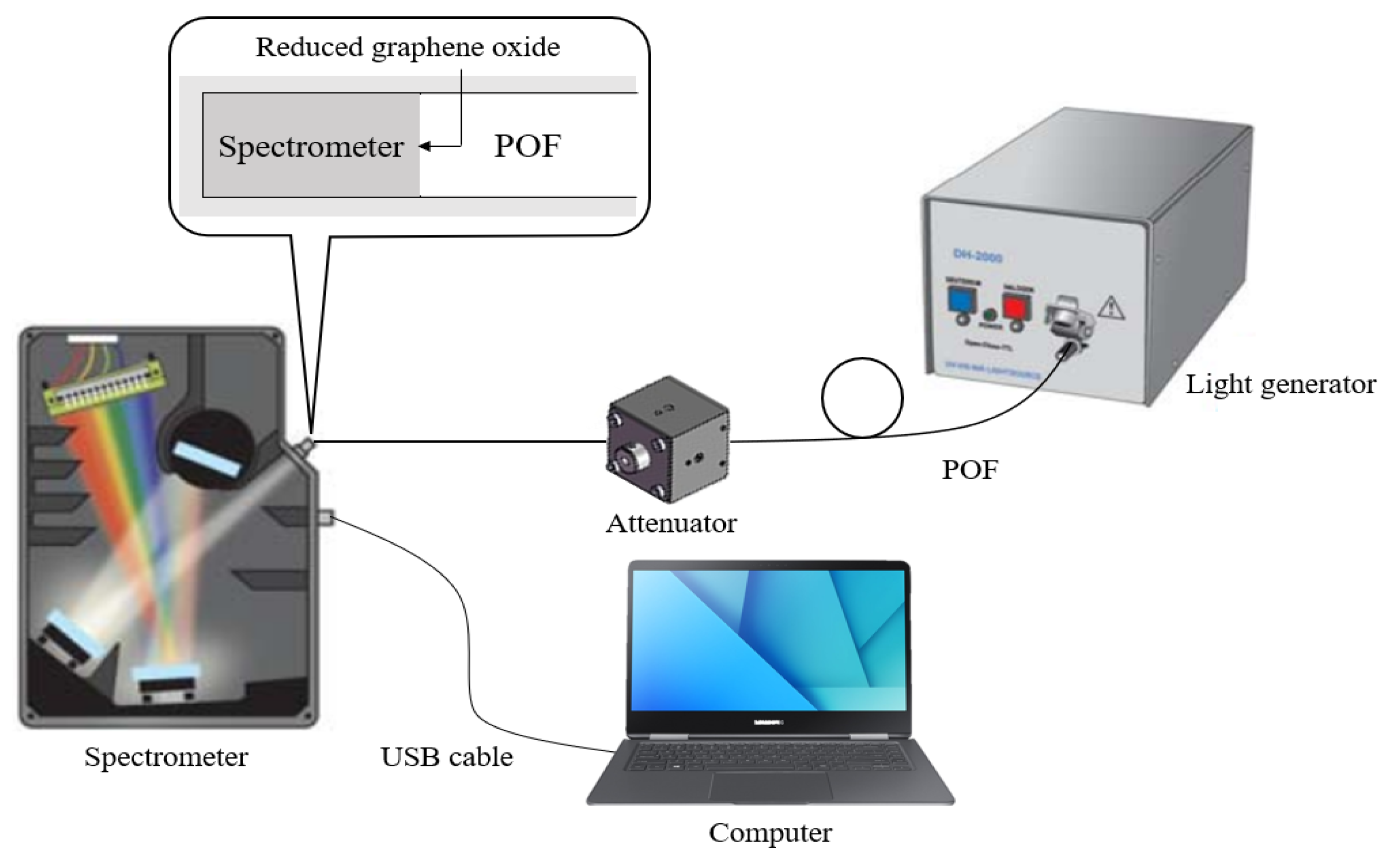
Figure 1 shows the experimental setup for measuring the variation in the light intensity of the POF via a light generator (FC5-LED, Prizmatix), a spectrometer (QE65000, Ocean Optics), and an optical attenuator (FLA-UV, Wyoptics). The light generator produced monoenergetic beams of 430, 535, and 630 nm. The length and diameter of the POF (CK120, Mitsubishi Rayon) were 1 m and 3.0 mm, respectively, and the core and cladding are made of polymethylmethacrylate (PMMA) resin and fluorinated polymer, respectively. The refractive indices of the core and cladding are 1.49 and 1.40, and the numerical aperture (NA) is 0.5. A 0.675 wt% RGO solution was coated at the end of a POF using a dip-coating machine (EF-4100, E-Flex); the number of coatings was 1, 3, 5, 10, 15, and 20. After RGO coating on the POF, the thicknesses of the RGO membranes were measured using a scanning electron microscope (SEM; S-3400N, Hitachi) according to the number of coatings.
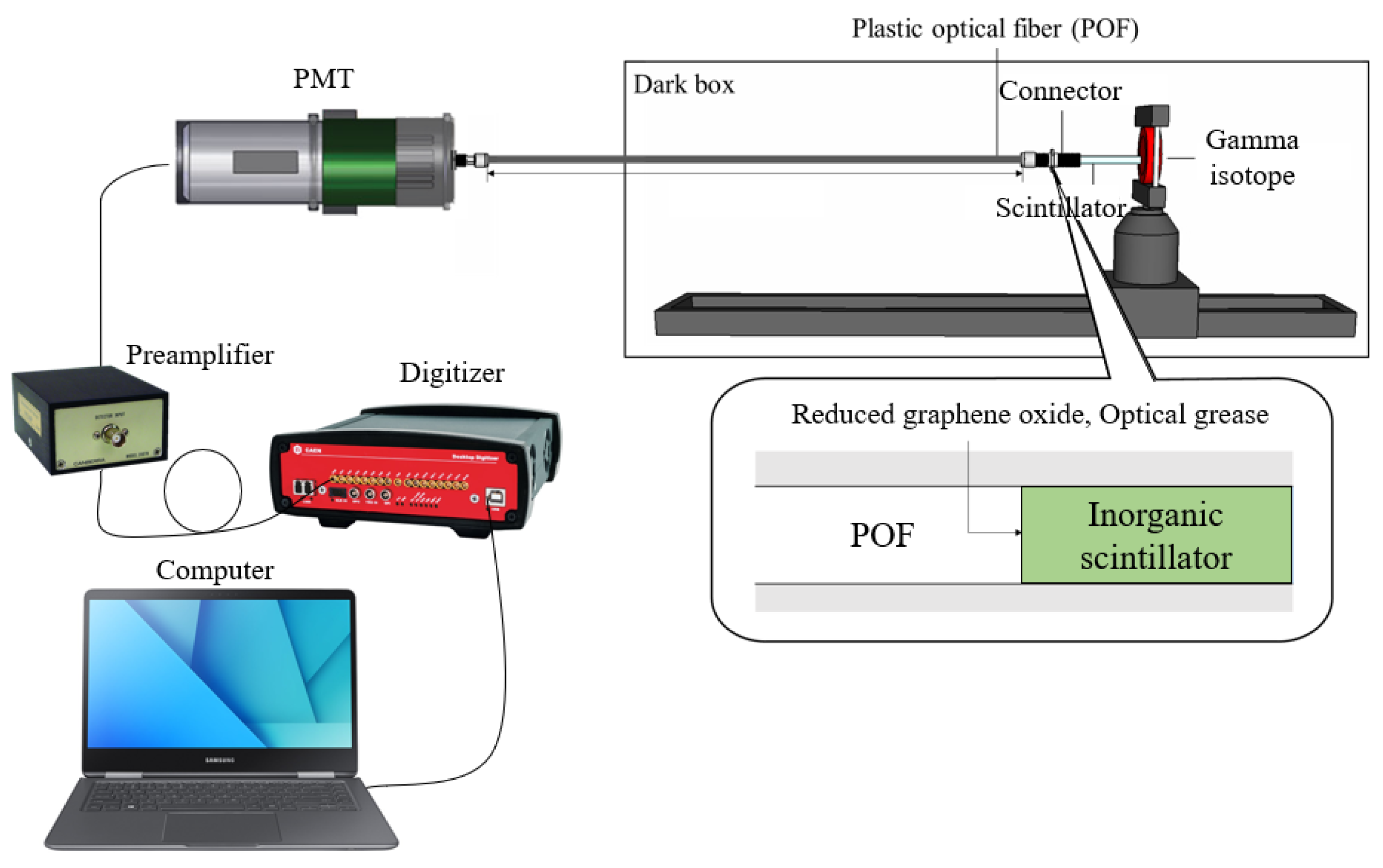
Figure 2 shows the experimental setup for capturing the gamma-ray energy spectra using an RGO-coated FORS. An RGO solution (0.675 wt%) was coated on the inorganic scintillators using the dip-coating method, and it coated 1, 3, 5, 10, 15, and 20 times. In the case of the connection, optical grease (EJ-550, ELJEN Tech.) and specially designed connectors were used to reduce scattered and leaky light between a POF and an inorganic scintillator. The lengths of the POF used in this experiment were 1, 2, 2.5, and 3 m, and the diameter was 3.0 mm. Variations in the amount of scintillating light and the energy resolution influence the length of the POF for remote gamma-ray spectroscopy.
Three types of inorganic scintillators, namely, cadmium tungstate (CdWO
4), cerium-doped lutetium yttrium orthosilicate (LYSO:Ce), and cerium-doped gadolinium aluminum gallium garnet (GAGG:Ce), were coated with RGO. They are suited for gamma-ray spectroscopy and RGO coatings owing to their high density, emission wavelength, and non-hygroscopicity. Each volume of the rectangular inorganic scintillator was 3 × 3 × 20 mm
3. The physical properties of the employed inorganic scintillators are listed in
Table 1 [
3,
16].
60Co and 137Cs were used as gamma-ray-emitting radioisotopes. The photopeak of gamma rays generated from 137Cs is 661.6 keV and 60Co produces two distinct gamma-ray peaks corresponding to 1173 and 1332 keV, respectively. The radioactivities of 60Co and 137Cs were 1.547 and 1.507 MBq, respectively.
For gamma-ray spectroscopy, a PMT (R6233-100, Hamamatsu Photonics), a preamplifier (2007 B, Canberra), and a digitizer (DT5725, Caen) were used to measure the amount of scintillating light and gamma-ray energy spectra [
17,
18].
3. Results
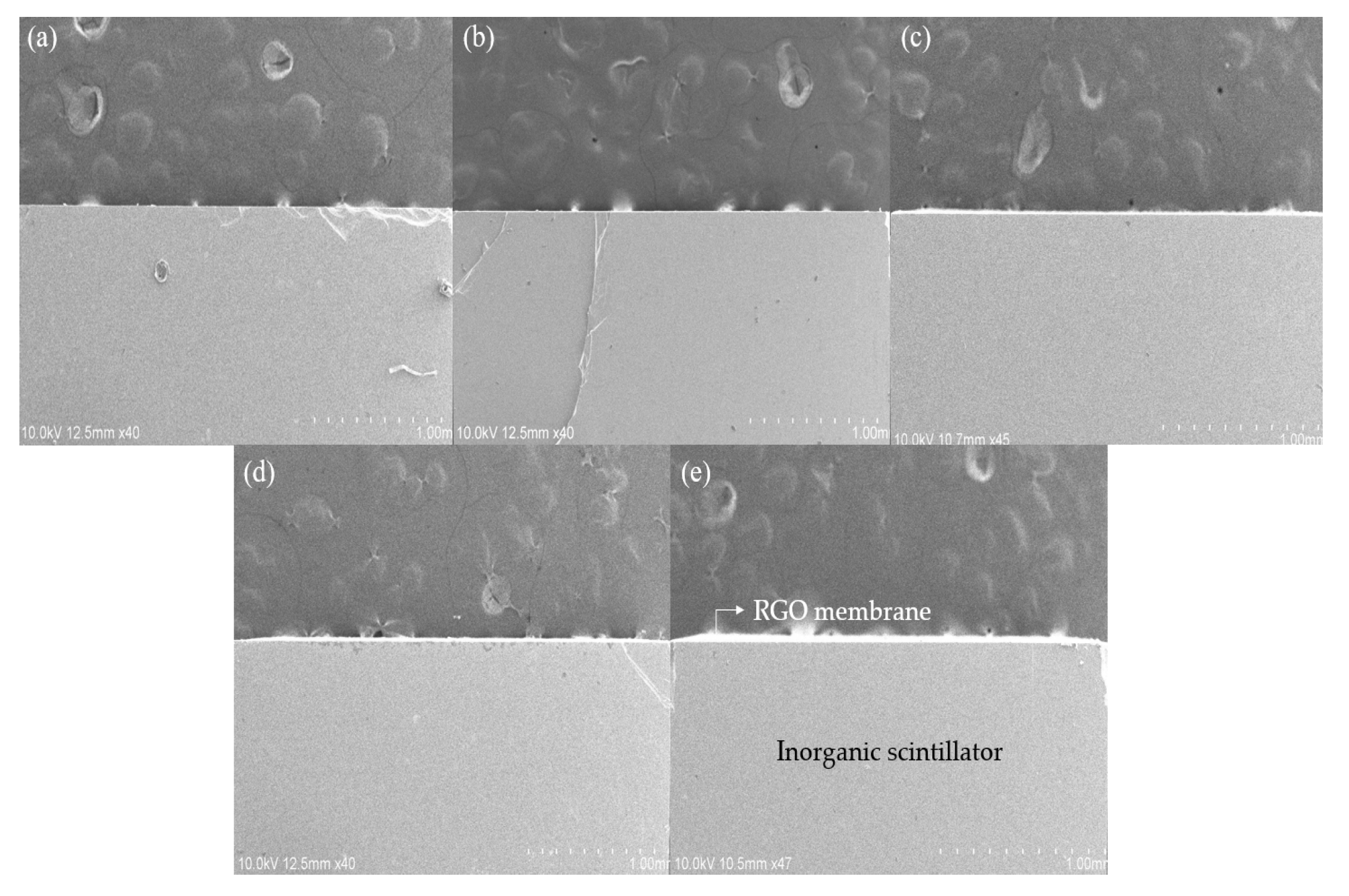
Three types of inorganic scintillators were coated with RGO solution using a dip-coating machine, as in the case of a POF, and the thicknesses of the RGO membranes on the scintillators were measured by microstructural analysis using SEM.
Figure 3 shows the SEM micrographs of the samples with the different number of coatings.
The white layer on the inorganic scintillator is the RGO membrane, which was not clearly visible when coated once but became visible when three layers were coated, as shown in
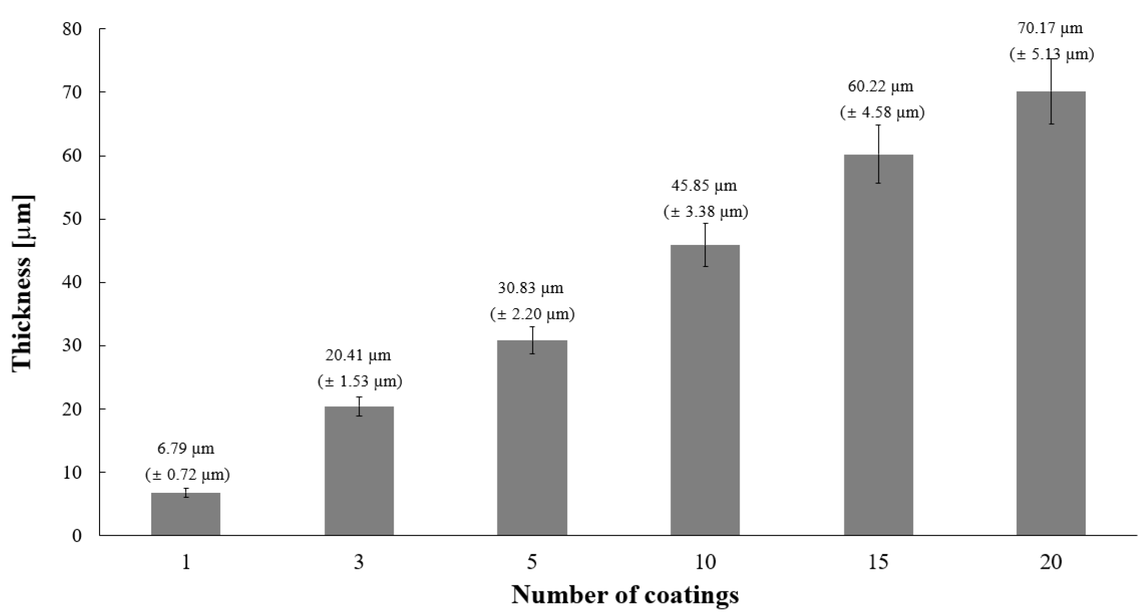
Figure 3c. The average thicknesses of the RGO membranes for the different number of coats are shown in
Figure 4.
As shown in
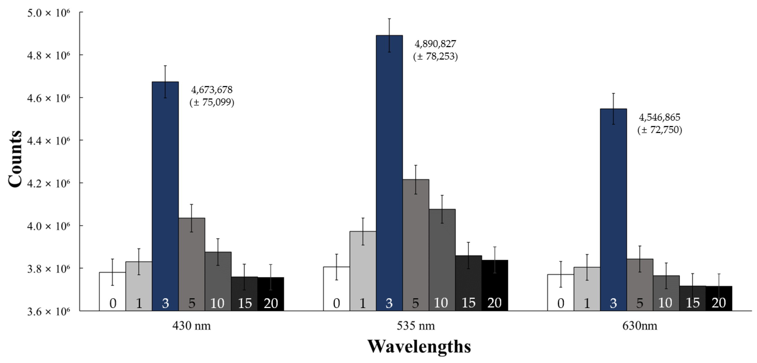
Figure 1, the transmitted light intensities were measured through the RGO membrane at the end of the POF according to the wavelengths of the emitted light, such as 430, 535, and 630 nm.
Figure 5 shows the variation in transmitted light intensities with the number of RGO coatings according to the three wavelengths of the emission lights. When the thickness of the RGO membrane was 20.41 µm corresponding to three coats, the transmitted light intensities become the highest among all three wavelengths of the emitted lights. In particular, the transmitted light intensity of the 535 nm emission wavelength increased by approximately 28.53% for a membrane thickness of 20.41 µm. Based on the results of this preliminary experiment, the three types of inorganic scintillators were coated with RGO thrice, and these were used in the gamma-ray spectroscopy in the FORS.
To evaluate the performance of the FORS for gamma-ray spectroscopy, RGOs were coated on the three types of inorganic scintillators, and the gamma-ray energy spectra of
60Co and
137Cs were measured using the experimental setup shown in
Figure 2. The total amount of scintillating light and the gamma-ray energy resolution of the photopeak from each RGO-coated inorganic scintillator were measured using the FORS, a PMT, a preamplifier, and a digitizer, and the results were compared with those of the FORS of uncoated inorganic scintillators. The measured total amount of scintillating light was calculated based on the total area of the gamma-ray spectrum, and the energy resolution was evaluated by the full width at the half maximum (FWHM) of a photopeak [
19].
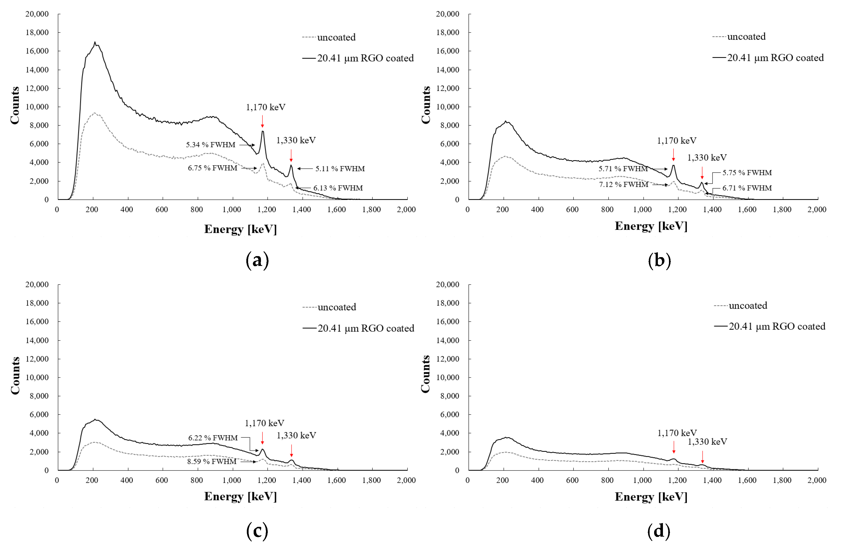
Figure 6 shows the gamma-ray energy spectra of 1.547 MBq
60Co radioisotopes, which were measured using three types of inorganic scintillators with a POF of one-meter length in the FORS. The measured total amount of scintillating lights and energy resolutions with FWHMs of the photopeaks using uncoated scintillators and scintillators with three-layer RGO coatings are also shown in
Figure 6. The largest total amount of scintillating light was measured using the RGO-coated GAGG:Ce scintillator, and it was approximately 28.6% greater than that measured by the uncoated scintillator. The energy resolutions of the photopeak were also increased, and the FWHMs were decreased by 1.41% and 1.02% in the photopeaks of 1170 and 1330 keV of the
60Co radioisotope, respectively. In all three RGO-coated scintillators, the total amount of scintillating light and energy resolution in the gamma-ray energy spectra of the
60Co radioisotope were enhanced, and they can be effectively used for remote gamma-ray spectroscopy using FORS. Improvements in the amounts of scintillating light and FWHMs are listed in
Table 2.
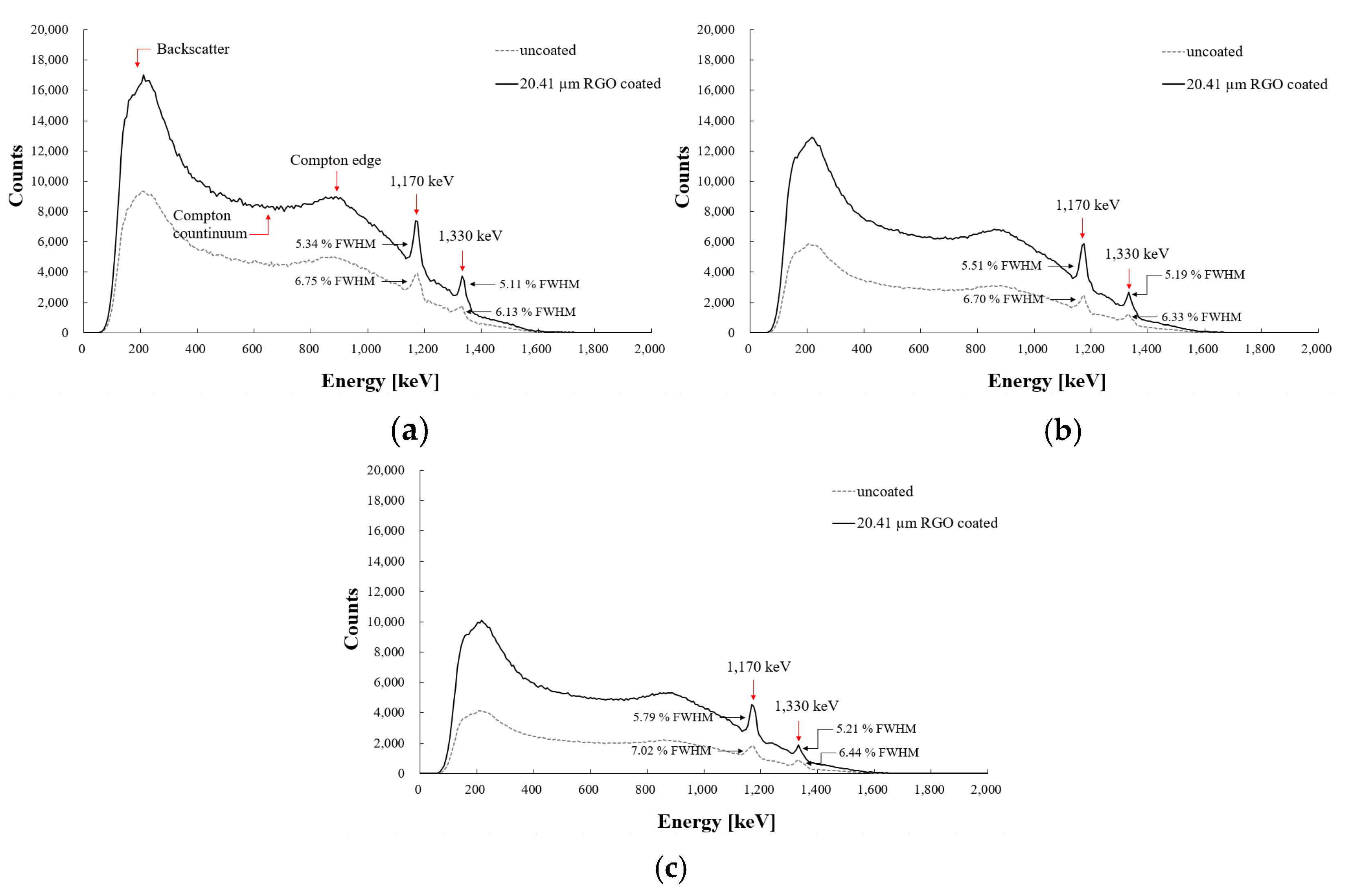
Figure 7 shows the measured gamma-ray energy spectra of the
137Cs radioisotope for the three types of RGO-coated and uncoated inorganic scintillators with 1 m-long POF using the FORS. The largest amount of scintillating light was also measured for the GAGG:Ce scintillator. The amount of scintillating light of the 20.41 µm RGO-coated GAGG:Ce for
137Cs was approximately 29.1% larger than that of the uncoated sample. The FWHM also decreased by 2.06% compared to the uncoated scintillator, and the energy resolution was enhanced. The improvements in the amounts of scintillating light and FWHMs of
137Cs in the gamma-ray energy spectra are listed in
Table 3. For the 20.41 µm RGO-coated LYSO:Ce and CdWO
4 scintillators, the amounts of scintillating light and energy resolutions were also enhanced compared to the uncoated scintillators, as shown in
Figure 7b,c.
It is well known that the intensity of light passing through optical fiber is attenuated, and the modal dispersion is significant in multimode step-index optical fibers. The measurement of the amount of scintillating light from a scintillator decreases as the length of the POF increases. Modal dispersion can be defined as the phenomenon in which the signal becomes spread over time because of different light propagation velocities for all modes in a multimode optical fiber. Because of this distortion mechanism, the gamma-ray energy spectra measured remotely using a FORS have a lower energy resolution in a photopeak than in the case of a direct gamma-ray spectroscopy system without a POF [
20,
21].
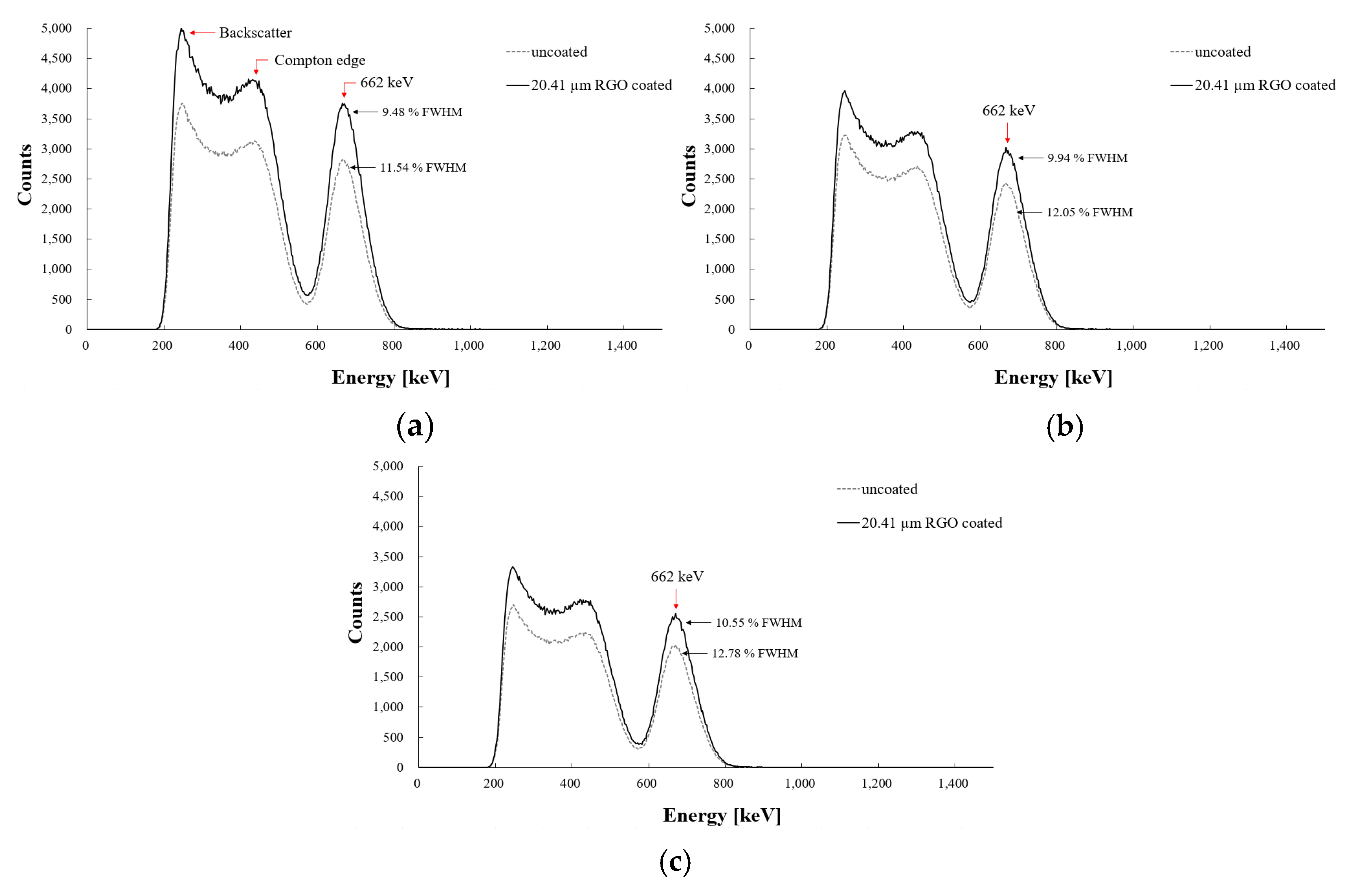
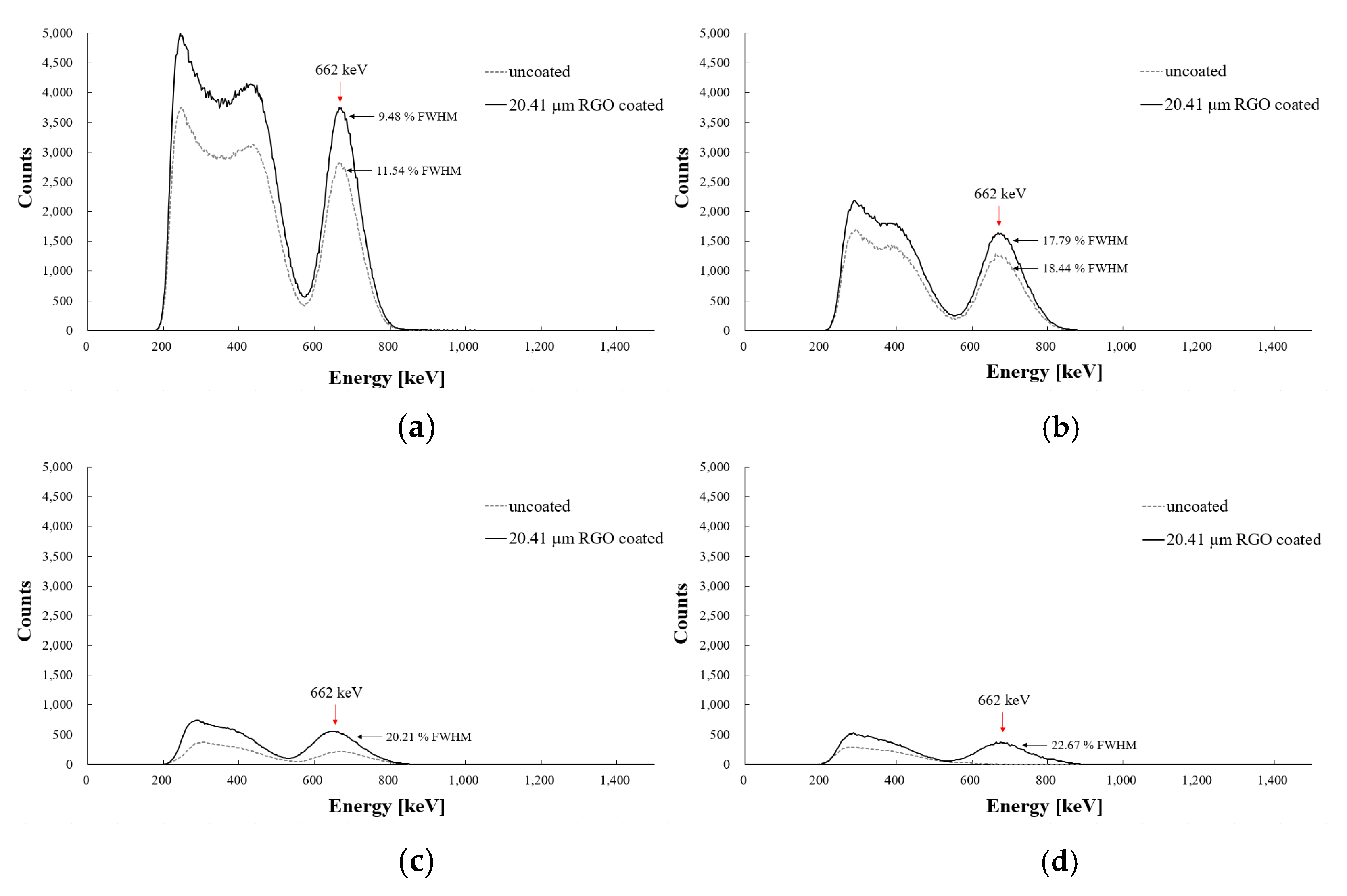
In a previous study on measuring the gamma-ray energy spectra using an FORS, the amount of scintillating light and energy resolution were reduced when the length of the POF was increased [
3]. In
Figure 8 and
Figure 9, the measured gamma-ray energy spectra of the
60Co and
137Cs radioisotopes, respectively, with the uncoated and RGO-coated GAGG:Ce scintillators using the FORS, which have different lengths of the POFs, are presented. As expected, the amounts of scintillating light and the energy resolutions of each photopeak were reduced in both cases. In particular, the gamma-ray photopeaks of
60Co and
137Cs radioisotopes were not measured in the FORS, which had an uncoated GAGG:Ce scintillator and a long POF, as shown in
Figure 8d and
Figure 9d. However, each photopeak was clearly observed in the gamma-ray energy spectra of the
60Co and
137Cs radioisotopes measured using the FORS, which is comprised of an RGO-coated GAGG:Ce scintillator and a long POF even though the energy resolution deteriorated significantly. These experiments show that RGO-coated inorganic scintillators can remotely detect photopeaks in the gamma-ray energy spectra of
60Co and
137Cs radioisotopes using the FORS from up to 3 m away.
4. Discussion
Generally, gamma-ray spectroscopy is widely used for the identification and quantification of radionuclides in the laboratory. However, it is necessary to directly measure the gamma-ray energy spectra in situ in areas contaminated by a nuclear accident or in the decommissioning of nuclear facilities [
22]. Generally, in-situ gamma-ray spectroscopy should have good performance, such as high counting efficiency and good energy resolution, and remote measurements are essential for the safety of personnel. Moreover, the detector system has a relatively small operating space, and the sensor should be compact enough for good spatial resolution to determine the exact points of contamination [
23].
In this study, an FORS was fabricated for remote gamma-ray spectroscopy, and an RGO-coated inorganic scintillator was used to enhance the performance of FORS for a high counting rate and better energy resolution. The amount of scintillating light increments can increase the counting rate in a scintillator-based radiation detector, and higher energy resolution is very important for radionuclide identification in the gamma-ray spectrum. Even though the energy resolution of the proposed FORS is much worse than that of a high-purity Ge (HPGe) detector, it has several advantages such as remote sensing, low cost, good portability, light weight, and no need for cooling [
5,
24].
In measuring the gamma-ray energy spectra of the 60Co and 137Cs radioisotopes using the FORS, the largest amount of scintillating light and highest energy resolution were measured using a GAGG:Ce scintillator, which was 20.41 µm thick coated with 0.675 wt% RGO solution.
The amount of scintillating light generated normally depends on the light yield of the scintillators. As listed in
Table 1, the light yield of GAGG:Ce was much higher than that of the LYSO:Ce and CdWO
4 inorganic scintillators, and the amount of scintillating light generated from the RGO-coated GAGG:Ce was larger. Additionally, the wavelength of emissions of GAGG:Ce was 530 nm, which is close to 535 nm and shows a higher transmitted light intensity through the RGO membrane, as shown in
Figure 5. Based on these results, an inorganic scintillator with a higher light yield and emission wavelength close to 535 nm would show better performance in gamma-ray spectroscopy after RGO coating.
For remote gamma-ray spectroscopy, the FORS fabricated and measured gamma energy spectra with different lengths of POF up to 3 m. However, the amount of scintillating light decreases, and energy resolution deteriorates owing to phenomena such as light attenuation and modal dispersion of the step index multimode POF. To minimize these obstacles, an optical fiber should have a low attenuation coefficient that increases the amounts of scintillating light measured and a graded index core that decreases the modal dispersion [
18,
19].
5. Conclusions
In this study, we developed a remote gamma-ray spectroscopy system based on FORS comprising an RGO-coated inorganic scintillator, a POF, a PMT, a preamplifier, and a digitizer. As a preliminary study, we determined the optimal thickness of the RGO membrane and the wavelength of light, which can increase the transmitted light intensity through the RGO coating. To evaluate the performance of the FORS, which has different types of inorganic scintillators, we measured the gamma-ray energy spectra of
60Co and
137Cs radioisotopes. In addition, these results were compared to the measured values using an FORS with an uncoated scintillator. For all three types of inorganic scintillators, namely, GAGG:Ce, LYSO:Ce, and CdWO
4, the amounts of generating scintillating light and the energy resolutions were enhanced after RGO coating compared to those for the uncoated scintillators. Moreover, a FORS with a POF of different lengths was fabricated and used to measure the gamma-ray energy spectra for remote gamma-ray spectroscopy. Even though the amount of scintillating light and energy resolution deteriorated owing to attenuation and modal dispersion as the length of the POF increased, we measured a photopeak in the energy spectrum using the RGO-coated scintillator and a 3 m long POF in the FORS. Without the RGO coating, photopeaks that can identify radionuclides were not observed in the gamma-ray energy spectra measured by the FORS incorporating a 3 m length of a POF for both
60Co and
137Cs radioisotopes. It was expected that inorganic scintillators coated with RGO in an FORS can be used to identify nuclides remotely in areas contaminated by a nuclear accident or in the decommissioning of nuclear facilities as an in-situ gamma-ray spectrometer. Furthermore, the proposed remote FORS based on RGO coating has potential applications in measuring cosmic-ray and gamma-ray energy spectra in a harsh environment such as space [
25,
26,
27,
28].

 ,
, 


