Generation of Photonic Hooks from Patchy Microcylinders

 ,
,  and
and 
Abstract
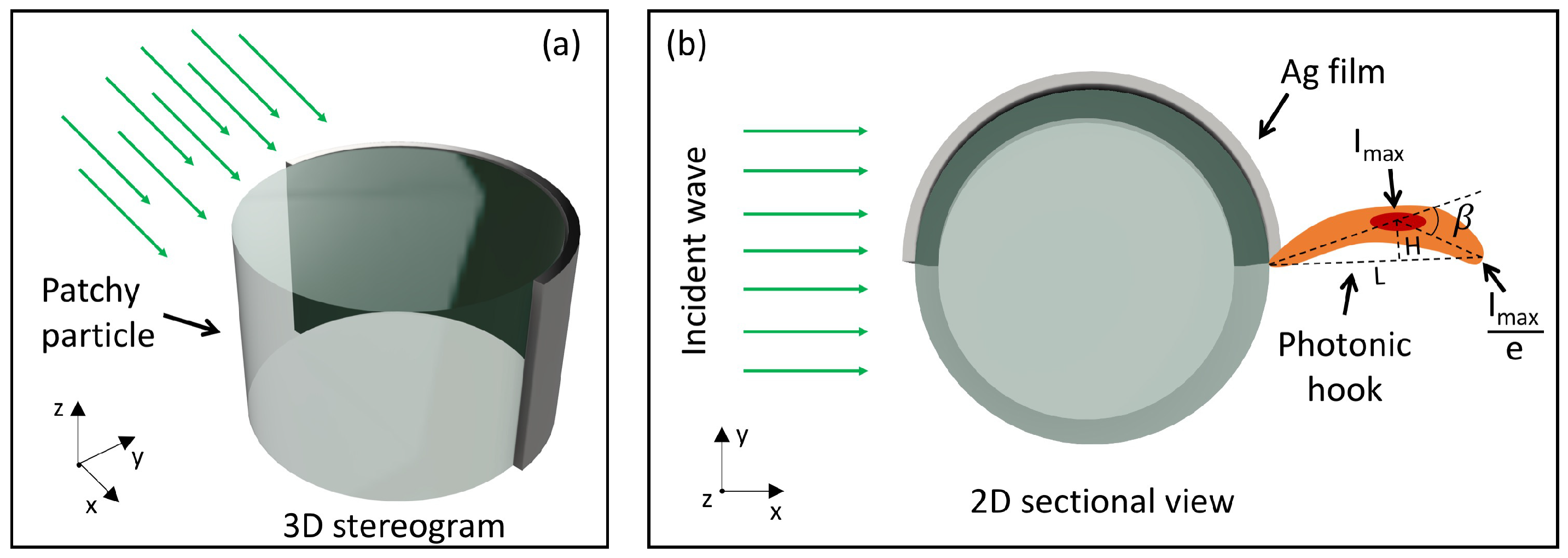
:1. Introduction
2. Simulation Method
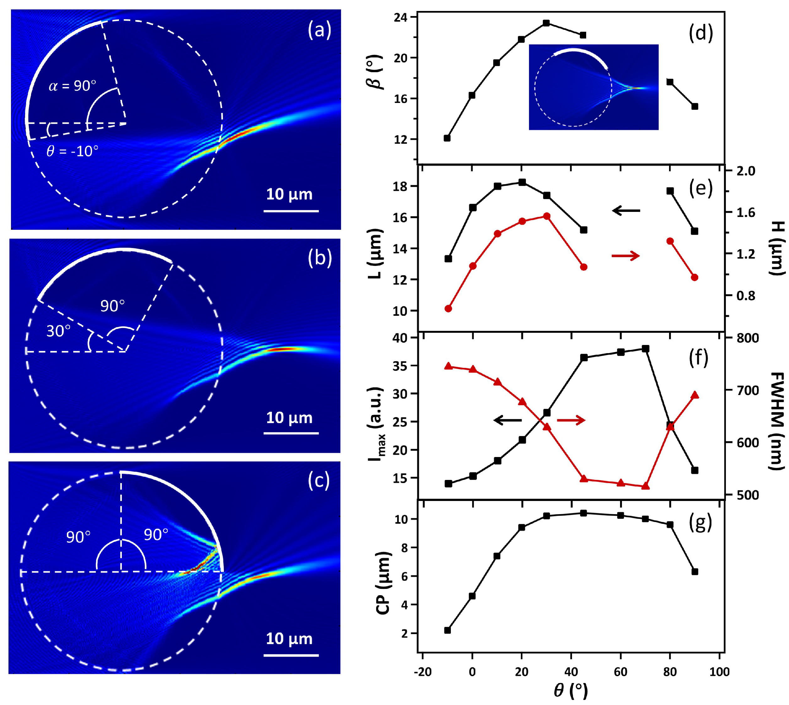
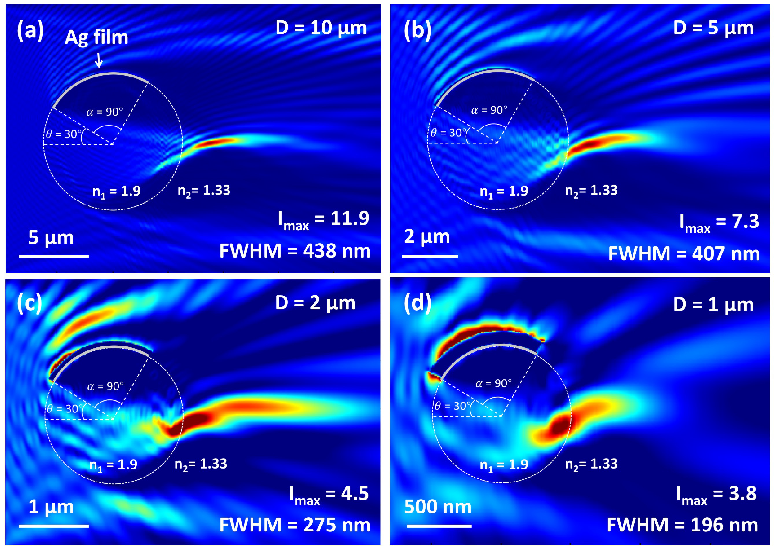
3. Results and Discussion
4. Conclusions
Author Contributions
Funding
Institutional Review Board Statement
Informed Consent Statement
Data Availability Statement
Acknowledgments
Conflicts of Interest
References
- Chen, Z.; Taflove, A.; Backman, V. Photonic nanojet enhancement of backscattering of light by nanoparticles: A potential novel visible-light ultramicroscopy technique. Opt. Express 2004, 12, 1214–1220. [Google Scholar] [CrossRef] [PubMed]
- Heifetz, A.; Kong, S.C.; Sahakian, A.V.; Taflove, A.; Backman, V. Photonic Nanojets. J. Comput. Theor. Nanosci. 2009, 6, 1979–1992. [Google Scholar] [CrossRef]
- Yan, B.; Yue, L.; Monks, J.N.; Yang, X.; Xiong, D.; Jiang, C.; Wang, Z. Superlensing plano-convex-microsphere (PCM) lens for direct laser nano-marking and beyond. Opt. Lett. 2020, 45, 1168–1171. [Google Scholar] [CrossRef]
- Li, Y.C.; Xin, H.B.; Lei, H.X.; Liu, L.L.; Li, Y.Z.; Zhang, Y.; Li, B.J. Manipulation and detection of single nanoparticles and biomolecules by a photonic nanojet. Light Sci. Appl. 2016, 5, e16176. [Google Scholar] [CrossRef] [PubMed]
- Yang, H.; Cornaglia, M.; Gijs, M.A.M. Photonic nanojet array for fast detection of single nanoparticles in a flow. Nano Lett. 2015, 15, 1730–1735. [Google Scholar] [CrossRef] [PubMed]
- Gu, G.; Song, J.; Chen, M.; Peng, X.; Liang, H.; Qu, J. Single nanoparticle detection using a photonic nanojet. Nanoscale 2018, 10, 14182–14189. [Google Scholar] [CrossRef]
- Wang, Z.; Guo, W.; Li, L.; Luk’yanchuk, B.; Khan, A.; Liu, Z.; Chen, Z.; Hong, M. Optical virtual imaging at 50 nm lateral resolution with a white-light nanoscope. Nat. Commun. 2011, 2, 218. [Google Scholar] [CrossRef]
- Ye, R.; Ye, Y.H.; Ma, H.F.; Ma, J.; Wang, B.; Yao, J.; Liu, S.; Cao, L.; Xu, H.; Zhang, J.Y. Experimental far-field imaging properties of a ∼5-μm diameter spherical lens. Opt. Lett. 2013, 38, 1829–1831. [Google Scholar] [CrossRef]
- Wang, F.; Liu, L.; Yu, H.; Wen, Y.; Yu, P.; Liu, Z.; Wang, Y.; Li, W.J. Scanning superlens microscopy for non-invasive large field-of-view visible light nanoscale imaging. Nat. Commun. 2016, 7, 13748. [Google Scholar] [CrossRef]
- Yang, H.; Trouillon, R.; Huszka, G.; Gijs, M.A.M. Super-resolution imaging of a dielectric microsphere is governed by the waist of its photonic nanojet. Nano Lett. 2016, 16, 4862–4870. [Google Scholar] [CrossRef]
- Luk’Yanchuk, B.S.; Raniagua-Domínguez, R.; Minin, I.; Minin, O.; Wang, Z. Refractive index less than two: Photonic nanojets yesterday, today and tomorrow. Opt. Mater. Express 2017, 7, 1820–1847. [Google Scholar] [CrossRef]
- Minin, I.V.; Liu, C.Y.; Geints, Y.E.; Minin, O.V. Recent advances in integrated photonic jet-based photonics. Photonics 2020, 7, 41. [Google Scholar] [CrossRef]
- Minin, I.; Minin, O. Diffractive Optics and Nanophotonics: Resolution below the Diffraction Limit; Springer: Berlin, Germany, 2015. [Google Scholar]
- Yue, L.; Minin, O.V.; Wang, Z.; Monks, J.N.; Shalin, A.S.; Minin, I.V. Photonic hook: A new curved light beam. Opt. Lett. 2018, 43, 771–774. [Google Scholar] [CrossRef] [PubMed]
- Minin, I.V.; Minin, O.V.; Katyba, G.M.; Chernomyrdin, N.V.; Kurlov, V.N.; Zaytsev, K.I.; Yue, L.; Wang, Z.; Christodoulides, D.N. Experimental observation of a photonic hook. Appl. Phys. Lett. 2019, 114, 031105. [Google Scholar] [CrossRef]
- Minin, I.V.; Minin, Q.V.; Liu, C.Y.; Wei, H.D.; Geints, Y.E.; Karabchevsky, A. Experimental demonstration of a tunable photonic hook by a partially illuminated dielectric microcylinder. Opt. Lett. 2020, 45, 4899–4902. [Google Scholar] [CrossRef] [PubMed]
- Minin, I.V.; Minin, O.V.; Ponomarev, D.S.; Glinskiy, I.A. Photonic hook plasmons: A new curved surface wave. Ann. Phys. 2018, 530, 1800359. [Google Scholar] [CrossRef]
- Yang, J.; Twardowski, P.; Gérard, P.; Duo, Y.; Fontaine, J.; Lecler, S. Ultra-narrow photonic nanojets through a glass cuboid embedded in a dielectric cylinder. Opt. Express 2018, 26, 3723–3731. [Google Scholar] [CrossRef]
- Gu, G.; Shao, L.; Song, J.; Qu, J.; Zheng, K.; Shen, X.; Peng, Z.; Hu, J.; Chen, X.; Chen, M.; et al. Photonic hooks from Janus microcylinders. Opt. Express 2019, 27, 37771–37780. [Google Scholar] [CrossRef]
- Geints, Y.E.; Minin, I.V.; Minin, O.V. Tailoring ’photonic hook’ from Janus dielectric microbar. J. Opt. 2020, 22, 065606. [Google Scholar] [CrossRef]
- Liu, C.Y.; Chung, H.J.; Minin, O.V.; Minin, I.V. Shaping photonic hook via well-controlled illumination of finite-size graded-index micro-ellipsoid. J. Opt. 2020, 22, 085002. [Google Scholar] [CrossRef]
- Gu, G.; Zhang, P.; Chen, S.; Zhang, Y.; Yang, H. Inflection point: A perspective on photonic nanojets. Photonics Res. 2021, 9, 1157–1171. [Google Scholar] [CrossRef]
- Liu, C.Y.; Chung, H.J.; E, H.P. Reflective photonic hook achieved by a dielectric-coated concave hemicylindrical mirror. J. Opt. Soc. Am. B Opt. Phys. 2020, 37, 2528–2533. [Google Scholar] [CrossRef]
- Geints, Y.E.; Zemlyanov, A.A.; Minin, I.V.; Minin, O.V. Specular-reflection photonic hook generation under oblique illumination of a super-contrast dielectric microparticle. J. Opt. 2021, 23, 045602. [Google Scholar] [CrossRef]
- Shen, X.; Gu, G.; Shao, L.; Peng, Z.; Hu, J.; Bandyopadhyay, S.; Liu, Y.; Jiang, J.; Chen, M. Twin photonic hooks generated by twin-ellipse microcylinder. IEEE Photonics J. 2020, 12, 6500609. [Google Scholar] [CrossRef]
- Zhou, S. Twin photonic hooks generated from two adjacent dielectric cylinders. Opt. Quantum Electron. 2020, 52, 389. [Google Scholar] [CrossRef]
- Zhou, S. Twin photonic hooks generated from two coherent illuminations of a micro-cylinder. J. Opt. 2020, 22, 085602. [Google Scholar] [CrossRef]
- Dholakia, K.; Bruce, G.D. Optical hooks. Nat. Photonics 2019, 13, 229–230. [Google Scholar] [CrossRef]
- Shang, Q.; Tang, F.; Yu, L.; Oubaha, H.; Caina, D.; Melinte, S.; Zuo, C.; Wang, Z.; Ye, R. Super-resolution imaging with patchy microspheres. arXiv 2021, arXiv:2108.06242. [Google Scholar]
- Sanchez, C.; Cristóbal, G.; Bueno, G.; Blanco, S.; Borrego-Ramos, M.; Olenici, A.; Pedraza, A.; Ruiz-Santaquiteria, J. Oblique illumination in microscopy: A quantitative evaluation. Micron 2018, 105, 47–54. [Google Scholar] [CrossRef] [PubMed]
- Fan, Y.; Li, J.; Lu, L.; Sun, J.; Hu, Y.; Zhang, J.; Li, Z.; Shen, Q.; Wang, B.; Zhang, R.; et al. Smart computational light microscopes (SCLMs) of smart computational imaging laboratory (SCILab). PhotoniX 2021, 2, 19. [Google Scholar] [CrossRef]
- Minin, O.V.; Minin, I.V. Terahertz microscopy with oblique subwavelength illumination in near field. Preprints 2021, 2021090024. [Google Scholar] [CrossRef]
- Pawar, A.B.; Kretzschmar, I. Patchy particles by glancing angle deposition. Langmuir 2008, 24, 355–358. [Google Scholar] [CrossRef] [PubMed]
- Pawar, A.B.; Kretzschmar, I. Multifunctional patchy particles by glancing angle deposition. Langmuir 2009, 25, 9057–9063. [Google Scholar] [CrossRef] [PubMed]


Publisher’s Note: MDPI stays neutral with regard to jurisdictional claims in published maps and institutional affiliations. |
© 2021 by the authors. Licensee MDPI, Basel, Switzerland. This article is an open access article distributed under the terms and conditions of the Creative Commons Attribution (CC BY) license (https://creativecommons.org/licenses/by/4.0/).
Share and Cite
Tang, F.; Shang, Q.; Yang, S.; Wang, T.; Melinte, S.; Zuo, C.; Ye, R. Generation of Photonic Hooks from Patchy Microcylinders. Photonics 2021, 8, 466. https://doi.org/10.3390/photonics8110466
Tang F, Shang Q, Yang S, Wang T, Melinte S, Zuo C, Ye R. Generation of Photonic Hooks from Patchy Microcylinders. Photonics. 2021; 8(11):466. https://doi.org/10.3390/photonics8110466
Chicago/Turabian StyleTang, Fen, Qingqing Shang, Songlin Yang, Ting Wang, Sorin Melinte, Chao Zuo, and Ran Ye. 2021. "Generation of Photonic Hooks from Patchy Microcylinders" Photonics 8, no. 11: 466. https://doi.org/10.3390/photonics8110466
APA StyleTang, F., Shang, Q., Yang, S., Wang, T., Melinte, S., Zuo, C., & Ye, R. (2021). Generation of Photonic Hooks from Patchy Microcylinders. Photonics, 8(11), 466. https://doi.org/10.3390/photonics8110466