Article

The Efficacy of Erbium-Ion, Diode, and CO2 Lasers in Debonding Attachments Used During Overlay Orthodontic Treatment and the Risk of Hard Tooth Tissue Damage Compared to Traditional Methods—An In Vitro Study

1
European Master Degree in Oral Laser Applications (EMDOLA), Wroclaw Medical University, 50-425 Wroclaw, Poland
2
Department of Mechanics, Materials and Biomedical Engineering, Faculty of Mechanical Engineering, Wroclaw University of Science and Technology, Smoluchowskiego 25, 50-370 Wroclaw, Poland
3
Faculty of Medicine and Dentistry Student, Wroclaw Medical University, 50-425 Wroclaw, Poland
4
Dental Surgery Department, Wroclaw Medical University, 50-425 Wroclaw, Poland
5
Laser Laboratory, Department of Integrated Dentistry, Wroclaw Medical University, 50-425 Wroclaw, Poland
6
Department of Periodontics, School of Dentistry, Virginia Commonwealth University (VCU), Richmond, VA 23284, USA
*
Authors to whom correspondence should be addressed.
Photonics 2025, 12(6), 621; https://doi.org/10.3390/photonics12060621
Submission received: 18 May 2025 / Revised: 7 June 2025 / Accepted: 10 June 2025 / Published: 18 June 2025
(This article belongs to the Special Issue Photonics: 10th Anniversary)

Abstract

:
Objective: This in vitro study evaluated the effectiveness of three laser systems—diode, CO2, and Er:YAG—for debonding composite attachments used in aligner orthodontic therapy. Materials and Methods: Fifty extracted human premolars with composite attachments were divided into five groups (n = 10): control, RT (rotary tools), diode laser (980 nm, irradiance was 4811 W/cm2), CO2 laser (10.6 µm, irradiance 1531 W/cm2), and Er:YAG laser (2940 nm, irradiance 471.7 W/cm2). Shear bond strength (SBS) testing measured debonding forces. Enamel surface changes were evaluated using micro-CT, optical profilometry, and stereomicroscopy. The Adhesive Remnant Index (ARI) assessed residual bonding material. Results: Laser treatment increased enamel roughness (p < 0.05). Er:YAG laser caused the highest roughness (Sa = 2.03 µm) and up to 0.17 mm enamel loss but left minimal adhesive remnants and no fractures. Diode laser preserved surface smoothness with moderate bond weakening. CO2 laser had intermediate effects. RT showed the highest SBS but resulted in greater enamel alteration. SBS was significantly reduced in the laser groups, lowest for Er:YAG (81.7 ± 45.5 MPa vs. control 196.2 ± 75.3 MPa). ARI indicated better adhesive removal in the laser-treated groups, with Er:YAG showing the highest percentage of clean enamel surfaces (67% vs. 25%). Conclusions: Er:YAG demonstrated the best balance between effective debonding and enamel preservation. Diode and CO2 lasers also offer viable alternatives to rotary tools. Further clinical studies are recommended.

1. Introduction

Clear aligner therapy has revolutionized modern orthodontics, offering an aesthetic, comfortable, and patient-friendly alternative to conventional fixed appliances. Unlike traditional braces that use metal brackets and wires, aligners are transparent, removable trays designed to apply controlled forces to teeth, gradually moving them into their desired positions [1,2,3]. Their discreet appearance and the ease of maintaining oral hygiene during treatment have contributed significantly to their popularity among both adolescents and adults [4,5,6,7]. However, despite their advantages, aligner therapy is primarily recommended for cases with mild-to-moderate malocclusions. The effectiveness of these systems often depends on adjunctive elements such as attachments, which support complex tooth movements by enhancing the mechanical retention of the aligner trays [8].
Attachments used during aligner therapy are composite-based structures bonded directly to the enamel. They serve as anchorage points, enabling movements such as rotations, extrusions, and torque control [8,9]. Despite their benefits, attachments present clinical challenges, particularly during the debonding phase at the end of treatment. These composite materials are often similar in optical properties to tooth enamel, complicating their identification and removal without harming the underlying hard tissue [10]. Conventional removal methods typically involve rotary instruments, which carry a risk of irreversible enamel loss and increased surface roughness. Clinicians are, therefore, actively seeking alternative techniques that can ensure safe, efficient, and minimally invasive debonding of these attachments [9].
Laser-assisted debonding has emerged as a promising alternative to mechanical removal techniques [10]. Lasers, including diode, Er:YAG, and CO2 types, operate by delivering concentrated energy that interacts selectively with the composite resin at the adhesive interface, either through thermal softening, photoablation, or thermal–mechanical disruption [11,12,13,14,15,16]. The mechanism of laser interaction with tissue is influenced by multiple factors, including wavelength, pulse duration, energy density, and tissue optical properties such as absorption and scattering coefficients [17,18]. By altering the adhesive’s integrity without physically contacting the enamel, laser systems offer the potential to reduce the risk of enamel microfractures, preserve tooth structure, and improve clinical efficiency during attachment removal procedures [10,19,20,21,22,23].
However, both conventional and laser-assisted debonding methods are associated with potential iatrogenic effects [23]. Rotary instrumentation can cause significant damage to enamel prisms, resulting in surface irregularities, microcracks, and increased susceptibility to plaque accumulation. Similarly, improper laser application—such as excessive energy levels or inappropriate wavelengths—may lead to thermal damage of the pulp, carbonization of the composite, or undesired alteration of enamel surface morphology [17,24,25,26,27,28,29,30,31]. Understanding these risks is critical to optimizing debonding protocols and minimizing adverse effects on tooth integrity. Studies suggest that although lasers can facilitate composite removal, further evidence is needed to validate their clinical safety and efficacy across different systems and settings.
Previous studies have partially addressed the effects of various debonding techniques on enamel integrity and bond strength. Yassaei et al. [32] found that conventional rotary instruments often result in significant enamel surface damage, including increased roughness and microcracks, while laser methods—particularly Er:YAG—demonstrated a more conservative impact. Similarly, Ajwa et al. [33] in a systematic review confirmed that Er:YAG and diode lasers are effective in reducing adhesive remnants without causing substantial enamel loss. In terms of bond strength, Khalil et al. [34] and Rechmann et al. [35] observed a marked decrease in shear bond strength following laser exposure, which facilitates safer removal but may also raise concerns about the stability of bonded attachments during treatment. Despite these findings, few studies have simultaneously compared surface morphology changes, adhesive residue, and mechanical performance across multiple laser systems using standardized methodology. The present study aims to address this gap by comprehensively evaluating Er:YAG, CO2, and diode lasers in comparison with rotary tools and untreated controls.
The primary objective of this in vitro study is to evaluate the efficacy of three different laser systems—diode, Er:YAG, and CO2—in debonding composite attachments used during aligner orthodontic therapy and to compare the potential risks of enamel damage relative to traditional rotary methods. According to our literature review and current evidence, no comprehensive comparative study to date has directly assessed the enamel surface morphology, shear bond strength, and risk of hard tissue damage across these three laser modalities in the context of aligner attachment removal. This research aims to fill that gap and contribute to evidence-based recommendations for safer orthodontic debonding practices.

2. Materials and Methods

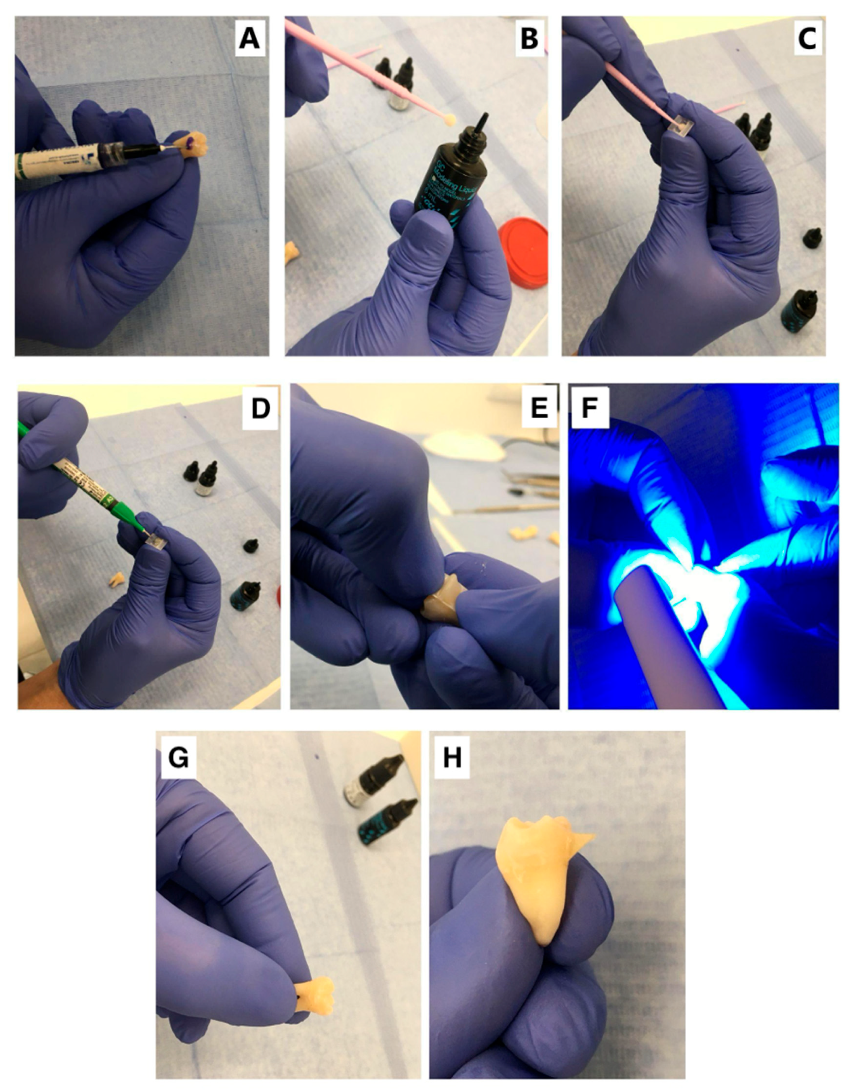
2.1. Sample Preparation

The study material consisted of 50 healthy premolars with intact buccal surfaces, extracted from adolescent and young adult patients (aged 15–25) as part of planned orthodontic treatment. Teeth that had undergone endodontic treatment, exhibited structural damage (e.g., cracks or fractures), showed carious lesions, or presented hypoplastic defects were excluded from the study. The study was conducted in accordance with the Declaration of Helsinki and approved by the Institutional Review Board and Ethics Committee (No. KB 423/2023N). Following extraction, the teeth were stored in a 0.1% thymol solution at room temperature (20 ± 5 °C) until further processing. To prepare the enamel surfaces, each tooth was brushed with Enamel Regeneration Oral-B toothpaste (Procter & Gamble, Cincinnati, OH, USA) using a Curaprox CS 5460 manual toothbrush (Curaden AG, Kriens, Switzerland) to remove surface debris. The teeth were then dried using compressed air. Enamel etching was performed using 37% orthophosphoric acid (Blue Etch, Cerkamed, Stalowa Wola, Poland), applied to the buccal surface with an AKZENTA applicator for 30 s (Figure 1A). The surface was subsequently rinsed with water at 20 °C and dried again with compressed air. To prevent adhesion of the composite to the matrix, the inner surface of the matrix was coated with Modeling Liquid (Figure 1B,C). The etched enamel was then treated with MTP Prime adhesive, which was applied and rubbed into the surface for 60 s using an AKZENTA applicator (Figure 1C). The adhesive was light-cured for 1 s using a FlashMax polymerization lamp (460 nm, 5000 mW/cm2). Next, the matrix was filled with Bulk Fill Flowable Restorative composite and positioned on the tooth surface (Figure 1D,E). Polymerization was carried out using the FlashMax lamp in three 3 s pulses, targeting the central, upper, and lower areas of the matrix (Figure 1F).

2.2. Study Groups

The 50 samples were randomly assigned to the five study groups (n = 10 per group) using a computer-generated sequence created with the online tool randomizer.org. The specimens (n = 50) were divided into five groups:
  • Control (Reference) Group (n = 10)
  • Group RT (n = 10): Attachment removal was performed using rotary tools (RT). Specimens in the RT group were prepared using Drill New Technology instruments (Edenta Etbl, Au SG, Switzerland). A tungsten carbide drill for contra-angle handpieces, with a short, rounded cone, was used in combination with a W&H Synea Fusion dental handpiece (Austria) operating at 40,000 rpm with water spray.
  • Group D (n = 10): Attachment debonding was carried out using a semiconductor diode laser (SMARTm PRO, Lasotronix, Piaseczno, Poland). The laser operated in pulsed mode with an average power of 3.4 W, an impulse time of 500 µs, and a recess time of 80 µs. It emitted a wavelength of 980 nm, with the pilot beam set to 10% power. The exposure duration was 11 s, and energy was delivered through a 300 µm spot diameter, totaling 37 J. Based on these parameters, the estimated fluence was 52,336 J/cm2, and the irradiance was 4811 W/cm2.
  • Group C (n = 10): Attachment debonding was performed using a CO2 laser (ADSS model FG 900S, ADSS Group Development Co. Ltd., Beijing, China). Each abutment was irradiated perpendicularly at the margin, targeting the composite near the attachment site. The laser operated in pulsed mode at 3 W, with a 10 ms pulse time, 10 ms pause time, and a frequency of 50 Hz. A 635 nm red diode pilot beam (<2 mW) was used for aiming. Irradiation was performed in spot mode with 1 mm spacing to ensure uniform coverage. Assuming a typical spot diameter of 500 µm, the irradiance was estimated at 1531 W/cm2. Fluence was not calculated due to the absence of total exposure time.
  • Group E (n = 10): Attachment debonding was performed using an Er:YAG laser (LiteTouch, Light Instruments Ltd., Yokne’am, Israel). This ablative fractional laser emits a focused beam at a wavelength of 2940 nm, which is highly absorbed in water, allowing for efficient ablation with minimal scatter and shallow penetration (within tens of micrometres). The laser’s core consists of an erbium-doped yttrium aluminum garnet crystal, activated by a xenon lamp. A 3.0 W beam was applied in two 10-s sequences, delivering 200 mJ per pulse at a frequency of 15 Hz. Irradiation was precisely directed along the attachment edge at 1 mm intervals. For a 900 µm spot diameter, the estimated fluence was 31.45 J/cm2, and irradiance was 471.7 W/cm2.
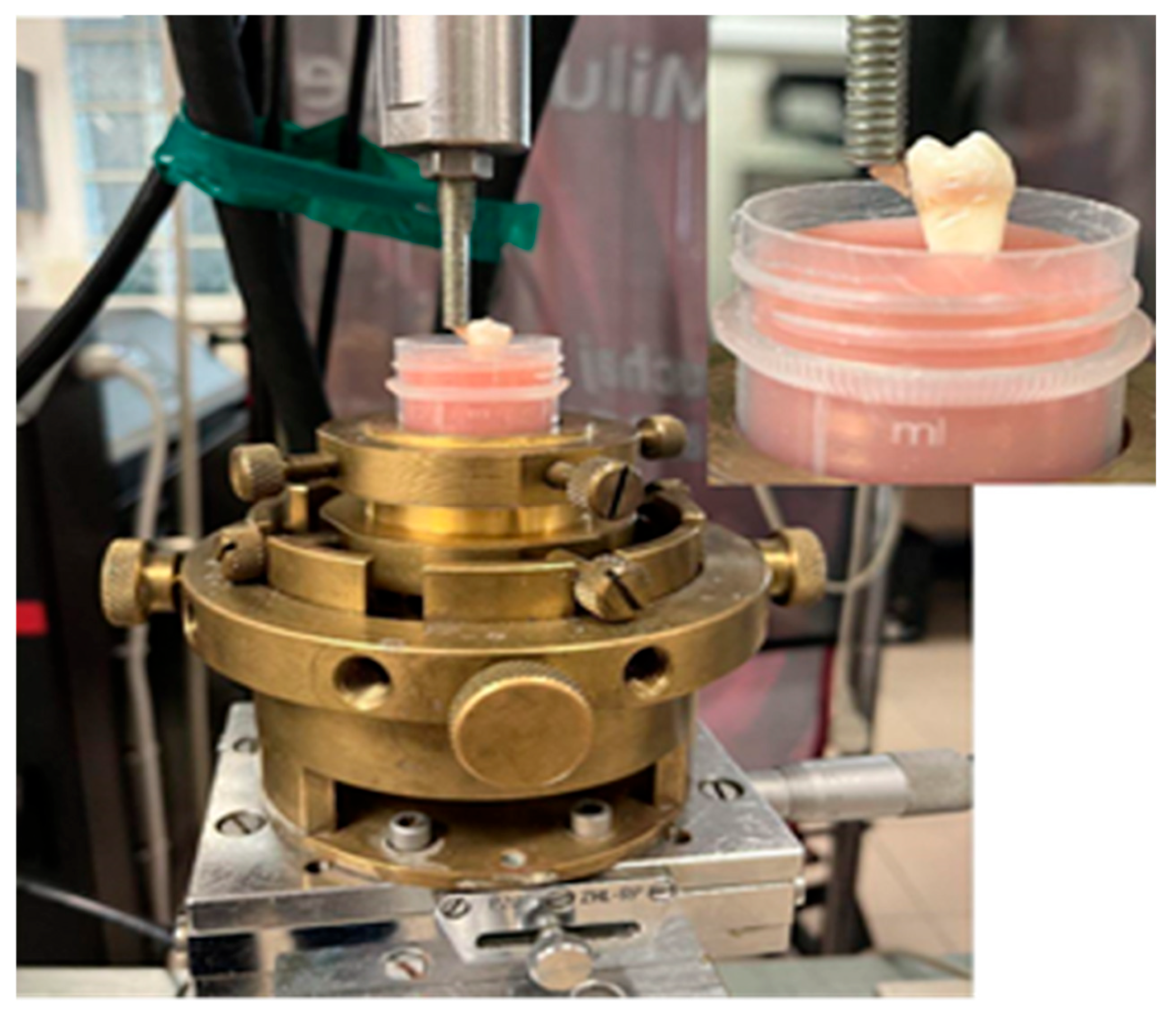
The laser fiber was positioned perpendicularly to the buccal surface at a distance of 1–2 mm, with the beam directed at the composite–enamel interface to ensure targeted ablation while minimizing enamel exposure (see Figure 2).

2.3. The Shear Test

The shear forces required to detach the attachments from the enamel surface were measured using an MTS 858 MiniBionix testing machine. Prior to testing, the prepared teeth were stored in a moist 0.1% thymol solution to maintain hydration. For stability during the test, each specimen was embedded in Duracryl™ Plus acrylic resin (SpofaDent, Markova, Jičín, Czech Republic). The shear test was performed using a cylindrical, flat-ended punch positioned perpendicularly to the bracket surface. A constant loading rate of 1 mm/min was applied. The assembly remained rigid throughout the procedure, which continued until complete detachment of the attachment from the enamel surface occurred (see Figure 3).

2.4. High-Resolution X-Ray Micro-Computed Tomography

High-resolution X-ray micro-computed tomography (micro-CT) was employed to evaluate the microstructural changes in the enamel before and after laser exposure. Imaging was performed using a SkyScan 1172 micro-CT system (Bruker, Kontich, Belgium), which consists of a tungsten anode X-ray source with an approximate focal spot size of 10 μm, a high-resolution radiation detector with a pixel size of 50 μm, and a rotating stage for sample positioning during scanning. Each specimen was scanned over a full 360° rotation, with image acquisition conducted at 0.41° rotation steps. Scanning parameters were set at 100 kV and 100 μA, achieving a spatial resolution of 6.8 μm. The resulting series of projection images (roentgenograms) were used to reconstruct detailed three-dimensional models of the enamel structure for comparative analysis.

2.5. Optical Profilometry

To evaluate the surface roughness of the enamel after treatment, optical profilometry was performed using a Leica® DCM8 profilometer (Leica Microsystems, Wetzlar, Germany). Surface roughness is a critical parameter influencing the adhesive properties of bonded joints, as it affects the mechanical interlocking and wettability of adhesives. The Leica® DCM8 utilizes confocal microscopy, which provides superior contrast and spatial resolution compared to conventional optical microscopy, owing to its use of point illumination and selective depth imaging.
During scanning, only light reflected from surface areas within the objective’s focal plane reaches the detector. The system acquires a series of optical sections at different depths, which are then compiled into a high-resolution three-dimensional image of the surface topography. The system achieves a lateral resolution of up to 140 nm and a vertical resolution down to 1 nm. Data analysis and surface reconstruction were carried out using Leica® Map 7.4 software, allowing for quantitative roughness assessment and comparative analysis between samples.

2.6. Evaluation of Adhesive Remnant (ARI) Score

To assess the amount of residual bonding material left on the enamel surface after debonding, the Adhesive Remnant Index (ARI) was used. The ARI score allows for classification of the enamel–attachment interface based on the extent of adhesive residue observed post-removal. Following the shear test, each specimen was visually inspected under magnification using a stereomicroscope (Discovery V.20, Zeiss, Oberkochen, Germany), and the enamel surface was photographed and evaluated.
The ARI classification applied in this study followed a five-level scale:
Score 0: No adhesive left on the tooth surface;
Score 1: Less than 50% of the adhesive left on the tooth;
Score 2: More than 50% of the adhesive left on the tooth;
Score 3: All adhesive remained on the tooth with a clear impression of the bracket base;
Score 4: Visible enamel fracture or damage.
Before the main evaluation, examiner calibration was performed to ensure consistency in ARI scoring. Two independent examiners were trained using a set of 10 representative images illustrating the full range of ARI scores (0–4). The images were jointly reviewed and discussed to standardize the interpretation of the scoring criteria. Following this, both examiners independently assessed the same 10 pilot specimens. Any discrepancies in scoring were resolved through discussion and consensus.

2.7. Statistical Analysis

The statistical analysis was performed using Statistica software (version 13.3.721.1, StatSoft, Tulsa, OK, USA). The normality of the data distribution was initially verified, and based on the results, parametric or non-parametric tests were applied accordingly. For the comparison of surface roughness parameters (Sa, Sq, Sz, Sp, and Sv) and mechanical properties (shear stress τ, detachment force F, and displacement l), one-way analysis of variance (ANOVA) was used. Statistically significant differences between groups were further analyzed using appropriate post hoc tests. The significance of differences in the distribution of the Adhesive Remnant Index (ARI) values among groups was assessed using the chi-square (χ2) test. A p-value less than 0.05 was considered statistically significant. All results are presented as the mean ± standard deviation (SD), unless stated otherwise.

3. Results

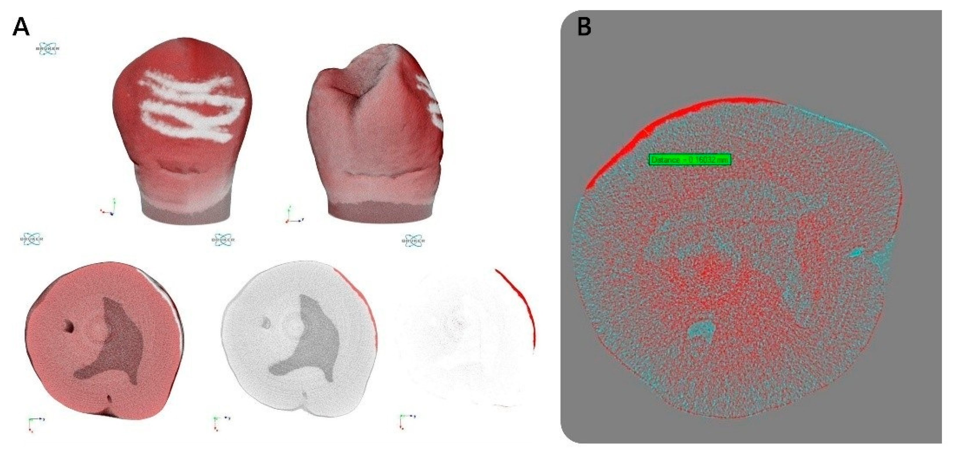
3.1. Effect of Laser Application on Enamel Microstructure (Micro-CT Analysis)

The comparative analysis of micro-CT reconstructions revealed clear differences in enamel surface microstructure depending on the type of laser used. Among the three tested energy sources, the Er:YAG laser caused the most pronounced changes in enamel. In this group, localized enamel loss reached depths of 0.13 to 0.17 mm, corresponding to approximately 9% of the total enamel thickness in the affected areas. This indicates a highly effective ablative interaction between the Er:YAG beam and dental hard tissue, which, while efficient for removal, may increase the risk of compromising enamel integrity if not properly controlled. In contrast, the CO2 laser produced moderate changes in enamel morphology. The affected zones were visibly altered, but without substantial depth of tissue loss. The structural differences were confined to the outer surface layers, suggesting a more superficial and thermally driven interaction. The diode laser had the least impact on enamel microstructure. Only minimal surface alterations were observed, with no measurable loss in enamel thickness. This suggests a less invasive mode of action, likely due to its lower absorption in hydroxyapatite and more selective effect limited to pigmented or soft tissues (see Figure 4).

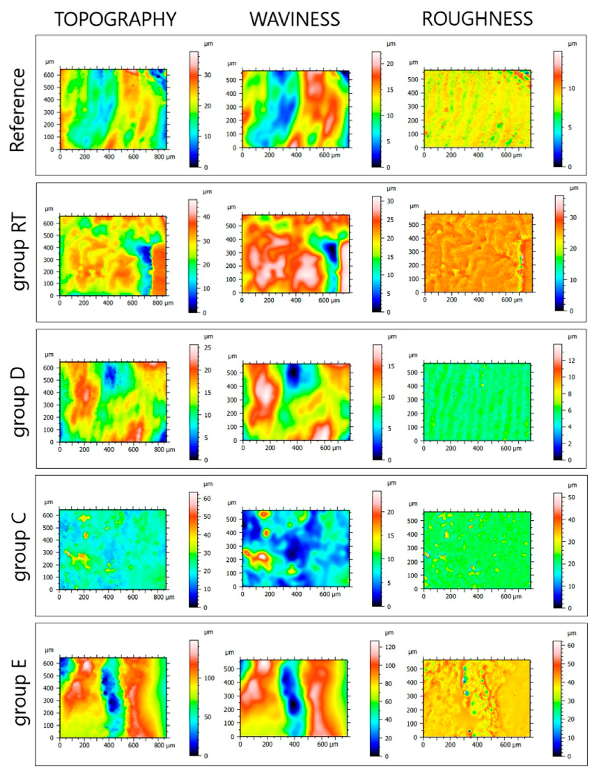
3.2. Surface Roughness Analysis (Optical Profilometry)

Quantitative analysis of enamel surface roughness revealed clear differences between the experimental groups, depending on the method of attachment removal. The RT group (rotary tools) exhibited only moderate surface alterations, with Sa and Sq values of 0.31 µm and 0.46 µm, respectively, indicating limited disruption of the enamel surface. Similarly, the diode laser group (D) showed comparably low roughness values (Sa = 0.30 µm; Sq = 0.42 µm), suggesting a minimally invasive effect on the enamel microtopography. In contrast, the CO2 laser group (C) demonstrated markedly increased surface irregularities, with Sa and Sq values rising to 0.79 µm and 1.24 µm, respectively. The corresponding Sz value reached 43.30 µm, indicating significant vertical height variation. This was accompanied by deeper valleys (Sv = 22.80 µm) and higher peaks (Sp = 20.50 µm) compared to the RT and D groups. The most pronounced surface alterations were observed in the Er:YAG laser group (E). This group recorded the highest values across all measured parameters, with Sa = 2.03 µm and Sq = 4.19 µm. The maximum surface height difference Sz exceeded 251 µm, with both valleys (Sv = 125.42 µm) and peaks (Sp = 125.80 µm) reaching extreme values. (see Table 1) These results indicate an aggressive ablative interaction with the enamel surface, leading to a highly irregular topography. Notably, changes in all roughness parameters were observed across all groups, regardless of the energy source used. While the Sa value increased only slightly (by approximately 7%) in the diode laser group, both the CO2 and Er:YAG lasers caused changes several times greater. The most significant differences were found in the Sz parameter, representing maximum surface height variation, which consistently reflected the severity of enamel alteration. Among all methods, the Er:YAG laser produced the most substantial surface modifications, as confirmed through both profilometric and microtomographic analyses. Differences in surface roughness between groups C and E and the reference group were statistically significant (p < 0.05, ANOVA) (see Figure 5).

3.3. Shear Bond Strength of Attachments to Enamel

The shear test results revealed significant differences in the mechanical performance of the attachment–enamel interface depending on the removal method applied prior to testing. The reference group exhibited the highest values of both shear stress (τ = 196.19 MPa) and maximum load-at-failure (F = 15.19 N), indicating the strongest bond strength and most effective attachment retention.
Among the experimental groups, the diode laser group (D) maintained relatively moderate values (τ = 126.01 MPa; F = 9.76 N), suggesting that diode laser application had a less detrimental effect on bond integrity compared to other laser-based methods.
More noticeable reductions in mechanical performance were observed in the CO2 laser group (C), where the average shear stress dropped to 116.36 MPa and the maximum load dropped to 9.29 N. While still functional, these values were significantly lower than the reference group, indicating partial compromise of enamel adhesion due to laser interaction.
The lowest bond strength was recorded in the Er:YAG laser group (E). This group showed a mean shear stress of 81.68 MPa and a maximum load-at-failure of only 6.08 N, with both values being significantly lower than the reference (p < 0.05). These findings point to a marked weakening of the adhesive interface following Er:YAG exposure, likely due to the pronounced microstructural changes and increased roughness documented in the profilometric and tomographic analyses.
In terms of displacement (l)—the distance the punch traveled before detachment—results reflected similar trends. The reference group showed the highest displacement (0.64 mm), while the Er:YAG group had the lowest value (0.29 mm), consistent with a stiffer and more brittle bond. The CO2 and diode groups showed intermediate values (0.40 mm and 0.41 mm, respectively) (see Table 2).

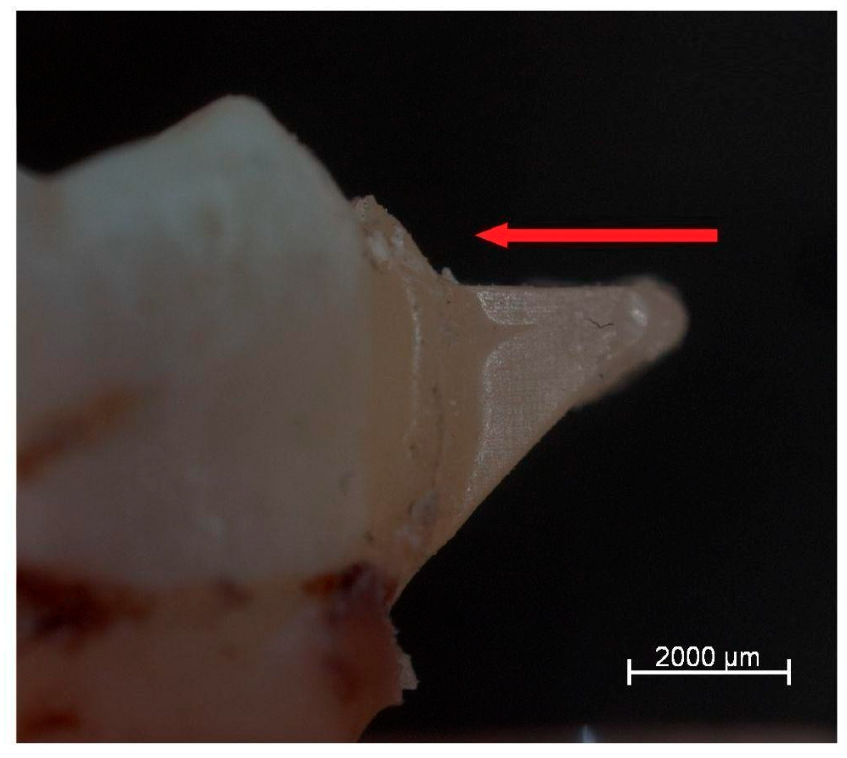
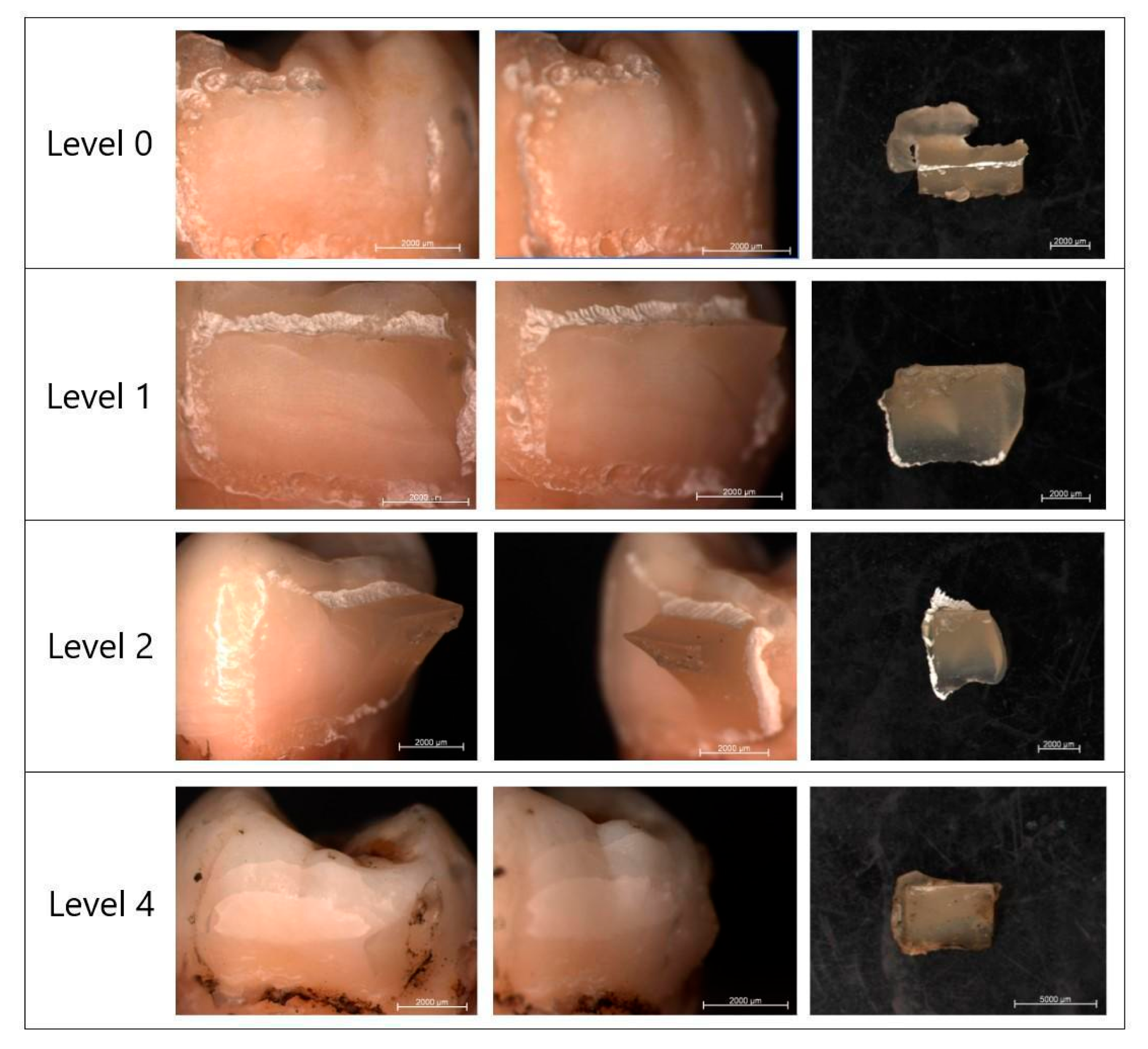
3.4. Morphological Evaluation of Enamel Surfaces After Attachment Removal

The final part of the analysis focused on assessing the condition of the enamel surface following the removal of orthodontic attachments. The extent of residual material was classified according to the Adhesive Remnant Index (ARI), which ranges from 0 (no residue) to 4 (enamel fracture) (see Figure 6). Although descriptive differences in ARI score distributions were observed among the groups, a chi-square test of independence did not reveal statistically significant differences (χ2 = 14.12, df = 9, p = 0.118). The results indicate some differences in the degree of residual adhesive left on the enamel among the examined groups. In the reference group, the ARI score distribution suggested a relatively high risk of enamel damage. Specifically, 19% of specimens showed signs of enamel fracture (score 4), and 38% retained less than half of the adhesive (score 1), with only 25% showing complete removal without residue (score 0). The diode laser group (D) presented a more favorable profile. The majority of specimens (67%) had less than half of the adhesive remaining (score 1), and 20% showed no residue at all (score 0). Enamel damage (score 4) was minimal, occurring in only 7% of cases. These results suggest that diode laser application tends to preserve enamel integrity while facilitating relatively clean detachment. In the CO2 laser group (C), the distribution was evenly spread among the first three ARI levels. One-third of specimens each fell into scores 0, 1, and 2, indicating moderate variability in bonding residue. No cases of enamel fracture were observed in this group. The Er:YAG laser group (E) showed the highest percentage of specimens with complete adhesive removal (67% scored 0), and no enamel damage was recorded. The remaining specimens had either less than half (11%) or more than half (22%) of the adhesive left. These results indicate that Er:YAG laser treatment was the most effective at minimizing both residual material and enamel damage during debonding (see Table 3).

4. Discussion

The primary goal of this study was to evaluate the effectiveness of three different laser systems—diode, CO2, and Er:YAG—in the debonding of composite attachments used during aligner orthodontic treatment, while also comparing the risk of enamel damage associated with these methods to that of traditional rotary instruments. The laser parameters used in this study were chosen based on manufacturer guidelines and the published literature to ensure clinically relevant and safe energy settings for effective composite removal while minimizing the risk of enamel damage [23,36]. The findings revealed significant variation between the groups in terms of both functional outcomes (shear bond strength and ARI score) and microstructural consequences (profilometry and micro-CT imaging). The Er:YAG laser emerged as the most effective tool for facilitating clean detachment of attachments with minimal damage to enamel, whereas traditional rotary methods—though providing the highest bond strength—were more likely to result in mechanical trauma to the enamel surface [36,37]. These observations suggest that while mechanical techniques remain effective from a strength perspective, their invasive nature may compromise the long-term integrity of dental hard tissues. Notably, the diode laser offered a middle-ground solution, enabling partial weakening of the adhesive interface with significantly less risk of enamel alteration [38]. Similar trends have been reported in prior research evaluating composite removal in orthodontics using laser technology, whereby laser-induced thermal or photoablative effects were shown to reduce iatrogenic damage when optimized correctly [39,40,41].
The microstructural effects of laser application were most clearly visualized using high-resolution X-ray micro-computed tomography (micro-CT). Among the tested modalities, Er:YAG laser irradiation resulted in the most profound changes, with enamel loss reaching up to 0.17 mm, which represented approximately 9% of the local enamel thickness in some regions. While this depth of removal suggests efficient ablation, it also highlights the importance of carefully controlling energy delivery to avoid unnecessary tissue loss [33,42,43,44]. In contrast, the CO2 laser produced moderate surface changes, largely confined to the outermost enamel layers, suggesting a more superficial thermal effect [45,46]. The diode laser, meanwhile, was associated with minimal structural impact, consistent with its lower affinity for hydroxyapatite and deeper penetration into pigmented soft tissue rather than mineralized enamel. These findings can be compared with earlier microtomographic studies by Dostalova et al. [47] and Fried et al. [48], who demonstrated similar patterns of localized tissue modification with Er:YAG and CO2 systems. Their results reinforce the interpretation that while Er:YAG is effective in facilitating detachment, its aggressive ablation can pose risks to enamel integrity if not carefully calibrated. As this study focused on energy application localized near the attachment, further research could explore the volumetric distribution of enamel changes in three dimensions.
Complementary data from optical profilometry further supported these observations by quantifying the extent of surface roughness changes post-debonding. In the RT group, surface disruption remained relatively limited (Sa = 0.31 µm), consistent with expectations for mechanical bur use under controlled conditions. Likewise, the diode laser group demonstrated minimal increase in roughness values (Sa = 0.30 µm), confirming its low-impact nature. However, the CO2 laser group showed significantly greater roughness (Sa = 0.79 µm; Sq = 1.24 µm), and the Er:YAG laser caused the most extensive changes (Sa = 2.03 µm; Sq = 4.19 µm), with Sz values exceeding 250 µm. This dramatic topographic variation reflects a highly ablative interaction, consistent with prior findings by Almeida et al. [49] and Yassaei et al. [32], who reported deep grooves and crater-like defects following laser application. Although this surface disruption can aid in mechanical detachment, excessive roughness poses risks such as increased bacterial adhesion, discoloration, and enamel erosion over time. Notably, the profilometric data correlated well with the micro-CT findings, further validating the method as a reliable technique for assessing enamel surface integrity post-debonding.
Mechanical testing via shear bond strength (SBS) provided further insight into the practical implications of these morphological changes. The highest bond strengths were observed in the reference group (τ = 196 MPa), wherein attachments were removed mechanically. However, this also corresponded with the greatest risk of enamel damage, suggesting that strong adhesion, while beneficial for clinical performance, may complicate safe removal. The diode laser group displayed a substantial reduction in bond strength (τ = 126 MPa), though still within acceptable clinical ranges, indicating partial thermal degradation of the bonding interface [50,51]. The CO2 laser group followed a similar trend (τ = 116 MPa), while the Er:YAG group recorded the lowest bond strength (τ = 82 MPa). These findings mirror those of Khalil et al. [34], who found that Er:YAG exposure significantly weakened the adhesive-to-enamel interface. The lowered resistance to shear may be advantageous in reducing trauma during debonding, provided that bond integrity during treatment remains uncompromised. Additionally, the displacement data (l) confirmed these mechanical patterns, with Er:YAG showing the shortest displacement prior to failure—suggesting a more brittle and predictable debonding mechanism.
To complement the SBS data, the Adhesive Remnant Index (ARI) was used to evaluate post-removal residue and enamel condition. The Er:YAG group once again performed best, with 67% of specimens showing no remaining adhesive (score 0) and no enamel fractures observed. This demonstrates the potential of Er:YAG in preserving enamel during detachment, likely due to its ability to break down the composite resin at the enamel–adhesive interface without compromising adjacent hard tissue. The diode and CO2 laser groups exhibited intermediate ARI profiles, with minimal enamel damage and variable residue [50]. In contrast, the reference group revealed the most unfavorable outcomes: only 25% scored 0, and 19% of samples presented visible enamel fracture (score 4). These results highlight the correlation between high bond strength and increased risk of cohesive failure or enamel damage. Similar patterns were noted by Morford et al. [51] and Rechmann et al. [52], who emphasized that lasers not only facilitate cleaner detachment but also minimize residual resin and reduce the need for invasive polishing or post-treatment correction.
Despite the promising results, this study has several limitations. Being an in vitro study, it does not fully replicate the complex intraoral environment, including factors such as humidity, saliva, occlusal forces, and biological variability. Additionally, while the sample size was sufficient for comparative purposes, larger multicenter studies are needed to generalize the findings. Another limitation concerns the lack of long-term evaluation of enamel after polishing and remineralization, which could affect interpretations of the clinical safety of various debonding methods. Although the laser parameters used in this study were selected based on previous research and clinical safety guidelines, no direct pulpal thermographic analysis was performed. Future in vitro or in vivo studies incorporating thermographic evaluation could provide additional confirmation of the thermal safety of these laser settings, especially in proximity to the pulp chamber. Advances in selective spectral feedback systems and shorter pulse-duration lasers may enable even more precise and tissue-specific composite removal protocols, as proposed by Chan et al. [53] and Dumore et al. [1], paving the way for safer and more predictable orthodontic procedures.

5. Conclusions

This in vitro study has demonstrated that laser-assisted debonding methods, particularly using the Er:YAG system, offer significant advantages over conventional rotary techniques in the removal of composite orthodontic attachments. While traditional methods provide the highest shear bond strength, they are also associated with the greatest risk of enamel surface damage and adhesive residue. In contrast, Er:YAG laser application facilitated efficient debonding, minimized residual material, and preserved enamel integrity, albeit at the cost of reduced bond strength.
The CO2 and diode lasers showed intermediate performance. The CO2 laser provided moderate surface alteration and bond strength reduction, while the diode laser caused minimal morphological changes and preserved acceptable adhesive strength. These findings support the potential of lasers, especially Er:YAG, as a safe and effective tool for clinical debonding procedures.
However, careful parameter selection and further research—particularly under in vivo conditions—are essential to ensure optimal outcomes. Future studies should focus on refining clinical protocols, evaluating patient-centered outcomes, and minimizing adverse thermal or structural effects on dental tissues.

Author Contributions

Conceptualization, J.M. and A.F.-M.; methodology, A.F.-M.; software, A.N.; validation, A.F.-M., A.N. and J.M.; formal analysis, K.G.-L.; investigation, A.F.-M. and A.N.; resources, A.F.-M.; data curation, A.N.; writing—original draft preparation, A.F.-M. and J.K.; writing—review and editing, J.M. and K.G.-L.; visualization, A.N.; supervision, K.G.-L. and J.M.; project administration, K.G.-L.; funding acquisition, K.G.-L. All authors have read and agreed to the published version of the manuscript.

Funding

This research received no external funding.

Institutional Review Board Statement

Bioethics Committee approval No. KB 423/2023N (25 January 2024).

Informed Consent Statement

Informed consent was obtained from all subjects involved in the study.

Data Availability Statement

Data are contained within the article.

Conflicts of Interest

The authors declare no conflicts of interest.

References

  1. Grassia, V.; Fiori, A.; Diodati, F.; Sayahpour, B.; Jamilian, A.; Armogida, N.G.; d’Apuzzo, F.; Nucci, L. Clear aligners: A network and bibliometric analysis of 50 pivotal articles. Dent. Med. Probl. 2025, 62, 161–171. [Google Scholar] [CrossRef]
  2. Hong, K.; Kim, W.-H.; Eghan-Acquah, E.; Lee, J.-H.; Lee, B.-K.; Kim, B. Efficient Design of a Clear Aligner Attachment to Induce Bodily Tooth Movement in Orthodontic Treatment Using Finite Element Analysis. Materials 2021, 14, 4926. [Google Scholar] [CrossRef]
  3. Ju, G.-Y.; Oh, S.; Lim, B.-S.; Lee, H.-S.; Chung, S.H. Effect of Simplified Bonding on Shear Bond Strength between Ceramic Brackets and Dental Zirconia. Materials 2019, 12, 1640. [Google Scholar] [CrossRef]
  4. Bae, G.-S. Clinical limitations and its solutions of the clear overlay appliance treatment. J. Korean Dent. Assoc. 2016, 54, 563–574. [Google Scholar] [CrossRef]
  5. Behnaz, M.; Farahnaki, A.; Rahimipour, K.; Mousavi, R.; Davoodi, N.S. Lingual Orthodontic Treatment: Efficacy and Complications. J. Adv. Oral Res. 2019, 10, 65–74. [Google Scholar] [CrossRef]
  6. Boyd, R.L. Esthetic Orthodontic Treatment Using the Invisalign Appliance for Moderate to Complex Malocclusions. J. Dent. Educ. 2008, 72, 948–967. [Google Scholar] [CrossRef]
  7. Breece, G.L.; Nieberg, L.G. Motivations for Adult Orthodontic Treatment. J. Clin. Orthod. 1986, 20, 166–171. [Google Scholar]
  8. Kim, H.; Choi, B.; Lim, S.; Gang, S. A Comparative Study about Bonding Strength of Customized Metalbase for Lingual Orthodontic Appliance Made by Non-Precious Metal. Oral Biol. Res. 2016, 40, 187–192. [Google Scholar]
  9. Kravitz, N.D.; Kusnoto, B.; BeGole, E.; Obrez, A.; Agran, B. How Well Does Invisalign Work? A Prospective Clinical Study Evaluating the Efficacy of Tooth Movement with Invisalign. Am. J. Orthod. Dentofac. Orthop. 2009, 135, 27–35. [Google Scholar] [CrossRef]
  10. Elkharashi, A.; Grzech-Leśniak, K.; Deeb, J.G.; Abdulmajeed, A.A.; Bencharit, S. Exploring the use of pulsed erbium lasers to retrieve a zirconia crown from a zirconia implant abutment. PLoS ONE 2020, 15, e0233536. [Google Scholar] [CrossRef]
  11. Lizarelli, R.D.F.; Moriyama, L.T.; Bagnato, V.S. Ablation of Composite Resins Using Er:YAG Laser—Comparison with Enamel and Dentin. Lasers Surg. Med. 2003, 33, 132–139. [Google Scholar] [CrossRef] [PubMed]
  12. McNamara, J.A.; Kramer, K.L.; Juenker, J.P. Invisible Retainers. J. Clin. Orthod. 1985, 19, 570–578. [Google Scholar] [PubMed]
  13. Meier, B.; Wiemer, K.B.; Miethke, R.-R. Invisalign--Patient Profiling. Analysis of a Prospective Survey. J. Orofac. Orthop. Der Kieferorthopadie 2003, 64, 352–358. [Google Scholar] [CrossRef] [PubMed]
  14. Melsen, B. Northcroft Lecture: How Has the Spectrum of Orthodontics Changed over the Past Decades? J. Orthod. 2011, 38, 134–143. [Google Scholar] [CrossRef]
  15. Locadia, M.; Sprangers, M. Kwaliteit van Leven. In Medische Psychologie; Bohn Stafleu van Loghum: Houten, The Netherlands, 2006; pp. 155–160. ISBN 978-90-313-4462-8. [Google Scholar]
  16. Lascala, N.; Moussalli, N. Laser Therapy in Periodontics. In Periodontal Therapeutic Compendium, 3rd ed.; Artes Médicas: São Paulo, Brazil, 1999; pp. 293–307. [Google Scholar]
  17. Rahimipour, K.; Mousavi, R.; Behnaz, M.; Dalaie, K.; Farahnaki, A.; Davoodi, N.S. Invisalign Orthodontic System: Treatment Efficacy, Complications, Patients’ Attitude. Orthod. Forum 2020, 15, 108–123. [Google Scholar] [CrossRef]
  18. Reinisch, L. Laser Physics and Tissue Interactions. Otolaryngol. Clin. N. Am. 1996, 29, 893–914. [Google Scholar] [CrossRef]
  19. Deeb, J.G.; Grzech-Leśniak, K.; Weaver, C.; Matys, J.; Bencharit, S. Retrieval of Glass Fiber Post Using Er:YAG Laser and Conventional Endodontic Ultrasonic Method: An In Vitro Study. J. Prosthodont. 2019, 28, 1024–1028. [Google Scholar] [CrossRef]
  20. Ryf, S.; Flury, S.; Palaniappan, S.; Lussi, A.; van Meerbeek, B.; Zimmerli, B. Enamel loss and adhesive remnants following bracket removal and various clean-up procedures in vitro. Eur. J. Orthod. 2012, 34, 25–32. [Google Scholar] [CrossRef]
  21. Deeb, J.G.; Skrjanc, L.; Kanduti, D.; Carrico, C.; Saturno, A.M.; Grzech-Leśniak, K. Evaluation of Er:YAG and Er,Cr:YSGG laser irradiation for the debonding of prefabricated zirconia crowns. Adv. Clin. Exp. Med. 2021, 30, 7–15. [Google Scholar] [CrossRef]
  22. Ireland, A.J.; Hosein, I.; Sherriff, M. Enamel loss at bond-up, debond and clean-up following the use of a conventional light-cured composite and a resin-modified glass polyalkenoate cement. Eur. J. Orthod. 2005, 27, 413–419. [Google Scholar] [CrossRef]
  23. Deeb, J.G.; Grzech-Leśniak, K.; Brody, E.R.; Matys, J.; Bencharit, S. Erbium Laser-assisted Ceramic Debonding: A Scoping Review. J. Prosthodont. 2022, 31, E100–E124. [Google Scholar] [CrossRef] [PubMed]
  24. Nelson, J.S.; Yow, L.; Liaw, L.H.; Macleay, L.; Zavar, R.B.; Orenstein, A.; Wright, W.H.; Andrews, J.J.; Berns, M.W. Ablation of Bone and Methacrylate by a Prototype Mid-Infrared Erbium:YAG Laser. Lasers Surg. Med. 1988, 8, 494–500. [Google Scholar] [CrossRef] [PubMed]
  25. Niemz, M.H. Laser-Tissue Interactions: Fundamentals and Applications, 4th ed.; Springer International Publishing AG: Cham, Switzerland, 2019; ISBN 978-3-030-11917-1. [Google Scholar]
  26. Oztoprak, M.O.; Nalbantgil, D.; Erdem, A.S.; Tozlu, M.; Arun, T. Debonding of Ceramic Brackets by a New Scanning Laser Method. Am. J. Orthod. Dentofac. Orthop. 2010, 138, 195–200. [Google Scholar] [CrossRef] [PubMed]
  27. Oztoprak, M.O.; Tozlu, M.; Iseri, U.; Ulkur, F.; Arun, T. Effects of Different Application Durations of Scanning Laser Method on Debonding Strength of Laminate Veneers. Lasers Med. Sci. 2012, 27, 713–716. [Google Scholar] [CrossRef]
  28. Glinkowski, W.; Pokora, L. Lasery w Terapii; Laser Instruments-Centrum Techniki Laserowej: Warszawa, Poland, 1993. [Google Scholar]
  29. Niemz, M.H. Laser–Tissure Interactions—Fundamentals and Applications, 3rd ed.; Springer Press: Heidelberg, Germany, 2003. [Google Scholar]
  30. Kuhn, K.; Schmid, C.U.; Luthardt, R.G.; Rudolph, H.; Diebolder, R. Er:YAG laser-induced damage to a dental composite in simulated clinical scenarios for inadvertent irradiation: An in vitro study. Lasers Med. Sci. 2022, 37, 1017–1030. [Google Scholar] [CrossRef]
  31. Yang, X.; Ye, S.; Wei, B.; Zeng, L. Removal of resin cement using a picosecond laser: An in vitro study. J. Prosthet. Dent. 2025, 133, 1058.e1–1058.e9. [Google Scholar] [CrossRef]
  32. Yassaei, S.; Aghili, H.; Joshan, N. Effects of Removing Adhesive from Tooth Surfaces by Er:YAG Laser and a Composite Bur on Enamel Surface Roughnessand Pulp Chamber Temperature. Dent. Res. J. 2015, 12, 254–259. [Google Scholar]
  33. Ajwa, N.; Alfayez, H.; Al-Oqab, H.; Melibary, R.; Alzamil, Y. The Effect of Erbium-Doped Yttrium Aluminum Garnet Laser in Debonding of Orthodontic Brackets: A Systematic Review of the Literature. Photobiomodulation Photomed. Laser Surg. 2021, 39, 725–733. [Google Scholar] [CrossRef]
  34. Khalil, A.S.; Tamish, N.M.; Elkalza, A.R. Assessment of Chemical, Ultrasonic, Diode Laser, and Er:YAG Laser Application on Debonding of Ceramic Brackets. BMC Oral Health 2022, 22, 79. [Google Scholar] [CrossRef]
  35. Rechmann, P.; Buu, N.C.H.; Rechmann, B.M.T.; Le, C.Q.; Finzen, F.C.; Featherstone, J.D.B. Laser All-Ceramic Crown Removal-a Laboratory Proof-of-Principle Study-Phase 1 Material Characteristics. Lasers Surg. Med. 2014, 46, 628–635. [Google Scholar] [CrossRef]
  36. Michalak, M.; Kiryk, S.; Kotela, A.; Wiśniewska, K.; Kiryk, J.; Zborowski, J.Z.; Matys, J.; Dobrzyński, M. Orthodontic Ceramic Bracket Removal Using Lasers: A Systematic Review. J. Funct. Biomater. 2025, 16, 123. [Google Scholar] [CrossRef] [PubMed]
  37. Paolone, G.; Mandurino, M.; Baldani, S.; Paolone, M.G.; Goracci, C.; Scolavino, S.; Gherlone, E.; Cantatore, G.; Gastaldi, G. Quantitative Volumetric Enamel Loss after Orthodontic Debracketing/Debonding and Clean-Up Procedures: A Systematic Review. Appl. Sci. 2023, 13, 5369. [Google Scholar] [CrossRef]
  38. Zheng, X.; Huang, X. Evaluation of the Re-Bond Strength of Debonded Metal and Ceramic Brackets Following Er: YAG Laser Treatment. BMC Oral Health 2024, 24, 710. [Google Scholar] [CrossRef] [PubMed]
  39. De Souza Henkin, F.; Macêdo, E.; da Silva Santos, K.; Schwarzbach, M.; Samuel, S.M.W.; Mundstock, K.S. In Vitro Analysis of Shear Bond Strength and Adhesive Remnant Index of Different Metal Brackets. Dent. Press J. Orthod. 2016, 21, 67–73. [Google Scholar] [CrossRef]
  40. Grzech-Leśniak, K.; Matys, J.; Żmuda-Stawowiak, D.; Mroczka, K.; Dominiak, M.; Brugnera Junior, A.; Gruber, R.; Romanos, G.E.; Sculean, A. Er:YAG Laser for Metal and Ceramic Bracket Debonding: An In Vitro Study on Intrapulpal Temperature, SEM, and EDS Analysis. Photomed. Laser Surg. 2018, 36, 595–600. [Google Scholar] [CrossRef]
  41. Indumathi, G.; Premkumar, S.; Amit, K. Assessment of Enamel Loss After Debonding of Ceramic, Composite Plastic And Metal Brackets-An In Vitro Study. J. Contemp. Orthod. 2019, 3, 1–10. [Google Scholar] [CrossRef]
  42. Vidor, M.M.; Felix, R.P.; Marchioro, E.M.; Hahn, L. Enamel Surface Evaluation after Bracket Debonding and Different Resin Removal Methods. Dent. Press J. Orthod. 2015, 20, 61–67. [Google Scholar] [CrossRef]
  43. Tocchio, R.M.; Williams, P.T.; Mayer, F.J.; Standing, K.G. Laser Debonding of Ceramic Orthodontic Brackets. Am. J. Orthod. Dentofac. Orthop. 1993, 103, 155–162. [Google Scholar] [CrossRef]
  44. Kiryk, J.; Matys, J.; Nikodem, A.; Burzyńska, K.; Grzech-Leśniak, K.; Dominiak, M.; Dobrzyński, M. The Effect of Er:YAG Laser on Shear Bond Strength of Orthodontic Brackets to Enamel—A Preliminary Study. Materials 2021, 14, 2093. [Google Scholar] [CrossRef]
  45. Chan, K.H.; Hirasuna, K.; Fried, D. Analysis of Enamel Surface Damage after Selective Laser Ablation of Composite from Tooth Surfaces. Photonics Lasers Med. 2014, 3, 37–45. [Google Scholar] [CrossRef]
  46. Obata, A.; Tsumura, T.; Niwa, K.; Ashizawa, Y.; Deguchi, T.; Ito, M. Super Pulse CO2 Laser for Bracket Bonding and Debonding. Eur. J. Orthod. 1999, 21, 193–198. [Google Scholar] [CrossRef] [PubMed]
  47. Dostalova, T.; Jelinkova, H.; Remes, M.; Šulc, J.; Němec, M. The Use of the Er:YAG Laser for Bracket Debonding and Its Effect on Enamel Damage. Photomed. Laser Surg. 2016, 34, 394–399. [Google Scholar] [CrossRef] [PubMed]
  48. Fried, W.A.; Chan, K.H.; Darling, C.L.; Fried, D. Use of a DPSS Er:YAG Laser for the Selective Removal of Composite from Tooth Surfaces. Biomed. Opt. Express 2018, 9, 5026–5036. [Google Scholar] [CrossRef] [PubMed]
  49. Almeida, H.C.; Vedovello Filho, M.; Vedovello, S.A.; Young, A.A.A.; Ramirez-Yanez, G.O. ER: YAG Laser for Composite Removal after Bracket Debonding: A Qualitative SEM Analysis. Int. J. Orthod. 2009, 20, 9–13. [Google Scholar]
  50. Mousa, M.M.; Al-Jannan, H.M.G.; Sultan, K.; Ajaj, M.A.; Hajeer, M.Y.; Al-Manadili, A.; Ammar, A.M.; Awawdeh, M. Effectiveness of Two Intensity Levels of Diode Laser in Debonding Metallic Brackets Regarding Enamel Surface Integrity and Pulpal Temperature: An Ex-Vivo Study. Cureus 2023, 15, e41372. [Google Scholar] [CrossRef]
  51. Matys, J.; Grzech-Leśniak, K.; Flieger, R.; Dominiak, M. Assessment of an Impact of a Diode Laser Mode with Wavelength of 980 Nm on a Temperature Rise Measured by Means of K-02 Thermocouple: Preliminary Results. Dent. Med. Probl. 2016, 53, 345–351. [Google Scholar] [CrossRef]
  52. Morford, C.K.; Buu, N.C.H.; Rechmann, B.M.T.; Finzen, F.C.; Sharma, A.B.; Rechmann, P. Er:YAG Laser Debonding of Porcelain Veneers. Lasers Surg. Med. 2011, 43, 965–974. [Google Scholar] [CrossRef]
  53. Chan, K.H.; Hirasuna, K.; Fried, D. Rapid and Selective Removal of Composite from Tooth Surfaces with a 9.3 µm CO2 Laser Using Spectral Feedback. Lasers Surg. Med. 2011, 43, 824–832. [Google Scholar] [CrossRef]
Figure 1. The subsequent stages of preparation are as follows: (A) Enamel etching with o-phosphoric acid; (B,C) application of modelling liquid to the matrix; (D) filling the mold with the composite; (E) bonding the attachment to enamel; (F) polymerization with a FlashMax lamp; (G,H) the final result with the attachment in place.
Photonics 12 00621 g001
Figure 2. Direction of laser irradiation during the debonding procedure. The red arrow indicates the horizontal path of the laser beam, directed perpendicularly to the buccal enamel surface along the edge of the composite attachment.
Photonics 12 00621 g002
Figure 3. Schematic of the test rig for measuring the detachment force of abutments from enamel.
Photonics 12 00621 g003
Figure 4. (A) Surface difference images of a premolar preparation after Er:YAG laser application. The red color indicates the difference. (B) Measurement of the thickness (CTAn, Kontich, Bruker) of the damaged enamel (shown in red) during Er:YAG laser preparation.
Photonics 12 00621 g004
Figure 5. Comparing the morphology components of the specimens: height distribution, waviness and roughness among groups.
Photonics 12 00621 g005
Figure 6. Examples of specimens illustrating the different ARI scores defined for the attachment–enamel interface.
Photonics 12 00621 g006
Table 1. Comparison of the values of the roughness parameters (Sa, Sq, Sz, Sp, and Sv) of the enamel surface for the different measurement groups.
Roughness ParametersReferenceRTGroup DGroup CGroup Ep-Value
Mean ± SDMean ± SDMean ± SDMean ± SDMean ± SD
Sa [µm]0.28 ± 0.040.31 ± 0.080.30 ± 0.030.79 ± 0.062.04 ± 0.08<0.05 for CvsE; C, EvsReference, RT, D
Sq [µm]0.39 ± 0.050.46 ± 0.140.42 ± 0.041.24 ± 0.104.19 ± 0.12<0.05 for CvsE; C, EvsReference, RT, D
Sz [µm]11.49 ± 3.8427.44 ± 17.3311.53 ± 3.6143.30 ± 3.23 *251.21 ± 3.23 *<0.05 for CvsE; C, EvsReference, RT, D; RTvsReference, D
Sp [µm]6.71 ± 3.409.02 ± 6.996.79 ± 2.3420.49 ± 0.37 *125.79 ± 1.17 *<0.05 for CvsE; C, EvsReference, RT, D
Sv [µm]4.78 ± 1.2318.44 ± 20.884.74 ± 1.2622.80 ± 3.13 *125.42 ± 2.06 *<0.05 for CvsE; C, EvsReference, RT, D; RTvsReference, D
Sa [µm]—arithmetic mean height of the surface; Sq [µm]—root mean square height of the surface; Sz [µm]—maximum surface height (peak-to-valley); Sp [µm]—maximum peak height; Sv [µm]—maximum valley depth; Mean ± SD—mean value and standard deviation; *—statistically significant difference versus the reference group, applicable only to groups C and E (p < 0.05).
Table 2. Comparison of the values of the mechanical parameters obtained in the shear test (τ, Fmax, and l) for the different groups of measurements.
Mechanical Parametersτ [MPa]F [N]l [mm]p-Value
Mean ± SDMean ± SDMean ± SD
Reference196.189 ± 75.2915.189 ± 4.570.637 ± 0.21<0.05
Group D126.008 ± 80.589.764 ± 6.060.413 ± 0.28<0.05
Group C116.360 ± 43.26 *9.292 ± 3.360.400 ± 0.13 *<0.05
Group E81.679 ± 45.52 *6.080 ± 2.97 *0.294 ± 0.13 *<0.05
p-value<0.05 for C; EvsReference, EvsC, D<0.05 for C; EvsReference, EvsC, D<0.05 for C; EvsReference, EvsC, D-
τ [MPa]—shear stress; F [N]—maximum detachment force; [mm]—displacement in millimeters; Mean ± SD—mean value and standard deviation); * p < 0.05.
Table 3. Distribution of ARI scores showing the degree of residual adhesive material left on the enamel surface across the study groups (χ2 = 14.12, df = 9, p = 0.118).
ARI Score01234
Reference25%38%19%0%19%
Group D20%67%7%0%7%
Group C33%33%33%0%0%
Group E67%11%22%0%0%
ARI—Adhesive Remnant Index.
Disclaimer/Publisher’s Note: The statements, opinions and data contained in all publications are solely those of the individual author(s) and contributor(s) and not of MDPI and/or the editor(s). MDPI and/or the editor(s) disclaim responsibility for any injury to people or property resulting from any ideas, methods, instructions or products referred to in the content.

