Review

Optical Fiber Sensing Technologies in Radiation Therapy

Department of Radiation Oncology, Cedars-Sinai Medical Center, Los Angeles, CA 90048, USA
*
Authors to whom correspondence should be addressed.
Photonics 2025, 12(11), 1058; https://doi.org/10.3390/photonics12111058
Submission received: 8 September 2025 / Revised: 20 October 2025 / Accepted: 24 October 2025 / Published: 25 October 2025

Abstract

Optical fiber technology is becoming essential in modern radiation therapy, enabling precise, real-time, and minimally invasive monitoring. As oncology moves toward patient-specific treatment, there is growing demand for adaptable and biologically compatible sensing tools. Fiber-optic systems meet this need by integrating into clinical workflows with highly localized dosimetric and spectroscopic feedback. Their small size and flexibility allow deployment within catheters, endoscopes, or treatment applicators, making them suitable for both external beam and internal therapies. This paper reviews the fundamental principles and diverse applications of optical fiber sensing technologies in radiation oncology, focusing on dosimetry, spectroscopy, imaging, and adaptive radiotherapy. Implementations such as scintillating and Bragg grating-based dosimeters demonstrate feasibility for in vivo dose monitoring. Spectroscopic techniques, such as Raman and fluorescence spectroscopy, offer real-time insights into tissue biochemistry, aiding in treatment response assessment and tumor characterization. However, despite such advantages of optical fiber sensors, challenges such as signal attenuation, calibration demands, and limited dynamic range remain. This paper further explores clinical application, technical limitations, and future directions, emphasizing multiplexing capabilities, integration and regulatory considerations, and trends in machine learning development. Collectively, these optical fiber sensing technologies show strong potential to improve the safety, accuracy, and adaptability of radiation therapy in personalized cancer care.

1. Introduction

Radiation therapy remains a cornerstone of cancer treatment, used in approximately 50% of all cancer cases, either as a standalone modality or in combination with surgery and chemotherapy [1]. Its fundamental aim is to eradicate malignant cells while preserving the function of surrounding healthy tissues. Over the past few decades, radiation oncology has witnessed profound technological innovation with the emergence of highly sophisticated modalities such as intensity-modulated radiation therapy (IMRT) [2], stereotactic body radiotherapy (SBRT) [3], image-guided radiotherapy (IGRT) [4], proton and heavy-ion therapies [5,6], and adaptive radiotherapy [7]. These advances have allowed clinicians to shape radiation beams with sub-millimeter precision and escalate doses to tumors with greater confidence. Despite advances in beam delivery and anatomical imaging, treatment effectiveness still faces limitations due to the biological heterogeneity of tumors and the lack of real-time functional feedback during therapy. These limitations hinder the ability to adapt treatment plans to spatially varying and temporally evolving tumor responses, increasing the risk of underdosing resistant regions or overdosing healthy tissue.
To address this gap, there has been a growing movement toward the integration of physiological and molecular data into the radiation therapy workflow [8,9]. Traditional imaging modalities, such as computed tomography (CT) and magnetic resonance imaging (MRI), provide structural or positional information with limited insight into the cellular responses of tissues. The introduction of positron emission tomography (PET)/CT imaging has provided functional or biological insights into tumor hypoxia and proliferation for modification of treatment target volumes [10,11]. However, due to its low signal-to-noise ratio and restricted resolution, micro disease may go undetected [12]. As a result, adaptive radiation therapy (ART) [13], which adjusts parameters in a treatment plan over the course of treatment based on observed changes in anatomy or tumor characteristics, is gaining attention. This technique requires tools capable of dynamic, in vivo monitoring of both radiation dose and biological markers, an area where optical fiber technologies could show significant promise.
Optical fibers offer a range of characteristics that make them ideal for clinical use in radiation oncology. Their miniature size allows for easy integration into treatment devices such as catheters, brachytherapy applicators, or even wearable sensors [14,15,16]. They are inherently biocompatible and exhibit strong resistance to electromagnetic interference, making them particularly suitable for use in high-field environments like MRI-guided radiotherapy [17]. Additionally, they can transmit light for sensing, imaging, and data acquisition over long distances with minimal signal loss. These properties enable optical fibers to support continuous, real-time monitoring of parameters such as radiation dose distribution, temperature fluctuations, and molecular changes in irradiated tissues [18]. Furthermore, optical fiber systems are uniquely compatible with emerging radiobiological monitoring approaches. Techniques such as Raman spectroscopy [19,20,21] and fluorescence sensing [22,23,24] can be implemented through fiber-optic probes [25], enabling biochemical assessments at the cellular level and differentiation of cancerous vs. normal tissues, without the need for invasive biopsies or exogenous contrast agents. Such information can provide early indicators of treatment efficacy or toxicity, allowing clinicians to adapt therapy plans on a patient-specific basis. By combining dosimetric accuracy with biological insight, optical fiber technologies stand to enhance the precision, responsiveness, and safety of radiation therapy, ultimately contributing to more personalized and effective cancer care.

2. Optical Fiber Dosimetry

Optical fiber-based dosimetry has emerged as a significant advancement in the real-time monitoring of radiation dose delivery. Unlike conventional dosimeters, such as ionization chambers, which are widely used for reference dosimetry due to their accuracy and reproducibility, fiber-optic dosimeters (FODs) offer unique advantages. For example, while ion chambers serve as the gold standard in clinical settings, they are also relatively large, fragile, and require high-voltage power supplies, which limit their application for in vivo or high-resolution spatial measurements. In contrast, FODs are miniaturized, immune to electromagnetic interference, flexible, and capable of remote sensing with low signal loss. A variety of FODs have been developed, utilizing these fiber properties, to facilitate measurement of the radiation dose [18,26,27]. Compared to ionization chambers, which offer excellent absolute dose measurement accuracy (~2% uncertainty) but limited spatial resolution (typically >2 mm), scintillating fiber dosimeters have demonstrated sub-millimeter spatial resolution (as low as ~0.5 mm) and dose linearity within ~2%, depending on the scintillator and readout method used [28,29,30,31]. Similarly, fiber Bragg gratings can detect temperature changes with ~0.1 °C sensitivity and resolve strain changes at the microstrain level, while Raman probes have demonstrated high signal-to-noise ratios (SNRs) in selected spectral bands (for example, SNR > 20 in the 1440 cm−1 band) [32,33,34]. These quantitative benchmarks suggest that while optical fiber systems may not yet replace ion chambers for reference dosimetry, they are well suited for high-resolution, real-time in vivo measurements in complex geometries or under MRI-guided conditions.

2.1. Scintillating Fiber Dosimeters

Scintillating fiber dosimeters (SFDs), as shown in Figure 1a, are an extensively studied class of FODs designed to measure ionizing radiation in real time [35]. These systems utilize scintillating materials, such as plastic scintillators [30,36,37] or inorganic materials [38] like Bismuth Germanate (BGO) [39], Lutetium Yttrium Orthosilicate (LYSO) [40], or Gadolinium Oxysulfide (Gd2O2S) [41], that emit photons in the visible spectrum when exposed to ionizing radiation. The emitted scintillation light is then transmitted via an optical fiber to a photodetector located outside the irradiation zone, where the measured light intensity is proportional to the absorbed dose.
These high-resolution, real-time dosimeters are especially advantageous when steep dose gradients and dynamic delivery protocols require accurate and immediate feedback. Their compact and flexible design enables easy integration into narrow applicators, catheters, or confined anatomical regions, supporting in vivo dosimetry in procedures such as brachytherapy and pediatric radiotherapy [42,43]. Unlike electronic detectors, they are immune to electromagnetic interference, allowing for safe use in MRI-guided radiotherapy. Furthermore, fibers can be multiplexed or arranged in arrays to provide spatially distributed, two or three-dimensional dose measurement in complex field settings [44,45].
Despite such advantages, SFDs also face certain limitations, most notably, Cherenkov radiation contamination (the “stem effect”) [17,18]. This effect arises from the generation of broadband light within optical fiber when high-energy charged particles travel faster than the speed of light in the medium, producing a background signal that can interfere with accurate dose readings. In practice, Cherenkov contamination can result in overestimation of absorbed dose by up to 20% in plastic scintillators if left uncorrected [46]. To mitigate this, dual-channel detection schemes, which subtract Cherenkov-only signal from the total light, can suppress unwanted signal in clinical beam conditions [47,48]. Similarly, spectral filtering has been demonstrated to reduce stem effect contributions by more than 80%, depending on the overlap between the emission spectrum of the scintillator and the Cherenkov background [49]. Time-gated acquisition further improves measurement fidelity to ~74–90% by rejecting early Cherenkov photons, and enables isolation of scintillation light in nanosecond-resolved systems [35,50,51]. These methods can reduce residual dose readout error to below 5–10%, enabling clinical-grade dosimetry under high-energy beams. Current research is also improving scintillator materials, fiber designs, and signal processing to enhance performance [48]. As these advancements progress, SFDs are better positioned to support high-precision, adaptive, and personalized radiation therapy.

2.2. Optically Stimulated Luminescence Fiber Dosimeters

Another FOD technique employs optically stimulated luminescence (OSL), as shown in Figure 1b, where specially doped fiber materials store radiation energy and later release it as light when stimulated by a secondary light source [52]. In these dosimeters, the optical fiber is doped with luminescent materials, commonly aluminum oxide (Al2O3:C) and other rare-earth-doped compounds, that can trap energy when exposed to ionizing radiation [53,54,55]. Upon later stimulation by a typically blue or green light source, the stored energy is released as luminescent light, with intensity proportional to absorbed dose. This feature allows for flexible, retrospective dosimetry, making OSL FODs useful in scenarios where real-time monitoring is not required or where stored-dose analysis is advantageous, such as environmental or personal monitoring, quality assurance, and treatment verification.
A significant benefit of OSL FODs lies in their potentially high signal stability, reusability, and wide dynamic range of detection (this term refers to the dosimeter’s ability to measure doses from sub-cGy levels to tens of Gy without saturation, offering compatibility with both low-dose and high-dose clinical scenarios). Unlike thermoluminescent dosimeters (TLDs), which require heating for readout, OSL systems can be optically stimulated without affecting the physical integrity of the dosimeter [56]. This makes them more suitable for repeated use and longitudinal measurements. Moreover, when coupled with fiber-optic transmission, OSL systems can be deployed in remote or sensitive anatomical locations and interrogated post-irradiation with minimal patient discomfort [57]. Their robustness and resistance to environmental perturbations also make them ideal for use in conditions where electronic detectors may be compromised, such as in MRI-compatible treatment rooms or high-dose environments.
Conventional OSL dosimeters (OSLDs), such as the widely used Landauer Luxel and Al2O3:C Dot detectors, are well-established in radiation oncology clinics for patient dose verification and personnel monitoring [58]. However, OSL fiber dosimeters offer added flexibility in geometry and integration. Their cylindrical form and bendability allow better conformity to irregular surfaces or internal cavities, enabling 3D dose mapping in phantoms or catheters, where planar badge-style OSLDs are difficult to deploy spatially.
Fiber-optic OSL dosimeters have shown promising performance in phantom and bench-top settings. For example, a miniaturized fiber dosimeter (125 µm optical fiber) was developed in 2019 coupling scintillator to the fiber end, and showed linear signal response with an SNR as high as 190 under 6 MV irradiation in air [14]. This demonstrates that compact fiber probes can maintain linearity in clinically relevant photon fields. In the domain of fiber-coupled OSL, fiber-coupled Al2O3:C with 15 m fiber cables was studied to find that delay of 1 h before readout introduces only ~0.5% variation in OSL signal, indicating system stability over typical treatment times [59]. Furthermore, BeO OSL detectors were characterized showing linearity comparable to ionization chambers over a clinical dose range and favorable temperature and angle behavior [60]. While none of these systems yet shows extremely wide dynamic ranges (e.g., >103) or long reuse cycles (>50), these studies offer concrete benchmark metrics (linearity, SNR, readout stability) that should guide future development of OSL-based fiber dosimeters.
Recent demonstrations in live and clinical settings are trending to support the translation of fiber-optic dosimetry toward in vivo use. For instance, in 2023, a ruby-based fiber optic probe was developed for real-time dose-rate measurement during internal β-radiation therapy (e.g., selective internal radiation therapy, SIRT). It achieved linear dose-rate response across a range of exposures, and after applying optical filtering to suppress Cherenkov/stem signals, the residual stem contribution was limited to 2.3% ± 1.1% [61]. Similarly, in 2023, a fiber-optical dosimetry sensor was proposed for radiation measurement in biological applications, demonstrating radioluminescence response under gamma irradiation with a compact geometry suitable for biological environments [62]. Another relevant study in 2024 investigated the biocompatibility of a fiber-optic dosimeter in biological media, which assessed sensor response stability, radiation-induced damage in surrounding media, and long-term operation in physiologic conditions [15]. While human in vivo trials remain sparse, optical-fiber-based real-time dosimeters have been used in phantom and catheter-mounted settings [63]. These results collectively suggest that fiber-based optical dosimetry is transitioning from proof-of-concept toward in situ deployment in biological environments.
Despite their advantages, the practical deployment of OSL fiber dosimeters still presents several challenges. The need for post-irradiation optical stimulation introduces logistical constraints in certain clinical workflows, particularly where immediate dose verification is essential [64]. Energy dependence of Al2O3:C is another consideration, especially in kilovoltage photon beams where the response can vary significantly with beam quality [58]. Additionally, while dose linearity is generally good over a broad range, nonlinear response may appear at very low doses or in ultra-high dose-rate settings, necessitating careful calibration. Moreover, the spatial resolution can be limited by the uniformity and geometry of the doped fiber materials [14]. However, recent advances in material science, such as the development of nanocrystalline OSL compounds and enhanced fiber doping techniques, are addressing these limitations [18]. Current research also explores integrating OSL sensing with fiber Bragg gratings (FBGs) for simultaneous measurement of dose, temperature, and mechanical stress, and utilizing multiplexed fiber arrays for spatially resolved dosimetry [16,18]. With continued innovations, OSL-based FODs are expected to play an increasingly important role in comprehensive, multi-parametric radiation monitoring for both clinical and research purposes.

2.3. Other Types of Fiber Dosimeters

In addition to scintillating and optically stimulated luminescence dosimeters, several other types of FODs have been developed to expand the capabilities of radiation monitoring. TLD fibers, for instance, incorporate traditional TLD materials [65], such as LiF or CaSO4:Dy, at the tip or along the length of optical fibers. These materials store radiation energy and emit light upon heating, with the emitted light transmitted through the fiber to a remote detector, improving the spatial precision of conventional TLD systems. Another emerging class involves Cherenkov fiber dosimeters, which measure the Cherenkov light generated directly in the fiber when high-energy charged particles travel faster than the speed of light in the medium [66,67]. While typically considered background noise in scintillating systems, Cherenkov light can be isolated and quantified as a surrogate for dose distribution when combined with time-gated detection and spectral filtering. However, due to the inherently low light yield and the need for sophisticated time-resolved and spectrally resolved detection systems, Cherenkov fiber dosimeters are not yet used routinely in radiation oncology clinics. Additionally, radioluminescent fiber dosimeters using rare-earth-doped materials, such as europium or terbium, offer enhanced radiation sensitivity and stability for high-precision applications [18,47,48]. Each of these dosimeter types brings unique benefits and trade-offs in terms of temporal resolution, sensitivity, and compatibility with various clinical workflows, and they continue to contribute to the expanding FOD toolkit in radiation therapy.
Currently, these diverse FOD types have undergone validation primarily in experimental and preclinical studies, with only a subset of SFDs, OSLDs and TLDs being routinely implemented in clinical radiation oncology. Typically, these systems are investigated for specific applications such as real-time beam monitoring, in vivo verification, and quality assurance in challenging environments (e.g., MRI-guided therapy or high-gradient fields). The main limitations for broader clinical deployment include the need for specialized readout equipment, signal stability under varying conditions, and lack of standardized protocols for integration into clinical workflows. However, the provided advantages of FODs, such as high spatial resolution, remote readout capabilities, and multi-parameter sensing, make them attractive candidates for future adaptive radiation therapy and biologically guided treatment paradigms.
As summarized in Table 1, main features of FODs vary with their types, with different principles, dose sensitivities, materials, advantages/disadvantages, and clinical suitability. Overall, FOD technologies are rapidly evolving, and continued validation, both technical and clinical, is needed to fully establish the role of FODs in routine radiation therapy practice.

3. Optical Fiber Spectroscopy

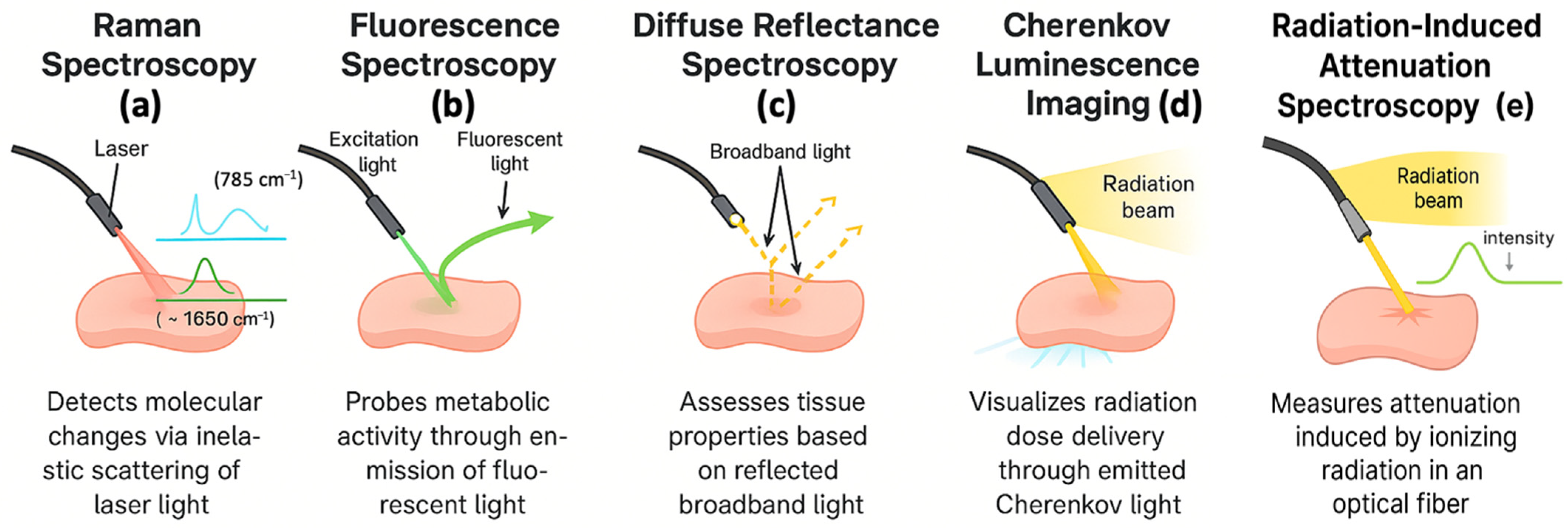
Optical spectroscopy has become a pivotal tool in radiation oncology for non-invasive, real-time assessment of biochemical and physiological changes in tissues exposed to ionizing radiation [68]. By capturing molecular signatures and metabolic alterations without the need for contrast agents or biopsies, these techniques offer a means of dynamic treatment monitoring that complements anatomical imaging. Among the primary modalities used are Raman spectroscopy [69], fluorescence spectroscopy [70,71], diffuse reflectance spectroscopy (DRS) [72], and Cherenkov luminescence imaging [73]. All of these are compatible with fiber-optic systems for in vivo integration, as shown in Figure 2 and summarized in Table 2.

3.1. Raman Spectroscopy

Raman spectroscopy, as shown in Figure 2a, uses inelastic scattering of monochromatic laser light to detect vibrational modes of biomolecules [74,75]. This produces a unique Raman spectrum, essentially a molecular fingerprint, that can differentiate tissue types and pathological states based on their biochemical composition. In radiation therapy, Raman spectroscopy enables detection of critical molecular changes such as DNA strand breaks, protein unfolding, and lipid peroxidation [76]. These changes manifest as altered intensities or shifts in characteristic Raman peaks, such as those associated with nucleic acids (~785 cm−1) or amide bonds in proteins (~1650 cm−1) [77]. Monitoring these shifts during or after irradiation provides insight into cellular damage mechanisms and can support adaptive treatment strategies by identifying radioresistant tumor subregions.

3.2. Fluorescence Spectroscopy

Fluorescence spectroscopy (Figure 2b), on the other hand, detects light emitted from intrinsic fluorophores such as NADH and FAD or from externally introduced dyes [78]. These fluorophores serve as metabolic indicators, reflecting mitochondrial activity and oxidative stress. Shifts in the fluorescence intensity or lifetime of these markers during radiation exposure can reveal disruptions in cellular respiration and redox balance, helping assess tumor response or early onset of tissue toxicity [79]. This method is particularly valuable for guiding fractionated or adaptive radiation therapy, where real-time feedback on tissue viability could inform mid-course treatment adjustments [80].

3.3. Diffuse Reflectance Spectroscopy

Diffuse reflectance spectroscopy, as shown in Figure 2c, extends these capabilities by evaluating the absorption and scattering of broadband light in tissues. It provides indirect but robust data on physiological parameters such as oxygen saturation, blood volume, and microvascular integrity [81]. The intensity and spectral characteristics of diffusely reflected light after multiple scattering and absorption events allow for the estimation of tissue optical properties and chromophore concentrations. Given the importance of tumor oxygenation in determining radiosensitivity, DRS can identify hypoxic regions that may require dose escalation or adjunctive therapy with radiosensitizers [82,83]. In preclinical models, fiber-optic DRS has successfully monitored vascular reoxygenation trends in radiation-sensitive versus radiation-resistant tumors following typical fractionated doses, supporting its application in adaptive therapy [84,85]. DRS offers a practical solution for minimally invasive and repeated bedside assessments throughout the course of therapy, enabling functional tissue monitoring that complements anatomical imaging modalities.

3.4. Cherenkov Luminescence Imaging

Cherenkov luminescence imaging, as shown in Figure 2d, is a relatively recent but promising addition to the suite of optical spectroscopy tools used in radiation therapy [73]. When high-energy photons or particles (such as in electron or photon beam therapy) exceed the phase velocity of light in tissue, Cherenkov light is emitted. This faint, broadband optical signal can be captured using time-gated or intensified cameras and is directly correlated with the instantaneous radiation dose deposition. Because Cherenkov light is produced only during treatment, it allows for real-time visualization of dose delivery on the patient’s surface or within shallow tissue depths [86]. Fiber-optic sensors can be used to collect this signal for localized monitoring, or imaging systems can track spatial dose distribution across the treatment field. Moreover, Cherenkov-excited luminescence and Cherenkov-induced fluorescence are being explored to enhance molecular imaging sensitivity and depth profiling during therapy [87].

3.5. Radiation-Induced Attenuation Spectroscopy

Radiation-induced attenuation (RIA) spectroscopy has been developed as a powerful fiber-based technique, particularly for in vivo dosimetry and environmental monitoring [88,89]. RIA occurs when ionizing radiation generates color centers or defects in optical fibers, leading to a measurable increase in light attenuation at specific wavelengths. This passive effect can be spectrally resolved and quantified using broadband light sources and optical spectrum analyzers, enabling distributed dose measurements along the fiber length [90]. RIA-based systems are compatible with harsh radiation environments, including proton and photon beams, and offer high dynamic range, repeatability, and immunity to electromagnetic interference [91,92].
Collectively, these optical spectroscopy techniques offer a layered view of radiation-induced effects, from molecular changes to physiological and dosimetric parameters. Their fiber-optic compatibility ensures minimally invasive implementation in vivo, even in anatomically challenging or sensitive regions. As radiation oncology moves toward biologically adaptive, precision-focused paradigms, the integration of Raman, fluorescence, DRS, RIA, and Cherenkov-based optical tools offers the potential to monitor and interpret radiation response in real time and at multiple biological scales [25,81]. Furthermore, the growing use of AI and machine learning to interpret complex data may enable automated, comprehensive decision-making, enhancing treatment accuracy and patient safety [93,94].

4. Applications of Optical Fiber Sensors in Radiation Therapy

4.1. Adaptive Radiation Therapy

Adaptive Radiation Therapy (ART) represents a significant shift from conventional static treatment paradigms to strategic, patient-specific protocols that adjust radiation delivery based on anatomical, physiological, or biological changes over the course of treatment [7,11]. While ART has traditionally relied on anatomical imaging modalities such as cone-beam CT and MRI to guide replanning, these systems often lack the ability to capture the molecular and metabolic alterations that underlie tumor response and treatment resistance. PET imaging adds valuable biological insight, particularly into tumor hypoxia and proliferation, but is limited by its low spatial resolution. Optical fiber technologies offer a novel solution to this limitation by enabling continuous, minimally invasive monitoring of biochemical and functional parameters during treatment, thereby enhancing the scope and effectiveness of ART [18,48].
Fiber-optic sensors can be embedded directly within treatment applicators, catheters, or patient-worn devices to monitor variables such as dose deposition, temperature, oxygenation, and metabolic activity with high temporal and spatial resolution [27]. For instance, FBGs can be used to track local thermal shifts that may signal inflammation or vascular disruption, while Raman and fluorescence probes can measure redox-related biomarkers like NADH and FAD, which provide insight into tumor hypoxia or radiation sensitivity [81]. These biological signs offer an early indication of treatment response or toxicity, often before anatomical changes become evident [95]. By integrating this real-time data into the ART workflow, clinicians can adapt fractionation schedules, modulate dose intensities, or adjust beam configurations with greater biological justification.
Moreover, the multiplexing capability of optical fibers allows multiple sensing points along a single strand, enabling simultaneous monitoring of diverse parameters across multiple tumor regions or organs at risk [27]. This feature is particularly advantageous in complex treatment areas such as the head and neck or pelvis, where tumor motion and organ deformation are common. When combined with artificial intelligence and machine learning algorithms, the spectral and dosimetric data collected by optical fiber systems can be rapidly analyzed to predict trends, identify deviations from planned responses, and guide personalized adaptation strategies [93].
In many cases, invasive placement of optical fiber systems into solid tumors can be clinically challenging. The development of non-invasive or minimally invasive strategies, such as surface-mounted or endoscopically guided delivery, will be critical. Implantable fiber sensors have also been explored in preclinical or interventional oncology settings. Future work must focus on robust, biocompatible integration methods for clinical deployment, possibly in combination with existing applicators or anatomical cavities, to extend sensing into deeper tissues without compromising treatment delivery. As the field moves toward biologically guided radiotherapy, the integration of optical fiber sensing into ART protocols has the potential to significantly improve treatment precision, reduce unnecessary toxicity, and enhance patient outcomes through truly responsive, data-driven intervention.

4.2. Real-Time Monitoring and Imaging

Real-time intrafraction monitoring during radiation therapy is important to ensure accurate and safe dose delivery. Optical fiber technology enables immediate detection of adverse tissue responses [63,96], with real-time sensing of clinically relevant parameters such as radiation dose, tissue temperature, and physiological indicators like oxygenation [27]. In treatments involving thermal modulation [97], such as hyperthermia therapy or thermal ablation, precise thermal control is crucial for therapeutic effectiveness and minimizing collateral damage. Fiber-optic temperature sensors, commonly based on FBGs or rare-earth doped fibers, provide accurate and stable temperature readings with sub-degree sensitivity [18,64].
Beyond sensing, optical fibers are also widely used in real-time imaging modalities, as summarized in Table 2. Endoscopic fiber bundles allow for the delivery and collection of light in narrow or curved anatomical regions. They support modalities like optical coherence tomography (OCT), which provides high-resolution cross-sectional images of tissue microarchitecture using low-coherence interferometry [98]. One emerging modality is photoacoustic imaging (PAI), which combines optical excitation with ultrasonic detection to visualize tissue structures based on optical absorption contrast [99]. In this approach, pulsed laser light delivered via optical fibers induces thermoelastic expansion in chromophore-rich tissues, generating ultrasound waves that are detected and reconstructed into functional images. PAI is particularly advantageous for visualizing vascular structures and monitoring tissue oxygenation, which is critical for radiotherapy planning and response assessment. Another complementary technique is Laser Speckle Contrast Imaging (LSCI), which analyzes dynamic speckle patterns formed by coherent laser light scattered from moving red blood cells to assess microvascular blood flow in tissue [100]. When integrated into fiber-optic probes, LSCI allows for minimally invasive, real-time mapping of perfusion changes, which is particularly valuable in identifying regions of ischemia or radiation-induced vascular disruption.
These monitoring and imaging techniques can enhance real-time intraoperative or interventional visualization and have the potential to improve the precision of tumor localization, margin assessment, and adaptive planning in procedures where spatial accuracy is critical. As fiber-based imaging technologies continue to evolve, as shown in Figure 3, their role in guiding and optimizing therapeutic interventions is expected to expand significantly.

4.3. Clinical Integration and Workflow Advantages

Integration of fiber optic systems into clinical radiation therapy offers transformative potential for improving workflow efficiency, treatment precision, and patient safety [18,48,64]. Thanks to their small diameter, mechanical flexibility and biocompatibility [15], optical fibers may be embedded into applicators, catheters, endoscopes, or surgical instruments, enabling real-time, in vivo monitoring during both external beam radiation therapy and brachytherapy [43,68]. Moreover, their non-metallic composition and immunity to electromagnetic interference allow safe operation within MRI-guided radiation therapy environments [17], where electronic components may fail due to the strong magnetic fields or radiofrequency noise. These features make fiber-optic sensors advantageous to deploy in anatomically constrained or sensitive sites, such as the prostate, cervix, oral cavity, and gastrointestinal tract, where traditional dosimeters may be too bulky or invasive.
Fiber-optic sensors also support distributed sensing, in which multiple sensing elements (e.g., FBGs or scintillating tips) can be arranged along a single fiber strand to monitor parameters such as dose deposition, temperature, oxygenation, and tissue response across multiple spatial locations simultaneously [18,27]. This capability is particularly advantageous in adaptive radiotherapy, where tumors and organs-at-risk may move or deform over time (e.g., in the head and neck or pelvic regions), and dynamic treatment planning may be required [101]. In these scenarios, fiber multiplexing may support spatially resolved measurements without adding significant hardware complexity.
Clinically, optical fiber dosimetry and sensing systems have been implemented in a variety of radiation therapy settings where real-time, in vivo, and minimally invasive monitoring is essential [48]. For example, in proton therapy, fiber-optic sensors have been used to track Bragg peak positions and range shifts in vivo by embedding sensors in phantoms or endoscopic probes, reducing range uncertainties and improving dose accuracy [102]. Fiber sensors have also been explored more extensively for dosimetric use in prostate cancer [64]. They have been applied in low dose rate (LDR) brachytherapy [63], high dose rate (HDR) brachytherapy [103], and external beam radiotherapy [104] across various source and applicator configurations [105,106]. Use of fiber sensor may provide us with real time in vivo monitoring of on entrance and exit dose during treatment while minimizing invasiveness, utilizing existing brachytherapy procedures or biopsy channels. These applications demonstrate the versatility of fiber-optic sensors across radiation treatment modalities and clinical scenarios.
Furthermore, as modern treatment platforms increasingly employ robotic guidance, motion tracking, and adaptive feedback control, the real-time output from fiber-optic sensors can be integrated into these systems to dynamically adjust beam delivery based on patient-specific anatomy [101]. For example, in a urethral dose brachytherapy clinical trial, a maximum of 9% deviation between in vivo measured dose from the planned dose was primarily attributed to small changes in patient anatomy [103]. Such deviations may be corrected using real-time measured values to better reflect the actual dose distribution. When paired with machine learning algorithms that analyze dosimetric or spectral trends, these data streams can possibly support automated decision-making, identifying deviations from expected responses and prompting timely plan adaptation [93,107,108]. In summary, fiber-optic sensing platforms hold great promise for real-time, in vivo, high-resolution, and spatially distributed dosimetric and spectroscopic monitoring in diverse radiation therapy scenarios. The continuously growing deployment of fiber sensors across various clinical settings demonstrates their substantial potential to enhance biological adaptability, treatment precision, and workflow integration in radiation oncology.

5. Challenges and Future Directions

5.1. Fiber-Optic Dosimetric Technical Challenges

Despite their many advantages, fiber optic sensors face a number of technical and clinical barriers that must be addressed before widespread adoption in radiation therapy [47,48]. On the technical front, one major limitation is signal contamination. In scintillating fiber systems, Cherenkov light generated within the fiber itself (or the “stem effect”, as reviewed in Section 2.1) can obscure the true scintillation signal, compromising dose accuracy [109]. Although dual-channel detection schemes and spectral filtering techniques have been developed to separate these signal components, their complexity limits scalability and ease of implementation in standard clinical settings.
Also, an important technical constraint lies in signal attenuation, particularly in fibers longer than 1–2 m or when bent near tight anatomical curves. Optical loss in silica fibers is typically <0.2 dB/km at 850–1550 nm, but radiation-induced defects (color centers) can increase attenuation significantly, especially under high-dose or pulsed-beam conditions [88]. RIA spectroscopy has been proposed not only as a sensing method but also as a tool to monitor these losses. Recent experimental data show that irradiation at elevated temperatures can reduce near-infrared RIA by up to a factor of four to six compared to room temperature, depending on wavelength and thermal conditions [89]. Phosphosilicate fibers, Ce-co-doping strategies, and alternative core compositions such as Al-doped fibers have been shown to stabilize RIA responses, mitigate temperature effects, and extend the usable dose range for distributed dosimetry [91,92]. Additionally, fiber fragility, especially in pure-silica or minimally shielded systems, can lead to mechanical failure after repeated clinical use. Development of polymer-clad or stretchable fibers is ongoing to improve long-term durability under bending and sterilization stress.
Calibration drift is another non-trivial concern in both fiber dosimetry and spectroscopy. Repeated thermal cycling, light exposure, and material-specific sensitivity changes can introduce calibration drift on the order of several percent in both TL and OSL systems, especially when annealing or re-setting steps are imperfectly controlled [26,56]. For example, temperature sensitivity in Al2O3-based dosimeters can shift light output by −0.2% to +0.6% per °C if uncorrected [110]. Active correction strategies now include dual-wavelength referencing, temperature-compensated algorithms, and internal calibration pulses integrated into the readout system [93]. Furthermore, signal normalization using reference scintillators or FBG-based thermal monitoring has shown promise in reducing environmental-induced uncertainty to <2% in clinical testing scenarios. Moreover, consistency remains a persistent challenge [108]. Environmental influences such as temperature, humidity, and ambient light may further complicate signal interpretation. Finally, the limited measurable range of some fiber-based sensing systems constrains their utility in high-dose-rate scenarios or in heterogeneous tissues with complex optical properties.

5.2. Fiber-Optic Spectroscopic Technical Challenges

Despite their sensitivity and molecular specificity, optical spectroscopic methods face several intrinsic technical limitations that could have hindered broad clinical adoption. Raman spectroscopy, for instance, suffers from inherently weak signal strength due to low scattering cross-sections (~10−6–10−10 of incident photons), which requires long acquisition times or high-powered lasers, raising safety concerns [111]. Similarly, autofluorescence from endogenous molecules (e.g., collagen, NADH) can obscure target-specific fluorescence signatures, particularly in the 400–600 nm range [112].
Photobleaching and phototoxicity (loss of fluorophore signal and tissue damage from prolonged light exposure) are also critical concerns during fluorescence-guided radiotherapy, especially in repetitive imaging scenarios. This limits the number of usable frames or timepoints within a fraction [113]. Moreover, the three spectroscopic techniques, Raman, fluorescence, and diffuse reflectance, are typically constrained to penetration depths ~< 3 mm, unless advanced fiber-based probes or time-resolved gating systems are used, which may further complicate the clinical workflow [68].
Compared to PET or MRI, which offer deep-tissue, tomographic, and often quantitative information on metabolism and anatomy, optical spectroscopy is currently best positioned as a complementary intraoperative or surface-level monitoring tool. For example, PET hypoxia tracers provide robust oxygenation maps [114] but lack the spatial and temporal resolution for real-time intra-fractional feedback. Conversely, Raman or fluorescence signals, while spatially localized and rapid, remain largely confined to accessible surfaces or inserted fiber regions. Hybrid approaches, such as integrating optical imagers into PET-compatible platforms, may have advantages to bridge this gap [115,116].

5.3. Clinical Translation and Adoption Challenges

Beyond engineering limitations, several clinical challenges remain [43]. One of the major challenges in clinical translation is the invasive placement of optical sensors for deep-seated tumors. While surface dosimetry and endoscopic access are relatively straightforward, interstitial applications (e.g., prostate, cervix) require specialized delivery mechanisms. Integration of optical fibers into HDR brachytherapy applicators, catheters, or biopsy channels has been explored, with some systems demonstrating sub-millimeter placement reproducibility and successful dose readout during clinical fractions [42,48]. These hybrid devices reduce insertion time and minimize patient discomfort but remain underutilized due to customization requirements and sterilization logistics.
The cost of specialty optical fibers, particularly rare-earth-doped variants (e.g., Ce-doped silica), is also a limiting factor, with individual sensor cost often restricting single-use deployment. Ongoing work in lower-cost polymer scintillating fibers, sol–gel–based doped preforms, and reusable probe architectures is aimed at reducing per-sensor expense [117,118]. Vendors are also exploring modular plug-and-play systems that allow routine sterilization and recalibration.
Workflow interruptions caused by manual sensor placement, alignment drift, and repeated probe insertions have been widely identified as barriers to clinical adoption of fiber-based dosimetry and optical readout systems. As a partial workaround, several groups have explored surface surrogate sensing, for example, skin-mounted fiber Bragg gratings or Cherenkov-emission imagers, to infer dose trends at depth when direct in vivo insertion is impractical [119,120]. Although accuracy depends strongly on anatomic geometry and calibration methodology, these approaches have demonstrated clinically relevant agreement with dose distributions in select scenarios [121,122].
Finally, data interpretation and integration into treatment planning systems remains underdeveloped, and the raw spectral or dosimetric data often require post-processing algorithm correction to become clinically actionable [26,93]. Recent reviews also highlight that coupling fiber-optic sensors with denoising models and algorithms reduces processing time and enhances predictive diagnostics [108]. To address these limitations, ongoing research is focused on real-time machine-learning-assisted signal analysis [123,124]. All these developments aim to make fiber dosimetry more clinically practical, scalable, and informative across diverse treatment modalities and anatomical sites.

5.4. Future Directions and Developments

The future of optical fiber technology in radiation oncology lies in its continued evolution toward multifunctionality, robustness, and intelligent integration with clinical systems [18]. Multiparameter sensors capable of simultaneously tracking radiation dose, temperature, biochemical markers, and tissue perfusion are under active development [125]. These hybrid sensors could offer comprehensive real-time feedback, allowing clinicians to adapt treatment plans based not only on anatomical changes but also on the tumor’s biological behavior and the patient’s physiological response [126,127]. For example, in head and neck cancer patients, embedded optical sensors could monitor redox-active species such as NADH and FAD, which reflect metabolic activity and radiation sensitivity in hypoxic tumor regions. This could allow clinicians to detect the early onset of treatment resistance or mucosal injury and adapt dose distributions accordingly. Similarly, in cervical cancer, fiber-based Raman and fluorescence spectroscopy might be used to detect pH shifts, lactate levels, or oxygen saturation in the tumor microenvironment as key indicators of hypoxia, acidosis, or necrosis, all of which are known to correlate with treatment resistance and recurrence risks.
Beyond sensing, the development of “smart fibers” that integrate microelectronics, nanostructures, or microfluidic channels into the fiber core offers exciting opportunities for multifunctional sensing and targeted drug delivery [128,129]. At the same time, advances in artificial intelligence and machine learning are enabling new approaches to spectral data interpretation, real-time anomaly detection, and outcome prediction. For example, classification algorithms trained on Raman or fluorescence spectra could eventually support automatic identification of tumor margins or early signs of toxicity [130,131]. However, real-time AI-assisted interpretation of optical spectra remains largely preclinical, as most current models are trained on curated datasets under controlled conditions [132,133,134], and their clinical translation will require rigorous validation, generalization, and regulatory approval. Likewise, smart fiber architectures that incorporate microfluidics or embedded electronics are still at the prototype stage and have not yet undergone systematic clinical trials [135,136,137]. While these emerging technologies hold significant promise, their integration into routine clinical workflows will need substantial translational development.
Finally, personalized dosimetry guided by continuous in vivo feedback is another emerging research area for precision oncology [69]. Such integration could help reduce treatment-related toxicity, improve local control, and streamline radiation therapy workflows. Realizing these advances will require close collaboration between physicists, engineers, oncologists, and regulatory experts to ensure patient safety, device reliability, and clinical translational feasibility.

6. Conclusions

The integration of optical fiber technology into radiation oncology offers a promising opportunity in the pursuit of precise, adaptive, and biologically informed cancer treatment. These technologies, characterized by their miniaturization, flexibility, and immunity to electromagnetic interference, offer substantial advantages over traditional monitoring systems. By enabling real-time measurement of critical treatment parameters, including radiation dose, temperature, oxygen saturation, and biochemical changes, optical fibers facilitate a more comprehensive and dynamic understanding of treatment delivery. Techniques such as Raman spectroscopy, fluorescence imaging, and Cherenkov emission detection extend the functional capabilities of optical fibers from conventional dosimetry to detailed molecular-level diagnostics.
These capabilities are particularly relevant in the evolving context of personalized and adaptive radiotherapy. As the field continues to shift from anatomically guided treatments toward biologically adaptive protocols, the need for technologies that can capture functional and metabolic responses in situ has become increasingly urgent. Optical spectroscopy allows for the direct monitoring of radiation-induced biochemical alterations, such as DNA strand breaks, protein denaturation, and redox imbalances, thereby offering predictive insight into tumor response and normal tissue toxicity. When coupled with advanced data analytics and machine learning, these systems may enable automated, data-driven treatment adaptation, enhancing both therapeutic efficacy and patient safety.
While the potential of optical fiber systems is considerable, several technical and clinical barriers must be addressed to enable routine clinical adoption. Challenges such as signal contamination from Cherenkov radiation, limited dynamic range, mechanical fragility, and calibration complexity require ongoing refinement in sensor design and signal processing methodologies. To better guide future comparative studies, standardized evaluation metrics such as dosimetric accuracy (e.g., <5% error), reproducibility (e.g., >95% agreement in repeated measures), and validation against reference standards (e.g., ionization chambers, PET) should be adopted. Integration burdens (cost, sterilization, system compatibility) are also vital for translational relevance. Current literature often lacks consistent reporting in these areas, hindering cross-platform comparisons. We recommend that future optical fiber sensing studies include these parameters in experimental protocols. Nevertheless, researchers are making substantial progress in overcoming these limitations, driven by interdisciplinary collaborations across engineering, physics, and clinical oncology. As these innovations continue to mature, optical fiber technologies are well-positioned to become an integral component of next-generation radiation therapy, contributing to more effective, responsive, and patient-centered oncologic care.

Funding

This research received no external funding.

Data Availability Statement

Data will be made available on request.

Conflicts of Interest

The authors declare no conflicts of interest.

References

  1. Baskar, R.; Lee, K.A.; Yeo, R.; Yeoh, K.-W. Cancer and Radiation Therapy: Current Advances and Future Directions. Int. J. Med. Sci. 2012, 9, 193. [Google Scholar] [CrossRef] [PubMed]
  2. Cho, B. Intensity-Modulated Radiation Therapy: A Review with a Physics Perspective. Radiat. Oncol. J. 2018, 36, 1. [Google Scholar] [CrossRef]
  3. Folkert, M.R.; Timmerman, R.D. Stereotactic Ablative Body Radiosurgery (SABR) or Stereotactic Body Radiation Therapy (SBRT). Adv. Drug Deliv. Rev. 2017, 109, 3–14. [Google Scholar] [CrossRef]
  4. De Los Santos, J.; Popple, R.; Agazaryan, N.; Bayouth, J.E.; Bissonnette, J.-P.; Bucci, M.K.; Dieterich, S.; Dong, L.; Forster, K.M.; Indelicato, D. Image Guided Radiation Therapy (IGRT) Technologies for Radiation Therapy Localization and Delivery. Int. J. Radiat. Oncol. Biol. Phys. 2013, 87, 33–45. [Google Scholar] [CrossRef]
  5. Mohan, R.; Grosshans, D. Proton Therapy–Present and Future. Adv. Drug Deliv. Rev. 2017, 109, 26–44. [Google Scholar] [CrossRef]
  6. Schardt, D.; Elsässer, T.; Schulz-Ertner, D. Heavy-Ion Tumor Therapy: Physical and Radiobiological Benefits. Rev. Mod. Phys. 2010, 82, 383–425. [Google Scholar] [CrossRef]
  7. Chetty, I.J.; Cai, B.; Chuong, M.D.; Dawes, S.L.; Hall, W.A.; Helms, A.R.; Kirby, S.; Laugeman, E.; Mierzwa, M.; Pursley, J. Quality and Safety Considerations for Adaptive Radiation Therapy: An ASTRO White Paper. Int. J. Radiat. Oncol. Biol. Phys. 2025, 122, 838–864. [Google Scholar] [CrossRef] [PubMed]
  8. Beaton, L.; Bandula, S.; Gaze, M.N.; Sharma, R.A. How Rapid Advances in Imaging Are Defining the Future of Precision Radiation Oncology. Br. J. Cancer 2019, 120, 779–790. [Google Scholar] [CrossRef]
  9. Fiz, F.; Iori, M.; Fioroni, F.; Biroli, M.; D’Agostino, G.R.; Gelardi, F.; Erba, P.A.; Versari, A.; Chiti, A.; Sollini, M. Molecular Guidance for Planning External Beam Radiation Therapy in Oncology. In Nuclear Oncology; Springer: Cham, Switzerland, 2022; pp. 1–40. ISBN 3319260677. [Google Scholar]
  10. Cai, B.; Banks, T.I.; Shen, C.; Prasad, R.; Bal, G.; Lin, M.-H.; Godley, A.; Pompos, A.; Garant, A.; Westover, K. Strategies for Offline Adaptive Biology-Guided Radiotherapy (BgRT) on a PET-Linac Platform. Cancers 2025, 17, 2470. [Google Scholar] [CrossRef] [PubMed]
  11. Dona Lemus, O.M.; Cao, M.; Cai, B.; Cummings, M.; Zheng, D. Adaptive Radiotherapy: Next-Generation Radiotherapy. Cancers 2024, 16, 1206. [Google Scholar] [CrossRef]
  12. Grégoire, V.; Haustermans, K.; Geets, X.; Roels, S.; Lonneux, M. PET-Based Treatment Planning in Radiotherapy: A New Standard? J. Nucl. Med. 2007, 48, 68S–77S. [Google Scholar]
  13. Glide-Hurst, C.K.; Lee, P.; Yock, A.D.; Olsen, J.R.; Cao, M.; Siddiqui, F.; Parker, W.; Doemer, A.; Rong, Y.; Kishan, A.U. Adaptive Radiation Therapy (ART) Strategies and Technical Considerations: A State of the ART Review from NRG Oncology. Int. J. Radiat. Oncol. Biol. Phys. 2021, 109, 1054–1075. [Google Scholar] [CrossRef]
  14. Suarez, M.A.; Lim, T.; Robillot, L.; Maillot, V.; Lihoreau, T.; Bontemps, P.; Pazart, L.; Grosjean, T. Miniaturized Fiber Dosimeter of Medical Ionizing Radiations on a Narrow Optical Fiber. Opt. Express 2019, 27, 35588–35599. [Google Scholar] [CrossRef]
  15. Elsharkawi, A.S.A.; Elazab, H.A.; Askar, M.A.; Abdelrahman, I.Y.; Arafa, A.A.; Gomma, L.R.; Lo, Y.-L. Biocompatibility and Radiosensitivity of a Fiber Optical-Based Dosimeter: Biological Applications. Biomed. Opt. Express 2024, 15, 3492–3506. [Google Scholar] [CrossRef] [PubMed]
  16. Guo, J.; Yang, C.; Dai, Q.; Kong, L. Soft and Stretchable Polymeric Optical Waveguide-Based Sensors for Wearable and Biomedical Applications. Sensors 2019, 19, 3771. [Google Scholar] [CrossRef] [PubMed]
  17. Madden, L.; Holloway, L.; Rosenfeld, A.; Li, E. Fibre-Optic Dosimetry for MRI-LINACs: A Mini-Review. Front. Phys. 2022, 10, 879624. [Google Scholar] [CrossRef]
  18. O’Keeffe, S.; McCarthy, D.; Woulfe, P.; Grattan, M.W.D.; Hounsell, A.R.; Sporea, D.; Mihai, L.; Vata, I.; Leen, G.; Lewis, E. A Review of Recent Advances in Optical Fibre Sensors for in Vivo Dosimetry during Radiotherapy. Br. J. Radiol. 2015, 88, 20140702. [Google Scholar] [CrossRef] [PubMed]
  19. Yang, W.; Knorr, F.; Latka, I.; Vogt, M.; Hofmann, G.O.; Popp, J.; Schie, I.W. Real-Time Molecular Imaging of near-Surface Tissue Using Raman Spectroscopy. Light Sci. Appl. 2022, 11, 90. [Google Scholar] [CrossRef]
  20. Chen, W.; Chen, Y.; Wu, C.; Zhang, X.; Huang, X. The Accuracy of Fiber-Optic Raman Spectroscopy in the Detection and Diagnosis of Head and Neck Neoplasm in Vivo: A Systematic Review and Meta-Analysis. PeerJ 2023, 11, e16536. [Google Scholar] [CrossRef]
  21. Wang, Y.; Fang, L.; Wang, Y.; Xiong, Z. Current Trends of Raman Spectroscopy in Clinic Settings: Opportunities and Challenges. Adv. Sci. 2024, 11, 2300668. [Google Scholar] [CrossRef]
  22. Li, Z.; Lan, N.; Cheng, Z.; Jin, F.; Song, E.; Xu, Z.; Zhang, Y.; Feng, Y.-Z.; Cai, X.; Ran, Y. In Vivo Fiber-Optic Fluorescent Sensor for Real-Time PH Monitoring of Tumor Microenvironment. Chem. Eng. J. 2024, 493, 152495. [Google Scholar] [CrossRef]
  23. Xie, H.; Xie, Z.; Mousavi, M.; Bendsoe, N.; Brydegaard, M.; Axelsson, J.; Andersson-Engels, S. Design and Validation of a Fiber Optic Point Probe Instrument for Therapy Guidance and Monitoring. J. Biomed. Opt. 2014, 19, 71408. [Google Scholar] [CrossRef]
  24. Thomas, T.P.; Myaing, M.T.; Ye, J.Y.; Candido, K.; Kotlyar, A.; Beals, J.; Cao, P.; Keszler, B.; Patri, A.K.; Norris, T.B. Detection and Analysis of Tumor Fluorescence Using a Two-Photon Optical Fiber Probe. Biophys. J. 2004, 86, 3959–3965. [Google Scholar] [CrossRef] [PubMed]
  25. Barik, A.K.; Lukose, J.; Upadhya, R.; Pai, M.V.; Kartha, V.B.; Chidangil, S. In Vivo Spectroscopy: Optical Fiber Probes for Clinical Applications. Expert Rev. Med. Devices 2022, 19, 657–675. [Google Scholar] [CrossRef] [PubMed]
  26. Zhang, J.; Xiang, Y.; Wang, C.; Chen, Y.; Tjin, S.C.; Wei, L. Recent Advances in Optical Fiber Enabled Radiation Sensors. Sensors 2022, 22, 1126. [Google Scholar] [CrossRef]
  27. Sporea, D. Optical Fiber Sensors in Ionizing Radiation Environments. In Handbook of Optical Fibers; Springer: Singapore, 2019; pp. 1913–1954. ISBN 9811070873. [Google Scholar]
  28. Ohta, T.; Nozawa, Y.; Ohira, S.; Nawa, K.; Yamashita, H.; Nakagawa, K. Characterization of a Practically Designed Plastic Scintillation Plate Dosimeter. Med. Phys. 2025, 52, e17904. [Google Scholar] [CrossRef]
  29. Bauer, C.J.; Schneider, F.; Göbel, I.D.; Oppitz, H.; Giordano, F.A.; Fleckenstein, J. Characterization of a Novel Plastic Scintillation Detector for in Vivo Electron Dosimetry. arXiv 2025, arXiv:2509.04933. [Google Scholar] [CrossRef]
  30. Ciarrocchi, E.; Ravera, E.; Cavalieri, A.; Celentano, M.; Del Sarto, D.; Di Martino, F.; Linsalata, S.; Massa, M.; Masturzo, L.; Moggi, A. Plastic Scintillator-Based Dosimeters for Ultra-High Dose Rate (UHDR) Electron Radiotherapy. Phys. Medica 2024, 121, 103360. [Google Scholar] [CrossRef]
  31. Fontbonne, J.M.; Iltis, G.; Ban, G.A.; Battala, A.; Vernhes, J.C.; Tillier, J.; Bellaize, N.; Le Brun, C.; Tamain, B.; Mercier, K. Scintillating Fiber Dosimeter for Radiation Therapy Accelerator. IEEE Trans. Nucl. Sci. 2002, 49, 2223–2227. [Google Scholar] [CrossRef]
  32. Wang, X.; Jiang, Y.; Xu, S.; Liu, H.; Li, X. Fiber Bragg Grating-Based Smart Garment for Monitoring Human Body Temperature. Sensors 2022, 22, 4252. [Google Scholar] [CrossRef]
  33. Kinet, D.; Mégret, P.; Goossen, K.W.; Qiu, L.; Heider, D.; Caucheteur, C. Fiber Bragg Grating Sensors toward Structural Health Monitoring in Composite Materials: Challenges and Solutions. Sensors 2014, 14, 7394–7419. [Google Scholar] [CrossRef]
  34. Fitzgerald, S.; Marple, E.; Mahadevan-Jansen, A. Performance Assessment of Probe-Based Raman Spectroscopy Systems for Biomedical Analysis. Biomed. Opt. Express 2023, 14, 3597–3609. [Google Scholar] [CrossRef] [PubMed]
  35. Moradi, F.; Bradley, D.A.; Tarif, Z.H.; Khodaei, A.; Basaif, A.; Ibrahim, S.A.; Abdul-Rashid, H.A. Time-Resolved Optical Fiber Measurements: A Review of Scintillator Materials and Applications. Radiat. Detect. Technol. Methods 2025, 9, 1–16. [Google Scholar] [CrossRef]
  36. Archambault, L.; Briere, T.M.; Pönisch, F.; Beaulieu, L.; Kuban, D.A.; Lee, A.; Beddar, S. Toward a Real-Time in Vivo Dosimetry System Using Plastic Scintillation Detectors. Int. J. Radiat. Oncol. Biol. Phys. 2010, 78, 280–287. [Google Scholar] [CrossRef]
  37. Kharzheev, Y.N. Radiation Hardness of Scintillation Detectors Based on Organic Plastic Scintillators and Optical Fibers. Phys. Part. Nucl. 2019, 50, 42–76. [Google Scholar] [CrossRef]
  38. Ding, L.; Wu, Q.; Wang, Q.; Li, Y.; Perks, R.M.; Zhao, L. Advances on Inorganic Scintillator-Based Optic Fiber Dosimeters. EJNMMI Phys. 2020, 7, 60. [Google Scholar] [CrossRef]
  39. Shi, Z.; Lv, S.; Tang, G.; Tang, J.; Jiang, L.; Qian, Q.; Zhou, S.; Yang, Z. Multiphase Transition toward Colorless Bismuth–Germanate Scintillating Glass and Fiber for Radiation Detection. ACS Appl. Mater. Interfaces 2020, 12, 17752–17759. [Google Scholar] [CrossRef]
  40. Park, C.H.; Lee, A.; Kim, R.; Moon, J.H. Evaluation of the Detection Efficiency of LYSO Scintillator in the Fiber-Optic Radiation Sensor. Sci. Technol. Nucl. Install. 2014, 2014, 248403. [Google Scholar] [CrossRef]
  41. Hu, Y.; Qin, Z.; Ma, Y.; Zhao, W.; Sun, W.; Zhang, D.; Chen, Z.; Wang, B.; Tian, H.; Lewis, E. Characterization of Fiber Radiation Dosimeters with Different Embedded Scintillator Materials for Radiotherapy Applications. Sens. Actuators A Phys. 2018, 269, 188–195. [Google Scholar] [CrossRef]
  42. Gierej, A.; Baghdasaryan, T.; Martyn, M.; Woulfe, P.; Mc Laughlin, O.; Prise, K.; Workman, G.; O’Keeffe, S.; Rochlitz, K.; Verlinski, S. Mass-Manufacturable Scintillation-Based Optical Fiber Dosimeters for Brachytherapy. Biosens. Bioelectron. 2024, 255, 116237. [Google Scholar] [CrossRef] [PubMed]
  43. Chow, J.C.L.; Ruda, H.E. In Vivo Dosimetry in Radiotherapy: Techniques, Applications, and Future Directions. Encyclopedia 2025, 5, 40. [Google Scholar] [CrossRef]
  44. Lee, B.; Jang, K.W.; Cho, D.H.; Yoo, W.J.; Shin, S.H.; Kim, H.S.; Yi, J.H.; Kim, S.; Cho, H.; Park, B.G. Measurement of Two-Dimensional Photon Beam Distributions Using a Fiber-Optic Radiation Sensor for Small Field Radiation Therapy. IEEE Trans. Nucl. Sci. 2008, 55, 2632–2636. [Google Scholar] [CrossRef]
  45. Terasawa, K.; Doke, T.; Hasebe, N.; Kikuchi, J.; Kudo, K.; Murakami, T.; Takeda, N.; Tamura, T.; Torii, S.; Yamashita, M. Scintillating Fiber Camera for Neutron Dosimetry in Spacecraft. Nucl. Instruments Methods Phys. Res. Sect. A Accel. Spectrometers Detect. Assoc. Equip. 2001, 457, 499–508. [Google Scholar] [CrossRef]
  46. Klavsen, M.F.; Ankjærgaard, C.; Behrens, C.P.; Vogelius, I.R.; Boye, K.; Hansen, R.H.; Andersen, C.E. Time-Resolved Plastic Scintillator Dosimetry in MR Linear Accelerators without Image Distortion. Radiat. Meas. 2022, 154, 106759. [Google Scholar] [CrossRef]
  47. Darafsheh, A.; Goddu, S.M.; Williamson, J.; Zhang, T.; Sobotka, L.G. Radioluminescence Dosimetry in Modern Radiation Therapy. Adv. Photonics Res. 2024, 5, 2300350. [Google Scholar] [CrossRef]
  48. Veronese, I.; Andersen, C.E.; Li, E.; Madden, L.; Santos, A.M.C. Radioluminescence-Based Fibre Optic Dosimeters in Radiotherapy: A Review. Radiat. Meas. 2024, 174, 107125. [Google Scholar] [CrossRef]
  49. Frelin, A.; Fontbonne, J.; Ban, G.; Colin, J.; Labalme, M.; Batalla, A.; Isambert, A.; Vela, A.; Leroux, T. Spectral Discrimination of Čerenkov Radiation in Scintillating Dosimeters. Med. Phys. 2005, 32, 3000–3006. [Google Scholar] [CrossRef]
  50. Archer, J.; Madden, L.; Li, E.; Carolan, M.; Petasecca, M.; Metcalfe, P.; Rosenfeld, A. Temporally Separating Cherenkov Radiation in a Scintillator Probe Exposed to a Pulsed X-Ray Beam. Phys. Medica 2017, 42, 185–188. [Google Scholar] [CrossRef]
  51. Archer, J.; Madden, L.; Li, E.; Carolan, M.; Rosenfeld, A. A Comparison of Temporal Cherenkov Separation Techniques in Pulsed Signal Scintillator Dosimetry. Biomed. Phys. Eng. Express 2018, 4, 44003. [Google Scholar] [CrossRef]
  52. McKeever, S.W.S. Optically Stimulated Luminescence: A Brief Overview. Radiat. Meas. 2011, 46, 1336–1341. [Google Scholar] [CrossRef]
  53. McKeever, S.W.S.; Blair, M.W.; Bulur, E.; Gaza, R.; Gaza, R.; Kalchgruber, R.; Klein, D.M.; Yukihara, E.G. Recent Advances in Dosimetry Using the Optically Stimulated Luminescence of Al2O3: C. Radiat. Prot. Dosim. 2004, 109, 269–276. [Google Scholar] [CrossRef] [PubMed]
  54. Lim, C.S.; Lee, S.B.; Jin, G.H. Performance of Optically Stimulated Luminescence Al2O3 Dosimeter for Low Doses of Diagnostic Energy X-Rays. Appl. Radiat. Isot. 2011, 69, 1486–1489. [Google Scholar] [CrossRef] [PubMed]
  55. Buranurak, S.; Andersen, C.E. Fiber-Coupled Al2O3: C Radioluminescence Dosimetry for Total Body Irradiations. Radiat. Meas. 2016, 93, 46–54. [Google Scholar] [CrossRef]
  56. McKeever, S.W.; Moscovitch, M. On the Advantages and Disadvantages of Optically Stimulated Luminescence Dosimetry and Thermoluminescence Dosimetry. Radiat. Prot. Dosim. 2003, 104, 263–270. [Google Scholar] [CrossRef]
  57. Magne, S.; Ferdinand, P. Fiber Optic Remote Gamma Dosimeters Based on Optically Stimulated Luminescence: State-of-the-Art at CEA. In Proceedings of the 11th International Congress of the International Radiation Protection Association (IRPA), Madrid, Spain, 23–28 May 2004. [Google Scholar]
  58. Cygler, J.E.; Yukihara, E.G. Optically Stimulated Luminescence (OSL) Dosimetry in Radiotherapy; Medical Physics Publishing: Madison, WI, USA, 2009. [Google Scholar]
  59. Andersen, C.E.; Edmund, J.M.; Damkjær, S.M.S. Precision of RL/OSL Medical Dosimetry with Fiber-Coupled Al2O3: C: Influence of Readout Delay and Temperature Variations. Radiat. Meas. 2010, 45, 653–657. [Google Scholar] [CrossRef]
  60. Santos, A.M.C.; Mohammadi, M.; Afshar V, S. Evaluation of a Real-time BeO Ceramic Fiber-coupled Luminescence Dosimetry System for Dose Verification of High Dose Rate Brachytherapy. Med. Phys. 2015, 42, 6349–6356. [Google Scholar] [CrossRef]
  61. Birajdar, S.; Zhang, W.; Santos, A.; Hickson, K.; Afshar Vahid, S. Real-Time in Vivo Dose Measurement Using Ruby-Based Fibre Optic Dosimetry during Internal Radiation Therapy. Phys. Eng. Sci. Med. 2023, 46, 1205–1213. [Google Scholar] [CrossRef] [PubMed]
  62. Elsharkawi, A.S.A.; Alazab, H.A.; Sayed, M.; Askar, M.A.; Abdelrahman, I.Y.; Arafa, A.A.; Saleh, H.I.; Gomaa, L.R.; Du, Y.-C. A Fiber-Optical Dosimetry Sensor for Gamma-Ray Irradiation Measurement in Biological Applications. Biosensors 2023, 13, 1010. [Google Scholar] [CrossRef]
  63. Woulfe, P.; Sullivan, F.J.; Kam, W.; O’Keeffe, S. Optical Fiber Dosimeter for Real-Time in-Vivo Dose Monitoring during LDR Brachytherapy. Biomed. Opt. Express 2020, 11, 4027–4036. [Google Scholar] [CrossRef]
  64. Woulfe, P.; Sullivan, F.J.; O’Keeffe, S. Optical Fibre Sensors: Their Role in in Vivo Dosimetry for Prostate Cancer Radiotherapy. Cancer Nanotechnol. 2016, 7, 1–16. [Google Scholar] [CrossRef]
  65. Fernández, S.D.S.; García-Salcedo, R.; Mendoza, J.G.; Sánchez-Guzmán, D.; Rodríguez, G.R.; Gaona, E.; Montalvo, T.R. Thermoluminescent Characteristics of LiF: Mg, Cu, P and CaSO4: Dy for Low Dose Measurement. Appl. Radiat. Isot. 2016, 111, 50–55. [Google Scholar] [CrossRef]
  66. Parks, A.; Hallett, J.; Niver, A.; Zhang, R.; Bruza, P.; Pogue, B.W. Review of Cherenkov Imaging Technology Advances in Radiotherapy: Single-Photon-Level Imaging in High Ambient Light and Radiation Backgrounds. Biophotonics Discov. 2024, 1, 20901. [Google Scholar] [CrossRef]
  67. Ashraf, M.R.; Rahman, M.; Zhang, R.; Williams, B.B.; Gladstone, D.J.; Pogue, B.W.; Bruza, P. Dosimetry for FLASH Radiotherapy: A Review of Tools and the Role of Radioluminescence and Cherenkov Emission. Front. Phys. 2020, 8, 328. [Google Scholar] [CrossRef]
  68. Kim, J.A.; Wales, D.J.; Yang, G. Optical Spectroscopy for in Vivo Medical Diagnosis—A Review of the State of the Art and Future Perspectives. Prog. Biomed. Eng. 2020, 2, 042001. [Google Scholar] [CrossRef]
  69. Kouri, M.A.; Spyratou, E.; Karnachoriti, M.; Kalatzis, D.; Danias, N.; Arkadopoulos, N.; Seimenis, I.; Raptis, Y.S.; Kontos, A.G.; Efstathopoulos, E.P. Raman Spectroscopy: A Personalized Decision-Making Tool on Clinicians’ Hands for in Situ Cancer Diagnosis and Surgery Guidance. Cancers 2022, 14, 1144. [Google Scholar] [CrossRef]
  70. Sun, Y.; Hatami, N.; Yee, M.; Phipps, J.; Elson, D.S.; Gorin, F.; Schrot, R.J.; Marcu, L. Fluorescence Lifetime Imaging Microscopy for Brain Tumor Image-Guided Surgery. J. Biomed. Opt. 2010, 15, 56022. [Google Scholar] [CrossRef]
  71. Butte, P.V.; Mamelak, A.N.; Nuno, M.; Bannykh, S.I.; Black, K.L.; Marcu, L. Fluorescence Lifetime Spectroscopy for Guided Therapy of Brain Tumors. Neuroimage 2011, 54, S125–S135. [Google Scholar] [CrossRef] [PubMed]
  72. Vishwanath, K.; Chang, K.; Klein, D.; Deng, Y.F.; Chang, V.; Phelps, J.E.; Ramanujam, N. Portable, Fiber-Based, Diffuse Reflection Spectroscopy (DRS) Systems for Estimating Tissue Optical Properties. Appl. Spectrosc. 2010, 65, 206–215. [Google Scholar] [CrossRef]
  73. Ciarrocchi, E.; Belcari, N. Cerenkov Luminescence Imaging: Physics Principles and Potential Applications in Biomedical Sciences. EJNMMI Phys. 2017, 4, 1–31. [Google Scholar] [CrossRef] [PubMed]
  74. Auner, G.W.; Koya, S.K.; Huang, C.; Broadbent, B.; Trexler, M.; Auner, Z.; Elias, A.; Mehne, K.C.; Brusatori, M.A. Applications of Raman Spectroscopy in Cancer Diagnosis. Cancer Metastasis Rev. 2018, 37, 691–717. [Google Scholar] [CrossRef] [PubMed]
  75. Desroches, J.; Jermyn, M.; Pinto, M.; Picot, F.; Tremblay, M.-A.; Obaid, S.; Marple, E.; Urmey, K.; Trudel, D.; Soulez, G. A New Method Using Raman Spectroscopy for in Vivo Targeted Brain Cancer Tissue Biopsy. Sci. Rep. 2018, 8, 1792. [Google Scholar] [CrossRef]
  76. Monaghan, J.F.; Byrne, H.J.; Lyng, F.M.; Meade, A.D. Radiobiological Applications of Vibrational Spectroscopy: A Review of Analyses of Ionising Radiation Effects in Biology and Medicine. Radiation 2024, 4, 276–308. [Google Scholar] [CrossRef]
  77. Mahadevan-Jansen, A.; Richards-Kortum, R.R. Raman Spectroscopy for the Detection of Cancers and Precancers. J. Biomed. Opt. 1996, 1, 31–70. [Google Scholar] [CrossRef]
  78. Wang, H.-W.; Wei, Y.-H.; Guo, H.-W. Reduced Nicotinamide Adenine Dinucleotide (NADH) Fluorescence for the Detection of Cell Death. Anti-Cancer Agents Med. Chem. (Former. Curr. Med. Chem. Agents) 2009, 9, 1012–1017. [Google Scholar] [CrossRef]
  79. Sibai, M.; Mehidine, H.; Devaux, B.; Abi Haidar, D. Characterization of a Bimodal Multi-Fibre Optic Clinical Probe for in Situ Tissue Diagnosis Based on Spectrally-and Temporally-Resolved Autofluorescence. Front. Phys. 2023, 11, 1120314. [Google Scholar] [CrossRef]
  80. Saha, A.; Barman, I.; Dingari, N.C.; McGee, S.; Volynskaya, Z.; Galindo, L.H.; Liu, W.; Plecha, D.; Klein, N.; Dasari, R.R. Raman Spectroscopy: A Real-Time Tool for Identifying Microcalcifications during Stereotactic Breast Core Needle Biopsies. Biomed. Opt. Express 2011, 2, 2792–2803. [Google Scholar] [CrossRef]
  81. Dadgar, S.; Rajaram, N. Optical Imaging Approaches to Investigating Radiation Resistance. Front. Oncol. 2019, 9, 1152. [Google Scholar] [CrossRef]
  82. Rickard, A.G.; Mikati, H.; Mansourati, A.; Stevenson, D.; Krieger, M.; Rocke, D.; Esclamado, R.; Dewhirst, M.W.; Ramanujam, N.; Lee, W.T. A Clinical Study to Assess Diffuse Reflectance Spectroscopy with an Auto-Calibrated, Pressure-Sensing Optical Probe in Head and Neck Cancer. Curr. Oncol. 2023, 30, 2751–2760. [Google Scholar] [CrossRef] [PubMed]
  83. Perekatova, V.; Kostyuk, A.; Kirillin, M.; Sergeeva, E.; Kurakina, D.; Shemagina, O.; Orlova, A.; Khilov, A.; Turchin, I. VIS-NIR Diffuse Reflectance Spectroscopy System with Self-Calibrating Fiber-Optic Probe: Study of Perturbation Resistance. Diagnostics 2023, 13, 457. [Google Scholar] [CrossRef] [PubMed]
  84. Wang, H.-W.; Putt, M.E.; Emanuele, M.J.; Shin, D.B.; Glatstein, E.; Yodh, A.G.; Busch, T.M. Treatment-Induced Changes in Tumor Oxygenation Predict Photodynamic Therapy Outcome. Cancer Res. 2004, 64, 7553–7561. [Google Scholar] [CrossRef] [PubMed]
  85. Dadgar, S.; Troncoso, J.R.; Siegel, E.R.; Curry, N.M.; Griffin, R.J.; Dings, R.P.M.; Rajaram, N. Spectroscopic Investigation of Radiation-Induced Reoxygenation in Radiation-Resistant Tumors. Neoplasia 2021, 23, 49–57. [Google Scholar] [CrossRef] [PubMed]
  86. Wang, X.; Li, L.; Li, J.; Wang, P.; Lang, J.; Yang, Y. Cherenkov Luminescence in Tumor Diagnosis and Treatment: A Review. Photonics 2022, 9, 390. [Google Scholar] [CrossRef]
  87. Tanha, K.; Pashazadeh, A.M.; Pogue, B.W. Review of Biomedical Čerenkov Luminescence Imaging Applications. Biomed. Opt. Express 2015, 6, 3053–3065. [Google Scholar] [CrossRef]
  88. De Michele, V.; Marcandella, C.; Vidalot, J.; Paillet, P.; Morana, A.; Cannas, M.; Boukenter, A.; Marin, E.; Ouerdane, Y.; Girard, S. Origins of Radiation-Induced Attenuation in Pure-Silica-Core and Ge-Doped Optical Fibers under Pulsed X-Ray Irradiation. J. Appl. Phys. 2020, 128, 103101. [Google Scholar] [CrossRef]
  89. Campanella, C.; De Michele, V.; Morana, A.; Guttilla, A.; Mady, F.; Benabdesselam, M.; Marin, E.; Boukenter, A.; Ouerdane, Y.; Girard, S. Temperature Dependence of Radiation Induced Attenuation of Aluminosilicate Optical Fiber. IEEE Trans. Nucl. Sci. 2022, 69, 1515–1520. [Google Scholar] [CrossRef]
  90. Di Francesca, D.; Vecchi, G.L.; Girard, S.; Alessi, A.; Reghioua, I.; Boukenter, A.; Ouerdane, Y.; Kadi, Y.; Brugger, M. Radiation-Induced Attenuation in Single-Mode Phosphosilicate Optical Fibers for Radiation Detection. IEEE Trans. Nucl. Sci. 2017, 65, 126–131. [Google Scholar] [CrossRef]
  91. Di Francesca, D.; Vecchi, G.L.; Girard, S.; Morana, A.; Reghioua, I.; Alessi, A.; Hoehr, C.; Robin, T.; Kadi, Y.; Brugger, M. Qualification and Calibration of Single-Mode Phosphosilicate Optical Fiber for Dosimetry at CERN. J. Light. Technol. 2019, 37, 4643–4649. [Google Scholar] [CrossRef]
  92. Vecchi, G.L.; Di Francesca, D.; Sabatier, C.; Girard, S.; Alessi, A.; Guttilla, A.; Robin, T.; Kadi, Y.; Brugger, M. Infrared Radiation Induced Attenuation of Radiation Sensitive Optical Fibers: Influence of Temperature and Modal Propagation. Opt. Fiber Technol. 2020, 55, 102166. [Google Scholar] [CrossRef]
  93. Venketeswaran, A.; Lalam, N.; Wuenschell, J.; Ohodnicki, P.R., Jr.; Badar, M.; Chen, K.P.; Lu, P.; Duan, Y.; Chorpening, B.; Buric, M. Recent Advances in Machine Learning for Fiber Optic Sensor Applications. Adv. Intell. Syst. 2022, 4, 2100067. [Google Scholar] [CrossRef]
  94. Zhou, Y.; Zhang, Y.; Yu, Q.; Ren, L.; Liu, Q.; Zhao, Y. Application of Machine Learning in Optical Fiber Sensors. Measurement 2024, 228, 114391. [Google Scholar] [CrossRef]
  95. Alhallak, K.; Jenkins, S.V.; Lee, D.E.; Greene, N.P.; Quinn, K.P.; Griffin, R.J.; Dings, R.P.M.; Rajaram, N. Optical Imaging of Radiation-Induced Metabolic Changes in Radiation-Sensitive and Resistant Cancer Cells. J. Biomed. Opt. 2017, 22, 60502. [Google Scholar] [CrossRef] [PubMed]
  96. Rahman, A.K.M.M.; Zubair, H.T.; Begum, M.; Abdul-Rashid, H.A.; Yusoff, Z.; Omar, N.Y.M.; Ung, N.M.; Mat-Sharif, K.A.; Bradley, D.A. Real-Time Dosimetry in Radiotherapy Using Tailored Optical Fibers. Radiat. Phys. Chem. 2016, 122, 43–47. [Google Scholar] [CrossRef]
  97. Paulides, M.M.; Verduijn, G.M.; Van Holthe, N. Status Quo and Directions in Deep Head and Neck Hyperthermia. Radiat. Oncol. 2016, 11, 21. [Google Scholar] [CrossRef]
  98. Gora, M.J.; Suter, M.J.; Tearney, G.J.; Li, X. Endoscopic Optical Coherence Tomography: Technologies and Clinical Applications. Biomed. Opt. Express 2017, 8, 2405–2444. [Google Scholar] [CrossRef]
  99. Zhou, J.; Jokerst, J.V. Photoacoustic Imaging with Fiber Optic Technology: A Review. Photoacoustics 2020, 20, 100211. [Google Scholar] [CrossRef]
  100. Mangraviti, A.; Volpin, F.; Cha, J.; Cunningham, S.I.; Raje, K.; Brooke, M.J.; Brem, H.; Olivi, A.; Huang, J.; Tyler, B.M. Intraoperative Laser Speckle Contrast Imaging for Real-Time Visualization of Cerebral Blood Flow in Cerebrovascular Surgery: Results from Pre-Clinical Studies. Sci. Rep. 2020, 10, 7614. [Google Scholar] [CrossRef]
  101. Colvill, E.; Booth, J.; Nill, S.; Fast, M.; Bedford, J.; Oelfke, U.; Nakamura, M.; Poulsen, P.; Worm, E.; Hansen, R. A Dosimetric Comparison of Real-Time Adaptive and Non-Adaptive Radiotherapy: A Multi-Institutional Study Encompassing Robotic, Gimbaled, Multileaf Collimator and Couch Tracking. Radiother. Oncol. 2016, 119, 159–165. [Google Scholar] [CrossRef]
  102. Penner, C.; Usherovich, S.; Andru, S.; Bélanger-Champagne, C.; Duzenli, C.; Stoeber, B.; Hoehr, C. A Multi-Point Optical Fibre Sensor for Proton Therapy. Electronics 2024, 13, 1118. [Google Scholar] [CrossRef]
  103. Suchowerska, N.; Jackson, M.; Lambert, J.; Yin, Y.B.; Hruby, G.; McKenzie, D.R. Clinical Trials of a Urethral Dose Measurement System in Brachytherapy Using Scintillation Detectors. Int. J. Radiat. Oncol. Biol. Phys. 2011, 79, 609–615. [Google Scholar] [CrossRef]
  104. Noor, N.M.; Hussein, M.; Bradley, D.A.; Nisbet, A. Investigation of the Use of Ge-Doped Optical Fibre for in Vitro IMRT Prostate Dosimetry. Nucl. Instrum. Methods Phys. Res. Sect. A Accel. Spectrometers Detect. Assoc. Equip. 2011, 652, 819–823. [Google Scholar] [CrossRef]
  105. Issa, F.; Hugtenburg, R.P.; Nisbet, A.; Bradley, D.A. Novel High Resolution 125I Brachytherapy Source Dosimetry Using Ge-Doped Optical Fibres. Radiat. Phys. Chem. 2013, 92, 48–53. [Google Scholar] [CrossRef]
  106. Andersen, C.E.; Nielsen, S.K.; Greilich, S.; Helt-Hansen, J.; Lindegaard, J.C.; Tanderup, K. Characterization of a Fiber-coupled Luminescence Dosimetry System for Online in Vivo Dose Verification during Brachytherapy. Med. Phys. 2009, 36, 708–718. [Google Scholar] [CrossRef]
  107. Bhardwaj, R.; Bhushan, M.; Khurana, S.; Jha, R. Advancements in Optical Fibre Sensors Using Artificial Intelligence Technology. In Optical Fiber Sensors and AI: Exploring the Fusion; Springer: Singapore, 2025; pp. 15–31. [Google Scholar]
  108. Bujugundla, R.S.; Pradhan, H.S. Emerging Technologies for Fiber-Optic Based Sensors in Biomedical Domain: A Review and Recent Developments. IEEE Trans. Instrum. Meas. 2024, 73, 7010332. [Google Scholar] [CrossRef]
  109. Beaulieu, L.; Beddar, S. Review of Plastic and Liquid Scintillation Dosimetry for Photon, Electron, and Proton Therapy. Phys. Med. Biol. 2016, 61, R305. [Google Scholar] [CrossRef] [PubMed]
  110. Edmund, J.M.; Andersen, C.E. Temperature Dependence of the Al2O3: C Response in Medical Luminescence Dosimetry. Radiat. Meas. 2007, 42, 177–189. [Google Scholar] [CrossRef]
  111. Yuan, X.; Song, Y.; Song, Y.; Xu, J.; Wu, Y.; Glidle, A.; Cusack, M.; Ijaz, U.Z.; Cooper, J.M.; Huang, W.E. Effect of Laser Irradiation on Cell Function and Its Implications in Raman Spectroscopy. Appl. Environ. Microbiol. 2018, 84, e02508-17. [Google Scholar] [CrossRef] [PubMed]
  112. Croce, A.C.; Bottiroli, G. Autofluorescence Spectroscopy and Imaging: A Tool for Biomedical Research and Diagnosis. Eur. J. Histochem. EJH 2014, 58, 2461. [Google Scholar]
  113. Boudreau, C.; Wee, T.-L.; Duh, Y.-R.; Couto, M.P.; Ardakani, K.H.; Brown, C.M. Excitation Light Dose Engineering to Reduce Photo-Bleaching and Photo-Toxicity. Sci. Rep. 2016, 6, 30892. [Google Scholar] [CrossRef]
  114. Lopci, E.; Grassi, I.; Chiti, A.; Nanni, C.; Cicoria, G.; Toschi, L.; Fonti, C.; Lodi, F.; Mattioli, S.; Fanti, S. PET Radiopharmaceuticals for Imaging of Tumor Hypoxia: A Review of the Evidence. Am. J. Nucl. Med. Mol. Imaging 2014, 4, 365. [Google Scholar]
  115. Liu, H.; Carpenter, C.M.; Jiang, H.; Pratx, G.; Sun, C.; Buchin, M.P.; Gambhir, S.S.; Xing, L.; Cheng, Z. Intraoperative Imaging of Tumors Using Cerenkov Luminescence Endoscopy: A Feasibility Experimental Study. J. Nucl. Med. 2012, 53, 1579–1584. [Google Scholar] [CrossRef]
  116. Li, C.; Yang, Y.; Mitchell, G.S.; Cherry, S.R. Simultaneous PET and Multispectral 3-Dimensional Fluorescence Optical Tomography Imaging System. J. Nucl. Med. 2011, 52, 1268–1275. [Google Scholar] [CrossRef] [PubMed]
  117. Jakubowski, K.; Huang, C.-S.; Boesel, L.F.; Hufenus, R.; Heuberger, M. Recent Advances in Photoluminescent Polymer Optical Fibers. Curr. Opin. Solid State Mater. Sci. 2021, 25, 100912. [Google Scholar] [CrossRef]
  118. Pilz, S.; Najafi, H.; Ryser, M.; Romano, V. Granulated Silica Method for the Fiber Preform Production. Fibers 2017, 5, 24. [Google Scholar] [CrossRef]
  119. Glaser, A.K.; Zhang, R.; Gladstone, D.J.; Pogue, B.W. Optical Dosimetry of Radiotherapy Beams Using Cherenkov Radiation: The Relationship between Light Emission and Dose. Phys. Med. Biol. 2014, 59, 3789. [Google Scholar] [CrossRef]
  120. Jarvis, L.A.; Zhang, R.; Gladstone, D.J.; Jiang, S.; Hitchcock, W.; Friedman, O.D.; Glaser, A.K.; Jermyn, M.; Pogue, B.W. Cherenkov Video Imaging Allows for the First Visualization of Radiation Therapy in Real Time. Int. J. Radiat. Oncol. Biol. Phys. 2014, 89, 615–622. [Google Scholar] [CrossRef]
  121. Moradi, F.; Ung, N.M.; Mahdiraji, G.A.; Khandaker, M.U.; See, M.H.; Taib, N.A.; Bradley, D.A. Evaluation of Ge-Doped Silica Fibre TLDs for in Vivo Dosimetry during Intraoperative Radiotherapy. Phys. Med. Biol. 2019, 64, 08NT04. [Google Scholar] [CrossRef] [PubMed]
  122. Le Deroff, C.; Pérès, E.A.; Ledoux, X.; Toutain, J.; Frelin-Labalme, A. In Vivo Surface Dosimetry with a Scintillating Fiber Dosimeter in Preclinical Image-guided Radiotherapy. Med. Phys. 2020, 47, 234–241. [Google Scholar] [CrossRef]
  123. Rai, S.; Shreya; Phogat, P.; Jha, R.; Singh, S. Machine Learning for Real-Time Data Analysis in Fiber Optic Sensing. In Optical Fiber Sensors and AI: Exploring the Fusion; Springer: Singapore, 2025; pp. 77–91. [Google Scholar]
  124. Katyal, J. AI Techniques for Signal Processing in Optical Fiber Sensors. In Optical Fiber Sensors and AI: Exploring the Fusion; Springer: Singapore, 2025; pp. 57–75. [Google Scholar]
  125. Correia, R.; James, S.; Lee, S.W.; Morgan, S.P.; Korposh, S. Biomedical Application of Optical Fibre Sensors. J. Opt. 2018, 20, 73003. [Google Scholar] [CrossRef]
  126. Wu, Y.; Chen, M.; Cai, J.; Xu, Z.; Jin, F.; Zhang, Y.; Wang, W.; Ran, Y.; Zhang, D.; Guan, B.-O. Sensitive and Efficient Fluorescent Fiber-Optic Sensor for in-Situ Hypoxia Detection in Solid Tumor. IEEE Sens. J. 2022, 22, 22646–22653. [Google Scholar] [CrossRef]
  127. De Vita, E.; De Landro, M.; Massaroni, C.; Iadicicco, A.; Saccomandi, P.; Schena, E.; Campopiano, S. Fiber Optic Sensors-Based Thermal Analysis of Perfusion-Mediated Tissue Cooling in Liver Undergoing Laser Ablation. IEEE Trans. Biomed. Eng. 2020, 68, 1066–1073. [Google Scholar] [CrossRef]
  128. Liu, Z.; Zhang, Z.F.; Tam, H.-Y.; Tao, X. Multifunctional Smart Optical Fibers: Materials, Fabrication, and Sensing Applications. Photonics 2019, 6, 48. [Google Scholar] [CrossRef]
  129. Massaroni, C.; Saccomandi, P.; Schena, E. Medical Smart Textiles Based on Fiber Optic Technology: An Overview. J. Funct. Biomater. 2015, 6, 204–221. [Google Scholar] [CrossRef]
  130. Blake, N.; Gaifulina, R.; Griffin, L.D.; Bell, I.M.; Thomas, G.M.H. Machine Learning of Raman Spectroscopy Data for Classifying Cancers: A Review of the Recent Literature. Diagnostics 2022, 12, 1491. [Google Scholar] [CrossRef]
  131. Kothari, R.; Jones, V.; Mena, D.; Bermúdez Reyes, V.; Shon, Y.; Smith, J.P.; Schmolze, D.; Cha, P.D.; Lai, L.; Fong, Y. Raman Spectroscopy and Artificial Intelligence to Predict the Bayesian Probability of Breast Cancer. Sci. Rep. 2021, 11, 6482. [Google Scholar] [CrossRef]
  132. Huang, L.; Sun, H.; Sun, L.; Shi, K.; Chen, Y.; Ren, X.; Ge, Y.; Jiang, D.; Liu, X.; Knoll, W. Rapid, Label-Free Histopathological Diagnosis of Liver Cancer Based on Raman Spectroscopy and Deep Learning. Nat. Commun. 2023, 14, 48. [Google Scholar] [CrossRef]
  133. Mannam, V.; Zhang, Y.; Yuan, X.; Ravasio, C.; Howard, S.S. Machine Learning for Faster and Smarter Fluorescence Lifetime Imaging Microscopy. J. Phys. Photonics 2020, 2, 42005. [Google Scholar]
  134. Xue, X.; Sun, H.; Yang, M.; Liu, X.; Hu, H.-Y.; Deng, Y.; Wang, X. Advances in the Application of Artificial Intelligence-Based Spectral Data Interpretation: A Perspective. Anal. Chem. 2023, 95, 13733–13745. [Google Scholar] [CrossRef] [PubMed]
  135. Hu, Y.; Minzioni, P.; Hui, J.; Yun, S.; Yetisen, A.K. Fiber Optic Devices for Diagnostics and Therapy in Photomedicine. Adv. Opt. Mater. 2024, 12, 2400478. [Google Scholar] [CrossRef]
  136. Li, L.; Zhang, Y.; Zhou, Y.; Zheng, W.; Sun, Y.; Ma, G.; Zhao, Y. Optical Fiber Optofluidic Bio-chemical Sensors: A Review. Laser Photon. Rev. 2021, 15, 2000526. [Google Scholar]
  137. Liu, X.; Miao, J.; Fan, Q.; Zhang, W.; Zuo, X.; Tian, M.; Zhu, S.; Zhang, X.; Qu, L. Recent Progress on Smart Fiber and Textile Based Wearable Strain Sensors: Materials, Fabrications and Applications. Adv. Fiber Mater. 2022, 4, 361–389. [Google Scholar]
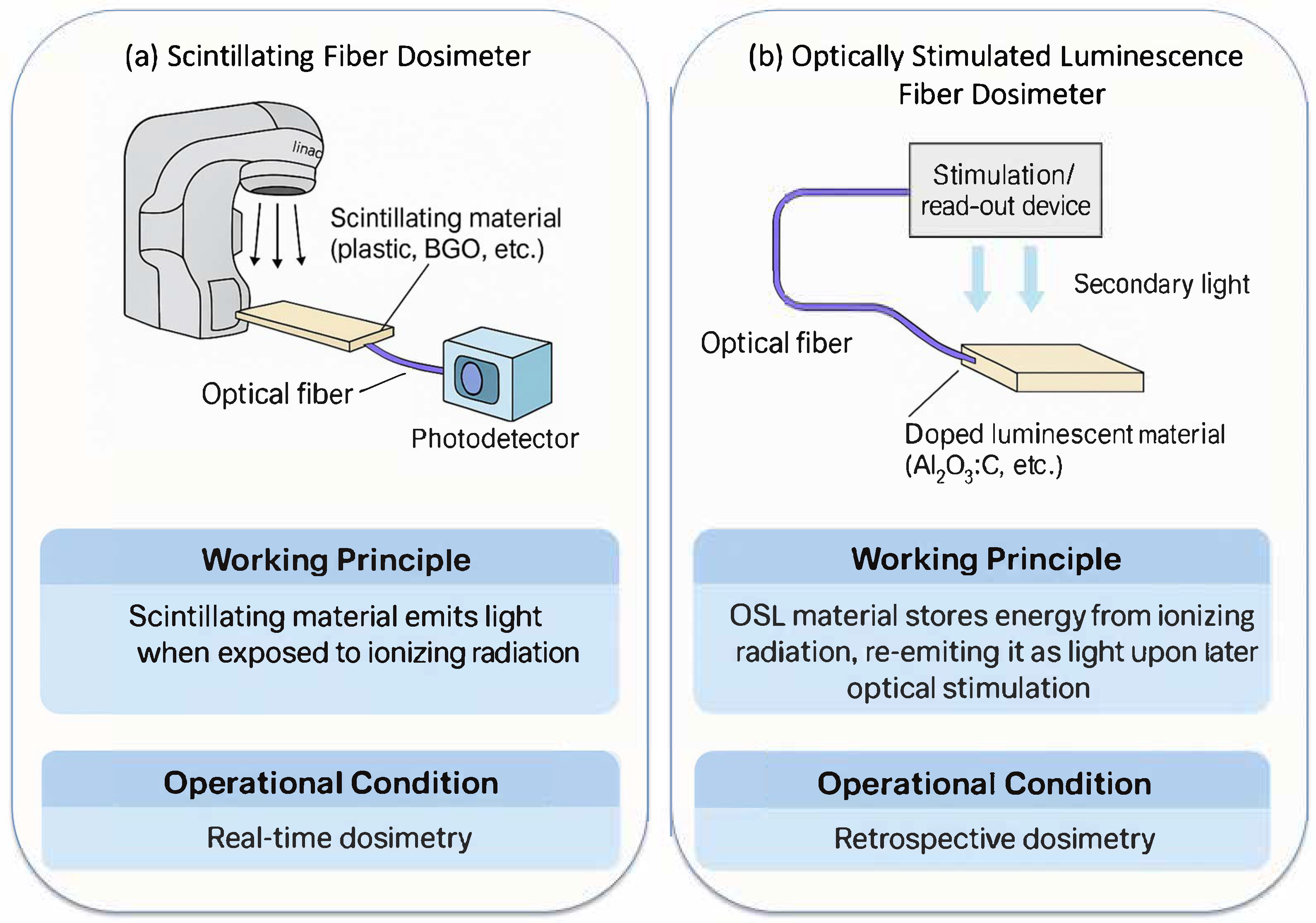
Figure 1. Optical fiber-based dosimetry. Setup, working principle, and operation condition of: (a) Scintillating Fiber Dosimeter; (b) Optically Stimulated Luminescence Fiber Dosimeter. To illustrate, relative sizes of scintillating and luminescent materials to optical fibers are exaggerated. SFD enables real-time readout, and OSL fiber dosimeter involves retrospective readout by light stimulation.
Photonics 12 01058 g001
Figure 2. Optical fiber spectroscopy in radiation oncology. Several examples shown here are: (a) Raman Spectroscopy, (b) Fluorescence Spectroscopy, (c) Diffuse Reflectance Spectroscopy, (d) Cherenkov Luminescence Imaging, and (e) Radiation-Induced Attenuation Spectroscopy. Optical spectroscopy has become pivotal tools in radiation oncology for non-invasive, real-time assessment of biochemical and physiological changes.
Photonics 12 01058 g002
Figure 3. Applications, technical challenges, and future directions of optical fiber sensors in radiation therapy. Optical fiber sensors offer advantages to potentially support adaptive radiation therapy, enable real-time monitoring and imaging of biochemical and physiological changes, and become integrated into the existing workflow with minimally invasive procedures and high compatibility. Meanwhile, fiber sensor technologies are evolving rapidly to address current technical limitations.
Photonics 12 01058 g003
Table 1. Main features of common type FODs and their comparisons.
Main
Feature
Scintillating Fiber DosimeterOptically Stimulated Luminescence Fiber DosimeterThermoluminescent Dosimeter FiberCherenkov Fiber
Dosimeter
Detection
Principle
Scintillation upon radiation exposureTraps radiation energy, later released by optical stimulationTraps radiation energy, released upon heatingDetects Cherenkov light emitted by high-energy charged particles
Real-time ReadoutYesNo (Delayed)No (Delayed)Yes
Radiation TypePhotons, electrons, protons, neutrons depending on scintillatorMainly photons, some sensitivity to electronsWide spectrum
depending on dopant
High-energy charged
particles (e.g., electrons, photons)
SensitivityHigh (depending on material)Moderate to high High Low to moderate
Key
Materials
Plastic scintillator, LYSO, BGO, Gd2O2SAl2O3:C, rare-earth doped materialsLiF, CaSO4:DyOptical fiber itself (e.g., PMMA or silica)
AdvantagesReal-time, high sensitivity, flexible integration and geometriesRetrospective readout, reusable, simple
structure
Established material base, thermal readoutReal-time, no added material needed, directly tied to dose delivery
DisadvantagesCherenkov contamination (stem effect),
fragility
Requires stimulation source, no real-time monitoringRequires heating, mechanical setup, sensitivity to environmental noiseLow signal intensity, needs high-sensitivity detectors
Clinical SuitabilityIn vivo dosimetry, real-time monitoring during RTQA, retrospective dose verification, environmental dosimetryQA, treatment verificationDose verification during LINAC, superficial
monitoring
Table 2. Comparison of major optical fiber spectroscopy and imaging techniques.
TechniquePrincipleTarget
Information
AdvantagesLimitationsFiber
Compatibility
Raman
Spectroscopy
Inelastic scattering of monochromatic light reveals vibrational modesMolecular composition (DNA, proteins, lipids)Label-free;
high chemical
specificity;
detects biochemical changes
Weak signal;
fluorescence interference;
slow acquisition
Excellent
(single-mode
fibers)
Fluorescence
Spectroscopy
Emission from intrinsic or extrinsic fluorophores upon excitationMetabolic activity (e.g., NADH, FAD); redox stateHigh sensitivity; real-time metabolic imagingRequires fluorophores; photobleaching; limited depthExcellent
(multi-mode
fibers)
Diffuse
Reflectance
Spectroscopy
Measured absorption and scattering of broadband lightBlood volume, oxygenation, scattering coefficientsSimple, low-cost; physiological parameters in real timeIndirect measurements; limited depth and resolutionExcellent
Cherenkov
Luminescence
Imaging
Light emitted by high-energy particles exceeding light speed in tissueDose deposition, real-time beam visualizationDirect correlation with radiation delivery; no added contrast neededShallow depth; weak signal; needs intensified/time-gated camerasModerate
(collection
via fiber)
Radiation-Induced Attenuation SpectroscopyIonizing radiation causing wavelength-dependent attenuation in fiberDose distribution, radiation field mappingPassive; label-free; works in high-radiation fields; distributed sensingRequires careful calibration; irreversible in some fiber typesExcellent
(especially for distributed sensing)
Optical
Coherence
Tomography
Low-coherence interferometry for cross-sectional imagingTissue microstructure and morphologyHigh-resolution; depth-resolved structural imaging; non-contactLimited penetration (~1–2 mm); less chemical specificityExcellent
(fiber bundles
or probes)
Photoacoustic
Imaging
Light-induced acoustic signal via thermoelastic expansionOptical absorption contrast (e.g., hemoglobin)Functional + structural imaging; deeper than optical-only methodsRequires laser source and ultrasound detectorGood
(hybrid probes)
Laser Speckle
Contrast Imaging
Analysis of dynamic speckle patterns from moving RBCsMicrovascular blood flowLabel-free; fast acquisition; sensitive to perfusion changesLimited to surface vasculature; sensitive to motion artifactsGood
(endoscopic probes)
Disclaimer/Publisher’s Note: The statements, opinions and data contained in all publications are solely those of the individual author(s) and contributor(s) and not of MDPI and/or the editor(s). MDPI and/or the editor(s) disclaim responsibility for any injury to people or property resulting from any ideas, methods, instructions or products referred to in the content.