Abstract
We present experimental results for the controlled mitigation of the electromagnetic pulses (EMPs) produced in the interactions of a 1 PW high-power 30 fs Ti:Sa laser VEGA-3 with solid-density targets transparent to laser-forward-accelerated relativistic electrons. This study aims at the band of very high frequencies (VHFs), i.e., those in the hundreds of MHz, which comprise the fundamental cavity modes of the rectangular VEGA-3 vacuum chamber. We demonstrate mode suppression by a tailoring of the laser-produced space charge distribution.
1. Introduction
Advances in laser technology have led to the emergence of high-power Ti:Sa laser systems [1,2,3,4,5,6,7,8,9] that are able to deliver laser pulses up to several PW at a high-repetition rate of 0.05 Hz–1 Hz. In principle, these systems allow for the generation of secondary sources that exist in a wide range, from ionizing radiation [10,11,12,13] to XUV- and THz-pulses [14,15,16,17], as well as current pulses [18] when they are focused to relativistic intensities. Recent developments toward high-repetition-rate ion sources [19,20] have aimed at the generation of pulsed, bright ion beams by well-known mechanisms such as Target Normal Sheath Acceleration (TNSA) [21,22], Radiation Pressure Acceleration [23], etc. [11], which are beneficial to isotope production [24,25], positron emission tomography [26], ion beam microscopy [27], particle-induced X-ray emission [28], as well as inertial confinement fusion [29]. The mechanisms rely on the build up of large accelerating potentials, which are the consequence of electron dynamics that are also the source of ultra-strong electromagnetic pulses (EMPs) [30].
When a focused laser pulse interacts with a μm-scale solid foil target at ultra-high intensities in the range of 1 × 10¹⁸ W cm−2 to 1 × 10²⁰ W cm−2, the extraction of electrons ensues from Brunel-type resonance absorption [31,32] and ponderomotive acceleration [33,34]. The electron dynamics give rise to the generation of intense EMPs with broad bandwidths, ranging from radio frequencies [35] to X-rays [36]. As they are particularly important for very high frequency (VHF) bands, the targets of the frequencies attain a strong positive net-charge due to laser-accelerated electrons that are able to escape the rising potential barrier [37]. These electrons predominantly propagate in a laser-forward direction [38], and they yield an asymmetric charge separation. They distribute a negative charge across the experimental setup when they stop on the -timescale of their time of flight. Return currents rise on the same timescale of [39], and resonances within the cavity of the experimental vacuum chamber can build up. The resulting broadband EMPs bear the geometry of the experimental setup in their mode structure [30,40].
The semiconductor electronics widely used in metrology devices or diagnostics are susceptible to EMPs in the VHV and ultra-high frequency bands [41]. The mitigation of EMPs is especially important in light of future applications that will require complex electronics systems for control and quality management. This work (i) proposes new means for measuring EMPs in the VHF band by deploying commercial broadband antennas, and it also (ii) demonstrates the efficient tailoring of electromagnetic modes by geometrical changes to the experimental setup.
2. Materials and Methods
For this work, EMPs were generated by the interaction of the -class VEGA-3 laser at Centro de Láseres Pulsados (CLPU, Villamayor, Spain) [42] with a double chirp pulse amplification Ti:Sa system that had a central wavelength of 800(10) nm.
2.1. Laser- and Target-System
A cross-polarized wave system was used to enhance the coherent contrast and reduce the throughput of the amplified spontaneous emission. The laser pulse was focused to relativistic intensities onto a 3.0(3) μm-thin, solid-density aluminum target. The target system holds 120 fresh surfaces for laser interaction in a matrix framed by aluminum plates that have recesses of a 1 mm diameter for each interaction zone, and these are exchanged by a 3D motorized mount [42]. The target system is vertically inclined by 12.5(5)° with respect to the laser axis to avoid back-reflection into the beamline. Before interaction, the laser pulse is amplified to a pulse energy up to 25 J per pulse, which is then compressed down to a 30 fs duration with a grating compressor and focused via a off-axis parabola to a focal spot size of a 12 μm Full-Width-at-Half-Maximum (FWHM). Both, the target positioning and the spatial characterization of the laser focal spot were conducted using a parallel imaging system. A motorized microscope objective picked up the laser pulse on-axis to the focal spot, and an achromatic doublet imaged the far-field of the beam onto a CCD camera. It was ensured that the numerical aperture of the imaging system was large enough to capture the laser pulse. The first Airy-disk enclosed 6.9 J, which was extrapolated from low-energy focus measurements, such as the laser intensity that reaching 1.3 × 10²⁰ W cm−2 on target. The normalized vector potential of the laser pulse calculated was as .
2.2. Space Charge Dynamics
The average energy that was transferred to an electron of the relativistic population approximately followed [43] with the ponderomotive scaling [22] of
where is the electron rest mass and c denotes the speed of light in a vacuum.
As the VEGA laser pulse showed no pre-pulses that were capable of inducing a density modulation across all of the 3 μm-thick target [42], the main acceleration mechanism of the charged particles was TNSA. In TNSA, the population of laser-heated relativistic electrons escape the target, and the successive potential dynamics lead to the formation of sheath fields that are capable of accelerating ionized surface contaminants up to several tens of MeV u−1 [44]. The ions co-propagate with slow electrons and form a quasi-neutral beam, which we will neglect in the following. The building up of a positive potential in the target through escaping relativistic electrons induces a return current toward the target. Note that the target’s only connection to the ground is via the motor block and the chamber itself. The deposition of electrons and return of the current lead to wall currents in the chamber, which, in turn, allow for resonant cavity modes to grow. With a focus on the MHz domain, the charging of the target on the timescale [37] is instantaneous: the target is initially at a net positive potential and electrons charge distributions are instantly deposited across the environment of the target.
2.3. Experimental Environment
The experimental setup is shown in Figure 1. The experimental environment was kept in a high-vacuum of 1 × 10⁻⁶ mbar, which allowed for (i) focusing the laser pulse to high intensities above the typical ionization threshold of matter at 1 × 10⁸ W cm−2 to 1 × 10¹² W cm−2 [45] and (ii) compressing the laser pulse beforehand without damage to the grating compressor of the VEGA-3 laser system. The interaction point was 31 cm off-center (i.e., toward the focusing parabola) with respect to the vacuum chamber. The vacuum chamber was made of aluminum, comprising dielectric view ports, and it was grounded via a spike earthing electrode.

Figure 1.
Experimental setup sketch (left) and computer-aided drawing (right) of the pulsed laser interaction with a solid-density target located off-center with respect to the rectangular vacuum vessel, which served as the interaction chamber. The EMPs were detected with a B-field antenna located behind the interaction region (in a laser forward direction) and above the path of the horizontal plane. A LiF crystal was located on a motorized stage to be moved in and out of the path of the laser-accelerated charged particles from the target rear side.
2.4. Cavity Modes
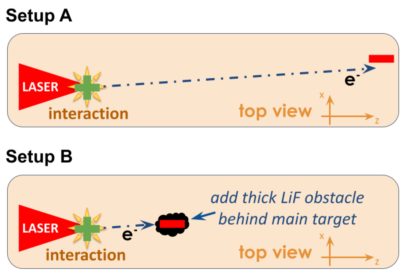
The inclination of the target leads to a deposition of negative charge in a wide opening angle ranging from one corner of the interaction chamber to the laser forward direction, see figure (Figure 2A). The relativistic electron beam is completely stopped in the thick wall of the vacuum chamber. Transverse and longitudinal electric fields are likely to build up. Return currents in the skin depth of the chamber walls then stream longitudinally and radially, causing a transverse azimuthal and radially toroidal magnetic field. The excited modes are expected to be , , and higher orders. The positive potential of the target and radial return currents can excite modes such as , , , , as well as higher orders.
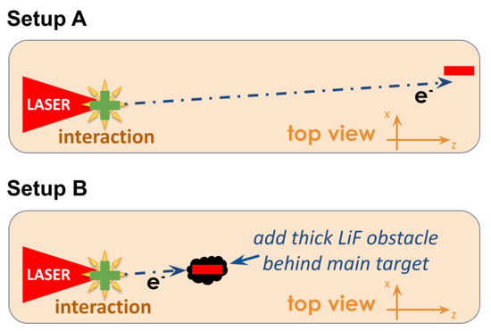
Figure 2.
A first setup (A) that comprises only the aluminum target and allows for a free propagation of accelerated species until they reach the chamber wall. The setup change (B) introduces an obstacle on the path of relativistic electrons, which are intended to change the seed space charge distribution and thus the build up of cavity modes.
In a medium with the speed of light c, resonant modes have the following frequencies:
The mode numbers correspond to dimensions for the width, height, and length of the vacuum vessel, respectively. The width in a laser-transverse direction is , the height is , and the length in a laser-forward direction is . Here, the lower bound is given by the rectangular inner surface. The outer bound is given by ports, flanges, and window surfaces, which are not expected to play a major role for resonances but will contribute to losses. The relevant cavity modes are
2.5. EMP Detection
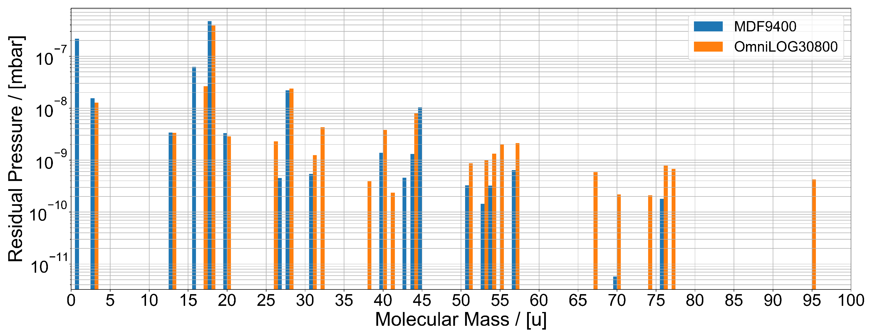
The primary EMP diagnostic used in this study was a commercial passive, calibrated B-field antenna with large bandwidth that ranged from 9 to 400 (Aaronia MDF9400). The outgassing of the antenna was tested before use, see Figure 3. The magnetic field antenna was positioned inside the vacuum chamber in the vertical plane of the laser propagation, which had a longitudinal value of behind the center of the cavity and above the horizontal center plane. The signal was transported via calibrated double-shielded SMA cables and was sent through a floating feed through. The conductive connection of the SMA cables and chamber was avoided completely, and the grounding was achieved via the wall plug of the data recorder. The waveforms were captured with an oscilloscope of a 1 bandwidth and sampling rate. The results displayed in this work were corrected for attenuation and the bandwidth limitations of the full measurement circuit, which was achieved by taking into account the transfer function of the circuit. In the frequency domain, the division of the oscilloscope measurement by the transfer function of the coaxial line between the antenna and analog digital converter will lead to an input signal by the antenna. The latter we call the corrected signal for clarity.
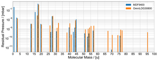
Figure 3.
The residual pressure of the detected molecules in a high vacuum chamber for commercially available EMP antennas from Aaronia.
3. Results
In order to unravel the changes to the mode structure in the VHF band, the standard mode structure in the vacuum chamber was first characterized, which was then followed by an obstacle being introduced to the path of the ejected electrons in order to modify the seed space charge distribution.
3.1. Standard Mode Structure in the VHF Band
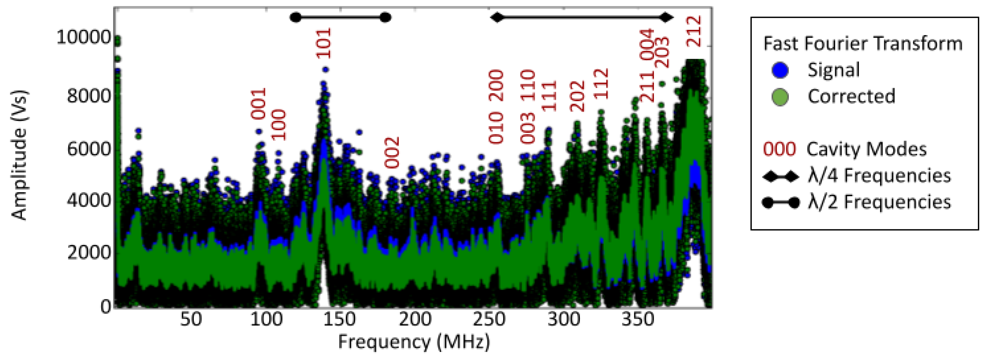
The time-integrated Fast Fourier Transform of the resulting time-resolved waveform data acquired during the experiment is shown in Figure 4. We defined the signal (blue points) as the measurement by the oscilloscope, and the corrected signal (green points) as the input signal from the antenna, which was calculated from the measurement. The latter was achieved by the division of the measurement by the transfer function of the coaxial line between the antenna and analog digital converter of the oscilloscope in the frequency domain. One notes the strong mode , which was selected by the initial diagonal space charge fields. The respective fundamentals and were clearly visible. In addition to the cavity modes, the cylindrical metallic -inch mounts of the optics with a length ranging from 20 to 30 produced spectral peaks with antenna fields in a range from 250 to 372 . Their resonance frequencies were defined by a match of multiples of with the length of the grounded monopole rod. Above these frequencies, there was a clear peak at 385(10) MHz. This corresponded to 19(1) , and might be related to the motorized translation stage holding the target.
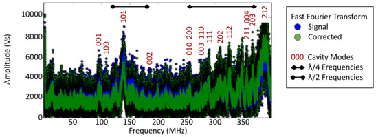
Figure 4.
The time-integrated spectrum of the magnetic field derived from the oscilloscope recordings of B-field antenna measurements in the VEGA-3 interaction chamber for shots on solid targets, with an indication of identified rectangular cavity modes and the resonance of the vertical stalks that hold optics (). Note that the signal denotes the measurement by the oscilloscope, and the corrected signal denotes the input signal from the antenna, which was calculated from the measurement.
3.2. Modified Mode Structure in the VHF Band
In order to alter the mode structure, a beam dumper from inorganic lithium fluoride (LiF) with a thickness of ≈ and a diameter of ≈ was positioned at 1 behind the aluminum target, see Figure 2B. The LiF and the axis were defined by the target normal and target center as being colinear. The crystal acted as an electron catcher, thereby removing part of the forward-propagating space charge. Due to a close positioning of the LiF catcher with respect to the target, practically the full solid angle in the laser-forward direction was covered. The arrangement was chosen to weaken the fundamental modes in the transverse and longitudinal directions. The stopping of electrons was simulated with the Monte Carlo simulation code CASINO [46] for a LiF crystal of a density of g cm−3. It stops all electrons up to an energy of , which presumably represent a significant part of the electron population.
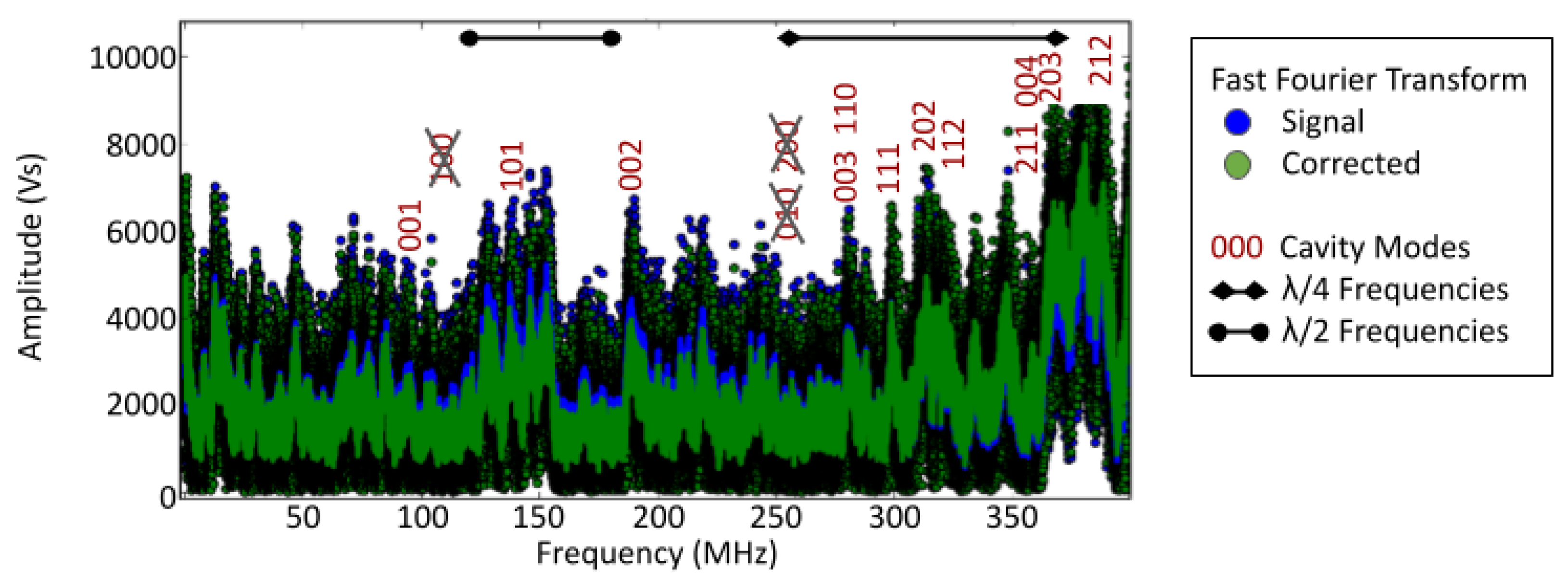
The mode structure with the LiF crystal installed is shown in Figure 5. We defined the signal (blue points) as the measurement by the oscilloscope, and the corrected signal (green points) as the input signal from the antenna, which was calculated from the measurement. The latter was achieved by division of the measurement by the transfer function of the coaxial line between the antenna and analog digital converter of the oscilloscope in the frequency domain. One notes a reduction in the fundamental mode in the longitudinal direction, a removal of the fundamental mode in the transverse direction, and a clear reduction in energy in mode . The return current in the mount of the beam dumper, which was about in height, manifested in a spectral peak that appeared at 366(4) MHz—on the left edge of the large peak, which might be attributed to the motorized translation stage that was discussed above.
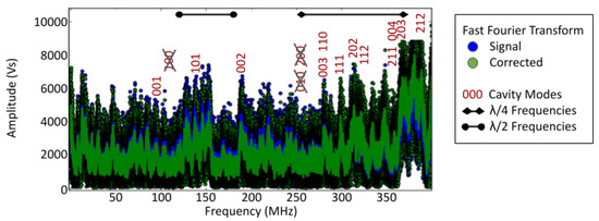
Figure 5.
The time-integrated spectrum of the magnetic field derived from the oscilloscope recordings of B-field antenna measurements in the VEGA-3 interaction chamber for shots on solid targets with an inserted LiF beam dump shortly behind the solid target, with an indication of the identified rectangular cavity modes and the resonance of the vertical stalks that hold optics (). Note that the signal denotes the measurement by the oscilloscope, and the corrected signal denotes the input signal from the antenna, which was calculated from the measurement.
4. Discussion
The results prove that a tailored geometrical modification of the experimental environment allows one to influence the VHF mode structure in the cavity. The build up of modes follows the geometry of the initial space charge distribution, which is the seed for cavity modes. The selection of suppressed modes is implicit; therefore, the method is only suitable if important geometrical modifications do not perturb the experiment, or if the presence of a huge obstacle is favorable.
In this work, a LiF crystal was used as a catcher in accordance with experiments that aim at a conversion of laser-accelerated ions to neutrons [47,48,49]. This scheme for EMP tailoring was not limited to the use of LiF as a catcher material, but it also has validity in other pitcher–catcher schemes [50]. The effectiveness of thick secondary catcher targets with respect to EMP tailoring aligns well with experimental requirements in many pitcher–catcher experiments, where thicker catcher targets represent good converters for the increased stopping of ion beam incidents.
5. Conclusions
Excited cavity modes were clearly detected by a compact EMP detection system based on a cheap commercial broadband range of 9 to 400 magnetic field antennas of a 15 diameter. The results show the respective fundamental modes of 125 and of 91 , as well as the antenna fields issued by cylindrical metallic -inch mounts in a range of 250 MHz to 372 MHz. Above these frequencies, there was a clear peak at 385(10) MHz, which might be related to the motorized translation stage holding the target.
This work shows that the EMP mode structure inside the vacuum cavity can be tailored by a modification of the experimental geometry. The introduction of a beam block to the propagation path of relativistic electrons suppresses the longitudinal mode and clearly reduces the mode. VHF EMPs are relevant in science and technology in many fields, ranging from homeland security [51] to medicine [52], due to their potential hazards and promising applications. Their controlled mitigation and generation will pave the ground for further studies in those fields.
Author Contributions
Conceptualization, M.E.; methodology, M.E.; software, M.E.; validation, M.E. and G.G.; formal analysis, M.E.; investigation, M.E. and J.I.A.; resources, M.D.R.-F., L.V. and G.G.; data curation, M.E.; writing—original draft preparation, M.E. and J.I.A.; writing—review and editing, M.E.; visualization, M.E.; supervision, M.E.; project administration, G.G.; funding acquisition, L.V. and G.G. All authors have read and agreed to the published version of the manuscript.
Funding
This research was funded by the European Union’s Horizon 2020 research and innovation program under grant agreement no. 871161.
Institutional Review Board Statement
Not applicable.
Informed Consent Statement
Not applicable.
Data Availability Statement
Data will be made available if a reasoanble request is addressed to the authors.
Acknowledgments
This work was supported by the European IMPULSE project with funding from the European Union’s Horizon 2020 research and innovation program under grant agreement no. 871161. The authors are grateful to Pilar Puyuelo-Valdes for organizing the underlying experiment as the responsible link scientist from the facility, as well as José Manuel Álvarez (CLPU) as the principal investigator for allowing us to conduct this parasitic study during his experimental campaign. We thank Pablo Vicente for his help with the computer-aided drawings. This work would not have been possible without the help of the laser and engineering teams at CLPU. Special thanks are given for the greatly appreciated support provided by the workshops of CLPU. We thank for the technical support by Juan Hernández for the vacuum measurements.
Conflicts of Interest
The authors declare no conflicts of interest.
References
- Jeong, T.M.; Lee, J. Femtosecond petawatt laser. Ann. Phys. 2014, 526, 157. [Google Scholar] [CrossRef]
- Hooker, C.J.; Blake, S.; Chekhlov, O.; Clarke, R.J.; Collier, J.L.; Divall, E.J.; Ertel, K.; Foster, P.S.; Hawkes, S.J.; Holligan, P.; et al. Commissioning the Astra Gemini Petawatt Ti:Sapphire Laser System. In Proceedings of the Conference on Lasers and Electro-Optics/Quantum Electronics and Laser Science Conference and Photonic Applications Systems Technologies, San Jose, CA, USA, 4–9 May 2008. CLEO (OSA Technical Digest), paper JThB2 (2008). [Google Scholar]
- Yu, T.J.; Lee, S.K.; Sung, J.H.; Yoon, J.W.; Jeong, T.M.; Lee, J. Generation of high-contrast, 30 fs, 1.5 PW laser pulses from chirped-pulse amplification Ti:sapphire laser. Opt. Express 2012, 20, 10807. [Google Scholar] [CrossRef]
- Leemans, W.P.; Daniels, J.; Deshmukh, A.; Gonsalves, A.J.; Magana, A.; Mao, H.S.; Mittelberger, D.E.; Nakamura, K.; Riley, J.R.; Syversrud, D. Bella laser and operations. In Proceedings of the PAC 2013, Pasadena, CA, USA, 29 September–4 October 2013; pp. 1097–1100. [Google Scholar]
- Sung, J.H.; Lee, H.W.; Yoo, J.Y.; Yoon, J.W.; Lee, C.W.; Yang, J.M.; Son, Y.J.; Jang, Y.H.; Lee, S.K.; Nam, C.H. 4.2 PW, 20 fs Ti: Sapphire laser at 0.1 Hz. Opt. Lett. 2017, 42, 2058. [Google Scholar] [CrossRef] [PubMed]
- Li, W.; Gan, Z.; Yu, L.; Wang, C.; Liu, Y.; Guo, Z.; Xu, L.; Xu, M.; Hang, Y.; Xu, J.; et al. 339 J high-energy Ti: Sapphire chirped-pulse amplifier for 10 PW laser facility. Opt. Lett. 2018, 43, 5681. [Google Scholar] [CrossRef]
- Lureau, F.; Matras, G.; Chalus, O.; Derycke, C.; Morbieu, T.; Radier, C.; Casagrande, O.; Laux, S.; Ricaud, S.; Rey, G.; et al. High-energy hybrid femtosecond laser system demonstrating 2 × 10 PW capability. High Power Laser Sci. Eng. 2020, 8, e43. [Google Scholar] [CrossRef]
- Danson, C.; Hillier, D.; Hopps, N.; Neely, D. Petawatt class lasers worldwide. High Power Laser Sci. Eng. 2015, 3, e3. [Google Scholar] [CrossRef]
- Wang, Y.; Wang, S.; Rockwood, A.; Luther, B.M.; Hollinger, R.; Curtis, A.; Calvi, C.; Menoni, C.S.; Rocca, J.J. 0.85 PW laser operation at 3.3 Hz and high-contrast ultrahigh-intensity λ = 400 nm second-harmonic beamline. Opt. Lett. 2017, 42, 3828–3831. [Google Scholar] [CrossRef] [PubMed]
- Daido, H.; Nishiuchi, M.; Pirozhkov, A.S. Review of laser-driven ion sources and their applications. Rep. Prog. Phys. 2012, 75, 056401. [Google Scholar] [CrossRef] [PubMed]
- Borghesi, M. Ion Acceleration: TNSA and Beyond. In Springer Proceedings in Physics; Springer: Berlin/Heidelberg, Germany, 2019; Volume 231. [Google Scholar]
- Tajima, T.; Malka, V. Laser plasma accelerators. Plasma Phys. Control. Fusion 2012, 62, 034004. [Google Scholar] [CrossRef]
- Norreys, P.A.; Santala, M.; Clark, E.; Zepf, M.; Watts, I.; Beg, F.N.; Krushelnick, K.; Tatarakis, M.; Dangor, A.E.; Fang, X.; et al. Observation of a highly directional λ-ray beam from ultrashort, ultraintense laser pulse interactions with solids. Phys. Plasmas 1999, 6, 2150–2156. [Google Scholar] [CrossRef]
- Lichters, R.; Meyer-ter-Vehn, J.; Pukhov, A. Short-pulse laser harmonics from oscillating plasma surfaces driven at relativistic intensity. Plasmas 1996, 3, 3425. [Google Scholar] [CrossRef]
- Corkum, P.B.; Krausz, F. Attosecond science. Nat. Phys. 2007, 3, 381. [Google Scholar] [CrossRef]
- Beg, F.N.; Bell, A.R.; Dangor, A.E.; Danson, C.N.; Fews, A.P.; Glinsky, M.E.; Hammel, B.A.; Lee, P.; Norreys, P.A.; Tatarakis, M. A study of picosecond laser-solid interactions up to 1019 Wcm−2. Phys. Plasmas 1997, 4, 447. [Google Scholar] [CrossRef]
- Bleko, V.; Karataev, P.; Konkov, A.; Kruchinin, K.; Naumenko, G.; Potylitsyn, A.; Vaughan, T. Coherent Cherenkov radiation as an intense THz source. J. Phys. Conf. Ser. 2016, 732, 012006. [Google Scholar] [CrossRef]
- Ehret, M.; Cikhardt, J.; Bradford, P.; Vladisavlevici, I.; Burian, T.; de Luis, D.; Henares, J.L.; Martín, R.H.; Apiñaniz, J.I.; Lera, R.; et al. High-repetition-rate source of nanosecond duration kA-current pulses driven by relativistic laser pulses. High Power Laser Sci. Eng. 2024. [Google Scholar] [CrossRef]
- Condamine, F.P.; Jourdain, N.; Hernandez, J.-C.; Taylor, M.; Bohlin, H.; Fajstavr, A.; Jeong, T.M.; Kumar, D.; Laštovička, T.; Renner, O.; et al. High-repetition rate solid target delivery system for PW-class laser–matter interaction at ELI Beamlines. Rev. Sci. Instruments 2021, 90, 063504. [Google Scholar] [CrossRef]
- Rehwald, M.; Assenbaum, S.; Bernert, C.; Brack, F.-E.; Bussmann, M.; Cowan, T.E.; Curry, C.B.; Fiuza, F.; Garten, M.; Gaus, L.; et al. Ultra-short pulse laser acceleration of protons to 80 MeV from cryogenic hydrogen jets tailored to near-critical density. Nat. Commun. 2023, 14, 4009. [Google Scholar] [CrossRef] [PubMed]
- Snavely, R.A.; Key, M.H.; Hatchett, S.P.; Cowan, T.E.; Roth, M.; Phillips, T.W.; Stoyer, M.A.; Henry, E.A.; Sangster, T.C.; Singh, M.S.; et al. Intense High-Energy Proton Beams from Petawatt-Laser Irradiation of Solids. Phys. Rev. Lett. 2000, 85, 2945. [Google Scholar] [CrossRef]
- Wilks, S.C.; Langdon, A.B.; Cowan, T.E.; Roth, M.; Singh, M.; Hatchett, S.; Key, M.H.; Pennington, D.; MacKinnon, A.; Snavely, R.A. Energetic proton generation in ultra-intense laser–solid interactions. Phys. Plasmas 2001, 8, 542. [Google Scholar] [CrossRef]
- Esirkepov, T.; Borghesi, M.; Bulanov, S.V.; Mourou, G.; Tajima, T. Highly Efficient Relativistic-Ion Generation in the Laser-Piston Regime. Phys. Rev. Lett. 2004, 92, 175003. [Google Scholar] [CrossRef]
- Nemoto, K.; Maksimchuk, A.; Banerjee, S.; Flippo, K.; Mourou, G.; Umstadter, D.; Bychenkov, V.Y. Laser-triggered ion acceleration and table top isotope production. Appl. Phys. Lett. 2001, 78, 595–597. [Google Scholar] [CrossRef]
- Ledingham, K.W.D.; McKenna, P.; McCanny, T.; Shimizu, S.; Yang, J.M.; Robson, L.; Zweit, J.; Gillies, J.M.; Bailey, J.; Chimon, G.N.; et al. High power laser production of short-lived isotopes for positron emission tomography. J. Phys. D Appl. Phys. 2004, 37, 2341. [Google Scholar] [CrossRef]
- Santala, M.I.K.; Zepf, M.; Beg, F.N.; Clark, E.L.; Dangor, A.E.; Krushelnick, K.; Tatarakis, M.; Watts, I.; Ledingham, K.W.D.; McCanny, T.; et al. Production of radioactive nuclides by energetic protons generated from intense laser-plasma interactions. Appl. Phys. Lett. 2001, 78, 19–21. [Google Scholar] [CrossRef]
- Merrill, F.E.; Golubev, A.A.; Mariam, F.G.; Turtikov, V.I.; Varentsov, D. HEDgeHOB Collaboration Proton Microscopy at FAIR. AIP Conf. Proc. 2009, 1195, 667–670. [Google Scholar] [CrossRef]
- Mirani, F.; Maffini, A.; Casamichiela, F.; Pazzaglia, A.; Formenti, A.; Dellasega, D.; Russo, V.; Vavassori, D.; Bortot, D.; Huault, M.; et al. Integrated quantitative PIXE analysis and EDX spectroscopy using a laser-driven particle source. Sci. Adv. 2021, 7, 3. [Google Scholar] [CrossRef] [PubMed]
- Roth, M.; Cowan, T.E.; Key, M.H.; Hatchett, S.P.; Brown, C.; Fountain, W.; Johnson, J.; Pennington, D.M.; Snavely, R.A.; Wilks, S.C.; et al. Fast Ignition by Intense Laser-Accelerated Proton Beams. Phys. Rev. Lett. 2001, 86, 436–439. [Google Scholar] [CrossRef] [PubMed]
- Consoli, F.; Tikhonchuk, V.T.; Bardon, M.; Bradford, P.; Carroll, D.C.; Cikhardt, J.; Cipriani, M.; Clarke, R.J.; Cowan, T.; Danson, C.N.; et al. Laser produced electromagnetic pulses: Generation, detection and mitigation. High Power Laser Sci. Eng. 2020, 8, e22. [Google Scholar] [CrossRef]
- Brunel, F. Not-so-resonant, resonant absorption. Phys. Rev. Lett. 1987, 59, 52. [Google Scholar] [CrossRef] [PubMed]
- Wilks, C.; Kruer, W.L. Absorption of ultrashort, ultra-intense laser light by solids and overdense plasmas. IEEE J. Quantum Electron. 1997, 33, 1954. [Google Scholar] [CrossRef]
- Wilks, S.C.; Kruer, W.L.; Tabak, M.; Langdon, A.B. Absorption of ultra-intense laser pulses. Phys. Rev. Lett. 1992, 69, 1383–1386. [Google Scholar] [CrossRef]
- Pukhov, A.; Meyer-ter, V.J. Relativistic laser-plasma interaction by multi-dimensional particle-in-cell simulations. Phys. Plasmas 1998, 5, 1880–1886. [Google Scholar] [CrossRef]
- Pearlman, J.S.; Dahlbacka, G.H. Charge separation and target voltages in laser-produced plasmas. Appl. Phys. Lett. 1977, 31, 414–417. [Google Scholar] [CrossRef]
- Courtois, C.; Fontaine, A.C.L.; Landoas, O.; Lidove, G.; Méot, V.; Morel, P.; Nuter, R.; Lefebvre, E.; Boscheron, A.; Grenier, J.; et al. Effect of plasma density scale length on the properties of bremsstrahlung X-ray sources created by picosecond laser pulses. Phys. Plasmas 2009, 16, 013105. [Google Scholar] [CrossRef]
- Poyé, A.; Hulin, S.; Ribolzi, J.; Bailly-Grandvaux, M.; Lubrano-Lavaderci, F.; Bardon, M.; Raffestin, D.; Santos, J.J.; Tikhonchuk, V. Thin target charging in short laser pulse interactions. Phys. Rev. E 2018, 98, 033201. [Google Scholar] [CrossRef]
- Rusby, D.R.; Wilson, L.A.; Gray, R.J.; Dance, R.J.; Butler, N.M.H.; MacLellan, D.A.; Scott, G.G.; Bagnoud, V.; Zielbauer, B.; McKenna, P.; et al. Measurement of the angle, temperature and flux of fast electrons emitted from intense laser-solid interactions. J. Plasma Phys. 2015, 81, 5. [Google Scholar] [CrossRef]
- Dubois, J.-L.; Lubrano-Lavaderci, F.; Raffestin, D.; Ribolzi, J.; Gazave, J.; Fontaine, A.C.L.; d’Humières, E.; Hulin, S.; Nicolaï, P.; Poyé, A.; et al. Target charging in short-pulse-laser–plasma experiments. Phys. Rev. E 2014, 89, 013102. [Google Scholar] [CrossRef]
- Nelissen, K.; Liszi, M.; Marco, M.D.; Ospina, V.; Drotár, I.; Gatti, G.; Kamperidis, C.; Volpe, L. Characterisation and Modelling of Ultrashort Laser-Driven Electromagnetic Pulses. Sci. Rep. 2020, 10, 3108. [Google Scholar] [CrossRef]
- Shurenkov, V.V.; Pershenkov, V.S. Electromagnetic effects and damage mechanism on the semiconductor electronics. Facta Univ. Ser. Electron. Energetics 2016, 29, 621–629. [Google Scholar] [CrossRef]
- Volpe, L.; Fedosejevs, R.; Gatti, G.; Pérez-Hernández, J.A.; Méndez, C.; Apiñaniz, J.; Vaisseau, X.; Salgado, C.; Huault, M.; Malko, S.; et al. Generation of high energy laser-driven electron and proton sources with the 200 TW system VEGA 2 at the Centro de Laseres Pulsados. High Power Laser Sci. Eng. 2019, 7, e25. [Google Scholar] [CrossRef]
- Kluge, T.; Cowan, T.; Debus, A.; Schramm, U.; Zeil, K.; Bussmann, M. Electron Temperature Scaling in Laser Interaction with Solids. Phys. Rev. Lett. 2011, 107, 205003. [Google Scholar] [CrossRef]
- Roth, M.; Schollmeier, M. Ion Acceleration—Target Normal Sheath Acceleration. In Proceedings of the 2014 CAS-CERN Accelerator School: Plasma Wake Acceleration, Geneva, Switzerland, 23–29 November 2014. [Google Scholar] [CrossRef]
- Vertes, A.; Wolf, M.D.; Juhasz, P.; Gijbels, R. Threshold conditions of plasma ignition in laser ionization mass spectrometry of solids. Anal. Chem. 1989, 61, 1029–1035. [Google Scholar] [CrossRef]
- Drouin, D.; Couture, A.R.; Joly, D.; Tastet, X.; Aimez, V.; Gauvin, R. CASINO V2.42—A Fast and Easy-to-use Modeling Tool for Scanning Electron Microscopy and Microanalysis Users. Scanning 2007, 29, 92–101. [Google Scholar] [CrossRef] [PubMed]
- Lancaster, K.L.; Karsch, S.; Habara, H.; Beg, F.N.; Clark, E.L.; Freeman, R.; Key, M.H.; King, J.A.; Kodama, R.; Krushelnick, K.; et al. Characterization of neutron yields from laser produced ion beams for fast neutron radiography. Phys. Plasmas 2004, 11, 3404–3408. [Google Scholar] [CrossRef]
- Higginson, D.P.; McNaney, J.M.; Swift, D.C.; Petrov, G.M.; Davis, J.; Frenje, J.A.; Jarrott, L.C.; Kodama, R.; Lancaster, K.L.; Mackinnon, A.J.; et al. Production of neutrons up to 18 MeV in high-intensity short-pulse laser matter interactions. Phys. Plasmas 2011, 18, 100703. [Google Scholar] [CrossRef]
- Deng, H.X.; Sha, R.; Hu, L.X.; Jiang, X.R.; Zhao, N.; Zou, D.B.; Yu, T.P.; Shao, F.Q. Pulsed neutron source from interaction of relativistic laser pulse with micro-structure assisted pitcher-catcher target. Plasma Phys. Control. Fusion 2022, 64, 085004. [Google Scholar] [CrossRef]
- Jiang, X.R.; Shao, F.Q.; Zou, D.B.; Yu, M.Y.; Hu, L.X.; Guo, X.Y.; Huang, T.W.; Zhang, H.; Wu, S.Z.; Zhang, G.B.; et al. Energetic deuterium-ion beams and neutron source driven by multiple-laser interaction with pitcher-catcher target. Nucl. Fusion 2020, 60, 076019. [Google Scholar] [CrossRef]
- Baker, G.H. Electromagnetic Pulse Resilience of United States Critical Infrastructure: Progress and Prognostics. J. Crit. Infrastruct. Policy 2021, 2, 1. [Google Scholar] [CrossRef]
- Kim, J.H.; Lee, J.; Kim, H.; Kim, K.; Kim, H.R. Possible Effects of Radiofrequency Electromagnetic Field Exposure on Central Nerve System. Biomol. Ther. 2019, 27, 265–275. [Google Scholar] [CrossRef]
Disclaimer/Publisher’s Note: The statements, opinions and data contained in all publications are solely those of the individual author(s) and contributor(s) and not of MDPI and/or the editor(s). MDPI and/or the editor(s) disclaim responsibility for any injury to people or property resulting from any ideas, methods, instructions or products referred to in the content. |
© 2024 by the authors. Licensee MDPI, Basel, Switzerland. This article is an open access article distributed under the terms and conditions of the Creative Commons Attribution (CC BY) license (https://creativecommons.org/licenses/by/4.0/).


