Review

Non-Pharmacological Therapies for Management of Temporomandibular Myofascial Pain Syndrome: Laser Photobiomodulation or Dry Needling? Meta-Analyses of Human Clinical Trials

by
Jumana Alsarhan
1,†,
Rita El Feghali
1,*,†,
Thaer Alkhudari
2 and
Stefano Benedicenti
1
1
Department of Surgical Sciences and Integrated Diagnostics, University of Genoa, 16132 Genoa, Italy
2
Department of Orthodontics, Istanbul Yeni Yüzyil University, Istanbul 34010, Turkey
*
Author to whom correspondence should be addressed.
These authors contributed to the manuscript equally.
Photonics 2024, 11(10), 965; https://doi.org/10.3390/photonics11100965
Submission received: 7 September 2024 / Revised: 29 September 2024 / Accepted: 9 October 2024 / Published: 14 October 2024
(This article belongs to the Section Biophotonics and Biomedical Optics)

Abstract

:
This review aims to compare the effect of photobiomodulation therapy (PBMT) using visible and near-infrared diode laser wavelengths to that of the dry needling technique (DNT) on the management of orofacial pain in patients with Temporomandibular Disorder Myofascial Pain Syndrome (TMD/MPS) in term of effectiveness, speed of recovery, and lasting of treatment. A systematic search of multiple electronic databases was carried out to identify the relevant clinical trials published between 1 January 2010 and 1 January 2024. The included studies were limited to human subjects who had orofacial pain associated with Axis 1 of TMD/MPS, involving two genders with age >18 years and were treated either with photobiomodulation using diode laser with wavelengths ranging from 600 up to 1200 nanometer (nm), or with the dry needling (DN) technique (superficial SDN or deep DDN), as a non-pharmacological therapies to decrease the intensity of orofacial pain associated with TMD/MPS. The risk of bias for included studies was assessed using the Cochrane RoB tool (for randomized studies). Three distinct meta-analyses were performed to quantify the pooled effects of PBM and DN in the management of TMD/MPS myofascial pain and deactivation of myofascial trigger points (MTPs). The meta-analyses were performed using Review Manager (RevMan) 5.3 from Cochrane. The confidence interval (CI) was established at 95% and p-values of less than 0.05 (p < 0.05) were considered statistically significant. Statistical heterogeneity was assessed using I2. Qualitative data were extracted and summarized in tables for each group study, while quantitative data were reported as Mean and Standard Deviation (SD) values for assessment variables in each sub-group study. The results among groups were systematically evaluated to draw the final conclusion. A rigorous electronic and manual search revealed 4150 possible articles. Following the application of the inclusion and exclusion criteria, twelve eligible studies were included in the analysis. Both PBMT and DNT were found to be effective in controlling the myalgia pain and primary symptoms associated with TMD/MPS, as well as deactivating the MTPs. DNT was statistically superior in terms of recovery time while PBMT was the more effective long-term therapy.

1. Introduction

Temporomandibular disorders (TMDs) encompass a range of conditions affecting the temporomandibular joint (TMJ), the masticatory muscles and their related structures [1]. These disorders are marked by pain, joint sounds, and limited movement of the jaw [2]. Orofacial pain refers to any discomfort that is specifically related to the soft and mineralized tissues of the oral cavity and face, including the skin, blood vessels, bones, teeth, glands, and muscles [3]. TMD is considered to be the most common cause of non-dental pain in the maxillofacial region [4]. Over the years several systems to classify TMDs have been proposed. The Research Diagnostic Criteria for Temporomandibular Disorders (RDC/TMD) classification has been extensively used as a diagnostic tool for TMD since it was released in 1992 [3]. It divides TMDs into three categories based on the common criteria among the diseases described in Table 1.
TMD can arise due to complications within the joint itself or in the encompassing musculature. Joint complications consist of arthritis, inflammation, and internal derangements. When the affected area is a muscle, the condition is referred to as myofascial pain syndrome (MPS) [6]. MPS is defined as a prevalent pain disorder characterized by the development of trigger points (TPs). This condition is characterized by pain emanating from the muscle of origin and extending beyond the masticatory muscle boundary [7]. Myofascial trigger points (MTPs) typically exhibit a distinct pattern of referred pain and hurt when compressed, stretched, overstretched, or squeezed [8]. A trigger point consists of many contraction knots, where individual muscle fibers contract and are unable to release [9]. The persistent contraction of muscle sarcomeres leads to the compression of nearby blood supply, causing a lack of energy in the affected area. The metabolic crisis triggers the activation of pain receptors, resulting in the development of a localized pain pattern that follows a specific pathway of nerves. The pain patterns are therefore constant and thoroughly established for different muscles [10]. Myofascial pain is a commonly recurring form of orofacial pain, affecting a significant portion of the population. It is observed that approximately half of individuals experience symptoms in the facial and masticatory muscles with higher incidence in females [11,12,13]. It is important to note that, based on Axis I and Diagnostic Criteria for Temporomandibular Disorders (DC/TMD), it is necessary to differentiate TMD/MPS from other temporo-mandibular disorders [8]. The precise etiology of MPS is still unknown, and the most conservative and strategic approach to treating the condition is still up for debate [8,14,15]. An extensive historical account and thorough clinical examination are used to identify the role of predisposing, perpetuating, and precipitating causes, medical comorbidities, and the level of involvement and depth of adaptation of different structures. This information allows the clinician to establish a diagnosis. Therefore, it is important to customize the treatment to the specific cause [16].
Extensive research has been conducted on various treatments, including occlusal splints and pharmacotherapies like anesthetics, antidepressants, anticonvulsants, non-steroidal anti-inflammatory drugs and, recently, the use of Botulinum toxin type A (BTX-A) [6,17]. Nevertheless, patients undergoing long-term treatments often face side effects due to adverse drug reactions [18]. The prevalence of musculoskeletal disorders, particularly those of myofascial origin, is expected to rise, necessitating the development of effective and safe therapeutic interventions. Conversely, as many as 40% of symptomatic patients experience spontaneous resolution of their symptoms without treatment, while 50–90% achieve relief through conservative therapy [15]. Consequently, self-care and noninvasive treatments represent viable options that should be pursued prior to considering invasive therapies [19]. Hence, alternative treatments like ultrasound, massage therapy, physiotherapy, acupuncture, exercise, transcutaneous electrical nerve stimulation (TENS), mesotherapy, wet needling, dry needling (DN) and photobiomodulation therapy (PBMT) were suggested [8,19,20].
Dry needling is a common efficient non-pharmacological therapeutic technique frequently used for TMD/MPS [21]. It is a procedure that aims to relieve pain by targeting trigger points to reduce tension or muscle cramps. It involves the insertion of monofilaments needles into specific trigger points inside muscles and tissues to help increase blood flow and oxygen and relieve pain and reduce stiffness [22]. Nevertheless, the mechanism by which dry needling alleviates pain in MPS remains unclear. Several theories have been presented, including the “gate control” hypothesis, change in the body’s natural painkillers, interruption of central sensitization, and the influence of placebo effects [23]. Dry needling is believed to inhibit pain signals by stimulating small-diameter nerve fibers and enhancing the release of neurotransmitters. It also has a suppressive impact on trigger points [24]. When a needle is inserted into a trigger point, it might cause a local twitch reaction. This involuntary contraction of the trigger point can also help facilitate physiological changes, such as lowering spontaneous electrical activity and decreasing the concentration of nociceptive and inflammatory substances, thereby promoting further relaxation of the trigger point [25]. Trigger-point dry needling can decrease the sensitivity of the central nervous system by diminishing the peripheral perception of pain linked with the trigger point, decreasing the activity of neurons in the dorsal horn, and regulating parts of the brainstem that are involved in pain. Nevertheless, the impacts are mostly seen in the immediate period, and the magnitudes of the effects are modest to tiny [9]. Like any form of therapy, dry needling may have a few infrequent but moderate adverse effects. The predominant adverse effects may encompass slight hemorrhaging, contusions, and transient discomfort (lasting around 24–48 h) [26]. Dry needling is not recommended for pregnant women, because there is a small risk it can start early labor. It is not advisable in patients with compromised immune system, ongoing malignancy, ongoing infection, or poor healing, since the body may not be capable of generating the same healing reaction to the needles. In addition, some blood thinners can increase the risk of bruising [27].
Photobiomodulation therapy (PBMT) is a non-invasive treatment that involves the use of non-ionizing electromagnetic light to stimulate photochemical reactions inside photoreceptive cellular structures [28]. Mitochondria and light-sensitive ion channels are especially susceptible to this mechanism [29]. It operates by modulating cellular homeostasis and metabolism subsequent to the transfer of photonic energy from visible and near-infrared light sources such as light-emitting diodes (LEDs), lasers, and/or broadband light [30]. The biostimulatory effects of PBM treatment have led to positive therapeutic results such as pain relief, inflammation reduction, immunomodulation, and the stimulation of wound healing and tissue regeneration [31]. Additionally, with a correct incident dose applied, laser-assisted PBM therapy has no appreciable thermal effects in the irradiated tissue [32]. PBMT demonstrates a biostimulating effect by enhancing cellular activity, adenosine triphosphate (ATP) production in the mitochondrial respiratory chain and increase in intracellular Ca++ [33]. Furthermore, it possesses analgesic properties that can be attributed to a range of mediators, including endorphins, acetylcholine, serotonin, and cortisol [34]. Low-level laser treatment induces angiogenesis, which in turn leads to an anti-inflammatory effect. PBMT also stimulates the release of growth factors and cytokines, enhancing replication mechanisms that promote cell repair processes and reduce oxidative stress [35].
The advantage of PBMT and DN in the treatment of TMD/MPS is controversial, and its benefits have been mentioned in some studies and denied in others. Multiple studies have demonstrated the efficacy of DN in inactivating trigger points in myofascial pain syndrome [36,37]. On the other side, Carrasco et al. found that the pain relief in patients with MPS of the masticatory and temporomandibular muscles in the groups receiving low-dose laser at different doses was similar to the groups receiving placebo laser [38]. In a study by Uemoto et al. on patients with MPS in the masseter muscle, the effect of dry needling was comparable to laser therapy [39].
Thus, up to this point, current research on the efficacy of laser treatment versus dry needling has yielded conflicting outcomes; see Figure 1.
Therefore, the aim of this study was to assess the impact of diode laser photobiomodulation and dry needling therapies on the management of trigger points in patients suffering from myofascial pain syndrome of temporomandibular joint disorder (TMJD/MPS) in terms of effectiveness, speed of recovery and lasting treatment.

2. Materials and Methods

2.1. Search Strategy for the Systematic Review

A systematic review of clinical trials (CTs) and randomized clinical trials (RCTs) was conducted in databases, and meta-analyses approaches were undertaken.
The review protocol is registered in PROSPERO (CRD42024519618), the International Prospective Register of Systematic Reviews.

2.1.1. Research Question

In order to conduct this systematic review and meta-analyses, the following questions were raised:
  • Is photobiomodulation treatment, which utilizes diode lasers with wavelengths ranging from 600 to 1200 nm, efficacious in treating myofascial pain associated with temporomandibular disorder (TMD) Axis 1?
  • Does the DN method effectively control TPs in TMJD/MPS?
  • Which therapy, PBMT or DNT, is more beneficial in treating myogenic pain of the temporomandibular joint (TMJ) in terms of efficacy, speed of recovery, and continuity of results?

2.1.2. Systematic Search Strategy

The electronic searches were conducted in the specified databases:
  • PubMed/Medline electronic database;
  • COCHRANE LIBRARY;
  • ScienceDirect;
  • Scopus;
  • Google Scholar.
A systematic search was conducted on electronic databases to identify interventional studies published in English between 1 January 2010 and 1 January 2024. These studies focused on the application of photobiomodulation (PBM) using diode lasers with wavelengths ranging from 600 to 1200 nm, or the DN technique, in patients experiencing symptoms related to temporomandibular disorders (TMDs) Axis 1 or temporomandibular joint/myofascial pain syndrome (TMJ/MPS).
The databases were queried using terms in either simple or multiple conjunctions, as outlined below: (Diode Laser Therapy OR Photo biomodulation OR Low-Level Laser Therapy AND TMJ Pain OR TMJ Analgesia), (Laser Photo biomodulation AND Myofascial Pain Syndrome). (Dry Needling AND TMJ Myofascial Pain), (DN OR PBM AND TMJ Pain OR MPS).
The selection of these keywords was based on the PICOS strategy; Population (adult patients with TMD); Intervention (PBM or DN); Comparison (compared or not with placebo group); Outcome (myofascial pain); and Study design (in vivo studies).
The criteria for inclusion/exclusion were applied as follows:
Inclusion Criteria:
  • Full-text clinical trials (CTs) or randomized clinical trials (RCTs) either blinded or not.
  • Studies published in the English language in peer-reviewed journals between 1 January 2010 and 1 January 2024.
  • Human studies involving both female and male genders > 18 years old.
  • Studies that have patients with myofascial pain resulting from Axis 1 of TMD or MPS according to the RDC/TMD classification, regardless of the quality of life.
  • Studies that compare extraoral photobiomodulation (PBM) therapy using diode lasers (600–1200 nm) with placebo or sham laser.
  • Studies that compare SDN or DDN therapy with placebo.
  • Studies that compare laser PBMT (600–1200 nm) with DNT in the management of TMJ myofascial pain.
Exclusion Criteria:
  • Systematic reviews and meta-analyses.
  • In vitro studies.
  • Studies that focus on specific age groups, such as adolescents or elders.
  • Studies that contain subjects younger than 18 years old.
  • Studies that have patients with a medical history of cancers or other syndromes in the head and neck region.
  • Studies that have patients with pain not related to Axis 1 of TMD according to the RDC/TMD classification.
  • Studies that use LEDs, or other laser equipment except diodes.
  • Studies that use different laser wavelengths except 600–1200 nm.
  • Studies that do not adopt the RDC/TMD classification in the diagnosis of TMJ diseases.
  • Comparative studies that compare PBM with any aspect of therapy except the DN technique.
  • Studies that adopt home PBM protocol.
  • Patents, degrees, or doctoral theses.
  • Republished articles or duplicated articles.

2.1.3. Study Selection and Data Extraction

Two independent reviewers examined the studies to assess their compliance with the selected criteria. The titles and abstracts were reviewed to ascertain the suitability of the studies. Disagreements were settled through debates with fellow authors. A final group of twelve eligible studies was selected.
The information and data are summarized in six tables, in the Results section, including the first author of the study, publication year, treatment protocol, evaluated variables, measurement scale for pain assessment, follow-up, and outcomes.

2.2. Study Quality Assessment

2.2.1. PRISMA Guidelines

A systematic search was conducted following the standards of the Preferred Reporting Items for Systematic Reviews and Meta-Analyses (PRISMA) statement [40].

2.2.2. Risk of Bias

The methodological quality of the research in all the included studies was evaluated through screening. The technique developed by the Cochrane Collaboration for evaluating the potential for bias in systematic reviews was utilized [41]. Each study included in the analysis had a thorough evaluation of bias, with a specific assessment conducted for each aspect. The final assessment categorized the total risk as either low, high, or unknown [42].

2.2.3. Meta Analysis of the Data

All the included studies were statistically analyzed to assess their results. Three distinct meta-analyses were performed to quantify the pooled effects of PBM and DN in the management of TMD/MPS myofascial pain and deactivation of myofascial trigger points (MTPs). The meta-analyses were performed using Review Manager (RevMan) 5.3 from Cochrane. The confidence interval (CI) was established at 95% and p-values of less than 0.05 (p < 0.05) were considered statistically significant. Statistical heterogeneity was assessed using I2. Qualitative data were extracted and summarized in tables for each group study, while quantitative data were reported as mean and standard deviation (SD) values for assessment variables in each sub-group study. The results among groups were systematically evaluated to draw the final conclusion.

3. Results

3.1. Literature Search Outcome

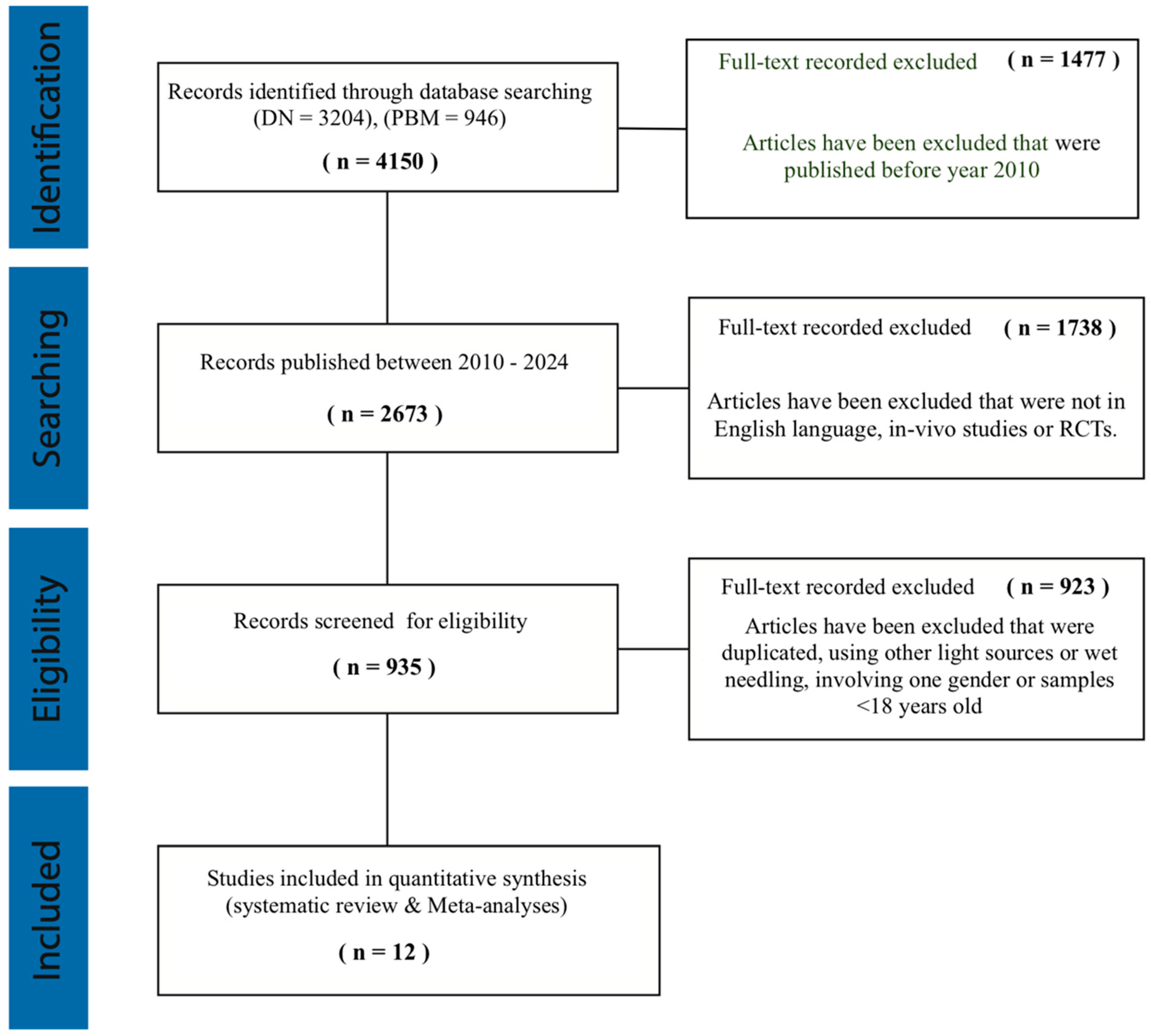
After conducting the initial electronic study, a preliminary database consisting of 4150 articles was generated. The titles and abstracts of the records were examined to ascertain the suitability of the investigations. After applying the inclusion and exclusion criteria, a total of twelve full texts were selected, which included studies [43,44,45,46,47,48,49,50,51,52,53,54] (Figure 2).

3.2. Risk of Bias

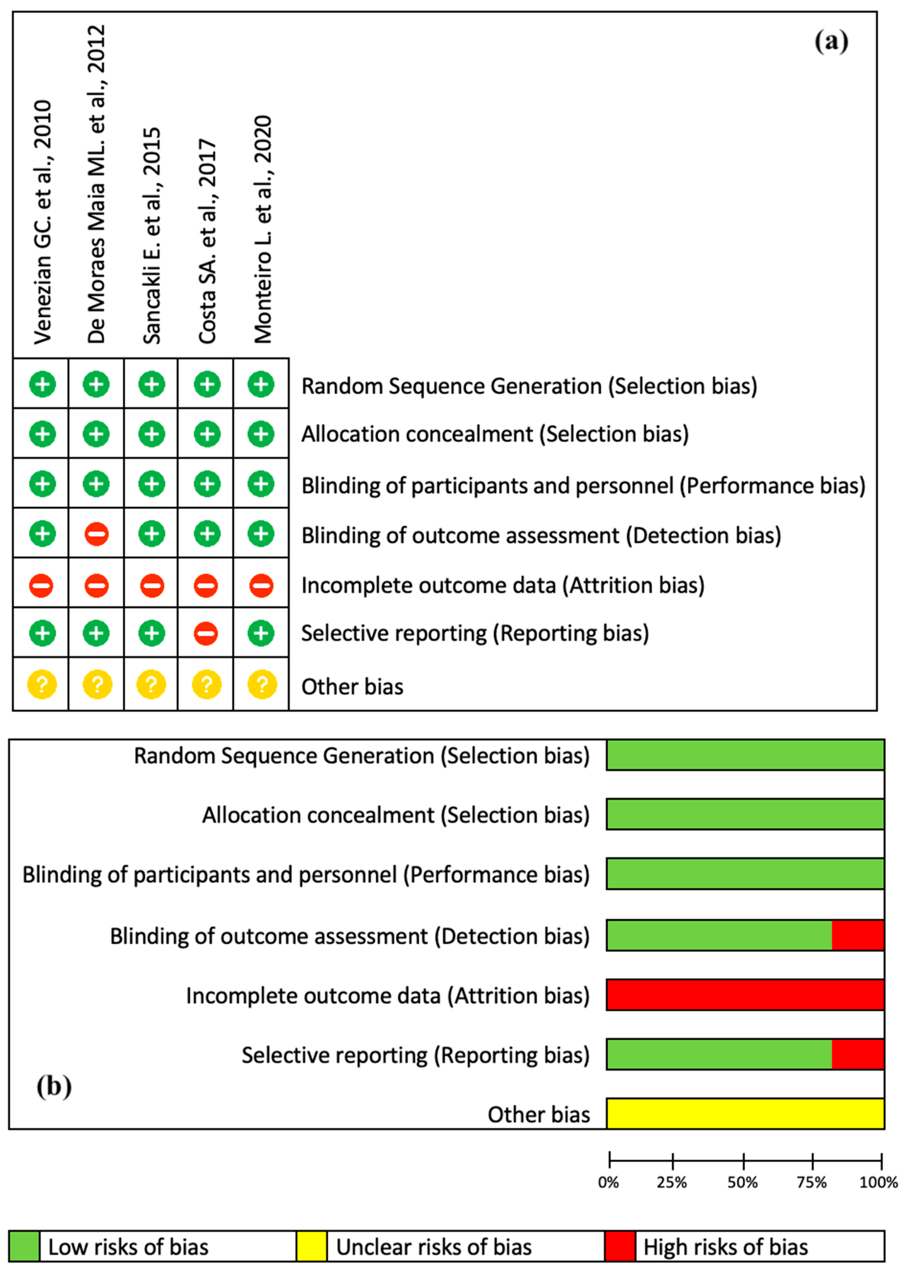
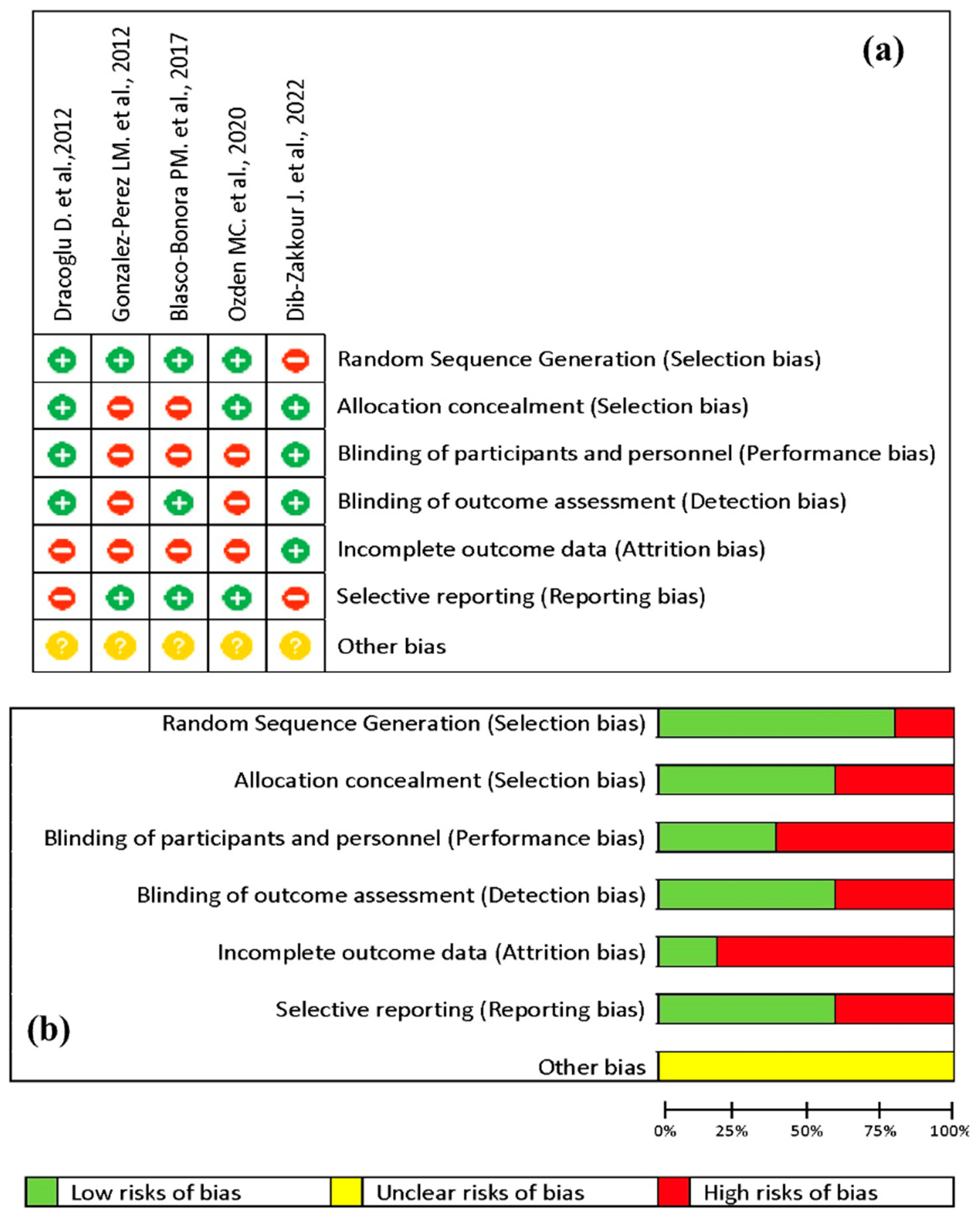
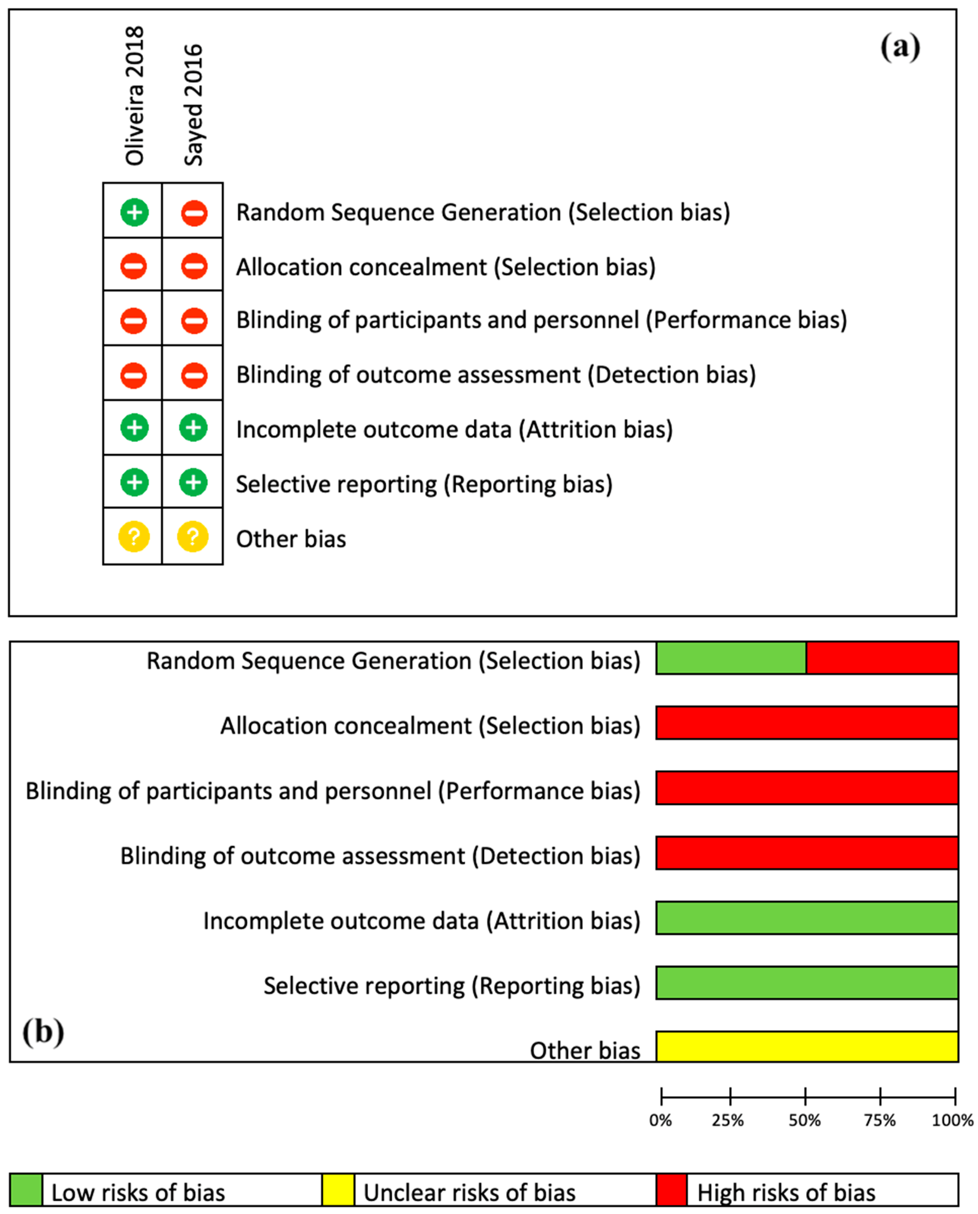
Twelve CTs [43,44,45,46,47,48,49,50,51,52,53,54] were evaluated for risk of bias using the Cochrane technique. The studies were categorized into three groups based on the therapy technique employed. The laser group consisted of five studies [43,44,45,46,47] that compared PBMT to placebo or sham laser. The DN group included five trials [48,49,50,51,52] that compared dry needling therapy (superficial and deep) to placebo. The third group consisted of two articles [53,54] that compared PBMT to DN treatment. Each group was individually assessed for bias as shown in Figure 3, Figure 4 and Figure 5.
Laser studies [43,44,45,46,47] had a “good quality” rating, indicating a low risk of bias. Two DN studies were rated as “good quality” [48,52], whereas the others were rated as “poor quality” [49,50,51]. The two included comparison studies [53,54] were classified as “fair quality” due to their moderate risk of bias. The majority of studies classified as being of “poor” or “fair” quality had incomplete outcome data (attrition bias), no participant blinding (performance bias), no outcome assessment blinding (detection bias), and no allocation concealment (selection bias); see Figure 3.

3.3. Study Characteristics

The primary outcome of this systematic review and meta-analysis was to assess myofascial pain and the secondary was to assess the pain pressure threshold (PPT) following PBMT or DN treatment associated with TMD/MPS. All the included studies are CTs published in English between January 2010 and January 2024 [43,44,45,46,47,48,49,50,51,52,53,54]. Table 2, Table 3, Table 4, Table 5, Table 6 and Table 7 provide a concise overview of the fundamental attributes of the twelve clinical trials that are presented. The studies were split into three categories based on the treatment approaches: the laser group [43,44,45,46,47], the DN group [48,49,50,51,52] and the comparative group associating both PBM and DN [53,54]. The included studies in the laser group had at least one test group and one placebo or sham laser control group. Every patient experienced pain in one or several TMJ masticatory muscles. The diagnosis was determined based on the RDC/TMD categorization. The number of treatment sessions varied and ranged from a single one [46] to twelve sessions [45], with evaluation at different time points. Pain assessment in all laser trials was conducted using the Visual Analog Scale (VAS). The five studies showed statistical analysis for their findings. However, they had no similarities either in the laser power parameters and application time or in technique/point of applications for PBM. Three out of the five studies displayed positive results and reported a significant reduction in pain intensity after completion of the treatment [44,46,47].
The DN group revealed no commonalities in technique or points of application, number of sessions, follow-up scheduling, degree of blindness, or whether deep or superficial application was used. Most DN studies presented positive results with statistically significant differences observed among the treatment groups [49,50,51,52], while study [48] showed no significant outcomes. Both studies in the comparative group did not find any statistically significant distinctions between the therapies at one week [54] and one month [53].

3.4. Meta-Analysis Results

Three distinct meta-analyses were performed to quantify the pooled effects of PBM and DN in the management of TMD/MPS myofascial pain and deactivation of myofascial trigger points (MTPs). Two common individual factors were assessed: pain and pain pressure threshold (PPT).
Review Manager (RevMan) 5.3 from Cochrane was used to perform statistical analysis. The effect size and total pooled estimates [with the 95% confidence interval (CI)] were computed for each included study. A random-effects model was employed. The I2 index, which assessed the degree of individual result heterogeneity, was used to quantify the heterogeneity of effect sizes. When the A I2 value was more than 75%, this indicated significant heterogeneity between the trials. The findings of the individual studies, as well as the pooled estimates, were graphically represented using forest plots. p-values less than 0.05 (p < 0.05) were considered to be statistically significant.

3.4.1. PBM Effect on Pain Measured by VAS

The results for PBM studies [43,44,46,47] showed that the pain decreased after laser treatment in both subgroups (immediately after and after one month) as shown in Figure 6. The overall effect of the treatment was statistically significant, as indicated by the Z-test and the confidence intervals. The heterogeneity between the subgroups was low, suggesting that the treatment effect was consistent across the subgroups.

3.4.2. PBM Effect on Pressure Pain Threshold

There was a significant improvement in the pressure pain after laser treatment, as shown in Figure 7.

3.4.3. Dry Needle Effect on Pain Measured by VAS

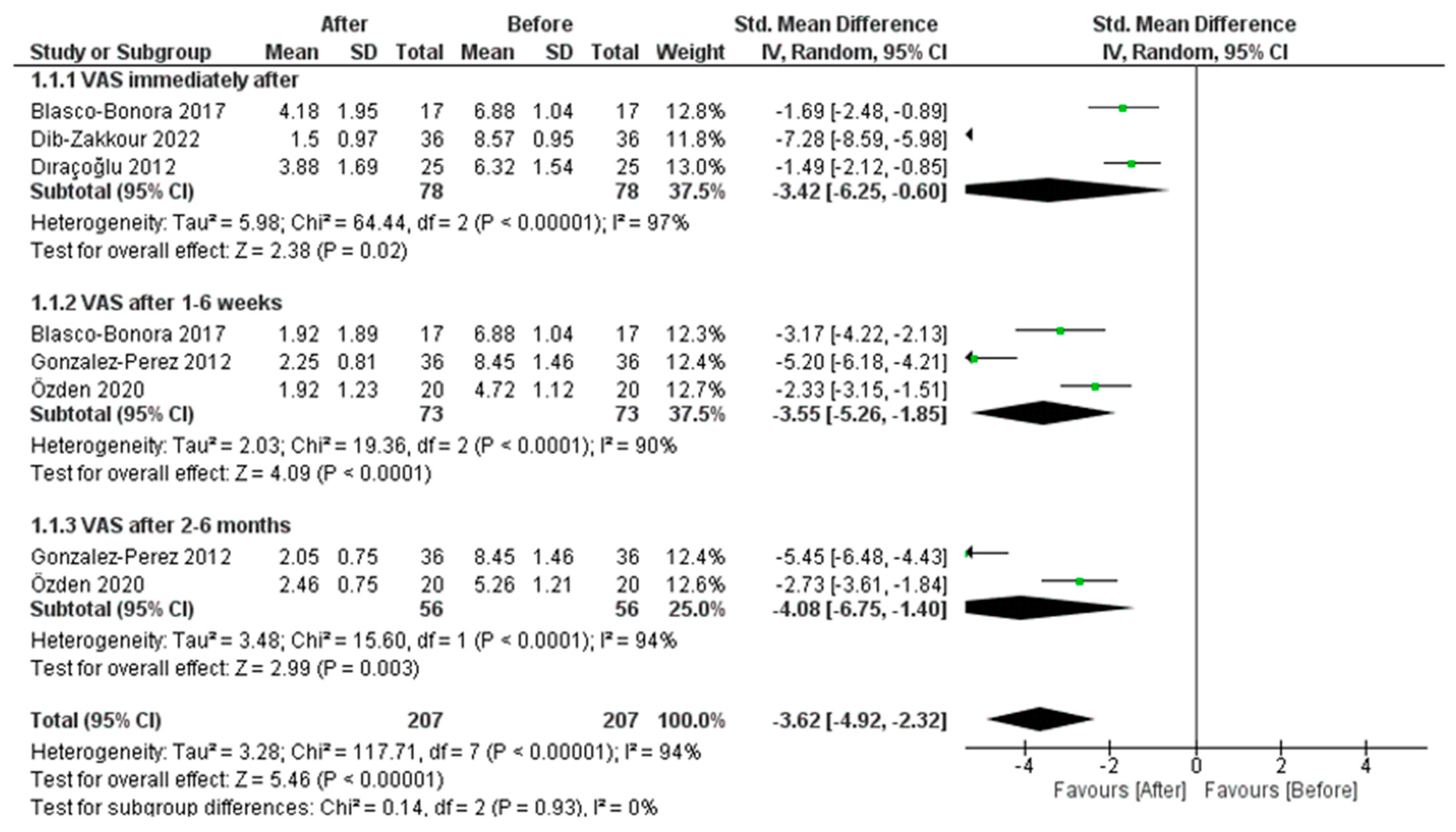
The findings of a meta-analysis of DN included studies [48,49,50,51,52] that used the visual analogue scale (VAS) to quantify pain ratings both before and after dry needle; these are shown in Figure 8. Along with the overall affect size for all studies combined, the figure displays the effect sizes for each subgroup of studies (immediately after, 1–6 weeks after, and 2–6 months after the intervention). The dry needle technique has a significant effect on reducing pain levels throughout all time intervals. There was significant heterogeneity between the included studies as measured by the significant chi square test of heterogeneity and the I2 value.

3.4.4. Dry Needle Effect on Pressure Pain Threshold

The result of the effect of dry needle on improvement in pressure pain was statistically insignificant as shown in Figure 9. Also, there was high heterogeneity between the studies as indicated by I2 value.

3.4.5. Comparison between Dry Needle and PBM Therapies

Both techniques have a positive effect on pain immediately after treatment, but this effect tends to decrease over time, and also becomes insignificant in the case of the dry needle technique. The pressure-pain-threshold variable was not assessed in the evaluated comparative studies [53,54].
Comparison between laser and dry needle effect on pain measured by VAS:
The results showed that there was no significant difference between the two techniques with respect to the pain measured by VAS (Figure 10). There was no heterogeneity between the studies included, as indicated by the non-significant chi square value and I2 = 0%.

3.5. Systematic Analysis of Results

The combined data of 430 participants who received laser PBM or DN interventions to manage myalgia pain from TMD or MPS showed statistical improvement for both interventions. After comparing the results of both meta-analyses for DN and PBM groups, we found both techniques have a positive effect immediately after the intervention, but this effect tends to decrease over time, and also becomes insignificant in the case of the DN technique. The higher statistically significant reduction in the level of pain and pain pressure threshold after the follow-up duration of the PBM intervention, compared to the DN technique, favored PBM therapy, particularly in the long term. The statistically significant improvement in the level of pain and pain pressure threshold immediately after the DN intervention, compared to PBM, favored the DN therapy, particularly in the deactivation of MTPs. However, the statistical results of comparative studies of PBM and DN techniques showed no significant differences between them, suggesting that the two therapies are quite efficient in decreasing pain values after the interventions and throughout the follow-up duration.

4. Discussion

4.1. Exploring the Mechanisms and Practical Uses of the Anti-Inflammatory Properties of Photobiomodulation

Since its inception, photobiomodulation therapy (PBMT) previously known as low-level laser therapy (LLLT) has been consistently surrounded by controversy, primarily due to the widespread use of various types of medical light sources and treatment protocols. These protocols include different parameters such as wavelength, pulse structure, fluence, irradiance, exposure and the lack of consensus on a standardized treatment course. Hence, the discrepancies in study designs resulted in a rise in the publication of negative trials and sparked controversy, despite the abundance of excellent clinical results that were also achieved. The shift in viewpoint that has taken place in recent years can be ascribed to various sources, with the progress gained in comprehending the mechanisms of action at a molecular, cellular, and tissue-based level being arguably the most significant among these variables [28]. Originally, red and near-infrared (NIR) light have been employed for therapeutic purposes. Nevertheless, current research suggests that additional wavelengths within the visible spectrum, such as blue and green light, may also have potential benefits [55]. Electromagnetic radiation with specific wavelengths ranging from visible to near infra-red (400–1100 nm) can cause photophysical and photochemical effects [56]. These effects can influence important biological processes to achieve therapeutic objectives, such as promoting cellular growth, improving mitochondrial function, and regulating inflammatory signaling [57]. Most of the literature indicates that red and near-infrared light (red: ~600–750 nm, NIR: ~750–1100 nm) have positive therapeutic benefits by improving tissue repair and decreasing inflammation and reducing pain [58,59,60]. This range of wavelengths (660–1100 nm), known as the “optical window”, allows for the greatest depth of light penetration into the tissue. When utilizing light therapy, it is crucial to take into account both the depth of light penetration and the specific wavelength of light that is absorbed by photo acceptors [61], i.e., the optical penetration depth, how far the energy penetrates into the tissue before being scattered to nothing or by being absorbed. It is seen that depending on the wavelength, the penetration depth into human mucous varies considerably. The depth is maximal in the spectral ranges 800–900 nm and 1000–1100 nm, where the optical radiation penetrates to depths of up to 6–6.5 mm. This is very important to take into consideration, because the penetration depth of light into a biological tissue is an important parameter for the correct determination of the irradiation dose in photothermal and photodynamic therapies of various diseases [62]. Light penetration into tissue is considered as being influenced by both absorption and scattering by molecules and structures present in tissue. So, both optical absorption and scattering play important roles in determining the spatial distribution of volumetric energy density deposited by laser radiation in tissue. For wavelengths below 450 nm and above 1800 nm, optical absorption, provided by hemoglobin and protein in UV and water in mid- and far-infrared (IR), dominates the optical properties [63]. Hence, PBM responses entail complicated events arising from photon absorption and scattering, as well as electromagnetic field creation [64]. From a medical perspective, the stimulation of cells and the activation of cellular pathways can lead to improvements in inflammation, tissue regeneration, and overall restoration of metabolic dysfunction in the tissues, resulting in homeostasis recovery [65]. These biological processes lead to the subsequent enhancement of cellular energy metabolism, an augmentation in tissue blood flow, the synthesis of chemicals like endorphins, and the suppression of inflammatory mediators such as bradykinin or prostaglandins [66]. The potential for photobiomodulation to induce analgesic and anti-inflammatory effects may be attributed to various mechanisms. Initially, PBM has the ability to impede the depolarization of C-fiber afferents and functions as a stabilizing agent for the resting membrane potential. It operates on the nerve endings and sustains the duration of analgesia [32]. Furthermore, PBM stimulates the generation and release of beta-endorphins, resulting in a decrease in pain. Additionally, it facilitates a decrease in the levels of bradykinin, substance P, and prostaglandins, leading to a reduction in inflammation and edema. Laser therapy enhances local circulation, including lymphatic and blood flow, which leads to muscle relaxation in areas experiencing muscular tension, so improving tissue analgesia [67,68].

4.2. Possible Management of Orofacial Pain with PBMT Compared to DNT

This review aims to compare the effect of photobiomodulation therapy (PBMT) using visible and near-infrared (NIR) diode laser wavelengths to that of the dry needling technique (DNT) on the management of orofacial pain in patients with temporomandibular disorder myofascial pain syndrome (TMD/MPS) in term of effectiveness, speed of recovery, and length of treatment. The research dealt with the range of visible and NIR diode wavelengths due to its implementation in the so-called “optical window”, where the optical penetration depth into tissue is maximal [69]. The prevalence of temporomandibular joint disorders is steadily rising, due to the simultaneous increase in stress levels and anxiety [70]. An early and accurate diagnosis is the initial stage in the process of healing. It is necessary to have a skilled expert who has a thorough understanding of anatomy in order to distinguish between normal and abnormal states and comprehend the intended goals of the available treatment options [71]. The study conducted by Silva A.M.R. et al. emphasized that successful treatment relies on a comprehensive understanding of the cause, symptoms, and accurate differential diagnosis [72]. Because of the intricate nature of the situation, there is currently no universally accepted treatment plan [73,74]. According to the literature, PBM and DN therapies are proposed as alternative treatments for patients with myofascial pain. These methods are successful and do not require intrusive procedures. Results of previous systematic reviews and meta-analysis on TMD/MPS have shown a significant clinical effect of lasers and DN in reducing pain when compared to placebo [75,76,77]; however, a restricted numbers of studies compared the two therapies. In our research, only two clinical comparative studies met the inclusion criteria and were subject to critical analysis [53,54]. Because of the limited numbers of included clinical studies, three distinct meta-analyses were performed to quantify the pooled effects. The results among groups were systematically evaluated, to draw the final conclusion. The strength of this meta-analysis lies in its provision of three meta-analyses inside a single review, as well as its rigorous technique for choosing the eligible studies to be included. Thus far, we have meticulously chosen comparable circumstances for each group trial, taking into account factors such as patient samples, disease severity, variable evaluation, scales, and objectives. All the studies included in the analysis [43,44,45,46,47,48,49,50,51,52,53,54] assessed the efficacy of diode laser PBMT and/or DNT in treating myofascial temporomandibular disorder (TMD) or myofascial pain syndrome (MPS) of the temporomandibular joint (TMJ).
Our meta-analysis findings indicate that there was no variation in the studies of the comparison group [53,54], and a small amount of heterogeneity in the research of the laser group [43,44,45,46,47]. Simultaneously, there was diversity among the investigations conducted on the DN group [48,49,50,51,52]. Within the PBM studies group, two out of five studies [43,44] found no significant disparities in pain reduction between the experimental group and placebo group when comparing before and after treatment. However, it is worth noting that the laser group experienced longer-lasting pain reduction and continued improvement at the end of the follow-up period, unlike the placebo group. Additionally, within the DN studies group, one clinical trial found no statistically significant differences between the experimental groups in terms of pain reduction when comparing pre- and post-treatment [48]. Conversely, the comparative trials [53,54] demonstrated that both treatments were equally helpful in reducing myofascial pain associated with TMD/MPS of the temporomandibular joint (TMJ), with no significant differences observed between them. After thoroughly examining and evaluating the results of all included studies, this meta-analysis indicates a substantial decrease in pain levels following the implementation of both therapies. The intervention of laser PBM demonstrated a long-term therapy effect that was generally more favorable, whereas the DN method showed a short-term treatment effect that was more beneficial, in terms of deactivating acute MTPs. Moreover, the unfavorable outcomes observed in certain studies included in this meta-analysis may be attributed to several factors, such as the distinction between acute and chronic pain. It is worth noting that non-pharmacological interventions are less efficacious in treating chronic pain compared to acute and emergency pain [78]. Furthermore, the utilization of the Visual Analog Scale (VAS) to assess pain introduces subjectivity, as patients self-report their pain levels. This subjective nature of the VAS poses a potential danger of either overestimating or underestimating pain when data are aggregated. In addition to the placebo effect, it has been suggested that placebo analgesia may trigger the activation of endogenous opioids involved in pain modulation [79]. Nevertheless, there exist certain constraints in this study, ranging from the restricted number of papers included to discrepancies found among the studies incorporated, such as the following:
  • Failing to assess the same set of masticatory muscles. Several studies examined the masseter muscle [51,52,53,54], while others investigated both the masseter and temporal muscles [43,44,45,46,47,48,50]. Additionally, one study specifically examined the lateral pterygoid muscle [49]. Monteiro et al. [47] evaluated the pain levels following laser PBM treatment in the masseter, temporal, lateral pterygoid muscles, and temporalis ligament. Furthermore, assessing the degrees of muscular pathology can be regarded as a complex task for evaluation.
  • In the DN studies [48,49,50,51,52], the authors treated the trigger points identified by patients without specifying the number of ones treated per patient, leading to an indeterminate number of needles used in each session. The investigations [48,49,52,53,54] did not describe the depth of needle entry, either. Likewise, the comparison of trigger-point locations and pain intensity levels among participants, as well as across various clinical trials, presents substantial obstacles that may compromise the integrity and reliability of the quality of the articles.
  • The studies [46,50,52] had a single treatment session, which may significantly impact the long-term viability of their findings. Other authors recommended using palliative methods such as thermotherapy and exercising following the treatment sessions. These procedures can potentially enhance the joint’s self-healing ability and may impact the accuracy of their findings [53,54].
  • The included studies dealt with both genders, except Oliveira et al. [54], who treated 10 women. Studies have shown that gender characteristics may have an important impact on the course and outcomes of the therapies. The prevalence of TMD decreases after menopause, indicating a significant relationship with the hormonal oscillation, and it is higher in women of reproductive age. Estrogen and prolactin, which are present in higher concentrations in women, can heighten the symptoms of TMJ dysfunction. These hormones can accelerate the breakdown of articular cartilage and bone, triggering a cascade of immunological reactions within these joints. One contributing factor is the higher prevalence of psychosomatic disorders in women, which is a direct result of their higher stress indices compared to men [8,11,12].
Despite meeting all the inclusion criteria, comparing the studies was challenging, due to the heterogeneity in PBM laser protocols and disparities in the outcomes:
  • The presence of diverse operating laser characteristics, as seen in Table 3 and Table 7, highlights the lack of consistency in delivering a valid, reliable, and accurate PBM protocol and doses. Furthermore, the variations in laser operational parameters, the manner of laser application, and other factors will undoubtedly impact the reliability and consistency of the results. For instance, a key element to take into account is the velocity of manual motion, which was not addressed in any of the research considered. The speed of the application is crucial, as it allows for enhanced regulation of the energy release onto the tissue. A gradual and slow motion of the hand enables a higher energy discharge per unit of surface area over a period of time. Conversely, a rapid movement may result in an inefficient transmission of photonic energy. Furthermore, the clinical experiments presented do not demonstrate consistency in the fluence or the amount of energy density supplied per trigger site. The energy density is a vital parameter that governs all interactions between lasers and tissues. It denotes the quantity of energy transferred to a given region within a specific timeframe. Typically, clinically evaluating this metric is usually quite challenging. The variables that have a direct correlation with this include the velocity of manual motion, the length of the procedure, the size of the fiber tip, and the concept of focusing or defocusing the laser beam in a Gaussian-profile delivery system [80]. Consequently, the general acceptance of PBM therapy as a viable treatment for managing severe disorders like TMD/MPS pain would be restricted. It is important to be aware that TMD/MPS disorders vary in intensity, and certain acute symptoms may improve on their own without treatment. Occasionally, they have a tendency to restrict themselves, and as a result, they may spontaneously improve without any external intervention in certain instances [6,8].
  • Another important factor to consider is the utilization of an optical power meter, which is a device used to measure the optical power (the amount of energy delivered per unit of time) in a light beam, such as a laser beam. It is widely recognized that light gradually dissipates its energy over time, and this principle also applies to laser light. Several factors can contribute to a decrease in power, such as unclean optics, electrical issues, and a limited lifespan [81]. The majority of the PBM research studies included (five out of seven) did not specify the utilization of a power meter. In these studies, the average power levels examined may be less precise.
  • Furthermore, none of the laser studies included provided any information regarding the specific properties of the beam profile, which undermines the ability to replicate the therapy in the selected research. The energy density delivered into the treated area is directly correlated with the beam profile. Using a traditional laser handpiece, the spatial beam profile naturally follows a Gaussian distribution. Typically, as the distance between the tip and the tissue increases, the energy density decreases [82]. This variable poses a significant difficulty for researchers and is a crucial consideration to take into account in PBM studies [83]. Utilizing a flat-top handpiece may effectively address and resolve these problems [80].
Due to the paucity of trials that directly compare both therapies, our meta-analysis only included two comparative studies [53,54], despite some discrepancies with our criteria for inclusion, as in the trial of Oliveira et al. [54], which exclusively focused on female participants. However, our inclusion criteria considered both genders in an attempt to mitigate the risk of bias of the included studies and yield more dependable and complete outcomes. Likewise, some research findings have compared the DN technique and diode laser PBMT with other aspects of treatment, such as Uemoto et al. [39], who conducted a clinical trial where they compared these techniques with the wet needling approach. The results showed that four sessions of needling with a 2% lidocaine injection or laser therapy at a dose of 4 J/cm2 were more effective than DNT in deactivating MTPs. In their study, Iibuldu et al. [84] examined the effectiveness of DNT compared to laser therapy. They found that the laser group showed a substantial improvement in comparison to the dry-needling group following the treatment. However, no improvement was observed over a 6-month review period.
The risk of bias assessment showed that the laser studies [43,44,45,46,47] were rated as having “good quality”, indicating a small risk of bias. Out of the DN studies, two were deemed to have “good quality” [48,52], while the remaining ones were considered to have “poor quality” [49,50,51]. The two comparative studies [53,54] that were included in the analysis were categorized as “fair quality”, because they had a modest risk of bias. The majority of studies categorized as “poor” or “fair” quality lacked comprehensive outcome data (attrition bias), no participant blinding (performance bias), no outcome assessment blinding (detection bias), and no allocation concealment (selection bias), which could potentially alter the effect estimates.
The meta-analyses evaluations revealed that the dry needle technique has a significant effect on reducing pain levels throughout all time intervals, although significant heterogeneity exists between the studies. The results for PBM studies [43,44,46,47] showed that the pain decreased after laser treatment in both subgroups (immediately after and after one month). The treatment had a consistent effect across all categories, as indicated by the statistically significant overall effect and low heterogeneity among the groupings. Following a comparative analysis of the results from both meta-analyses for the DN and PBM groups, it was shown that both strategies yield a beneficial impact immediately following the intervention. However, over time, this benefit diminishes and becomes statistically insignificant, specifically in the case of the DN technique. PBM therapy demonstrated a greater and statistically significant decrease in pain level and pain pressure threshold compared to the DN approach, during the follow-up period. This advantage was particularly evident in the long term. The DN intervention resulted in a statistically significant improvement in pain level and pain pressure threshold, compared to PBM. This effect was particularly notable in the deactivation of MTPs. Nevertheless, the statistical findings from comparison studies on PBM and DN methods revealed no substantial disparities between them, indicating that both treatments are highly effective in reducing pain levels during the interventions and during the follow-up period.
Furthermore, extensive evidence supports the notion that optimal PBM therapy should result in the intended clinical outcome without inducing any local temperature rise or tissue destruction. None of the included studies reported any adverse results or thermal collateral harm. Consequently, the utilization of visible and NIR diode laser wavelengths (600–1200 nm) in PBM is considered to be both safe and successful, as it fulfills the majority of the requirements for an optimal PBM therapy.

5. Conclusions

Based on our findings, both diode laser PBM and DN methods exhibit immediate positive effect following the therapy. However, over time, these effects tend to diminish and become negligible in the case of DN. Nevertheless, research results indicate that the DN approach provides rapid pain alleviation for myofascial trigger points (MTPs), despite its brief duration. PBMT has demonstrated superior efficacy compared to DN, making it the preferred treatment option for people who experience needle phobia. Moreover, there is a significant paucity of clinical trials that directly compare these two procedures. Additional rigorous research with a larger sample size, comparable evaluated factors such as age, gender, and severity of the disease, a reliable non-sensitive PBM operator technique, and extensive follow-up periods, is required to substantiate the superior effectiveness of these options and provide the optimal treatment protocol.

Author Contributions

Conceptualization, R.E.F. and J.A.; methodology, R.E.F. and J.A.; software, R.E.F. and J.A.; validation, S.B.; formal analysis, R.E.F. and J.A.; investigation, J.A., R.E.F. and T.A.; resources, S.B.; data curation, R.E.F. and J.A.; writing—original draft preparation, R.E.F. and J.A.; writing—review and editing, R.E.F., J.A., T.A. and S.B.; supervision, R.E.F.; project administration, R.E.F.; funding acquisition, S.B. All authors have read and agreed to the published version of the manuscript.

Funding

This research received no external funding.

Data Availability Statement

Not applicable.

Conflicts of Interest

The authors declare no conflicts of interest.

References

  1. Desai, A.P.; Roy, S.K.; Semi, R.S.; Balasundaram, T. Efficacy of Low-Level Laser Therapy in Management of Temporomandibular Joint Pain: A Double-Blind and Placebo-Controlled Trial. J. Maxillofac. Oral Surg. 2022, 21, 948–956. [Google Scholar] [CrossRef] [PubMed]
  2. Alsarhan, J.; El Feghali, R.; Alkhudari, T.; Benedicenti, S.; Pasquale, C. Can Photobiomodulation Support the Management of Temporomandibular Joint Pain? Molecular Mechanisms and a Systematic Review of Human Clinical Trials. Photonics 2022, 9, 420. [Google Scholar] [CrossRef]
  3. Schiffman, E.; Ohrbach, R.; Truelove, E.; Look, J.; Anderson, G.; Goulet, J.P.; List, T.; Svensson, P.; Gonzalez, Y.; Lobbezoo, F.; et al. Diagnostic Criteria for Temporomandibular Disorders (DC/TMD) for Clinical and Research Applications: Recommendations of the International RDC/TMD Consortium Network* and Orofacial Pain Special Interest Group. J. Oral Facial Pain Headache 2014, 28, 6–27. [Google Scholar] [CrossRef] [PubMed]
  4. Vier, C.; Almeida, M.B.; Neves, M.L.; Santos, A.R.S.D.; Bracht, M.A. The effectiveness of dry needling for patients with orofacial pain associated with temporomandibular dysfunction: A systematic review and meta-analysis. Braz. J. Phys. Ther. 2019, 23, 3–11. [Google Scholar] [CrossRef] [PubMed]
  5. Chaurand, J.; Pacheco-Ruíz, L.; Orozco-Saldívar, H.; López-Valdés, J. Efficacy of botulinum toxin therapy in treatment of myofascial pain. J. Oral Sci. 2017, 59, 351–356. [Google Scholar] [CrossRef]
  6. Kalladka, M.; Young, A.; Khan, J. Myofascial pain in temporomandibular disorders: Updates on etiopathogenesis and management. J. Bodyw. Mov. Ther. 2021, 28, 104–113. [Google Scholar] [CrossRef]
  7. Peck, C.C.; Goulet, J.P.; Lobbezoo, F.; Schiffman, E.L.; Alstergren, P.; Anderson, G.C.; de Leeuw, R.; Jensen, R.; Michelotti, A.; Ohrbach, R.; et al. Expanding the taxonomy of the diagnostic criteria for temporomandibular disorders. J. Oral Rehabil. 2014, 41, 2–23. [Google Scholar] [CrossRef]
  8. Golanska, P.; Saczuk, K.; Domarecka, M.; Kuć, J.; Lukomska-Szymanska, M. Temporomandibular Myofascial Pain Syndrome-Aetiology and Biopsychosocial Modulation. A Narrative Review. Int. J. Environ Res. Public Health 2021, 18, 7807. [Google Scholar] [CrossRef]
  9. Fernández-De-Las-Peñas, C.; Nijs, J. Trigger point dry needling for the treatment of myofascial pain syndrome: Current perspectives within a pain neuroscience paradigm. J. Pain Res. 2019, 12, 1899–1911. [Google Scholar] [CrossRef]
  10. Fernández-De-Las-Peñas, C.; Dommerholt, J. Myofascial trigger points: Peripheral or central phenomenon? Curr. Rheumatol. Rep. 2014, 16, 395. [Google Scholar] [CrossRef]
  11. Alrizqi, A.H.; Aleissa, B.M. Prevalence of Temporomandibular Disorders Between 2015–2021: A Literature Review. Cureus 2023, 15, e37028. [Google Scholar] [CrossRef] [PubMed]
  12. Fleckenstein, J.; Zaps, D.; Rüger, L.J.; Lehmeyer, L.; Freiberg, F.; Lang, P.M.; Irnich, D. Discrepancy between prevalence and perceived effectiveness of treatment methods in myofascial pain syndrome: Results of a cross-sectional, nationwide survey. BMC Musculoskelet. Disord. 2010, 11, 32. [Google Scholar] [CrossRef] [PubMed]
  13. Fernández-de-las-Peñas, C.; Svensson, P. Myofascial Temporomandibular Disorder. Curr. Rheumatol. Rev. 2015, 12, 40–54. [Google Scholar] [CrossRef] [PubMed]
  14. Kuć, J.; Szarejko, K.D.; Gołębiewska, M. Evaluation of soft tissue mobilization in patients with temporomandibular disorder-myofascial pain with referral. Int. J. Environ. Res. Public Health 2020, 17, 9576. [Google Scholar] [CrossRef]
  15. List, T.; Jensen, R.H. Temporomandibular disorders: Old ideas and new concepts. Cephalalgia 2017, 37, 692–704. [Google Scholar] [CrossRef]
  16. Fricton, J. Myofascial Pain: Mechanisms to Management. Oral Maxillofac. Surg. Clin. N. Am. 2016, 28, 289–311. [Google Scholar] [CrossRef]
  17. Brighenti, N.; Battaglino, A.; Sinatti, P.; Abuín-Porras, V.; Sánchez Romero, E.A.; Pedersini, P.; Villafañe, J.H. Effects of an Interdisciplinary Approach in the Management of Temporomandibular Disorders: A Scoping Review. Int. J. Environ. Res. Public Health. 2023, 20, 2777. [Google Scholar] [CrossRef]
  18. Herranz-Aparicio, J.; Vázquez-Delgado, E.; Arnabat-Domínguez, J.; España-Tost, A.; Gay-Escoda, C. The use of low-level laser therapy in the treatment of temporomandibular joint disorders. Review of the literature. Med. Oral Patol. Oral Cir. Bucal. 2013, 18, e603–e612. [Google Scholar] [CrossRef]
  19. Wieckiewicz, M.; Boening, K.; Wiland, P.; Shiau, Y.Y.; Paradowska-Stolarz, A. Reported concepts for the treatment modalities and pain management of temporomandibular disorders. J. Headache Pain 2015, 16, 106. [Google Scholar] [CrossRef]
  20. Budakoti, A.; Puri, N.; Dhillon, M.; Ahuja, U.; Rathore, A.; Choudhary, A.; Kour, M. A comparative evaluation of the effectiveness of low-level laser therapy, ultrasound therapy, and transcutaneous electric nerve stimulation in the treatment of patients with TMDs: A prospective study. Laser Dent. Sci. 2019, 3, 257–267. [Google Scholar] [CrossRef]
  21. Yehoshua, I.; Rimon, O.; Mizrahi Reuveni, M.; Peleg, R.; Adler, L. Dry needling for the treatment of acute myofascial pain syndrome in general practitioners’ clinics: A cohort study. BMC Prim. Care 2022, 23, 339. [Google Scholar] [CrossRef] [PubMed]
  22. Costa, A.D.; Bavaresco, C.S.; Grossmann, E. The use of acupuncture versus dry needling in the treatment of myofascial temporomandibular dysfunction. Rev. Dor. 2017, 18, 342–349. [Google Scholar] [CrossRef]
  23. Halle, J.S.; Halle, R.J. Pertinent dry needling considerations for minimizing adverse effects—Part One. Int. J. Sports Phys. Ther. 2016, 11, 651–662. [Google Scholar] [PubMed]
  24. Ansari, M.; Baradaran Mahdavi, S.; Vahdatpour, B.; Lahijanian, A.; Khosrawi, S. Effects of Dry Needling and Low-Power Laser for the Treatment of Trigger Points in the Upper Trapezius Muscle: A Randomized Clinical Trial. J. Chiropr. Med. 2022, 21, 288–295. [Google Scholar] [CrossRef] [PubMed]
  25. Gattie, E.; Cleland, J.A.; Snodgrass, S. The Effectiveness of Trigger Point Dry Needling for Musculoskeletal Conditions by Physical Therapists: A Systematic Review and Meta-analysis. J. Orthop. Sports Phys. Ther. 2017, 47, 133–149. [Google Scholar] [CrossRef]
  26. Brady, S.; McEvoy, J.; Dommerholt, J.; Doody, C. Adverse events following trigger point dry needling: A prospective survey of chartered physiotherapists. J. Man. Manip. Ther. 2014, 22, 134–140. [Google Scholar] [CrossRef]
  27. Boyce, D.; Wempe, H.; Campbell, C.; Fuehne, S.; Zylstra, E.; Smith, G.; Wingard, C.; Jones, R. Adverse events associated with therapeutic dry needling. Int. J. Sports Phys. Ther. 2020, 15, 103–113. [Google Scholar] [CrossRef]
  28. Hamblin, M.R. Photobiomodulation or low-level laser therapy. J. Biophotonics 2016, 9, 1122–1124. [Google Scholar] [CrossRef]
  29. Amaroli, A.; Ferrando, S.; Benedicenti, S. Photobiomodulation affects key cellular pathways of all life-forms: Considerations on old and new laser light targets and the calcium issue. Photochem. Photobiol. 2019, 95, 455–459. [Google Scholar] [CrossRef]
  30. Dompe, C.; Moncrieff, L.; Matys, J.; Grzech-Leśniak, K.; Kocherova, I.; Bryja, A.; Bruska, M.; Dominiak, M.; Mozdziak, P.; Skiba, T.H.I.; et al. Photobiomodulation—Underlying mechanism and clinical applications. J. Clin. Med. 2020, 9, 1724. [Google Scholar] [CrossRef]
  31. Anders, J.J.; Lanzafame, R.J.; Arany, P.R. Low-level light/laser therapy versus photobiomodulation therapy. Photomed. Laser Surg. 2015, 33, 183–184. [Google Scholar] [CrossRef] [PubMed]
  32. El Feghali, R.; Tatarian, K.; Zogheib, C.; Benedicenti, S.; Pasquale, C.; Amaroli, A. The 1064-nm Nd: YAG photobiomodulation vs. 20% benzocaine topical gel in inducing mucosal anaesthetic effect: A double-blind randomized clinical trial. Photonics 2022, 9, 519. [Google Scholar] [CrossRef]
  33. Ferrando, S.; Agas, D.; Mirata, S.; Signore, A.; De Angelis, N.; Ravera, S.; Utyuzh, A.S.; Parker, S.; Sabbieti, M.G.; Benedicenti, S.; et al. The 808 nm and 980 nm infrared laser irradiation affects spore germination and stored calcium homeostasis: A comparative study using delivery hand-pieces with standard (Gaussian) or flat-top profile. J. Photochem. Photobiol. B. 2019, 199, 111627. [Google Scholar] [CrossRef]
  34. Amaroli, A.; Agas, D.; Laus, F.; Cuetri, V.; Hanna, R.; Sabbieti, M.G.; Benedicenti, S. The effects of photobiomodulation of 808 nm diode laser therapy at higher fluence on the in vitro osteogenic differentiation of bone marrow stromal cells. Front. Physiol. 2018, 9, 123. [Google Scholar] [CrossRef]
  35. Assis, L.; Moretti, A.I.; Abrahao, T.B.; Cury, V.; Souza, H.P.; Hamblin, M.R.; Parizotto, N.A. Low-level laser therapy (808 nm) reduces inflammatory response and oxidative stress in rat tibialis anterior muscle after cryolesion. Lasers Surg. Med. 2012, 44, 726–735. [Google Scholar] [CrossRef]
  36. Gonzalez-Perez, L.M.; Infante-Cossio, P.; Granados-Nunez, M.; Urresti-Lopez, F.J.; Lopez-Martos, R.; Ruiz-Canela-Mendez, P. Deep dry needling of trigger points located in the lateral pterygoid muscle: Efficacy and safety of treatment for management of myofascial pain and temporomandibular dysfunction. Med. Oral Patol. Oral Cir. Bucal. 2015, 20, e326–e333. [Google Scholar] [CrossRef] [PubMed]
  37. Fernández-Carnero, J.; La Touche, R.; Ortega-Santiago, R.; Galan-del-Rio, F.; Pesquera, J.; Ge, H.Y.; Fernández-de-Las-Peñas, C. Short-term effects of dry needling of active myofascial trigger points in the masseter muscle in patients with temporomandibular disorders. J. Orofac. Pain 2010, 24, 106–112. [Google Scholar]
  38. Carrasco, T.G.; Guerisoli, L.D.; Guerisoli, D.M.; Mazzetto, M.O. Evaluation of low intensity laser therapy in myofascial pain syndrome. Cranio 2009, 27, 243–247. [Google Scholar] [CrossRef]
  39. Uemoto, L.; Garcia, M.A.; Gouvêa, C.V.; Vilella, O.V.; Alfaya, T.A. Laser therapy and needling in myofascial trigger point deactivation. J. Oral Sci. 2013, 55, 175–181. [Google Scholar] [CrossRef]
  40. Moher, D.; Liberati, A.; Tetzlaff, J.; Altman, D.G. The PRISMA Group. Preferred Reporting Items for Systematic Reviews and Meta-analyses: The PRISMA Statement. PLoS Med. 2009, 6, 1000097. [Google Scholar] [CrossRef]
  41. Berger, V.W.; Alperson, S.Y. A general framework for the evaluation of clinical trial quality. Rev. Recent. Clin. Trials 2009, 4, 79–88. [Google Scholar] [CrossRef] [PubMed]
  42. Higgins, J.; Savovic, J.; Page, M.; Elbers, R.; Sterne, J. Assessing Risk of Bias in Randomized Trials. In Cochrane Handbook for Systematic Reviews of Interventions, 2nd ed.; John Wiley & Sons: Chichester, UK, 2019; pp. 205–228. [Google Scholar]
  43. Venezian, G.C.; da Silva, M.A.M.R.; Mazzetto, R.G.; Oliveira Mazzetto, M. Low level laser effects on pain to palpation and electromyographic activity in TMD patients: A double-blind, randomized, placebo-controlled study. Cranio 2010, 28, 84–91. [Google Scholar] [CrossRef]
  44. de Moraes Maia, M.L.; Ribeiro, M.A.; Maia, L.G.; Stuginski-Barbosa, J.; Costa, Y.M.; Porporatti, A.L.; Conti, P.C.; Bonjardim, L.R. Evaluation of low-level laser therapy effectiveness on the pain and masticatory performance of patients with myofascial pain. Lasers Med. Sci. 2012, 29, 29–35. [Google Scholar] [CrossRef] [PubMed]
  45. Sancakli, E.; Gökçen-Röhlıg, B.; Balık, A.; Öngül, D.; Kıpırdı, S.; Keskın, H. Early results of low-level laser application for masticatory muscle pain: A double-blind randomized clinical study. BMC Oral Health 2015, 15, 131. [Google Scholar] [CrossRef]
  46. Costa, S.A.P.; Florezi, G.P.; Artes, G.E.; Costa, J.R.D.; Gallo, R.T.; Freitas, P.M.; Witzel, A.L. The analgesic effect of photobiomodulation therapy (830 nm) on the masticatory muscles: A randomized, double-blind study. Braz. Oral Res. 2017, 31, e107. [Google Scholar] [CrossRef] [PubMed]
  47. Monteiro, L.; Ferreira, R.; Resende, T.; Pacheco, J.J.; Salazar, F. Effectiveness of photobiomodulation in temporomandibular disorder-related pain using a 635 nm diode laser: A randomized, blinded, and placebo-controlled clinical trial. Photobiomodul. Photomed. Laser Surg. 2020, 38, 280–288. [Google Scholar] [CrossRef]
  48. Dıraçoğlu, D.; Vural, M.; Karan, A.; Aksoy, C. Effectiveness of Dry Needling for the Treatment of Temporomandibular Myofascial Pain: A Double-blind, Randomized, Placebo Controlled Study. J. Back Musculoskelet. Rehabil. 2012, 25, 285–290. [Google Scholar] [CrossRef] [PubMed]
  49. Gonzalez-Perez, L.M.; Infante-Cossio, P.; Granados-Nuñez, M.; Urresti-Lopez, F.J. Treatment of temporomandibular myofascial pain with deep dry needling. Med. Oral Patol. Oral Cir. Buca. 2012, 17, e781–e785. [Google Scholar] [CrossRef]
  50. Blasco-Bonora, P.M.; Martín-Pintado-Zugasti, A. Effects of myofascial trigger point dry needling in patients with sleep bruxism and temporomandibular disorders: A prospective case series. Acupunct. Med. 2017, 35, 69–74. [Google Scholar] [CrossRef]
  51. Özden, M.C.; Atalay, B.; Özden, A.V.; Çankaya, A.; Kolay, E.; Yıldırım, S. Efficacy of dry needling in patients with myofascial temporomandibular disorders related to the masseter muscle. Cranio 2020, 38, 305–311. [Google Scholar] [CrossRef]
  52. Dib-Zakkour, J.; Flores-Fraile, J.; Montero-Martin, J.; Dib-Zakkour, S.; Dib-Zaitun, I. Evaluation of the Effectiveness of Dry Needling in the Treatment of Myogenous Temporomandibular Joint Disorders. Medicina 2022, 58, 256. [Google Scholar] [CrossRef] [PubMed]
  53. Sayed, S.; Refai, H.; Hassanin, N.; Zaky, A. Low-Level Laser Therapy versus Dry Needling for Inactivation of Myofascial Trigger Points. Egypt. Dent. J. 2016, 62, 539–545. [Google Scholar] [CrossRef]
  54. Oliveira, D.A.; Pinto, R.; Reis, L.; Dias, I.; Leite, I.; Leite, F. Clinical effectiveness evaluation of laser therapy and dry needling in the treatment of patients with myofascial pain in masseter muscle. Int. J. Orofac. Myol. 2018, 44, 22–41. [Google Scholar] [CrossRef]
  55. Serrage, H.; Heiskanen, V.; Palin, W.M.; Cooper, P.R.; Milward, M.R.; Hadis, M.; Hamblin, M.R. Under the spotlight: Mechanisms of photobiomodulation concentrating on blue and green light. Photochem. Photobiol. Sci. 2019, 18, 1877–1909. [Google Scholar] [CrossRef]
  56. Wang, Y.; Huang, Y.Y.; Wang, Y.; Lyu, P.; Hamblin, M.R. Photobiomodulation (blue and green light) encourages osteoblastic-differentiation of human adipose-derived stem cells: Role of intracellular calcium and light-gated ion channels. Sci. Rep. 2016, 6, 33719. [Google Scholar] [CrossRef]
  57. Hamblin, M.R. Mechanisms and applications of the anti-inflammatory effects of photobiomodulation. AIMS Biophys. 2017, 4, 337–361. [Google Scholar] [CrossRef]
  58. Cardoso, F.d.S.; Mansur, F.C.B.; Araújo, B.H.S.; Gonzalez-Lima, F.; da Silva, S.G. Photobiomodulation Improves the Inflammatory Response and Intracellular Signaling Proteins Linked to Vascular Function and Cell Survival in the Brain of Aged Rats. Mol. Neurobiol. 2022, 59, 420–428. [Google Scholar] [CrossRef] [PubMed]
  59. Salman, S.; Guermonprez, C.; Peno-Mazzarino, L.; Lati, E.; Rousseaud, A.; Declercq, L.; Kerdine-Römer, S. Photobiomodulation Controls Keratinocytes Inflammatory Response through Nrf2 and Reduces Langerhans Cells Activation. Antioxidants 2023, 12, 766. [Google Scholar] [CrossRef]
  60. Ketz, A.K.; Byrnes, K.R.; Grunberg, N.E.; Kasper, C.E.; Osborne, L.; Pryor, B.; Tosini, N.L.; Wu, X.; Anders, J.J. Characterization of Macrophage/Microglial Activation and Effect of Photobiomodulation in the Spared Nerve Injury Model of Neuropathic Pain. Pain Med. 2017, 18, 932–946. [Google Scholar] [CrossRef]
  61. Huang, Y.Y.; Chen, A.C.; Carroll, J.D.; Hamblin, M.R. Biphasic dose response in low level light therapy—An update. Dose Response 2011, 9, 602–618. [Google Scholar] [CrossRef]
  62. Bashkatov, A.N.; Genina, E.A.; Kochubey, V.I.; Tuchin, V.V. Optical properties of human skin, subcutaneous and mucous tissues in the wavelength range from 400 to 2000 nm. J. Phys. D Appl. Phys. 2005, 38, 2543–2555. [Google Scholar] [CrossRef]
  63. Vogel, A.; Venugopalan, V. Mechanisms of pulsed laser ablation of biological tissues. Chem. Rev. 2003, 103, 577–644. [Google Scholar] [CrossRef] [PubMed]
  64. Amaroli, A.; Benedicenti, S.; Bianco, B.; Bosco, A.; Vargas, M.R.C.; Hanna, R.; Ramakrishnan, P.K.; Raffetto, M.; Ravera, S. Electromagnetic Dosimetry for Isolated Mitochondria Exposed to Near-Infrared Continuous-Wave Illumination in Photobiomodulation Experiments. Bioelectromagnetics 2021, 42, 384–397. [Google Scholar] [CrossRef] [PubMed]
  65. Colombo, E.; Signore, A.; Aicardi, S.; Zekiy, A.; Utyuzh, A.; Benedicenti, S.; Amaroli, A. Experimental and Clinical Applications of Red and Near-Infrared Photobiomodulation on Endothelial Dysfunction: A Review. Biomedicines 2021, 9, 274. [Google Scholar] [CrossRef]
  66. Del Vecchio, A.; Floravanti, M.; Boccassini, A.; Gaimari, G.; Vestri, A.; Di Paolo, C.; Romeo, U. Evaluation of the efficacy of a new low-level laser therapy home protocol in the treatment of temporomandibular joint disorder-related pain: A randomized, double-blind, placebo-controlled clinical trial. Cranio 2021, 39, 141–150. [Google Scholar] [CrossRef]
  67. da Cunha, L.A.; Firoozmand, L.M.; da Silva, A.P.; Camargo, S.E.; Oliveira, W. Efficacy of low-level laser therapy in the treatment of temporomandibular disorder. Int. Dent. J. 2008, 58, 213–217. [Google Scholar] [CrossRef] [PubMed]
  68. Silveira, P.C.; Scheffer, D.d.L.; Glaser, V.; Remor, A.P.; Oinho, R.A.; Aguiar Junior, A.S.; Latini, A. Low-level laser therapy attenuates the acute inflammatory response induced by muscle traumatic injury. Free Radic. Res. 2016, 50, 503–513. [Google Scholar] [CrossRef]
  69. Douplik, A.; Saiko, G.; Schelkanova, I.; Tuchin, V.V. The response of tissue to laser light. In Lasers for Medical Applications Diagnostics, Therapy and Surgery; Jelinkova, H., Ed.; Woodhead Publishing: Cambridge, UK, 2013; pp. 47–109. [Google Scholar] [CrossRef]
  70. Luo, L.Y.; Lee, J.; Li, K.Y.; Leung, Y.Y.; Li, D.T. Psychological outcomes on anxiety and depression after interventions for temporomandibular disorders: A systematic review and meta-analysis. Diagnostics 2023, 13, 653. [Google Scholar] [CrossRef]
  71. Wilkie, G.; Al-Ani, Z. Temporomandibular joint anatomy, function and clinical relevance. Br. Dent. J. 2022, 233, 539–546. [Google Scholar] [CrossRef]
  72. da Silva, M.A.; Botelho, A.L.; Turim, C.V.; da Silva, A.M. Low level laser therapy as an adjunctive technique in the management of temporomandibular disorders. Cranio 2012, 30, 264–271. [Google Scholar] [CrossRef]
  73. Barbero, M.; Schneebeli, A.; Koetsier, E.; Maino, P. Myofascial pain syndrome and trigger points: Evaluation and treatment in patients with musculoskeletal pain. Curr. Opin. Support Palliat. Care 2019, 13, 270–276. [Google Scholar] [CrossRef] [PubMed]
  74. Brochado, F.T.; Jesus, L.H.; Martins, M.D.; Chaves, K.D. Non-invasive therapies for management of temporomandibular disorders: A systematic review. Clinic. Biomed. Res. 2019, 39, 230–243. [Google Scholar] [CrossRef]
  75. Chen, J.; Huang, Z.; Ge, M.; Gao, M. Efficacy of low-level laser therapy in the treatment of TMDs: A meta-analysis of 14 randomised controlled trials. J. Oral Rehabil. 2015, 42, 291–299. [Google Scholar] [CrossRef]
  76. Zhang, Y.; Qian, Y.; Huo, K.; Liu, J.; Huang, X.; Bao, J. Efficacy of laser therapy for temporomandibular disorders: A systematic review and meta-analysis. Complement. Ther. Med. 2023, 74, 102945. [Google Scholar] [CrossRef]
  77. Tesch, R.S.; Macedo, L.C.D.S.P.; Fernandes, F.S.; Goffredo Filho, G.S.; Goes, C.P.Q.F. Effectiveness of dry needling on the local pressure pain threshold in patients with masticatory myofascial pain. Cranio 2021, 39, 171–179. [Google Scholar] [CrossRef] [PubMed]
  78. Salmos-Brito, J.A.; de Menezes, R.F.; Teixeira, C.E.; Gonzaga, R.K.; Rodrigues, B.H.; Braz, R.; Bessa-Nogueira, R.V.; Gerbi, M.E. Evaluation of low-level laser therapy in patients with acute and chronic temporomandibular disorders. Lasers Med. Sci. 2013, 28, 57–64. [Google Scholar] [CrossRef] [PubMed]
  79. Benedetti, F.; Pollo, A.; Colloca, L. Opioid-mediated placebo responses boost pain endurance and physical performance: Is it doping in sport competitions? J. Neurosci. 2007, 27, 11934–11939. [Google Scholar] [CrossRef]
  80. Amaroli, A.; Arany, P.; Pasquale, C.; Benedicenti, S.; Bosco, A.; Ravera, S. Improving Consistency of Photobiomodulation Therapy: A Novel Flat-Top Beam Hand-Piece versus Standard Gaussian Probes on Mitochondrial Activity. Int. J. Mol. Sci. 2021, 22, 7788. [Google Scholar] [CrossRef] [PubMed]
  81. Parker, S.; Cronshaw, M.; Anagnostaki, E.; Bordin-Aykroyd, S.R.; Lynch, E. Systematic Review of Delivery Parameters Used in Dental Photobiomodulation Therapy. Photobiomodul. Photomed. Laser Surg. 2019, 37, 784–797. [Google Scholar] [CrossRef]
  82. Hanna, R.; Agas, D.; Benedicenti, S.; Ferrando, S.; Laus, F.; Cuteri, V.; Lacava, G.; Sabbieti, M.G.; Amaroli, A. A Comparative Study Between the Effectiveness of 980 nm Photobiomodulation Delivered by Hand-Piece with Gaussian vs. Flat-Top Profiles on Osteoblasts Maturation. Front. Endocrinol. 2019, 10, 92. [Google Scholar] [CrossRef]
  83. Sommer, A.P.; Pinheiro, A.L.; Mester, A.R.; Franke, R.P.; Whelan, H.T. Biostimulatory windows in low-intensity laser activation: Lasers, scanners, and NASA’s light-emitting diode array system. J. Clin. Laser Med. Surg. 2001, 19, 29–33. [Google Scholar] [CrossRef] [PubMed]
  84. Ilbuldu, E.; Cakmak, A.; Disci, R.; Aydin, R. Comparison of laser, dry needling, and placebo laser treatments in myofascial pain syndrome. Photomed. Laser Surg. 2004, 22, 306–311. [Google Scholar] [CrossRef] [PubMed]
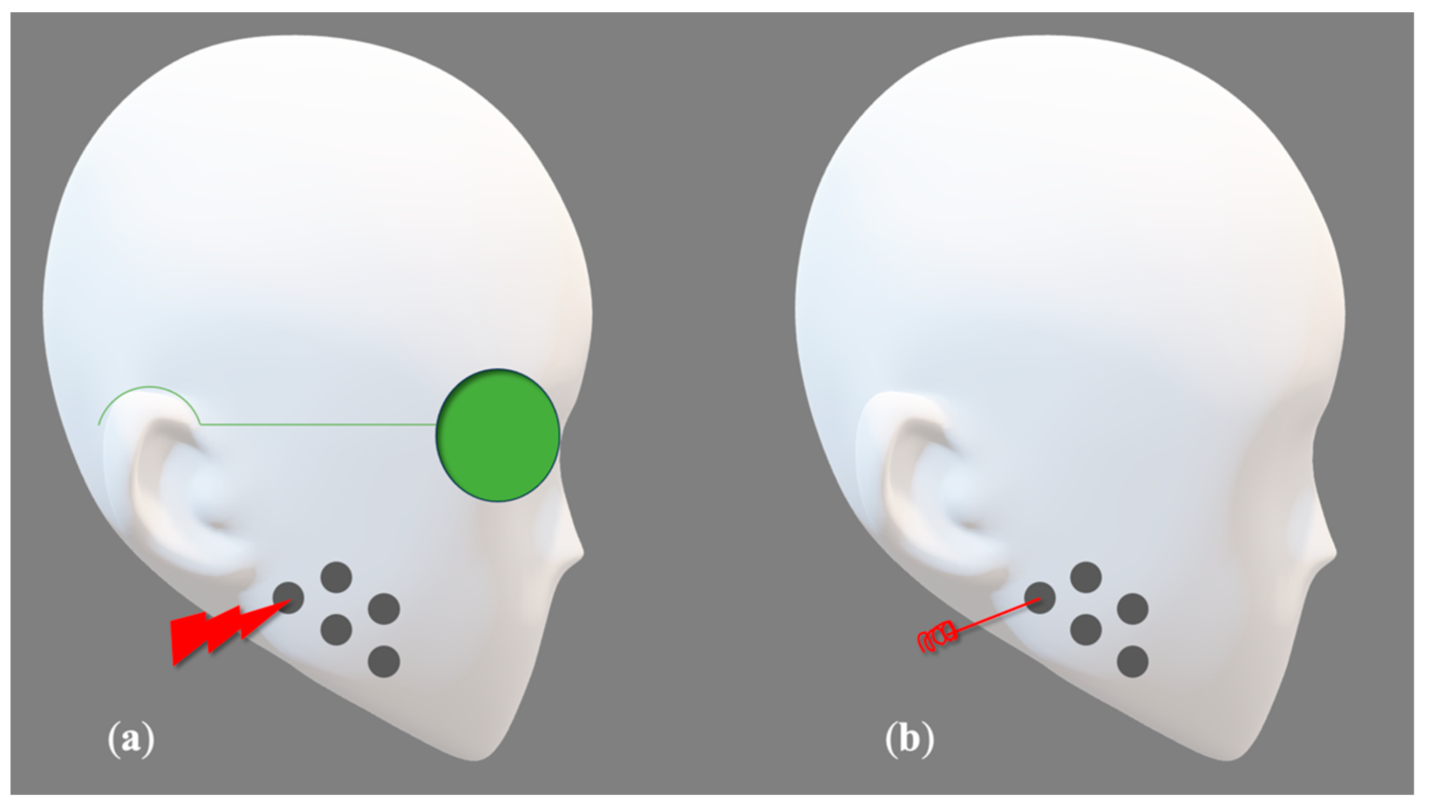
Figure 1. Myofascial trigger points marked with ballpoint pen according to Uemoto et al. [39]. (a), PBM therapy, (b) DN application. Image created with PowerPoint.
Photonics 11 00965 g001
Figure 2. Flowchart illustrating the process of literature search in accordance with PRISMA principles.
Photonics 11 00965 g002
Figure 3. (a) Risk-of-bias summary: review authors’ judgments about each risk-of-bias item for Laser PBM-included-studies group. (b) Risk-of-bias graph: review authors’ judgment about each risk-of-bias item presented as percentages across all included.
Photonics 11 00965 g003
Figure 4. (a) Risk-of-bias summary: review authors’ judgments about each risk-of-bias item for DN-included-studies group. (b) Risk-of-bias graph: review authors’ judgment about each risk-of-bias item presented as percentages across all included.
Photonics 11 00965 g004
Figure 5. (a) Risk-of-bias summary: review authors’ judgments about each risk-of-bias item for comparative studies of laser and dry needling methods. (b) Risk-of-bias graph: review authors’ judgment about each risk-of-bias item presented as percentages across all included.
Photonics 11 00965 g005
Figure 6. Meta-analysis result of PBM effect measured by VAS.
Photonics 11 00965 g006
Figure 7. Meta-analysis result of PBM effect on pressure pain threshold.
Photonics 11 00965 g007
Figure 8. Meta-analysis result of DN effect measured by VAS.
Photonics 11 00965 g008
Figure 9. Meta-analysis result of DN effect on Pressure Pain Threshold.
Photonics 11 00965 g009
Figure 10. Meta-analysis result of dry needle versus PBM effects measured by VAS.
Photonics 11 00965 g010
Table 1. RDC (research diagnostic criteria) of temporal mandibular disorder/TMD classification [5].
RDC/TMD Classification
Axis 1: Muscular disorders, a—(with pain), b—(with pain and limited opening)
Axis 2: Disc Displacement disorders, a—(disc displacement with reduction) b—(disc displacement without reduction)
Axis 3: Any joint pain (arthralgia, osteoarthritis)
Table 2. Summary of laser-PBM-studies group. PBM, photobiomodulation; VAS, visual analog scale; MMO, maximum mouth opening; P, protrusion; LL, left laterality; RL, right laterality; UM, upper masseter; MM, middle masseter; LM, lower masseter; EMG, electromyographic; PPT, pain pressure threshold; MP, masticatory performance.
Author/YearGroupsNumber of Patients
Gender
Age
Number of ApplicationPoints of ApplicationVariablesScaleFollow UpOutcomes
Venezian et al.
(2010)
[43]
1. Diode (PBM)
2. Placebo.
3. Diode (PBM)
4. Placebo
43 women
5 men
18–60 years
2 times/week
4 weeks
Extra-orally
1 pt/Anterior Temporalis
3 pts/ Masseter (UM, MM, LM)
Pain
surface electromyograph charging
VAS
Electromyographic (EMG) device
Pain:
Before
After the treatment
After 30 days of last treatment
EMG:
Before
After the treatment
The results showed no significant statistical differences between groups for both variables.
De Moraes Maia et al.
(2012)
[44]
1. Diode
(PBM)
2. Placebo.
19 women
2 men
Mean of ages
27.76 + 10.44
2 times/week
4 weeks
Extra-Orally
5 pts/Masseter
5 pts/ Anterior Temporal
Pain intensity
PPT
MP
VAS
Analog Compression Dynamometer
Optocal tablets
Baseline
Weekly
End of the therapy
30 days of final session
The results showed no significant difference between the groups according to pain intensity.
Sancakli et al.
(2015)
[45]
1. Diode
(PBM)
2. Diode
(PBM)
3. Placebo
21 women
9 men
18–60 years
3 times/week
4 weeks
Extra-orally
3pts/masseter
3pts/temporal
Pain intensity
PPT
MP
VAS
Muscle palpation
Baseline
End of the therapy
Laser groups showed significant reduction of pain and improvement in other Variables compared to placebo group.
Costa et al.
(2017)
[46]
1. Diode
(PBM)
2. Placebo
54 women
6 men
18–76 years
Single sessionExtra-orally:
3 pts/temporal
2 pts/ masseter
Pain
Pain during muscular palpation
MMO
VAS
Muscular AlgometerDigital Caliper
Before
After the treatment
The laser group showed a significant differences and improvement in pain reported with palpation compared to placebo group, while there was no significant improvement in range of mandibular movements.
Monteiro et al.
(2020)
[47]
1. Diode
(PBM)
2. Placebo
32 women
10 men
age > 18
1 time /week
4 weeks
Extra-orally:
Trigger points determined by patients
Pain
MMO
Pain tenderness
Mandibular movement
(P, LL, RL)
VAS
Patchmeter
Muscular Palpation
Baseline
1 month after the last session
The laser group showed significant improvement for all variables compared to placebo group.
Table 3. Details of laser PBM group parameters. NM, not mentioned; Ga Al As, gallium–aluminum–arsenide; nm, nanometer; W, watt; Mw, milliwatt; S, second; mm, millimeter; CW, continuous wave; J/cm2, joules per centimeter square.
StudyWavelengthPowerTip DiameterIrradiation TimeSpeed of MovementTip-tissue DistanceDelivery ModeContact/
Non-Contact
Energy DensityPower Meter
Venezian et al.
(2010)
[43]
780 nm
(Ga Al As)
50 mW
60 mW
NM20 s
40 s
NM0 mmCWContact25 J/cm2
60 J/cm2
No
De Moraes Maia et al.
(2012)
[44]
808 nm
(Ga Al As)
100 mWNM19 s/pointNM0 mmCWContact70 J/cm2Yes
Sancakli et al.
(2015)
[45]
820 nm
(Ga Al As)
300 mW6 mm10 sNM2 mmCWNon-Contact3 J/cm2Yes
Costa et al.
(2017)
[46]
830 nm100 mWNM28 s/pointNM0 mmCWContact100 J/cm2Yes
Monteiro et al.
(2020)
[47]
635 nm200 mW8 mm20 sNM0 mmCWContact8 J/cm2Yes
Table 4. Summary of DN studies group. VAS, visual analog system; PPT, pain pressure threshold; MMO, maximal mouth opening; MO, mouth opening; L, laterality; P, protrusion; MPA, mechanical pressure algometer; MA, manual algometer.
Author/YearGroupsNumber of Patients Gender AgeNumber of ApplicationPoints of ApplicationVariablesScaleFollow UpOutcomes
Dracoglu et al.
(2012)
[48]
1. DN
2. Placebo
45 women
7 men
18–57 years
1 time/week
3 weeks
Trigger points determined by patients in the masseter and temporalis musclesPain intensity
PPT
MO
VAS
Pressure algometry
Millimeter ruler
Baseline
After one week of last session
The results showed there were no differences between the groups in terms of pain and mouth opening, while the needling group showed significant improvement in PPTcompared to placebo group.
Gonzales-Perez et al.
(2012)
[49]
1. DN
No control group
30 women
6 men
1 time/week
3 weeks
Trigger points determined by patients in the external pterygoid musclePain
Range of mandibular movement
(MO, L, P)
VAS
Therabite System
Before
After 2 weeks
After 1 month
After 2 months
After 6 months of last session
The results showed significant improvements for the variables After the therapeutic intervention
Blasco-Bonora et al.
(2017)
[50]
1. DN
No control group
11 women
6 men
23–66 years
Single sessionTrigger points determined by patients in the masseter and temporalis musclesPain
PPT
MMO
Jaw disability
VAS
MPA
Millimeter ruler
JDC list of RDC/TMD
Baseline
After treatment for all Variables except jaw disability which was assessed after 1 week of the treatment.
The results showed significant improvements in the study group for all variables after the treatment.
Ozden et al.
(2018)
[51]
1. SDN
2. DDN
3.Control group
31 women
29 men
18–65 year
1 time/week
3 weeks
Points of Application
Trigger points determined by patients in the masseter muscle
Pain
PPT
MJO
VAS
MA
Before
At the third week
At the sixth week of the last intervention
The results showed significant improvements in both groups.
While SDN group showed significantly better Pain reduction compared to DDN group.
Dib-Zakkour et al.
(2022)
[52]
1. DDN
2. Control group
36 patients
18–40 years
Single sessionTrigger points determined by patients in the masseter musclePain
Muscular palpation
MO
Articular sounds
Tone of masseter muscle
VAS
Algometer
Digital caliper
Auscultate
Electromyography
Before
After 10 min of the session
After 15 days of the intervention
The results showed significant reduction in the fascial pain and muscle activity in the study group compared to controlling group.
Table 5. Details of the needling technique used in the DN-included studies. NM, not mentioned; mm, millimeter; SDN, superficial dry needling; DDN, deep dry needling.
StudyNeedle SizeNeedling Method
(Superficial/Deep)
Penetration DepthNumber of Needles in One Session
Dracoglu et al.
(2012)
[48]
0.22 × 30 mm(Deep/Superficial)
NM
NMNM
Gonzales-Perez et al.
(2012)
[49]
0.25 × 40 mmDeepNMNM
Blasco-Bonora et al.
(2017)
[50]
0.16 × 25 mmNM15–25 mmNM
Ozden et al.
(2018)
[51]
0.25 × 25 mmSDN
DDN
SDN 5–10 mm
DDN > 10 mm
NM
Dib-Zakkour et al.
(2022)
[52]
0.30 × 30 mmNMNMNM
Table 6. Summary of the comparative group. P, power; E, energy; T, time; ED, energy density; AP, average power; CW, continuous wave; mW, milliwatts; J, joule; J/cm2, joules per centimeter square; nm, nanometer; NRS, numerical rating scale; MMO, maximum mouth opening.
Author/YearGroupsNumber of Patients Gender AgeLaser ParametersNeedling MethodNumber of ApplicationVariable/ScaleFollow UpOutcomes
Sayed S. et al.
(2016)
[53]
1. PBM
2. DN
17 women
1 man
18–42 years
Wavelength (980 nm)
P (0,2 W)
E (12 J)
T (50 SEC)
23 ×1.5 inch
Depth 1–2 cm
1 time/week
4 weeks
Pain intensity /NRS
MMO/in mm using Vernier graduated caliper
Before
After 2 weeks
After 4 weeks
The results showed insignificant difference between the groups.
Oliveira D.A. et al.
(2018)
[54]
1. PBM
2. DN
10 women
18–70 years
Wavelength (660 nm)
ED (40 J/cm2)
AP (40 mW)
E (1.6 J)
T (40 sec)
CW
0.25× 30 mm
Length 5 cm
Time 1 min
1 time/week
12 weeks
Pain/VAS
MO/in millimeter ruler
Before
After 1 week
The results showed insignificant difference between the groups.
Table 7. Details of laser group parameters used in the comparative group. Details of laser parameters of comparative studies. NM, not mentioned; nm, nanometer; mW, milliwatt; s, second; mm, millimeter; CW, continuous wave; J/cm2, joules per centimeter square.
StudyWavelengthPowerTip DiameterIrradiation TimeSpeed of MovementTip-Tissue DistanceDelivery ModeContact/
Non-Contact
Energy DensityPower Meter
Sayed et al.
(2016)
[53]
980 nm200 mWNM50 s/sessionNMNMNMNMNMNM
Oliveira et al.
(2018)
[54]
660 nm40 mWNM40 s/pointNM0 mmCWContact40 J/cm2NM
Disclaimer/Publisher’s Note: The statements, opinions and data contained in all publications are solely those of the individual author(s) and contributor(s) and not of MDPI and/or the editor(s). MDPI and/or the editor(s) disclaim responsibility for any injury to people or property resulting from any ideas, methods, instructions or products referred to in the content.