Abstract
Mg4Ta2O9 single crystals doped with 0.01, 0.1, and 1% Er were grown using the floating zone method, and their photoluminescence and scintillation properties were studied. X-ray diffraction analysis confirmed that all samples had a hexagonal structure of Mg4Ta2O9. The samples showed that the emission peak at 1550 nm was due to 4f−4f transitions of Er3+ ions, with quantum yields of 32.9, 46.6, and 18.0% for 0.01, 0.1, and 1% doping concentrations, respectively. All samples showed scintillation with a broad emission band at 350 nm due to charge transfer from Ta5+ to O2− ions and emission peak at 1550 nm due to 4f−4f transition of Er3+ ions. The dose rate response functions showed that the detection limit for all samples was 0.06 Gy/h for scintillation detector applications.
1. Introduction
Radiation detectors play an important part in various fields, such as space exploration [1,2], environmental radiation dosimetry [3,4], well-logging [5,6], and medical imaging [7,8]. In these applications, scintillators are mostly used due to their high detection efficiency and ease of use. Scintillators are classified as phosphors that immediately emit ultraviolet (UV), visible (Vis), and near-infrared (NIR) photons under irradiations of ionizing radiations [9,10,11]. The required characteristics for scintillators are radiation resistance, high density (ρ), large effective atomic number (Zeff), and high light yield (LY) [12]. To meet the above characteristics, various scintillators, such as Bi4Ge3O12 (BGO) [13], CaWO4 [14], Tl:NaI [15], and Tl:CsI, have been developed [16]. Because photodetectors based on bi-alkali photocathode have wavelength sensitivity in the range of 300–600 nm, the development of scintillators has mainly been conducted with materials showing emission wavelengths in the UV-Vis range.
In recent years, research and development of NIR scintillators have progressed because of the development of InGaAs-based photodetectors [17,18,19,20]. NIR scintillators have drawn attention for dose monitoring systems in high-dose areas [21,22]. In such applications, scintillation detectors, consisting of a NIR emitting scintillator and an optical fiber, have been proposed [23]. The system using NIR emitting scintillators has two advantages. Firstly, under high dose conditions, Cherenkov light appears in the range of UV-Vis [24]. Therefore, the wavelength of Cherenkov light (noise) overlaps with UV-Vis photons (signal) from the common scintillators, and it is difficult to distinguish these two components. In contrast, since NIR scintillators emit photons in a different wavelength range from Cherenkov light, it is easy to distinguish. Secondly, NIR photons penetrate optical fibers more efficiently than UV-Vis light [25]. The light transmission efficiency of optical fibers degrades in high dose areas, but the extent of degradation is milder in the NIR region than in the UV-Vis region. Therefore, when NIR-emitting scintillators are used, it enables us to efficiently guide emitted scintillation light to a photodetector.
In this study, Mg4Ta2O9 is selected as a host. In recent years, Ta-based compounds have been investigated in a variety of fields, including phosphors [26], microwave dielectrics [27], and photo-catalysis [28]. Among them, Mg4Ta2O9 has been attracting attention as a candidate material for scintillators, owing to its relatively large Zeff (64.3) and high density (6.20 g/cm3) [29,30,31]. In our previous work, undoped Mg4(Ta,Nb)2O9 single crystals were investigated, and they showed high LY and low afterglow levels comparable with commercial BGO crystal in the UV-Vis range [32]. In terms of luminescence center, we focused on Er3, which is an intriguing luminescent center that can emit NIR photons [33]. Phosphors doped with Er, until now, have been intensively researched as laser materials for optical communication [34,35,36] because transmission loss in a glass fiber is low in the range of 1400–1600 nm [25]. While the optical properties of Mg4Ta2O9 doped with Mn2+ and Cr3+ have been investigated in previous studies [30,37], the scintillation properties of Mg4Ta2O9 doped with Er3+ have not been examined at all. Er-doped Mg4Ta2O9 single crystals were fabricated using the floating zone (FZ) method, and their PL and scintillation characteristics were analyzed.
2. Materials and Methods
Mg4Ta2O9 single crystals were fabricated using a FZ furnace (FZD0192) from Canon Machinery. The selected Er concentrations were 0.01, 0.1, and 1%. The starting materials were MgO (High Purity Chemicals, 99.99%), Ta2O5 (Rare Metallic, 99.99%), and Er2O3 (Furuuchi Chemical, 99.99%). The powders were mixed homogeneously using an agate mortar and then formed into cylindrical rods by hydrostatic pressure of 10 MPa for 10 min. The cylindrical rods were sintered in air at 1400 °C for 8 h for crystal growth. In crystal growth, the pull-down speed was 5 mm/h, and the rotational speed was 3 rpm. After the growth was finished, the polycrystalline part was removed from the resulting crystalline rod by using pliers, and the part (5–7 mm) forming single crystals was taken out. The polisher (MetaServ 250) from Buehler was used to mechanically grind the large upper and lower surfaces so that they were parallel. To identify crystal phases of the synthesized sample, powder X-ray diffraction (PXRD) patterns were assessed in the 10–90° range with a diffractometer (MiniFlex 600) from Rigaku.
The Quantaurus-QY Plus device (C13534) from Hamamatsu Photonics was used to measure the PL excitation and emission spectrum, as well as the quantum yield (QY). The measurements were conducted using a Xenon lamp as the source of excitation light and with two different types of linear image sensors for photodetection: a silicon sensor capable of detecting UV-Vis light and an Indium Gallium Arsenide (InGaAs) sensor capable of detecting NIR light. Additionally, the PL decay time profile was measured using the Quantaurus-τ (C11367) from Hamamatsu Photonics.
Our custom-built setup was used to measure the X-ray-induced scintillation spectra [38]. For this, a X-ray generator (XRB80N100/CB) from Spellman was set to 80 kV for bias voltage and 1.2 mA for tube current as the X-ray source. The scintillation light was then transmitted to a spectrometer consisting of a monochromator (163) from Shamrock and a Si-based line camera (DU-420-BU2) from Andor for monitoring wavelength at 200–700 nm and an InGaAs-based line camera (DU492A) from Andor for monitoring wavelength at 700–1600 nm. Our own setup was used to measure the X-ray-induced scintillation decay time profiles and the afterglow profiles [39]. Additionally, to evaluate the dose rate characteristics as a NIR scintillation detector, the emission intensity under different X-ray dose rates were measured by using our own setup [40]. For this measurement, the tube voltage supplied to the generator (XRB80P&N200X4550) from Spellman was fixed at 40 kV, while the tube current was varied from 5.2 to 0.052 mA. The NIR photon was detected with an InGaAs PIN photodiode (G12180–250A) from Hamamatsu Photonics through an optical fiber (FP600ERT) from Thorlabs.
3. Results
3.1. Sample Conduction
Figure 1 shows photographs of the 0.01, 0.1, and 1% Er-doped Mg4Ta2O9 used for the subsequent characterizations. The samples had a size of approximately ~13 × 4 × 1.5 mm. Since near-infrared scintillators have not been practically implemented yet, the optimal sample size for measurements has not been determined. Hence, the samples were processed into sufficient size to measure the scintillation properties. The 0.1 and 0.01% Er-doped samples were colorless and transparent, whereas the 1% Er-doped sample exhibited a pale pink color, which is typical for Er-doped materials. Because of the significant presence of cracks in the fabricated samples, transmission spectra could not be measured. However, the 0.1% sample was more transparent than the 1% sample, and no opaque cores due to impurity were observed.
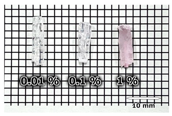
Figure 1.
Picture of Mg4Ta2O9 single crystals doped with 0.01, 0.1, and 1% Er.
Figure 2 shows the PXRD patterns of three different concentrations of Er-doped Mg4Ta2O9 and the reference patterns of Mg4Ta2O9. The reference data are from the Crystallography Open Database (COD) 2002422. The diffraction peaks observed in the Er-doped samples in Figure 2a were found to be identical to those of the reference pattern, indicating that the samples consisted of a single phase of Mg4Ta2O9 with a hexagonal structure. No additional peaks, corresponding to impurity phases, were detected in the samples. These results suggest that all the Er-doped Mg4Ta2O9 single crystals possessed a well-defined crystal structure [41,42]. Figure 2b shows an enlarged view of the strongest diffraction peaks. Peak shifts due to Er-doping were not clearly observed in the PXRD patterns. This is because Er concentrations were too low to detect the peak shifts in our XRD measurements.
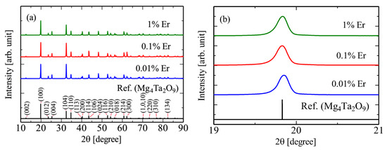
Figure 2.
PXRD patterns of Mg4Ta2O9 single crystals doped with 0.01, 0.1, and 1% Er and references in the range of (a) 10–90° and (b) 19–21°. The Miller indices of relatively high intensity peaks are shown in Figure 2a.
3.2. PL Properties
Figure 3 shows the PL emission and excitation contour graph of the 0.1% Er-doped Mg4Ta2O9 monitoring wavelengths at (a) 220–950 nm and (b) 950–1685 nm. The excitation wavelength of the intrinsic luminescence of Mg4Ta2O9 is 220 nm [32], which is outside the measurable range of the instrument used in this study, so no emission was observed in Figure 3a. In Figure 3b, an emission peak at 1550 nm under excitation light at 380 nm and 520 nm was observed, which was attributed to the 4f−4f transitions of Er3+ [43,44]. The spectral characteristics of the other samples were consistent with that of the Mg4Ta2O9 doped with 0.1% Er. Therefore, only the 0.1% sample was selected as the representative one for this measurement. The QYs monitored between 1450–1650nm for samples doped with 0.01%, 0.1%, and 1% Er under excitation at 520 nm were 32.9%, 46.6%, and 18.0%, respectively. The highest QY values were observed at 0.1% Er-doped sample, while the QY of 1% Er-doped sample was lower than that of 0.1% Er-doped sample. It is likely due to concentration quenching.
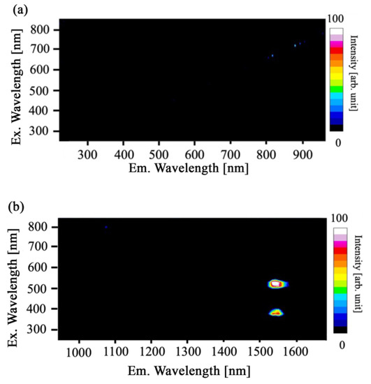
Figure 3.
PL excitation and emission spectrum of Mg4Ta2O9 single crystals doped with 0.1% Er under the excitation wavelength at 250–850 nm in the emission range of (a) 220–950 nm and (b) 950–1685 nm. The intensity of the emitted light is represented by a color bar, with white indicating high intensity and blue indicating low intensity. The horizontal and vertical axes are emission and excitation wavelengths, respectively.
3.3. Scintillation Properties
Figure 4 shows the X-ray-induced scintillation spectra of the 0.01, 0.1, and 1% Er-doped Mg4Ta2O9 in the range of (a) 250–700 nm and (b) 700–1625 nm. The maximum signal intensity at ~1540 nm was 1,855 for the 0.01% sample, 21,153 for the 0.1% sample, and 21,056 for the 1% sample. Therefore, it is suggested that the emission intensity of the 0.01% sample is significantly lower than those of the 0.1% and 1% samples. In the range of 250–700 nm, a broad emission band due to the charge transfer (CT) from Ta5+ to O2− was observed [29]. Furthermore, a partial decrease in the emission spectrum with increasing Er concentration was observed at 300–500 nm, which coincided with the excitation wavelengths shown in Figure 3 and was found to be due to the absorption of the 4f−4f transitions of Er3+. Furthermore, emission peaks originated from the 4f−4f transitions of Er3+ were observed at 550 and 560 nm [45,46]. However, the emission peaks were hardly observed owing to the weak emission intensity. In the 700–1625 nm range, sharp emission peaks were observed at 980 nm and 1550 nm, which were attributed to the 4f−4f transitions of Er3+ [43,47]. The broad peak at 1200 nm in the 0.01% sample is noise. This is observed due to the weak intensity of the Er-derived emission.
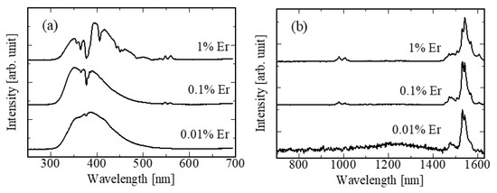
Figure 4.
X-ray-induced scintillation spectra of Mg4Ta2O9 single crystals doped with 0.01, 0.1, and 1% Er in the range of (a) 250–700 nm and (b) 700–1625 nm.
X-ray-induced scintillation decay time profiles of the 0.01, 0.1, and 1% Er-doped Mg4Ta2O9 single crystals are shown in Figure 5. All the decay curves were approximated by a single exponential function. The decay time constants of the 0.01, 0.1, and 1% Er-doped Mg4Ta2O single crystals were 6.11, 5.66, and 5.65 μs, respectively. The obtained decay time constants were nearly the same as those due to the CT from Ta5+ to O2− in Mg4Ta2O9 [29]. According to previous studies [33], emission from CT at 300–500 nm overlaps with the excitation band of Er3+ ions (Figure 3), suggesting that the energy transfer from CT to Er3+ ions caused a decrease in the decay time constant.
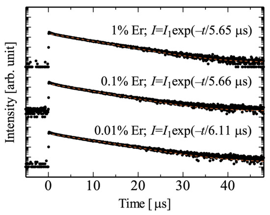
Figure 5.
Scintillation decay time profiles induced by X-rays for Mg4Ta2O9 single crystals doped with 0.01, 0.1, and 1% Er. Each fitted curve is represented by a dashed line.
Figure 6 displays the afterglow profiles of single crystals of Mg4Ta2O9 doped with 0.01, 0.1, and 1% Er after being exposed to X-ray irradiation for 2 ms. The afterglow level at 20 ms after X-ray irradiation (AGL20) can be expressed as the ratio of the difference between the signal intensity at 20 ms after X-ray irradiation (I20) and the background signal intensity (Ibg), to the signal intensity during X-ray irradiation (Imax). In other words, the formula for AGL20 can be rewritten as: AGL20 = (I20 − Ibg) / (Imax − Ibg). The afterglow levels of Mg4Ta2O9 doped with 0.01, 0.1, and 1% Er were found to be 267, 247, and 138 ppm, respectively. These values were lower than those of Tl:CsI scintillator, which were reported to have an afterglow level of around 300 ppm in the UV-Vis range when evaluated using the same evaluation system [48].
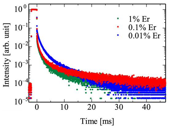
Figure 6.
Afterglow profiles of Mg4Ta2O9 single crystals doped with 0.01, 0.1, and 1% Er.
To evaluate the performance of the Er-doped Mg4Ta2O9 as a scintillation detector, the correlation between NIR emission intensity and X-ray irradiation dose rate was measured for samples doped with 0.01, 0.1, and 1% Er in Mg4Ta2O9, as shown in Figure 7. The y-axis is defined as the difference between the signal detected during X-ray irradiation and the background signal. All the samples showed a good linearity in the dose rate range of 0.06–6 Gy/h. For comparison, the Zeff, density, dose–response characteristics, and photodetector are summarized in Table 1. The dose rate response functions of Er-doped BGO and Bi4Si3O12 (BSO) NIR scintillators evaluated in the same measurement system have been reported to show a lower detection limit of 0.006 Gy/h [44,49]. Previous studies reported a lower detection limit of 0.8 Gy/h in measurement systems combining commercially available Pr-doped Gd2O2S and Si-based photodetectors, 0.3 Gy/h for a system combining a Pr-doped BaTi4O9 and an InGaAs-based photodetector, and 0.06 Gy/h for a Nd-doped SrY2O4 and InGaAs-based system. Hence, the lower detection limit of measurement of 0.06 Gy/h is still sufficient for radiation monitoring in nuclear facilities [23]. Therefore, Er-doped Mg4Ta2O9 is considered to be a candidate for NIR emitting scintillators.
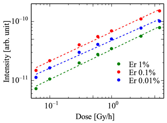
Figure 7.
Relationship between NIR emission intensity and X-ray irradiation dose rate for Mg4Ta2O9 single crystals doped with 0.01, 0.1, and 1% Er.

Table 1.
Zeff, density, dose–response properties, and type of photodetector for the reported NIR-emitting scintillators.
4. Conclusions
The 0.01%, 0.1%, and 1% Er-doped Mg4Ta2O9 single crystals were synthesized by the FZ method. The PL characteristics of all the Er-doped Mg4Ta2O9 exhibited luminescence attributed to the 4f−4f transition of Er3+ ions. In addition, the PL QY in the NIR region of the 0.1% Er-doped Mg4Ta2O9 single crystal was the highest value of 46.6% among the samples presented. under X-ray irradiation, all the Er-doped samples exhibited similar luminescence characteristics to PL, and additional luminescence due to the CT from Ta5+ to O2− in Mg4Ta2O9 was observed. The afterglow levels of 0.01%, 0.1%, and 1% Er-doped Mg4Ta2O were 267.0, 247.0, and 137.9 ppm, respectively. The relationship between NIR luminescence intensity and X-ray dose rate showed good linearity from 0.06–6 Gy/h, confirming sufficient coverage for process radiation monitoring in nuclear facilities. These results suggest that Er-doped Mg4Ta2O9 single crystals are promising candidates for NIR emission scintillators for high-dose rate monitoring applications. The 0.1% samples were more transparent compared with the 1% sample, and no visible impurity cores were observed. In addition, they showed higher QY values and scintillation light intensities in the NIR range compared with the 0.01% sample. These results suggest that the 0.1% concentration offers the most favorable results in terms of optical response and quality.
Author Contributions
Conceptualization, T.Y.; methodology, T.H., D.N., K.O. and T.Y.; validation, D.N. and N.K.; formal analysis, T.H. and D.N.; investigation, T.H., K.I. and K.O.; resources, D.N. and T.Y.; data curation, T.H.; writing—original draft preparation, T.H.; writing—review and editing, K.I., D.N. and T.K; visualization, T.H. and T.K.; supervision, T.Y.; funding acquisition, T.K., D.N., N.K. and T.Y. All authors have read and agreed to the published version of the manuscript.
Funding
This work was supported by Grants-in-Aid for Scientific A (22H00309), Grants-in-Aid for Scientific B (22H03872, 22H02939, 21H03733, and 21H03736), and Challenging Exploratory Research (22K18997) from the Japan Society for the Promotion of Science. The Cooperative Research Project of Research Center for Biomedical Engineering, A-STEP (JPMJTM22DN) from JST, Konica Minolta Science and Technology Foundation, Nakatani Foundation, and Kazuchika Okura Memorial Foundation are also acknowledged.
Institutional Review Board Statement
Not applicable.
Informed Consent Statement
Not applicable.
Data Availability Statement
Data will be made available on request.
Conflicts of Interest
The authors declare that they have no known competing financial interests or personal relationships that could have appeared to influence the work reported in this paper’s conceptualization, investigation, or writing.
References
- Rieder, R.; Economou, T.; Wänke, H.; Turkevich, A.; Crisp, J.; Brückner, J.; Dreibus, G.; McSween, H.Y. The Chemical Composition of Martian Soil and Rocks Returned by the Mobile Alpha Proton X-Ray Spectrometer: Preliminary Results from the X-Ray Mode. Science 1997, 278, 1771–1774. [Google Scholar] [CrossRef] [PubMed]
- Dong, T.; Zhang, Y.; Ma, P.; Zhang, Y.; Bernardini, P.; Ding, M.; Guo, D.; Lei, S.; Li, X.; De Mitri, I.; et al. Charge Measurement of Cosmic Ray Nuclei with the Plastic Scintillator Detector of DAMPE. Astropart. Phys. 2019, 105, 31–36. [Google Scholar] [CrossRef]
- Gold, R. Meson Dosimetry for the Natural Environment. Radiat. Res. 1973, 56, 413. [Google Scholar] [CrossRef] [PubMed]
- Tsutsumi, M.; Tanimura, Y. LaCl3(Ce) Scintillation Detector Applications for Environmental Gamma-Ray Measurements of Low to High Dose Rates. Nucl. Instrum. Methods Phys. Res. A 2006, 557, 554–560. [Google Scholar] [CrossRef]
- Melcher, C.L.; Schweitzer, J.S.; Manente, R.A.; Peterson, C.A. Applications of Single Crystals in Oil Well Logging. J. Cryst. Growth 1991, 109, 37–42. [Google Scholar] [CrossRef]
- Nikitin, A.; Bliven, S. Needs of Well Logging Industry in New Nuclear Detectors. In Proceedings of the IEEE Nuclear Science Symposuim & Medical Imaging Conference, Knoxville, TN, USA, 30 October–6 November 2010; IEEE: Piscataway, NJ, USA, 2010; pp. 1214–1219. [Google Scholar]
- van Eijk, C.W.E. Inorganic Scintillators in Medical Imaging. Phys. Med. Biol. 2002, 47, R85–R106. [Google Scholar] [CrossRef]
- Lecoq, P. Development of New Scintillators for Medical Applications. Nucl. Instrum. Methods Phys. Res. A 2016, 809, 130–139. [Google Scholar] [CrossRef]
- Liu, Y.; Sowerby, B.D.; Tickner, J.R. Comparison of Neutron and High-Energy X-Ray Dual-Beam Radiography for Air Cargo Inspection. Appl. Radiat. Isot. 2008, 66, 463–473. [Google Scholar] [CrossRef]
- Yanagida, T. Study of Rare-Earth-Doped Scintillators. Opt. Mater. 2013, 35, 1987–1992. [Google Scholar] [CrossRef]
- Ichiba, K.; Okazaki, K.; Takebuchi, Y.; Kato, T.; Nakauchi, D.; Kawaguchi, N.; Yanagida, T. X-Ray-Induced Scintillation Properties of Nd-Doped Bi4Si3O12 Crystals in Visible and Near-Infrared Regions. Materials 2022, 15, 8784. [Google Scholar] [CrossRef]
- Yanagida, T. Inorganic Scintillating Materials and Scintillation Detectors. Proc. Jpn. Acad. Ser. A Math. Sci. Ser. B 2018, 94, 75–97. [Google Scholar] [CrossRef] [PubMed]
- Farukhi, M.R. Bi4Ge3O12 (BGO)—A Scintillator Replacement for NaI(Tl). MRS Proc. 1982, 16, 115. [Google Scholar] [CrossRef]
- Coltman, J.W. The Scintillation Counter. Proc. IRE 1949, 37, 671–682. [Google Scholar] [CrossRef]
- Hofstadter, R. Alkali Halide Scintillation Counters. Phys. Rev. 1948, 74, 100–101. [Google Scholar] [CrossRef]
- Grassmann, H.; Lorenz, E.; Moser, H.-G. Properties of CsI(TI)—Renaissance of an Old Scintillation Material. Nucl. Instrum. Methods. Phys. Res. A 1985, 228, 323–326. [Google Scholar] [CrossRef]
- Moses, W.W.; Weber, M.J.; Derenzo, S.E.; Perry, D.; Berdahl, P.; Boatner, L.A. Prospects for Dense, Infrared Emitting Scintillators. IEEE Trans. Nucl. Sci. 1998, 45, 462–466. [Google Scholar] [CrossRef]
- Bressi, G.; Carugno, G.; Conti, E.; Noce, C.D.; Iannuzzi, D. New Prospects in Scintillating Crystals. Nucl. Instrum. Methods Phys. Res. A 2001, 461, 361–364. [Google Scholar] [CrossRef]
- Yanagida, T.; Fujimoto, Y.; Ishizu, S.; Fukuda, K. Optical and Scintillation Properties of Nd Differently Doped YLiF4 from VUV to NIR Wavelengths. Opt. Mater. 2015, 41, 36–40. [Google Scholar] [CrossRef]
- Awater, R.H.P.; Alekhin, M.S.; Biner, D.A.; Krämer, K.W.; Dorenbos, P. Converting SrI2:Eu2+ into a near Infrared Scintillator by Sm2+ Co-Doping. J. Lumin. 2019, 212, 1–4. [Google Scholar] [CrossRef]
- Xiong, L.-Q.; Chen, Z.-G.; Yu, M.-X.; Li, F.-Y.; Liu, C.; Huang, C.-H. Synthesis, Characterization, and in Vivo Targeted Imaging of Amine-Functionalized Rare-Earth up-Converting Nanophosphors. Biomaterials 2009, 30, 5592–5600. [Google Scholar] [CrossRef]
- Ning, Y.; Chen, S.; Chen, H.; Wang, J.-X.; He, S.; Liu, Y.-W.; Cheng, Z.; Zhang, J.-L. A Proof-of-Concept Application of Water-Soluble Ytterbium(III) Molecular Probes in in Vivo NIR-II Whole Body Bioimaging. Inorg. Chem. Front. 2019, 6, 1962–1967. [Google Scholar] [CrossRef]
- Takeda, E.; Kimura, A.; Hosono, Y.; Takahashi, H.; Nakazawa, M. Radiation Distribution Sensor with Optical Fibers for High Radiation Fields. J. Nucl. Sci. Technol. 1999, 36, 641–645. [Google Scholar] [CrossRef]
- Watanabe, K.; Yanagida, T.; Nakauchi, D.; Kawaguchi, N. Scintillation Light Yield of Tb:Sr2Gd8(SiO4)6O2. Jpn. J. Appl. Phys. 2021, 60, 106002. [Google Scholar] [CrossRef]
- Peters, K. Polymer Optical Fiber Sensors—A Review. Smart Mater. Struct. 2011, 20, 013002. [Google Scholar] [CrossRef]
- Wachtel, A. Self-Activated Luminescence of M2+ Niobates and Tantalates. J. Electrochem. Soc. 1964, 111, 534. [Google Scholar] [CrossRef]
- Wu, H.T.; Li, L.X.; Zou, Q.; Liao, Q.W.; Ning, P.F.; Zhang, P. Synthesis, Characterization, and Microwave Dielectric Properties of Mg4Nb2O9 Ceramics Produced through the Aqueous Sol–Gel Process. J. Alloys Compd. 2011, 509, 2232–2237. [Google Scholar] [CrossRef]
- Zhang, H.; Sun, X.; Wang, Y.; Xu, X. Switching on Wide Visible Light Photocatalytic Activity over Mg4Ta2O9 by Nitrogen Doping for Water Oxidation and Reduction. J. Catal. 2019, 377, 455–464. [Google Scholar] [CrossRef]
- Yuan, D.; Moretti, F.; Perrodin, D.; Bizarri, G.; Shalapska, T.; Dujardin, C.; Bourret, E. Modified Floating-Zone Crystal Growth of Mg4Ta2O9 and Its Scintillation Performance. CrystEngComm 2020, 22, 3497–3504. [Google Scholar] [CrossRef]
- Stevels, A.L.N.; Vink, A.T. Fine Structure in the Low Temperature Luminescence of Zn2SiO4:Mn and Mg4Ta2O9:Mn. J. Lumin. 1974, 8, 443–451. [Google Scholar] [CrossRef]
- Carone, D.; Jacobsohn, L.G.; Breton, L.S.; zur Loye, H.-C. Synthesis, Structure, and Scintillation of Rb4Ta2Si8O23. Solid. State. Sci. 2022, 127, 106861. [Google Scholar] [CrossRef]
- Hayashi, T.; Ichiba, K.; Nakauchi, D.; Watanabe, K.; Kato, T.; Kawaguchi, N.; Yanagida, T. Evaluation of Scintillation Properties of Mg4(Ta,Nb)2O9 Single Crystals. J. Lumin. 2023, 255, 119614. [Google Scholar] [CrossRef]
- Petit, L.; Cardinal, T.; Videau, J.J.; Le Flem, G.; Guyot, Y.; Boulon, G.; Couzi, M.; Buffeteau, T. Effect of the Introduction of Na2B4O7 on Erbium Luminescence in Tellurite Glasses. J. Non. Cryst. Solids 2002, 298, 76–88. [Google Scholar] [CrossRef]
- Girard, S.; Laurent, A.; Pinsard, E.; Robin, T.; Cadier, B.; Boutillier, M.; Marcandella, C.; Boukenter, A.; Ouerdane, Y. Radiation-Hard Erbium Optical Fiber and Fiber Amplifier for Both Low- and High-Dose Space Missions. Opt. Lett. 2014, 39, 2541. [Google Scholar] [CrossRef] [PubMed]
- Thomas, J.; Myara, M.; Troussellier, L.; Burov, E.; Pastouret, A.; Boivin, D.; Mélin, G.; Gilard, O.; Sotom, M.; Signoret, P. Radiation-Resistant Erbium-Doped-Nanoparticles Optical Fiber for Space Applications. Opt. Express 2012, 20, 2435. [Google Scholar] [CrossRef] [PubMed]
- Stange, H.; Petermann, K.; Huber, G.; Duczynski, E.W. Continuous Wave 1.6 μm Laser Action in Er Doped Garnets at Room Temperature. Appl. Phys. B Lasers Opt. 1989, 49, 269–273. [Google Scholar] [CrossRef]
- Wang, S.; Pang, R.; Tan, T.; Wu, H.; Wang, Q.; Li, C.; Zhang, S.; Tan, T.; You, H.; Zhang, H. Achieving High Quantum Efficiency Broadband NIR Mg4Ta2O9: Cr3+ Phosphor Through Lithium-Ion Compensation. Adv. Mater. 2023, 35, 2300124. [Google Scholar] [CrossRef]
- Yanagida, T.; Kamada, K.; Fujimoto, Y.; Yagi, H.; Yanagitani, T. Comparative Study of Ceramic and Single Crystal Ce: GAGG Scintillator. Opt. Mater. 2013, 35, 2480–2485. [Google Scholar] [CrossRef]
- Yanagida, T.; Fujimoto, Y.; Ito, T.; Uchiyama, K.; Mori, K. Development of X-Ray-Induced Afterglow Characterization System. Appl. Phys. Express 2014, 7, 062401. [Google Scholar] [CrossRef]
- Akatsuka, M.; Kimura, H.; Onoda, D.; Shiratori, D.; Nakauchi, D.; Kato, T.; Kawaguchi, N.; Yanagida, T. X-Ray-Induced Luminescence Properties of Nd-Doped GdVO4. Sens. Mater. 2021, 33, 2243. [Google Scholar] [CrossRef]
- Fu, Z.; He, Y. Sintering Behavior, Phase Composition and Microwave Dielectric Characteristics OfMg4Nb2O9 Ceramics Doped with ZnO-B2O2-SiO2 Glass. Integr. Ferroelectr. 2021, 221, 161–167. [Google Scholar] [CrossRef]
- Sun, D.C.; Senz, S.; Hesse, D. Topotaxial Formation of Mg4Ta2O9 and MgTa2O6 Thin Films by Vapour-Solid Reactions on MgO (001) Crystals. J. Eur. Ceram. Soc. 2004, 24, 2453–2463. [Google Scholar] [CrossRef]
- Dickinson, S.K.; Hilton, R.M.; Lipson, H.G. Czochralski Synthesis and Properties of Rare-Earth-Doped Bismuth Germanate (Bi4Ge3O12). Mater. Res. Bull. 1972, 7, 181–191. [Google Scholar] [CrossRef]
- Ichiba, K.; Okazaki, K.; Takebuchi, Y.; Kato, T.; Nakauchi, D.; Kawaguchi, N.; Yanagida, T. Visible–Near Infrared Scintillation Properties of Er-Doped Bi4Si3O12 Single Crystals. ECS J. Solid State Sci. Technol. 2023, 12, 046001. [Google Scholar] [CrossRef]
- Amin, J.; Dussardier, B.; Schweizer, T.; Hempstead, M. Spectroscopic Analysis of Er3+ Transitions in Lithium Niobate. J. Lumin. 1996, 69, 17–26. [Google Scholar] [CrossRef]
- Dorenbos, P.; van Loef, E.V.D.; Vink, A.P.; van der Kolk, E.; van Eijk, C.W.E.; Krämer, K.W.; Güdel, H.U.; Higgins, W.M.; Shah, K.S. Level Location and Spectroscopy of Ce3+, Pr3+, Er3+ and Eu3+ in LaBr3. J. Lumin. 2006, 117, 147–155. [Google Scholar] [CrossRef]
- Talewar, R.A.; Mahamuda, S.; Swapna, K.; Venkateswarlu, M.; Rao, A.S. Sensitization of Er3+ NIR Emission Using Yb3+ Ions in Alkaline-Earth Chloro Borate Glasses for Fiber Laser and Optical Fiber Amplifier Applications. Mater. Res. Bull. 2021, 136, 111144. [Google Scholar] [CrossRef]
- Nakauchi, D.; Kato, T.; Kawaguchi, N.; Yanagida, T. Characterization of Eu-Doped Ba2SiO4, a High Light Yield Scintillator. Appl. Phys. Express 2020, 13, 122001. [Google Scholar] [CrossRef]
- Okazaki, K.; Fukushima, H.; Nakauchi, D.; Okada, G.; Onoda, D.; Kato, T.; Kawaguchi, N.; Yanagida, T. Investigation of Er:Bi4Ge3O12 Single Crystals Emitting near-Infrared Luminescence for Scintillation Detectors. J. Alloys Compd. 2022, 903, 163834. [Google Scholar] [CrossRef]
- Fukushima, H.; Akatsuka, M.; Kimura, H.; Onoda, D.; Shiratori, D.; Nakauchi, D.; Kato, T.; Kawaguchi, N.; Yanagida, T. Optical and Scintillation Properties of Nd-doped Strontium Yttrate Single Crystals. Sens. Mater. 2021, 33, 2235. [Google Scholar] [CrossRef]
- Kimura, H.; Akatsuka, M.; Nakauchi, D.; Kato, T.; Kawaguchi, N.; Yanagida, T. Optical and radioluminescence properties of Pr-doped BaTi4O9 crystals synthesized by the floating zone method. Jpn. J. Appl. Phys. 2022, 61, SB1006. [Google Scholar] [CrossRef]
Disclaimer/Publisher’s Note: The statements, opinions and data contained in all publications are solely those of the individual author(s) and contributor(s) and not of MDPI and/or the editor(s). MDPI and/or the editor(s) disclaim responsibility for any injury to people or property resulting from any ideas, methods, instructions or products referred to in the content. |
© 2023 by the authors. Licensee MDPI, Basel, Switzerland. This article is an open access article distributed under the terms and conditions of the Creative Commons Attribution (CC BY) license (https://creativecommons.org/licenses/by/4.0/).