Article

Analysis of Compounding and Broadband Extinction Properties of Novel Bioaerosols

1
State Key Laboratory of Pulsed Power Laser Technology, National University of Defense Technology, Hefei 230037, China
2
Anhui Province Key Laboratory of Electronic Restriction, National University of Defense Technology, Hefei 230037, China
3
Key Laboratory of Ion Beam Bioengineering, Hefei Institutes of Physical Science, Chinese Academy of Sciences, Hefei 230001, China
*
Author to whom correspondence should be addressed.
Photonics 2023, 10(4), 357; https://doi.org/10.3390/photonics10040357
Submission received: 26 November 2022 / Revised: 16 March 2023 / Accepted: 16 March 2023 / Published: 23 March 2023
(This article belongs to the Section Biophotonics and Biomedical Optics)

Abstract

:
Artificially prepared microbial spores have excellent electromagnetic attenuation properties due to their special composition and structure. At present, studies on the optical properties of microbial spores have mainly focused on those with a single band or a single germplasm, which has limitations and cannot reveal the optical properties comprehensively. In this paper, 3 kinds of laboratory-prepared microbial spores were selected for compounding, and the spectral reflectivities of single-germplasm biospores and compound biospores were measured in the wavebands of 0.25–2.4 and 3–15 μm. The complex refractive indices (CRIs) were calculated in combination with the Kramers–Kronig (K-K) algorithm. Relying on the smoke box broadband test system, the transmittance of single-germplasm bioaerosols and compound bioaerosols from the ultraviolet (UV) band to the far-infrared (FIR) band was measured, and the mass extinction coefficients were calculated. The results indicate that the trend of the complex refractive indices of the compound spores is consistent with that of the single-germplasm spores with a larger particle size. For the single-germplasm bioaerosols, the lowest transmittance values were 2.21, 5.70 and 6.27% in the visible (VIS), near-infrared (NIR) and middle-infrared (FIR) bands, and the mass extinction coefficients reached 1.15, 0.87 and 0.84 m2/g, respectively. When AO and BB spores were compounded at 4:1, the extinction performance of the bioaerosols somewhat improved in all wavebands. These results can help to comprehensively analyze the optical properties of bioaerosols and provide ideas for the development of new extinction materials.

1. Introduction

Bioaerosols are widely distributed in the atmospheric environment and play an important role in the Earth’s ecosystem [1]. They mainly include bacteria, fungi, viruses, mold spores and pollen [2,3]. With the characteristics of general aerosols also come some biological implications, such as infectivity and sensitization [4,5]. Among these, 80% of plant diseases worldwide are associated with fungi, while bacteria trigger the development of various human diseases. Several biosensors have been reported for the detection of plant pathogens. For example, a highly sensitive SPR immunosensor was developed to detect pseudocercospora fijiensis in real samples of leaf extracts in the early stages of the disease [6]. The use of an electrochemical enzyme-linked immunoassay increases the sensitivity of the detection of the cucumber mosaic virus [7]. A study presented a sensitive and selective electrochemical immunosensor able to detect Plum pox virus particles in a simple-to-prepare plant extract [8]. In biosensor applications for bacterial detection, Kumar et al. [9] presented a fiber-optic localized surface plasmon resonance-based sensitive biosensor for the detection of the Shigella bacterial species. Kaushik et al. [10] developed a MoS2-nanosheet-functionalized fiber-optic SPR biosensor for the sensitive detection of E. coli. Biosensors have the advantages of specific analytical targets, high sensitivity, simplicity and convenience, and will have a wide range of applications in clinical diagnosis, agriculture, environmental monitoring, food safety, drug development and analysis, bioprocessing, etc., in the future.
Since its outbreak, coronavirus disease 2019(COVID-19) has had strong transmissibility and pathogenicity as it is mainly transmitted through the droplets of bioaerosols [11,12,13,14]. Bioaerosols have the potential hazard of spreading viruses on a large scale and have an important impact on human health [15]. Techniques for the collection, detection and identification of bioaerosols have gradually emerged as research hotspots in recent years [16,17]. In particular, studies applying spectroscopic techniques to investigate bioaerosols have not only explained the sources of bioaerosol pollution but have also realized early warnings of harmful bioaerosols as well as monitored the dynamic range of aerosols in real time [18,19,20]. The single attenuation mechanism of traditional electromagnetic attenuation materials makes it difficult to realize broadband attenuation. While artificially prepared microbial spores are rich in species, diverse in composition and complex in structure, they are also both safe and environmentally friendly. When released to form bioaerosols, microbial spores can absorb and scatter electromagnetic waves in a wide range of wavelengths. The application of bioaerosols in the extinction field shows great prospects. At present, related research works have been conducted on the optical properties of bioaerosols: Yabushit et al. [21] investigated the extinction properties of microorganisms in the cosmic environment in the ultraviolet, visible and infrared bands; Gurton et al. [22] determined the infrared extinction spectrum of 3–13 μm Bacillus subtilis spores in a laboratory smoke box and interpreted the experimental transmission data; Sun Dujuan et al. [23] employed the FDTD method to compare a biological cell model’s RCS in the far-field and analyzed the influences of a cell’s sub-microstructures, including organelles, nucleoli and cellular stroma, on its scattering properties; Li Le et al. [24,25] studied the extinction performance of Aspergillus niger spores and pear pollen in the infrared band, with the conclusion that Aspergillus niger spores have a good extinction performance within the waveband of 2.5–15 μm compared with common inorganic compounds; and Zhao Xinying et al. [26] measured the spectral reflectance of 3 eukaryotic and prokaryotic microbes in the waveband from 0.25 to 15 μm and then calculated the complex refractive indices based on the Kramers–Kronig (K-K) algorithm.
At present, in the various studies on extinction materials, the main extinction materials used in the visible band are red phosphorus, white phosphorus, oil mist, titanium tetrachloride, etc. Among these, red phosphorus [27] and white phosphorus are the most commonly used and the most effective visible light extinction materials. However, red phosphorus is toxic and can easily and spontaneously combust in high storage conditions, thus becoming a pollutant when released into the air. White phosphorus, relative to the performance of red phosphorus, is stable and less toxic. The advantages of oil mist include the fact that it is inexpensive with a better matting effect, while its disadvantages are that it is more toxic and more complex to use. In the infrared band, the main extinction materials used are metallic copper powder and expanded graphite [28], the latter of which can act in both the infrared and millimeter wavebands. However, there are certain problems with its extinction duration and stability. It was found that biomaterials have the following properties: 1. Biological materials are novel materials with diverse material properties and multiple chemical compositions. 2. A single bioaerosol can achieve a wide interference band, from UV to FIR. 3. Bioaerosols have stable atmospheric colloidal characteristics. 4. Bioaerosols are environmentally friendly materials, non-toxic and non-polluting. Therefore, the material studied in this paper can be used as a new broadband extinction material with good application prospects.
Most of the current studies on the extinction performance of microbial spores are limited to single germplasms. The studied wavelengths are also limited to the infrared band and rarely involve the ultraviolet and visible wavelengths, although the ultraviolet, visible and infrared bands are the most concentrated bands of atmospheric radiation energy. As they are an important part of the atmospheric environment, the study of the optical properties of microorganisms should also cover these three wavebands. In a previous study, the authors tested the dynamic extinction performance of some single-germplasm bioaerosols at some wavelengths. The results showed that bioaerosols have different extinction abilities at specific wavelengths and that different germplasms of bioaerosols can cover specific optical wavelengths in order to broaden the range of bands covered by the bioaerosols and to achieve their broadband extinction. This research selected three kinds of laboratory-prepared microbial spores for compounding and analyzed the optical properties of single-germplasm and compounded bioaerosols from the ultraviolet to FIR wavebands with the use of static and dynamic tests. After that, we performed a comprehensive analysis of the extinction performance of bioaerosols, with the complex refractive index, transmittance and mass extinction coefficient employed as the main parameters. Finally, we explored the application of compounding technology in order to enhance the broadband extinction performance of bioaerosols.

2. Materials and Methods

2.1. Materials Prepared

The microbial spores were provided by the Key Laboratory of Ion Beam Bioengineering, Chinese Academy of Sciences. The operating steps were as follows: bacterial species activation, flask culture shaking, large-scale tank fermentation, centrifugation, pure water cleaning, drying in a vacuum freeze dryer, and ultra-fine crushing in a Chinese medicine crusher. Then, the microbial spores were stored in desiccators containing a silica gel absorbent and were subsequently sealed and bagged at room temperature. The microscopic structural morphologies of the microbial spores were observed with a scanning electron microscope (SEM), as shown in Figure 1. The morphologies of the three microbial spores were regular, with concentrated particle size distributions. Among the 3 spores, the AO spores were spherical with depressions on the surface, and the particle size range was 3–5 μm. The AN spores were similar in size to the AO spores and had a pumpkin-shaped appearance with obvious protrusions on the surface. The BB spores were smaller in size than the AO and AN spores, and the particle size range was 1–2 μm, with a flat and smooth surface. We compounded the AO with the BB spores and the AN with the BB spores at ratios of 1:1, 3:2, 7:3 and 4:1, respectively. The main process of compounding involved mixing the weighed spores with a stirrer and storing them in biological test tubes, after which they were placed in a drying oven at 80 °C for 2 h. Finally, they were sealed and bagged at room temperature and placed on a drying dish for storage.

2.2. Principle

2.2.1. Rayleigh Scattering and Mie Scattering

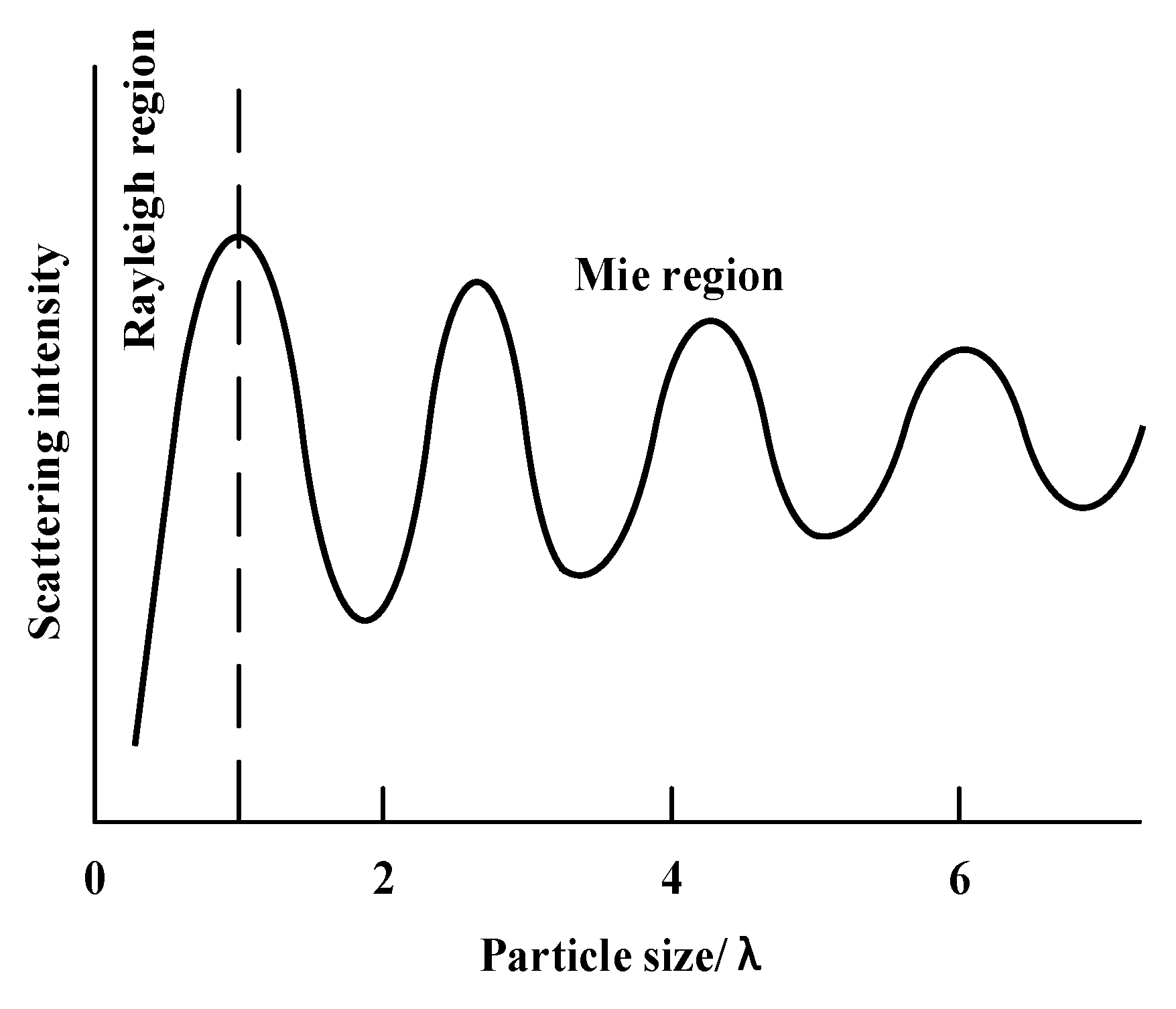
The attenuation of electromagnetic waves by bioaerosols is mainly due to the absorption and scattering effects of the biological particles in the aerosols. The types of scattering of electromagnetic waves caused by these particles are mainly Rayleigh scattering and Mie scattering. When the particle size is smaller than the incident wavelength, the scattering intensity of the smaller-scale particle is much smaller than that of the larger-scale particle; this is known as Rayleigh scattering. Mie scattering occurs when the particle size is similar to the wavelength value. If the particles have both scattering and absorbing effects on the electromagnetic waves, a better extinction performance will be obtained. Judging from the sizes of the three biological particles, the AO and AN spores have a better extinction effect in the MIR band, while the BB spores are more effective at extinction in the visible and NIR wavebands. The relationship between particle size and scattering intensity is shown in Figure 2.

2.2.2. Complex Refractive Index

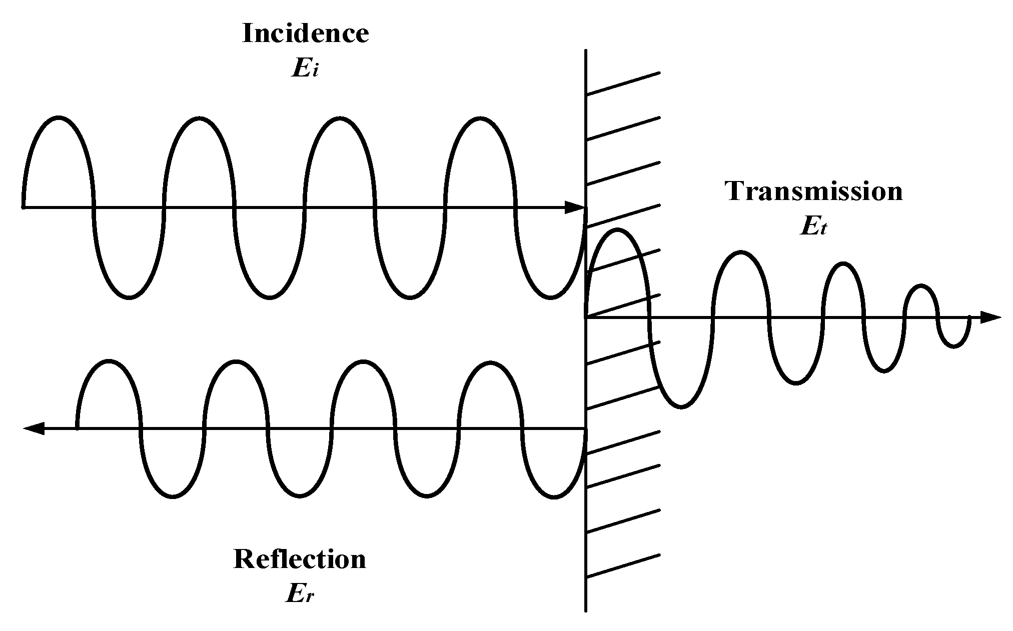
As the most dominant optical constant of an absorbing optical medium, the complex refractive index is crucial for the application of optical materials and the design of optoelectronic devices [29,30]. The reflection spectroscopy method is suitable for the measurement of the smooth surfaces of opaque bulk materials [31,32,33]. In this research, the reflectance spectrum of microbial spores was measured and then combined with the Kramers–Kronig [34] relationship to obtain the complex refractive index, which was then used as a parameter in the analysis of the optical properties of the microbial spores. As shown in Figure 3, the electromagnetic wave is reflected and refracted at the interface formed by two different materials.
We assume that the complex refractive index of a material is expressed as m= n + ik, where m is the complex refractive index, n is the real part of the complex refractive index and k is the imaginary part of the complex refractive index. Thus, according to the Fresnel formula in the case of a perpendicular incidence,
r = n + i k 1 n + i k + 1 = | r | e i θ
where θ represents the reflection phase shift and | r | represents the reflection coefficient. Then, we perform the following logarithmic operations on the Fresnel formula:
In r = In | r | i θ
according to the K-K algorithm. For a vertically incident electromagnetic wave, the reflected phase shift θ(λ) is calculated as follows [35]:
θ ( λ ) = λ π P 0 In R ( λ ) ( λ 2 λ 2 ) d λ
where P is the Cauchy principal value function, λ is the wavelength and R(λ) is the vertical reflectivity. It is known that R(λ) should be obtained over the full bandwidth to be able to calculate the complex refractive index. However, in actual experimental measurements, R(λ) can be measured only within the limited bandwidth. The constant extrapolation method and empirical formulas were used to expand the range of reflectivity. The reflection phase shift θ(λ) can be calculated using the following equation [25]:
θ ( λ ) = λ π P ( 0 λ 1 ln R ( λ ) ( λ 2 λ 2 ) d λ + λ 1 λ 2 ln R ( λ ) ( λ 2 λ 2 ) d λ + λ 2 ln R ( λ ) ( λ 2 λ 2 ) d λ )
where the real part n(λ) and the imaginary part k(λ) of the complex refractive index can be expressed as the reflectance R = | r | 2 , and the reflection phase shift θ(λ) can be calculated using the following equations [26]:
n ( λ ) = 1 R ( λ ) 1 + R ( λ ) + 2 R ( λ ) cos θ ( λ )
k ( λ ) = 2 R ( λ ) sin θ ( λ ) 1 + R ( λ ) + 2 R ( λ ) cos θ ( λ )

2.2.3. Transmittance and Mass Extinction Coefficient

Transmittance is one of the most commonly used indicators to analyze a material’s extinction performance. It is defined as the rate of change of the target radiation power before and after the release of the material; the smaller the transmittance, the better the extinction performance of the material. Transmittance T and attenuation rate E can be equated as E = 1 − T. If the target radiation power intensity in the experiment can be read directly with the power meter, then we can use the radiation power P to calculate the transmittance directly. If not, it can be transformed into other readable parameters proportional to the radiation power, such as the radiation brightness I. This is proportional to the gray value or the radiation emission degree M, which is proportional to the thermal temperature. Both the mass extinction coefficient and transmittance can measure the extinction performance of a material, but the mass extinction coefficient is more reflective of the material’s own properties and is not affected by the shape and size of the material’s particles. The mass extinction coefficient is defined as the shaded area per unit mass; the higher the mass extinction coefficient, the better the extinction performance of the material. The mass extinction coefficient can be expressed as follows:
α = In T c L
where T is the transmittance; α is the mass extinction coefficient in units of m2/g; c is the mass concentration in units of g/m2; and L is the light distance in units of m.

2.3. Methods

2.3.1. Static Testing

The investigation into the broadband extinction performance of bioaerosols was mainly based on static and dynamic testing. Static testing was conducted by measuring the spectral reflectance of pressed tablets made from the biospores, and the complex refractive indices of the spores were calculated based on the K-K relationship. We first weighed single-germplasm and compound biospores, each of which had a weight of 2.5 g. A 10,000 ppm balance was used. Then, using a HY-15 tablet press, the spores were pressed into tablets with a diameter of 4 cm and a thickness of 1.5–2.5 mm. The pressure of the tablet press was controlled at 10 MPa, and the holding time was 30 s. Some of the biological spores and pressed tablets are shown in Figure 4. The spectral reflectance of the pressed tablets in the 0.25–2.4 μm waveband was measured by applying a Hitachi U-4100 ultraviolet–near-infrared spectrophotometer, and the spectral reflectance in the 3–15 μm waveband was measured using a BRUKER TENSOR 37 infrared spectrometer with a BRUKER HYPERION microscope and a SONY SSC-DC83P lens. The instruments are shown in Figure 5. (The name of the manufacturer, city, and country from where the equipment was sourced in the paper can be seen in the supplementary material)

2.3.2. Dynamic Testing

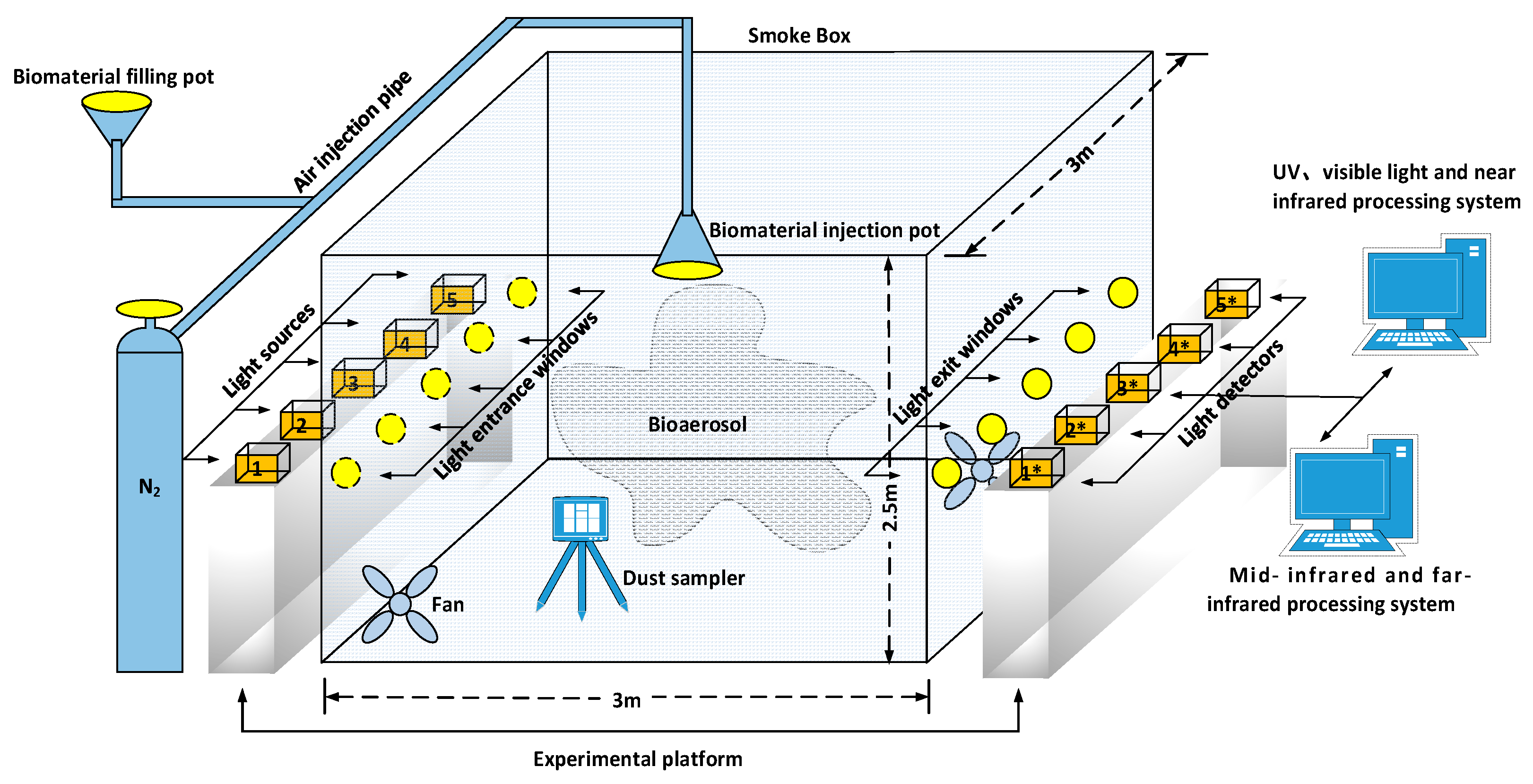
The dynamic test mainly investigated the extinction abilities of the bioaerosols in the broadband under steady atmospheric suspension, and it further analyzed the differences between the extinction performances of the single-germplasm bioaerosols and the compound bioaerosols in various wavebands. We constructed a broadband extinction performance test system using a smoke box, as shown in Figure 6. An amount of 40 g of each kind of biomaterial was weighed on an electronic balance and poured into the filling port of the smoke box for every experiment. N2 gas (10 MPa) was used to inject the biomaterial into the smoke box. Two fans were installed in the corners of the box to speed up the gas flow inside and accelerate the uniform diffusion of the bioaerosols. At the same time, a dust sampler was installed to measure the aerosol concentration inside the box. It was estimated that when the bioaerosols in the smoke box started floating evenly and steadily, the mass of the smoke floating in the box could account for approximately 60–70% of the initial smoke material. We used 70% of the initial smoke material as the actual floating mass in the process of calculating the mass extinction coefficients. A large exhaust air system was connected to the outside part of the box in order to completely drain the bioaerosol in the box after each experiment. Ultraviolet, visible, NIR, MIR and FIR light sources were placed in front of the 5 incident optical windows on the left side of the box, marked as 1, 2, 3, 4 and 5. The 5 outgoing optical windows on the right side were equipped with ultraviolet, visible, NIR, MIR and FIR detectors, marked as 1*, 2*, 3*, 4* and 5*. The light sources and detector modes with their spectral ranges are listed in Table 1.
The main steps of the broadband dynamic testing for the bioaerosols were as follows: installation and commissioning of the instruments and equipment, weighing and placement of filter membranes, weighing and loading of materials, bioaerosol release diffusion, experimental data recording and bioaerosol emission. Among these, the detectors of ultraviolet, visible and NIR bands read the power value directly, and the transmittance is calculated based on the power value before and after the bioaerosol release with the passing of time. The MIR and FIR wavebands convert the data recorded with the thermal imaging camera into grayscale image values. Figure 7 shows the variations in the mid and FIR thermal camera images before and after the bioaerosol release.

3. Results and Discussions

3.1. Analysis of Complex Refractive Index

3.1.1. Reflectivity

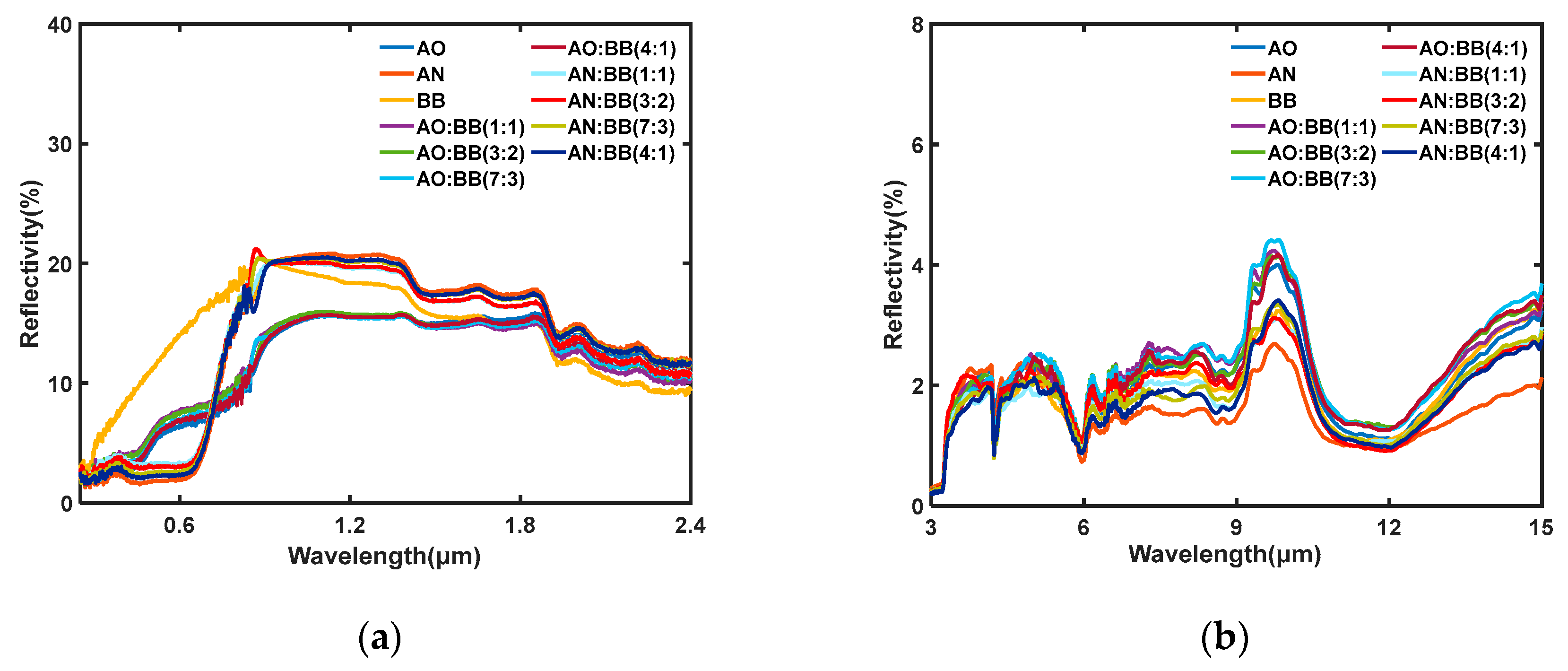
The reflectivities of single and composite biospores in the wavebands of 0.25–2.4 and 3–15 μm were obtained through spectrophotometer and infrared spectrometer experiments. Figure 8a,b shows the spectral reflectivities of the single-germplasm and compound biospores in the 0.25–2.4 and 3–15 μm wavebands, respectively. Among these, the reflectance of the BB spores is significantly greater than that of the AO and AN spores in the ultraviolet–visible range. In the 3–15 μm band, the overall reflectance trends of the single-germplasm and compound spores are close to each other. On the whole, the spectral reflectance of the biospores in the 0.25–2.4 μm band is greater than that in the 3–15 μm band and fluctuates more smoothly.

3.1.2. The Real Part n(λ)

The real part of the complex refractive indices n(λ) of the biospores in the 0.25–2.4 μm and 3–15 μm wavebands were calculated based on the spectral reflectivity combined with the K-K algorithm, as shown in Figure 9. For the absorbing medium, the real part of the complex refractive index determines the propagation velocity of light waves in the medium. In the 0.25–2.4 μm band in Figure 9a,b, the n(λ) varies mainly in the range of 1–2.5, which fluctuates significantly in the range of the 0.24–1.4 μm waveband and moderately in the range of the 1.4–2.4 μm waveband. In the 0.24–1.2 μm waveband, the n(λ) of the BB spores is significantly larger than that of the AO and AN spores, indicating that the scattering intensity of the BB spores in this band is stronger than that of the AO and AN spores. It can be further observed that the n(λ) values of the AN spores and the AN:BB compound spores have obvious trough points at 0.7 μm, indicating that the scattering intensities of these biospores are weak at this wavelength, while none of the other spores have obvious extreme points. In the 3–15 μm band, the n(λ) values of the biospores show a similar trend overall with more obvious fluctuations, with 3 peak points at 3.8 μm, 5 μm and 9.8 μm, and two trough points at 4.2 μm and 6 μm. Combining the two wavebands, the n(λ) values of the biospores are larger in the range of 1.2–1.8 μm than in the other bands.

3.1.3. The Imaginary Part k(λ)

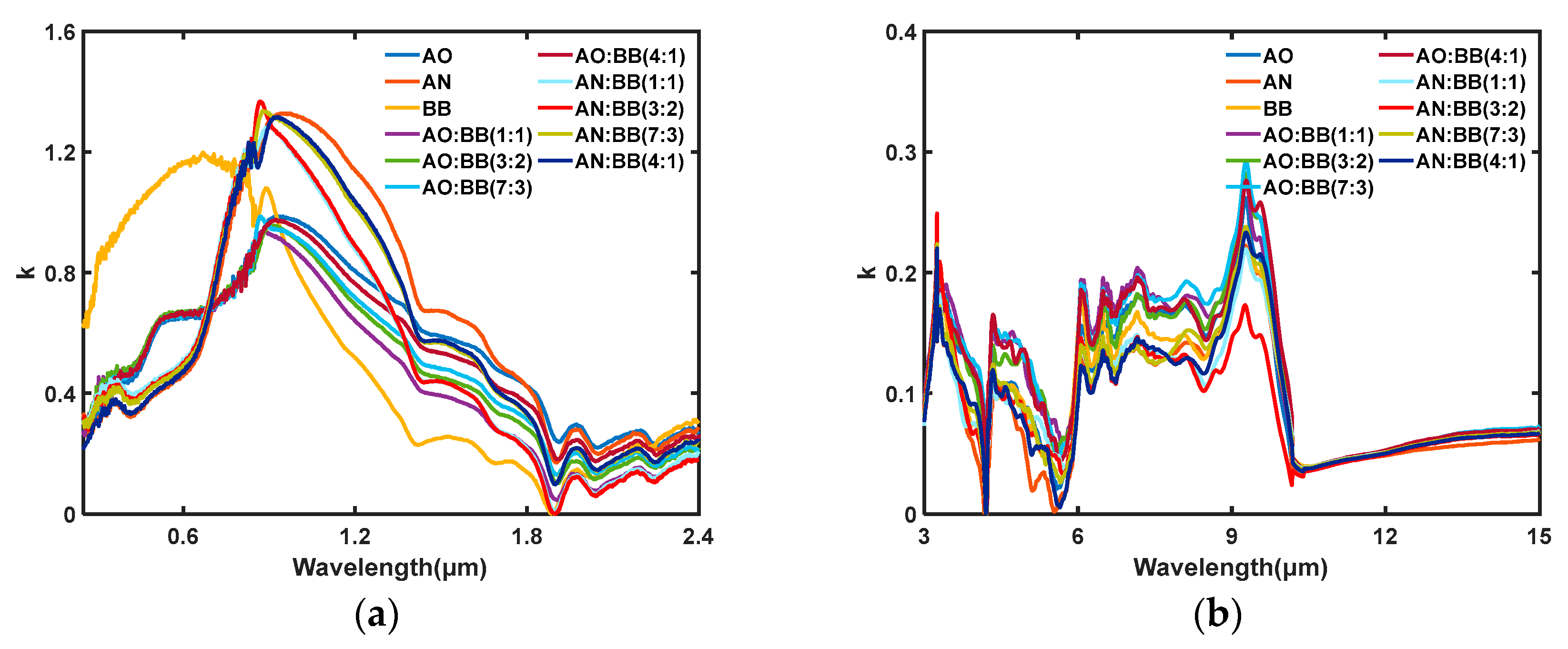
The imaginary part k(λ) of the complex refractive indices of the biospores, with wavelengths in the ranges of 0.25–2.4 μm and 3–15 μm, is shown in Figure 10. For the absorbing medium, the imaginary part of the complex refractive index determines the attenuation of the light wave propagation in the medium, as shown in Figure 10a,b. The microbial spores have similar absorption peaks, which are mainly determined by the absorption characteristics of the microbial cell components such as water, protein, lipid, nucleic acid, etc.
It is noteworthy that in terms of both the spectral reflectance and complex refractive index, the compound spores show high similarity to the AO and AN spores, which have a larger particle size but are different from the BB spores; this phenomenon did not change even when the two spores were compounded at a 1:1 equal-mass ratio. There are two reasons for this analysis: Firstly, since the particle sizes of the AO and AN spores are about twice as large as that of the BB spores, in the spectral reflectance test, the absorption and scattering of light were more affected by the spores with a larger particle size. Secondly, the mass density of the BB spores is smaller than that of the AO and AN spores; therefore, even if the composite is of an equal mass, the volume occupied by the BB spores in the compound spores is smaller than that of the AO and AN spores, resulting in their dissimilar performance in the reflectance test.

3.2. Analysis of Transmittance

3.2.1. The Transmittance of Single-Germplasm Aerosols

Firstly, we tested the extinction performance of the three single-germplasm bioaerosols in different wavebands with the use of the broadband test system. The average transmittance values and mass extinction coefficients are shown in Table 2. As can be seen in Table 2, the extinction properties of the three bioaerosols vary in different wavebands. In the ultraviolet band, the average transmittance values of the AO, AN and BB aerosols are less than 30%, and the mass extinction coefficients are 0.43, 0.36 and 0.50 m2/g, respectively. In the visible band, the 3 single-germplasm bioaerosols have excellent extinction performances, with transmittance values of 2.46, 6.49 and 2.21% and mass extinction coefficients of 1.12, 0.83 and 1.15 m2/g, respectively, which are much larger than 0.5 m2/g. In the NIR band, the transmittance value of the BB aerosol is 5.7%, and its mass extinction coefficient reaches 0.87m2/g; it is the best among the 3 single-germplasm bioaerosols as the mass extinction coefficient of both the AO and AN aerosols is about 0.5m2/g. In the MIR band, the AO aerosol has the best extinction performance, with a transmittance value and mass extinction coefficient of 6.27% and 0.84 m2/g, respectively. In the FIR band, the transmittance values of all 3 bioaerosols are more than 20%, and the mass extinction coefficients are less than 0.5 m2/g.
In summary, the order of the extinction performance is BB > AO > AN in the ultraviolet, visible and NIR wavebands, while it is AO > AN > BB in the MIR and FIR bands. Among these performance results, the extinction performance of the AO aerosol is better than that of the AN aerosol in all wavebands, while the BB aerosol has obvious advantages over the other two bioaerosols in the NIR wavelengths but lacks extinction ability in the MIR and FIR bands. The mass extinction coefficients of the AO and BB aerosols in ultraviolet and visible light differ by 0.07 and 0.03 m2/g, but in the MIR and FIR bands, they differ by 0.41 and 0.11 m2/g. Therefore, the broadband extinction performance of the AO aerosol is more balanced than that of the other two single-germplasm aerosols.

3.2.2. The Transmittance of Compound Aerosols

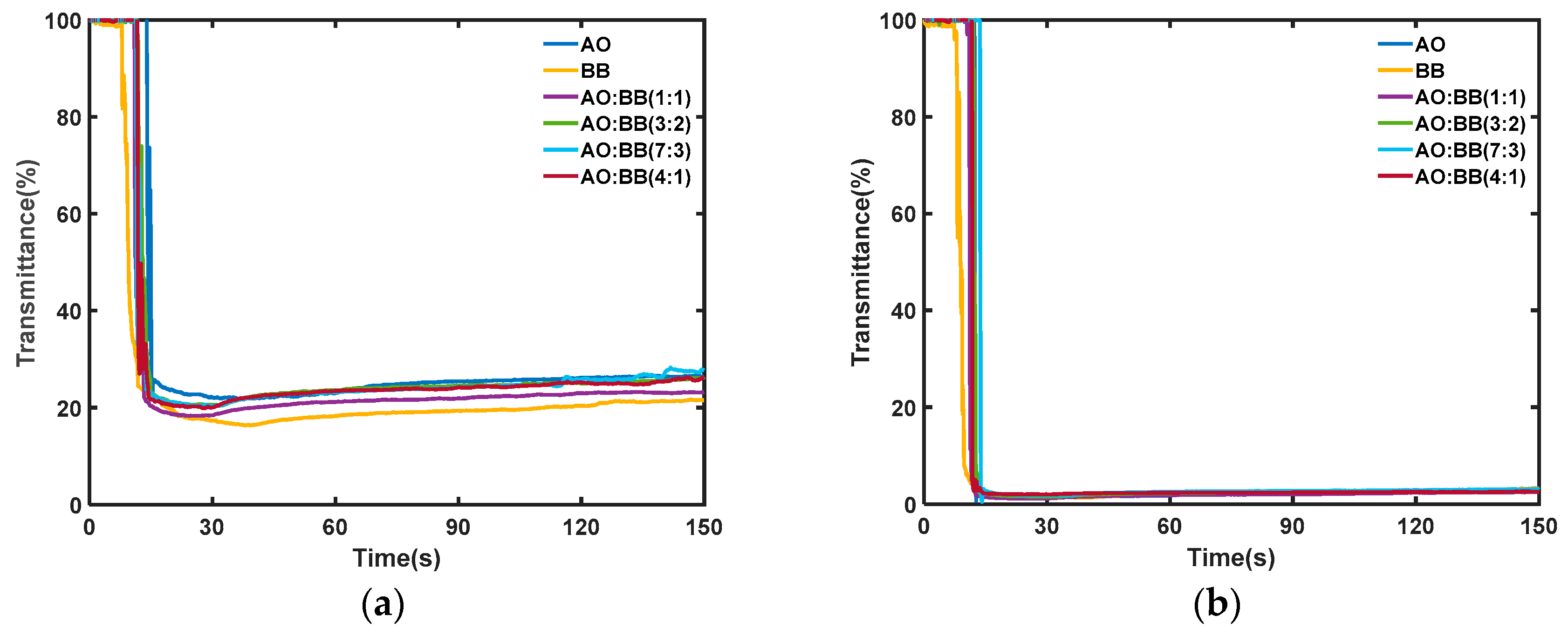
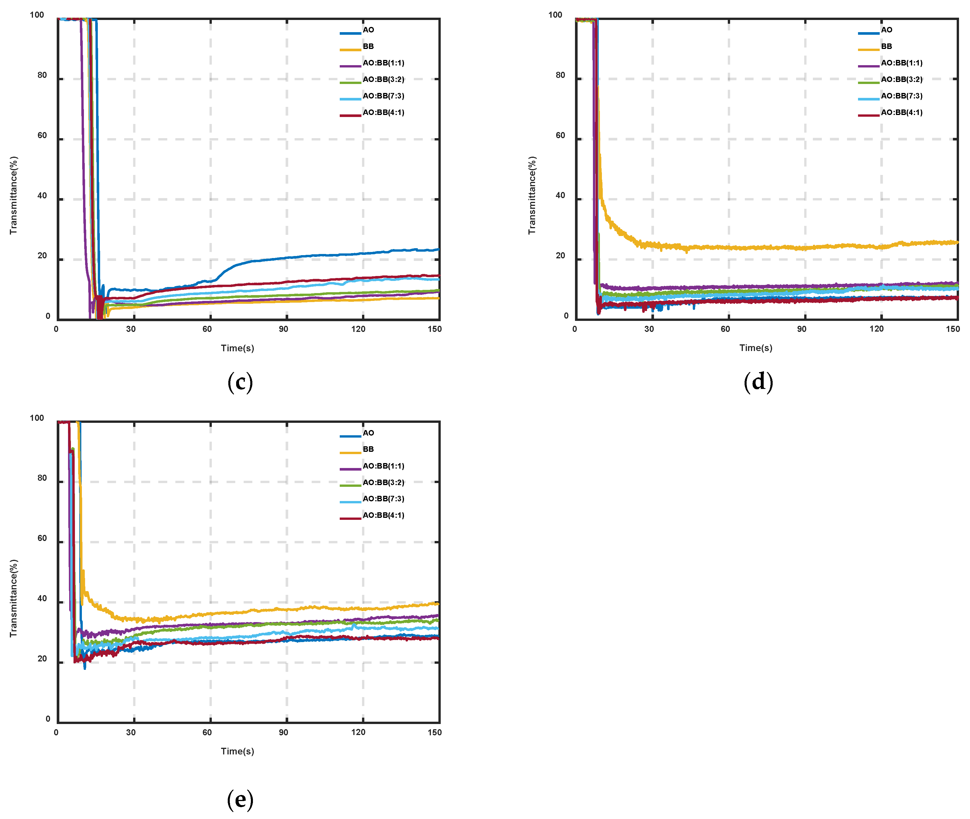
In order to further improve the broadband extinction performance of the bioaerosols, we gave priority to the compounding of the AO and BB spores and tested the broadband extinction experimental transmittance of the compound aerosols using the extinction test system. The results are shown in Figure 11. The test results show that in the ultraviolet band, the transmittance values of the AO and compound aerosols are about 20–25%, while that of the BB aerosol is less than 20%. In the visible band, both the single-germplasm and compound aerosols show an excellent extinction performance, with transmittance values of less than 5%. In the NIR band, the transmittance values of the compound aerosols are about 10%, which is lower than 15% for the AO aerosol and higher than 5% for the BB aerosol. In the MIRMIR band, the transmittance values of the AO and compound aerosols are about 10%, while that of the BB aerosol is more than 20%. In the FIR band, the transmittance of both the single and compound aerosols is more than 20%. The effective action times of the bioaerosols are all greater than two minutes, indicating that the aerosols have good atmospheric suspension properties and can be continuously extinguished.

3.3. Analysis of Mass Extinction Coefficients

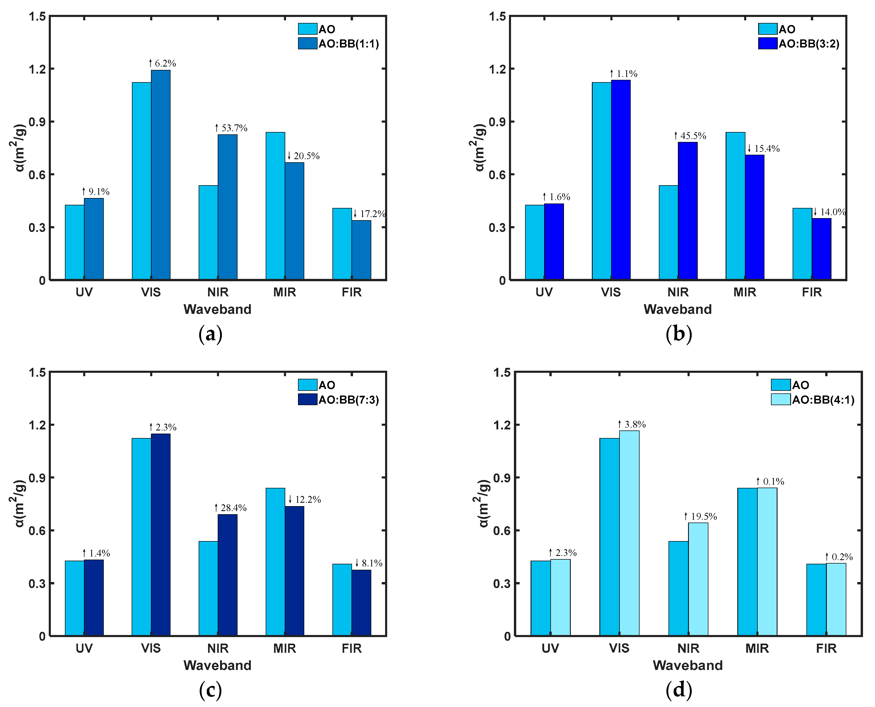
We compared the compound aerosol with the AO aerosol, which has a good extinction performance in all wavebands. Then, we calculated the mass extinction coefficients and visualized the results to quantitatively analyze the changes in the extinction abilities of the bioaerosols with different compound ratios, as shown in Figure 12. When the compounding ratio is 1:1, compared with the AO aerosols, the mass extinction coefficient of the compounded aerosols is increased by 9.1% and 6.2% in the ultraviolet and visible bands, respectively, while it is improved by 53.7% in the NIR band, which is related to the good extinction performance of the BB aerosols in the NIR band. In the MIR and FIR bands, it decreased by 20.5% and 17.2%, respectively, which is also caused by the lack of extinction ability of the BB aerosols in the mid and FIR bands. When the compounding ratio is 3:2, the mass extinction coefficient of the compounded aerosols remains basically unchanged compared with that of the AO aerosols in the ultraviolet and visible wavebands. Meanwhile, it increases by 45.5% in the NIR waveband and decreases by 15.4% and 14.0% in the MIR and FIR bands, respectively, and the magnitude of the variation is reduced compared with the 1:1 ratio. When the compounding ratio is 7:3, the magnitude of the change in the mass extinction coefficient is further reduced. When the compounding ratio is 4:1, the mass extinction coefficient of the compounded material is slightly increased in the UV, VIS, MIR and FIR bands, while it is 19.5% in the NIR band.
After adding a small amount of the BB spores to the AO spores, spores with a small particle size acted as dispersants in the compound aerosols, which enhanced the effective usage rate of the compound material in the smoke box and compensated for the loss caused by the reduction in AO spores. When sampling the concentration of aerosols in the box, it was found that when a small amount of BB spores was added to the AO spores, the effective concentration of the compound aerosols in the box was higher than that of the same mass of AO spores. On the other hand, the surfaces of the AO spores were depressed and had a high oil content and viscosity, which could easily generate electrostatic force due to friction. This promoted the adhesion of the AO and BB spores to each other in order to form aggregate particles. The aggregated particles also increased the mass extinction coefficient in the infrared band to a certain extent.

3.4. Discussions

As an important component of the atmosphere, the optical properties of bioaerosols directly affect atmospheric radiation properties. In this paper, the reflectance spectra of the biospores and the K-K algorithm were combined to calculate the complex refractive indices of the 3 biospores as well as the compound spores in the 0.25~2.4 and 3~15 μm bands, and the optical properties of the microbial spores in the 0.25~2.4 and 3~15 μm bands were analyzed with the spectra. The dynamic extinction properties of the single-species bioaerosols and compound bioaerosols were tested using a broadband test system, and based on the above experimental results and analysis, the following can be seen:
1. In the 0.25–2.4 μm band, the k(λ) values of the biospores show a trend that increases and then decreases with the wavelength, and the values vary between 0 and 1.4. The k(λ) values of the single-germplasm and compound spores have peak points at 0.85 μm and trough points at 1.9 μm, except for the BB spores, which indicates that the above spores have a strong absorption ability at 0.85 μm and a weak ability at 1.9 μm. In the 3–15 μm band, the peaks of the different types of biospores were superimposed with the peak points taken at 3.3 μm, 4.4 μm, 6.1 μm and 9.5 μm, and with the trough points taken at 4.2 μm, 5.6 μm and 10.2 μm, respectively. The trend of the complex refractive indices of compound spores is consistent with that of the single-germplasm spores with a larger particle size.
2. In this paper, we compared the mass extinction coefficients of the biospores with those of commonly used extinction materials in the MIR and FIR wavebands, and the results are shown in Table 3. In the MIR waveband, the mass extinction coefficient of AO spores is less than that of aluminum powder while being greater than those of other materials. In the FIR waveband, the extinction performance of AO spores is at an intermediate level. Furthermore, the biospores studied in this paper showed a particular extinction performance in the UV–FIR bands. Based on the two indices of transmittance and mass extinction coefficients, the bioaerosols had an excellent extinction performance in several bands and achieved the effect of the broadband extinction of a single material. Based on the good extinction performance of the biospores, the extinction performance of bioaerosols in the ultraviolet–FIR wavelength range can be effectively improved by compounding different species of biospores at specific ratios.

4. Conclusions

The artificially prepared microbial materials were proven to have good electromagnetic attenuation properties due to their special compositions and structures, which can be applied to the field of extinction. In this research, the effect of compounding factors on the extinction performance was investigated by testing the static and dynamic extinction performance of single-germplasm and compounded biospores, and the following conclusions were drawn:
1. The spectral reflectance method was used to calculate the complex refractive indices of the single-germplasm and compound spores in the 0.25–2.4 and 3–15 μm wavebands, and the optical properties of the biological spores were analyzed with the use of the complex refractive index. This showed that the absorption characteristics of the compound biospores were highly similar to those of the spores with a larger particle size in the fraction. In the 3–15 μm band, different types of bioaerosol peaks were superimposed with the peak points taken at 3.3 μm, 4.4 μm, 6.1 μm and 9.5 μm and the trough points taken at 4.2 μm, 5.6 μm and 10.2 μm.
2. Based on the results of the broadband testing, from the perspective of transmittance, the single-germplasm aerosols showed an excellent extinction performance in the visible, NIR and MIR bands, with the lowest transmittance values being 2.21, 5.70 and 6.27% and mass extinction coefficients reaching 1.15, 0.87 and 0.84 m2/g, respectively.
3. The mass extinction coefficients of the compound aerosols were compared with those of the AO aerosol in various wavelengths. At a compound ratio of 4:1, the mass extinction coefficients of the compound aerosols were improved compared with those of the AO aerosol in all wavelengths. The improvement in the NIR band was 19.5%, which showed the best broadband extinction performance.
The results of this study can provide ideas for the development and application of new extinction materials.

Supplementary Materials

The following supporting information can be downloaded at https://www.mdpi.com/article/10.3390/photonics10040357/s1, Table S1: The name of the manufacturer, city, and country from where the equipment was sourced.

Author Contributions

Conceptualization, X.C. and Y.H.; methodology, X.C. and Y.H.; validation, X.C. and X.W.; formal analysis, X.C.; investigation, X.C. and X.W.; resources, P.W.; data curation, Y.H.; writing—original draft preparation, X.C.; writing—review and editing, Y.H. and Y.G.; visualization, X.C. and X.W.; supervision, Y.H. and Y.G.; project administration, Y.H.; funding acquisition, Y.H. All authors have read and agreed to the published version of the manuscript.

Funding

This research was funded by the Research Plan Project of the National University of Defense Technology (grant no. ZK18-01-02), the National Natural Science Foundation of China (grant no. 61871389), the Postgraduate Scientific Research Innovation Project of Hunan Province (grant no. CX20220007), the National Natural Science Foundation of China (grant no. 62075241) and the Advanced Laser Technology Laboratory Foundation of Anhui Province of China (grant no. 20191003).

Institutional Review Board Statement

Not applicable.

Informed Consent Statement

Not applicable.

Data Availability Statement

Not applicable.

Conflicts of Interest

The authors declare no conflict of interest.

References

  1. Li, M.; Wang, L.; Qi, W.; Liu, Y.; Lin, J. Challenges and Perspectives for Biosensing of Bioaerosol Containing Pathogenic Microorganisms. Micromachines 2021, 12, 798. [Google Scholar] [CrossRef] [PubMed]
  2. Després, V.; Huffman, J.A.; Burrows, S.M.; Hoose, C.; Safatov, A.; Buryak, G.; Fröhlich-Nowoisky, J.; Elbert, W.; Andreae, M.; Pöschl, U.; et al. Primary biological aerosol particles in the atmosphere: A review. Tellus B Chem. Phys. Meteorol. 2012, 64, 15598. [Google Scholar] [CrossRef] [Green Version]
  3. Mirskaya, E.; Agranovski, I.E. Sources and mechanisms of bioaerosol generation in occupational environments. Crit. Rev. Microbiol. 2018, 44, 739–758. [Google Scholar] [CrossRef] [PubMed]
  4. Gormley, M.; Aspray, T.J.; Kelly, D.A. Aerosol and bioaerosol particle size and dynamics from defective sanitary plumbing systems. Indoor Air 2021, 31, 1427–1440. [Google Scholar] [CrossRef]
  5. Mbareche, H.; Morawska, L.; Duchaine, C. On the interpretation of bioaerosol exposure measurements and impacts on health. J. Air Waste Manag. Assoc. 2019, 69, 789–804. [Google Scholar] [CrossRef] [PubMed] [Green Version]
  6. Moreno, D.L.; Álvarez, A.S.; Flores, I.I.; Canche, B.C.; Pech, M.C.; Chiu, J.F.; Delgado, M.R. Early detection of the fungal banana black sigatoka pathogen pseudocercospora fijiensis by an SPR immunosensor method. Sensors 2019, 19, 465. [Google Scholar] [CrossRef] [Green Version]
  7. Jiao, K.; Sun, W.; Zhan, S.S. Sensitive detection of a plant virus by electrochemical enzyme-linked immunoassay. Fresenius J. Anal. Chem. 2000, 367, 667–671. [Google Scholar] [CrossRef]
  8. Jarocka, U.; Wasowicz, M.; Radecka, H.; Malinowski, T.; Michalczuk, L.; Radecki, J. Impedimetric immunosensor for detection of Plum Pox Virus in plant extracts. Electroanalysis 2011, 23, 2197–2204. [Google Scholar] [CrossRef]
  9. Kumar, S.; Zhu, G.; Singh, R.; Wang, Q.-L.; Zhang, B.-Y.; Cheng, S.; Liu, F.-Z.; Marques, C.; Kaushik, B.-K.; Jha, R. MoS2 Functionalized Multicore Fiber Probes for Selective Detection of Shigella Bacteria based on Localized Plasmon. J. Light. Technol. 2021, 39, 4069–4081. [Google Scholar] [CrossRef]
  10. Kaushik, S.; Tiwari, U.K.; Pal, S.S.; Sinha, R.K. Rapid detection of Escherichia coli using fiber optic surface plasmon resonance immunosensor based on biofunctionalized Molybdenum disulfide(MoS2) nanosheets. Biosens. Bioelectron. 2019, 126, 501–509. [Google Scholar] [CrossRef]
  11. Elder, A.; Paterson, C. Sharps injuries in UK health care: A review of injury rates, viral transmission and potential efficacy of safety devices. Occup. Med. 2006, 56, 566–574. [Google Scholar] [CrossRef] [PubMed] [Green Version]
  12. Kenarkoohi, A.; Noorimotlagh, Z.; Falahi, S.; Amarloei, A.; Mirzaee, S.A.; Pakzad, I.; Bastani, E. Hospital indoor air quality monitoring for the detection of SARS-CoV-2 (COVID-19) virus. Sci. Total Environ. 2020, 748, 141324. [Google Scholar] [CrossRef] [PubMed]
  13. Li, X.; Jiang, J.; Wang, D.; Deng, J.; He, K.; Hao, J. Transmission of Coronavirus via Aerosols and Influence of Environmental Conditions on Its Transmission. Environ. Sci. 2021, 42, 3091–3098. [Google Scholar]
  14. Wilson, N.M.; Norton, A.; Young, F.P.; Collins, D.W. Airborne transmission of severe acute respiratory syndrome coronavirus-2 to healthcare workers: A narrative review. Anaesthesia 2020, 75, 1086–1095. [Google Scholar] [CrossRef] [PubMed]
  15. Li, W.; Xu, T.; Tang, H.; Jiang, J.; Fu, Y. Technologies for microbial aerosol sampling and identification: A review and current perspective. Acta Microbiol. Sin. 2022, 62, 1345–1361. [Google Scholar]
  16. Kaliszewski, M.; Trafny, E.A.; Lewandowski, R.; Włodarski, M.; Bombalska, A.; Kopczyński, K.; Antos-Bielska, M.; Szpakowska, M.; Młyńczak, J.; Mularczyk-Oliwa, M.; et al. A new approach to UVAPS data analysis towards detection of biological aerosol. J. Aerosol Sci. 2013, 58, 148–157. [Google Scholar] [CrossRef]
  17. Taketani, F.; Kanaya, Y.; Nakamura, T.; Koizumi, K.; Moteki, N.; Takegawa, N. Measurement of fluorescence spectra from atmospheric single submicron particle using laser-induced fluorescence technique. J. Aerosol Sci. 2013, 58, 1–8. [Google Scholar] [CrossRef]
  18. Agustin, I.; Avishai, B.; Richader, G. Estimating the limit of bio-aerosol detection with passive infrared spectroscopy. Int. J. High Speed Electron. Syst. 2008, 18, 701–711. [Google Scholar]
  19. Feng, M.-C.; Xu, L.; Gao, M.-G.; Jiao, Y.; Wei, X.-L.; Jin, L.; Cheng, S.-Y.; Li, X.-X.; Feng, S.-X. Optical properties research of Bacillus subtilis spores by Fourier transform infrared spectroscopy. Spectrosc. Spectr. Anaysis 2012, 32, 3193–3196. [Google Scholar]
  20. Tiange, L.; Wei, X.; Yonghua, F.; Dacheng, L.; Yueming, Y. Study on Passive Detection of Biological Aerosol with Fourier-Transform Infrared Spectroscopic Technique. Acta Opt. Sin. 2010, 30, 1656–1661. [Google Scholar] [CrossRef]
  21. Yabushita, S.; Wada, K.; Takai, T.; Inagaki, T.; Young, D.; Arakawa, E.T. A spectroscopic study of the microorganism model of interstellar grains. Astrophys. Space Sci. 1986, 124, 377–388. [Google Scholar] [CrossRef]
  22. Gurton, K.P.; Ligon, D.; Kvavilashvili, R. Measured Infrared Spectral Extinction for Aerosolized Bacillus subtilis var. niger Endospores from 3 to 13 mum. Appl. Opt. 2001, 40, 4443–4448. [Google Scholar] [CrossRef] [PubMed]
  23. Sun, D.-J.; Hu, Y.-H.; Wang, Y.; Li, L.; Li, L. Sub-microstructures’ Influences on Cell′s Scattering Prosperities. Acta Photonica Sin. 2013, 42, 710–714. [Google Scholar]
  24. Li, L.; Hu, Y.; Gu, Y.; Chen, W. Infrared extinction performance of Aspergillus niger spores. Infrared Laser Eng. 2014, 43, 2175–2179. [Google Scholar]
  25. Li, L.; Hu, Y.-H.; Gu, Y.-L.; Chen, W.; Zhao, Y.-Z.; Chen, S.-J. Measurement and analysis on complex refraction indices of pear pollen in infrared band. Spectrosc. Spectr. Anaysis 2015, 35, 89–92. [Google Scholar]
  26. Zhao, X.; Hu, Y.; Gu, Y.; Chen, X.; Wang, X.; Wang, P.; Dong, X. Analysis of optical properties of bio-smoke materials in the 0.25–14 μm band. Chin. Phys. B 2019, 28, 034201. [Google Scholar] [CrossRef]
  27. Lin, N. Survey of foreign red phosphorus smoke agent. Initiat. Pyrotech. 1996, 1, 42–46. [Google Scholar]
  28. Zhou, M.-S.; Xu, M. Numerical calculation of 3 mm wave extinction for expanded. Acta Phys. Sin. 2013, 9, 378–384. [Google Scholar]
  29. Qi, H.; Zhang, X.; Jiang, M.; Yang, L.; Li, D. Optical constants of polyacrylamide solution in infrared spectral region. Optik 2017, 146, 27–32. [Google Scholar] [CrossRef]
  30. Wang, L.; Liu, H.; Li, S.; Jiang, C.; Ji, Y.; Chen, D. Study on the composite dispersion model of optical constants of metal-oxide films in the range from ultraviolet to near infrared. Optik 2018, 168, 892–900. [Google Scholar] [CrossRef]
  31. Dou, Z.-W.; Li, X.-X.; Zhao, J.-J. Complex refraction indices of expanded graphite deduced from its reflection spectra in infrared band. Acta Armamentarii 2011, 32, 498–502. [Google Scholar]
  32. Wang, M.-J.; Wu, Z.-S.; Li, Y.-L.; Xiang, N.-J. Numerical inversion of the optical characteristics of film material based on Kramers-Kronig relations. Infrared Laser Eng. 2010, 39, 120–123. [Google Scholar]
  33. Zamiri, R.; Rebelo, A.; Zamiri, G.; Adnani, A.; Kuashal, A.; Belsley, M.S.; Ferreira, J.M.F. Far-infrared optical constants of ZnO and ZnO/Ag nanostructures. RSC Adv. 2014, 4, 20902–20908. [Google Scholar] [CrossRef]
  34. Barber, P.W. Absorption and scattering of light by small particles. J. Colloid Interface Sci. 1984, 98, 290–291. [Google Scholar] [CrossRef]
  35. Segal-Rosenheimer, M.; Linker, R. Impact of the non-measured infrared spectral range of the imaginary refractive index on the derivation of the real refractive index using the Kramers–Kronig transform. J. Quant. Spectrosc. Radiat. Transf. 2009, 110, 1147–1161. [Google Scholar] [CrossRef]
Figure 1. SEM images of microbial spores (10,000× magnification, scale 5μm). (a) AO spores; (b) AN spores; and (c) BB spores.
Photonics 10 00357 g001
Figure 2. The relationship between particle size and scattering intensity.
Photonics 10 00357 g002
Figure 3. Reflection and refraction of perpendicularly incident light on a plane.
Photonics 10 00357 g003
Figure 4. Biological spores and pressed tablets.(The tablets are stored in sealed bags)
Photonics 10 00357 g004
Figure 5. (a) U-4100 spectrophotometer; (b) BRUKER TENSOR 37 infrared spectrometer.
Photonics 10 00357 g005
Figure 6. Broadband extinction performance test system for bioaerosols.
Photonics 10 00357 g006
Figure 7. MIR and FIR thermal images before and after bioaerosol release. (a) MIR thermal images before and after bioaerosol release; (b) MIR thermal images before and after bioaerosol release.
Photonics 10 00357 g007
Figure 8. Spectral reflectivities of single-germplasm biospores and compound bioaerosols. (a) Reflectivities in the 0.25–2.4 μm wavelength range. (b) Reflectivities in the 3–14 μm wavelength range.
Photonics 10 00357 g008
Figure 9. The real part of the complex refractive indices n(λ) of single-germplasm biospores and compound bioaerosols. (a) n(λ) values in the 0.25–2.4 μm wavelength range. (b) n(λ) values in the 3–14 μm wavelength range.
Photonics 10 00357 g009
Figure 10. The imaginary part of the complex refractive indices k(λ) of single-germplasm biospores and compound bioaerosols. (a) k(λ) values in the 0.25–2.4 μm wavelength range. (b) k(λ) values in the 3–14 μm wavelength range.
Photonics 10 00357 g010
Figure 11. The broadband experimental transmittance of compound bioaerosols. (a) UV band. (b) Vis band. (c) NIR band. (d) MIR band. (e) FIR band.
Photonics 10 00357 g011aPhotonics 10 00357 g011b
Figure 12. Comparison of the mass extinction coefficients of different ratios of compound aerosols with the AO aerosol. (a) 1:1 compounding; (b) 3:2 compounding; (c) 7:3 compounding; and (d) 4:1 compounding.
Photonics 10 00357 g012
Table 1. Light sources and detectors in the broadband extinction performance test system.
BandLight SourceDetector
Light Source ModelEmission Spectral RangeDetector ModelResponse Spectral Range
UVNBeT Merc-500
mercury lamp
180~500 nmNewport 843-R
(Probe model 818UV)
200~1100 nm
VISMBL-FN-473
BK 11367
473 nmOPHIR Starlite
(Probe model PD300)
350~1100 nm
NIRMIL-H-1064
BH 81394
1.064 μmOPHIR Starlite
(Probe model PD300)
350~1100 nm
MIRFuyuan black body
HFX-300A
Temperature
5~400 °C
FLIR SC70003.8~5.1 μm
FIRFuyuan black body
HFX-300A
Temperature
5~400 °C
VARIO CAM HD8~14 μm
Table 2. The average transmittance values and mass extinction coefficients of the three single-germplasm bioaerosols.
TypeUVVISNIRMIRFIR
τ (%)α (m2/g)τ (%)α (m2/g)τ (%)α (m2/g)τ (%)α (m2/g)τ (%)α (m2/g)
AO24.510.432.461.1216.970.536.270.8426.010.41
AN29.950.366.490.8320.070.4811.330.6628.790.38
BB19.230.502.211.155.700.8724.510.4336.910.30
Table 3. Comparison of mass extinction coefficients.
TypeAOGraphite PowderSilica PowderAluminum PowderCopper PowderIron PowderRed Phosphorus Powder
α (m2/g)MIR0.840.800.171.030.390.390.66
FIR0.410.530.490.790.390.320.14
Disclaimer/Publisher’s Note: The statements, opinions and data contained in all publications are solely those of the individual author(s) and contributor(s) and not of MDPI and/or the editor(s). MDPI and/or the editor(s) disclaim responsibility for any injury to people or property resulting from any ideas, methods, instructions or products referred to in the content.