Article

Preparation and Antioxidant Activities of Phenylethanoids from Dracocephalum heterophyllum

1
Center for Mitochondria and Healthy Aging, College of Life Sciences, Yantai University, Yantai 264005, China
2
Key Laboratory of Tibetan Medicine Research, Chinese Academy of Sciences, Xining 810008, China
3
Qinghai Provincial Key Laboratory of Tibetan Medicine Research, Xining 810008, China
4
College of Pharmacy, Qinghai Nationalities University, Xining 810007, China
5
Sinochem Lantian Trading Co., Ltd., Hangzhou 310051, China
*
Authors to whom correspondence should be addressed.
Separations 2022, 9(5), 111; https://doi.org/10.3390/separations9050111
Submission received: 10 April 2022 / Revised: 26 April 2022 / Accepted: 27 April 2022 / Published: 28 April 2022
(This article belongs to the Section Analysis of Food and Beverages)

Abstract

:
The health benefits of Dracocephalum heterophyllum are widely reported in traditional Tibetan medicines, but the reported chemical composition is limited, probably due to difficulties in separating and purifying compounds. In this study, antioxidative phenylethanoids were isolated from an extract of Dracocephalum heterophyllum using medium- and high-pressure liquid chromatography, coupled with on-line HPLC–1,1-diphenyl-2-picrylhydrazyl recognition. Firstly, crude samples (1.3 kg) of Dracocephalum heterophyllum were pretreated via silica gel medium-pressure liquid chromatography to yield 994.0 g of Fr2, of which 10.8 g was then pretreated via MCI GEL®CHP20P medium-pressure liquid chromatography. The resulting Fr23 and Fr25 were further separated and purified using high-pressure liquid chromatography, and yielded 8.08 mg of Fr2391, 9.76 mg of Fr2551, 16.09 mg of Fr2581, and 8.75 mg of Fr2582. Furthermore, analysis of the purity and structures of the phenylethanoids suggested that Fr2391, Fr2551, Fr2581, and Fr2582 corresponded to decaffeoylverbascoside, rosmarinic acid, acteoside, and 2′-O-acetylplantamajoside, respectively, with all being over 95% pure. Finally, the antioxidant potential of the compounds was explored based on their ability to scavenge 1,1-diphenyl-2-picrylhydrazine, as well as through molecular docking of proteins related to antioxidant pathways. Altogether, our findings revealed that the proposed method is promising for separating pure antioxidative phenylethanoids from other natural compounds.

Graphical Abstract

1. Introduction

In aerobic organisms, free radicals are generated during mitochondrial production of adenosine triphosphate and, in moderate amounts, these radicals are useful for cell signaling transduction as well as immune activity [1]. However, when produced excessively, they can lead to oxidative stress, which can be responsible not only for cell damage, but also for neurodegenerative and cardiovascular diseases, aging, inflammation, or even cancer [2,3,4,5]. In this context, exogenous scavengers can be useful for mitigating the negative impacts of excessive free radicals [6,7], but due to the potential toxic effects of synthetic antioxidants, many natural products are being considered for this purpose [8]. The in vitro antioxidant potential of natural products therefore prompted this study’s focus on the isolation and identification of additional antioxidative compounds.
Dracocephalum heterophyllum (D. heterophyllum), from the family Lamiaceae, is a perennial aromatic herb that grows on sandy, gravelly slopes as well as the alpine meadows of high mountains [9,10]. Being highly popular in traditional Tibetan medicine (TTM), this herb has been used for treating hypertension, jaundice, coughs, and mouth ulcers [11,12]. Until now, several compounds—including flavonoids, phenylpropanoids, terpenes, steroids, and alkaloids—had been isolated from D. heterophyllum [13,14,15], but recent studies indicated that its extract could also inhibit hepatitis while displaying antiviral and antioxidant activities [16,17]. For instance, in studying the in vitro antioxidant potential of D. heterophyllum extracts, Shi et al. found that ethyl-acetate-based extracts showed potent free radical scavenging activity [18]. As a result, it would be worth further exploring the antioxidant active substances in D. heterophyllum.
Traditionally, before testing antioxidant activities, antioxidant compounds are purified from natural products using separation techniques based on open-column chromatography. However, in addition to the need for specialized skills, this approach is also labor-intensive and time-consuming [19]. Therefore, it has become necessary for rapid, convenient, and efficient methods to be developed so as to successfully isolate and identify antioxidants from complex natural products, with screening methods such as the on-line HPLC-1,1-diphenyl-2-picrylhydrazyl (HPLC–DPPH) system being recognized as both effective and rapid for this purpose [20,21]. This system makes use of HPLC-based separation of compounds, and is followed by derivatization using DPPH prepared in ethanol. Eventually, when reading absorbance values at 517 nm, lower values are indicative of antioxidant activity. The efficiency of this approach is already well established, especially for identifying antioxidant compounds from different sources of natural products.
With preparative HPLC being known for its excellent performance and reproducibility, it can be a method of choice for isolating specific components from mixtures. In addition, the separation process can also be automated, while offering the possibility to detect compounds in real time [22,23,24]. However, one major drawback of preparative HPLC is that the stationary phase can be easily contaminated. Consequently, instead of being injected directly, crude extracts need to be pretreated for removing non-target ingredients and enriching the target compounds before being injected into the HPLC system. Medium-pressure liquid chromatography is commonly used for this purpose, with pretreatment performed using different stationary phases, such as silica gel, gel polyamide, or MCI GEL®CHP20P [25,26]. In this study, selecting a suitable sample pretreatment material was important for enriching target compounds from D. heterophyllum, as well as for subsequent purification.
In recent years, the antioxidant capacity of natural compounds was measured by 1,1-diphenyl-2-picrylhydrazyl (DPPH) assays [27], which are based on the fact that when a DPPH radical solution is mixed with antioxidant molecules, it produces a reduced form that loses its violet color [28]. Although highly popular and convenient, this assay still remains limited, as it does not use physiological radicals. The nuclear factor erythroid 2-related factor 2 (Nrf2)/heme oxygenase 1 (HO-1) is an anti-injury defense mechanism against external stimuli that has developed during the evolution of organisms, and it also represents the main signaling pathway for antioxidative stress [29]. In mammals, Nrf2 is a transcription factor that regulates phase II detoxification responses and protective antioxidant levels, while maintaining a balance in cellular redox reactions. HO-1, an inducible 32-kDa protein, is among those genes regulated by Nrf2, and it can be upregulated after stimulation by cytokines, growth factors, heavy metals, nitric oxide, and heme. This protein can also exert cytoprotective effects, while being involved in the development of oxidation-related diseases [30]. Therefore, the antioxidant activity of antioxidants can be initially explored through DPPH scavenging experiments and molecular docking with Nrf2/HO-1-pathway-related proteins and factors.
Previous work performed by the authors of this paper involved the targeted isolation of antioxidative compounds from D. heterophyllum using liquid–liquid extraction, middle chromatogram isolated gel open-column chromatography, reversed-phase liquid chromatography, and hydrophilic interaction chromatography, along with an on-line high-performance liquid chromatography-1,1-diphenyl-2-picrylhydrazyl (HPLC–DPPH)-based recognition to isolate eight antioxidative compounds (caffeoyl-β-D-glucopyranoside, ferruginoside B, verbascoside, 2′-O-acetylplantamajoside, sibiricin A, luteolin, rosmarinic acid, and methyl rosmarinate) [31]. However, this work applies visible chromatographic separation techniques, in combination with on-line HPLC-DPPH recognition, to aid in the comprehensive and efficient exploration of powerful antioxidants from D. heterophyllum. The overall separation steps can be seen in Figure S1 of the Supplementary Materials. Medium-pressure liquid chromatography was subsequently used to enrich the antioxidative peak fractions and remove non-antioxidant ones. This was followed by the separation and purification of antioxidative compounds from peak fractions using high-pressure liquid chromatography, before eventually determining the antioxidant capacity of isolated antioxidants through DPPH scavenging assays as well as molecular docking. The activity-directed recognition and full-range chromatographic separation techniques reported in this work would very likely aid in the similar recognition and separation of antioxidants from other sources of natural products.

2. Materials and Methods

2.1. Instrumentation and Reagents

For preparative liquid chromatography (Hanbon Science & Technology Co., Huaian, China), a compatible UV–Vis detector (NU3000), an LC workstation, two prep-HPLC pumps (NP7000), and a 5 mL manual injector were used, with the on-line HPLC-DPPH screening subsequently performed using Essentia LC-16 (Shimadzu Instruments, Shanghai, China) and LC-10AD instruments (Shimadzu Instruments, Kyoto, Japan). Each of the two HPLCs included two binary gradient pumps, the LC workstation, the UV–Vis detector, a column thermostat, and an autosampler, and both HPLCs were then connected to a triple valve coupled with a polyether ether ketone reaction coil (18.0 m × 0.25 mm i.d.). HPLC analysis was performed on the LC-16, while the DPPH screening chromatogram was acquired from the LC-10AD. In addition, ESI-MS analysis as well as both 1H and 13C NMR were performed using a Waters QDa ESI mass spectrometer (Waters Instruments Co., Milford, Massachusetts, USA) and a 600 MHz Bruker Avance (Bruker Instruments Co., Karlsruhe, Baden-Württemberg, Germany), with MeOH-d4 used as a solvent for the NMR. Eventually, a ReadMax 1900 microplate reader (Flash, Shanghai, China) was used to read UV absorbance values.
The silica (100–200 mesh) required for the medium-pressure column (49 × 460 mm) was purchased from Qingdao Ocean Chemical Corporation (Shandong, China), while the MCI GEL®CHP20P (120 μm) separation material was obtained from Mitsubishi Chemical Corporation (Tokyo, Japan). Preparative Click XIon (20 × 250 mm, 5 μm) and Click XIon analytical (4.6 × 250 mm, 5 μm) columns were provided by ACCHROM Corporation (Beijing, China). Two Kromasil 100-5 Phenyl columns (4.6 × 250 mm, 5 μm and 20 × 250 mm, 5 μm) and the analytical column ReproSil-Pur C18 AQ (4.6 × 250 mm, 5 μm) were purchased from Nouryon Kromasil Corporation (Bohus, Sweden) and Maisch Corporation (Munich, Germany), respectively.
Ethanol, methanol (CH3OH), dichloromethane (CH2Cl2), and acetonitrile (ACN) of analytical grade, along with HPLC-grade ACN and ethanol, were purchased from Kelon Chemical Reagent Factory (Chengdu, China). DPPH was obtained from Sigma-Aldrich (Steinheim, Germany), and a water purifier from Moore (Chongqing, China) was used for preparing HPLC-grade H2O.

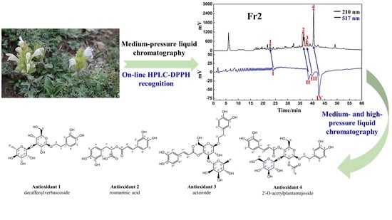
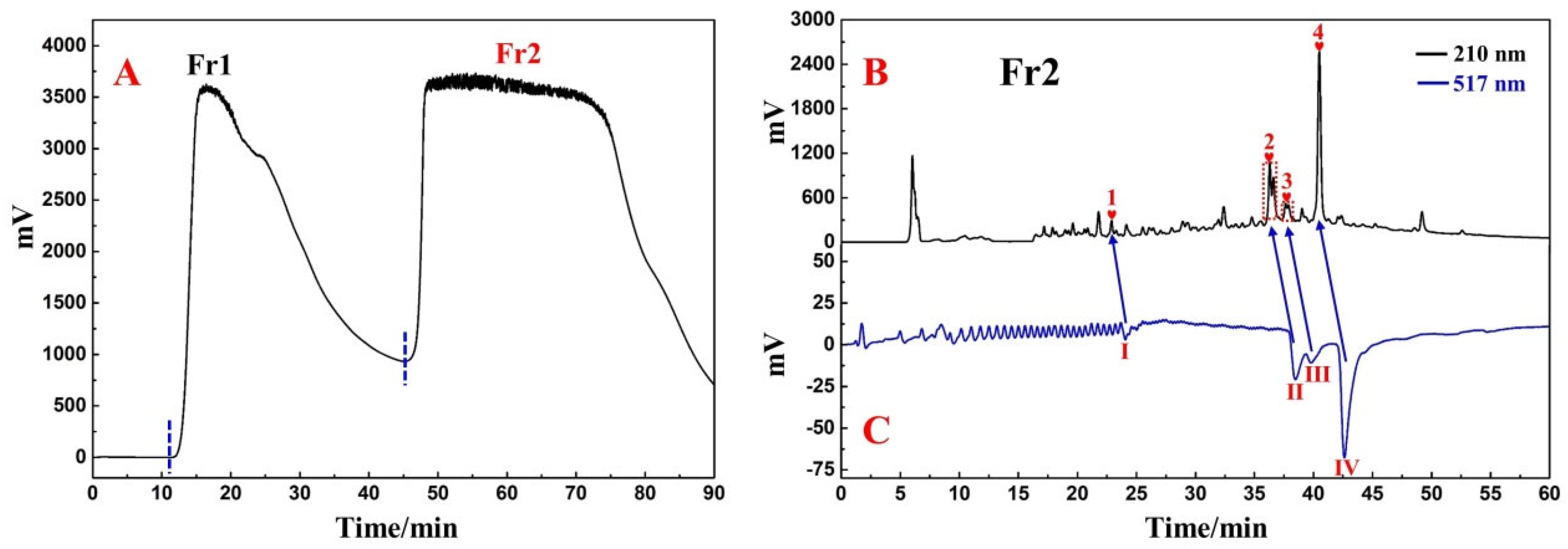
2.2. Plant Sample Preparation and Pretreatment via Medium-Pressure Liquid Chromatography

Whole D. heterophyllum plants were collected from the North Mountain in Huzhu, Qinghai, prior to validation by Prof. Lijuan Mei from the Northwest Institute of Plateau Biology, and subsequent storage of a sample (nwipb-2016-10-10) in the Qinghai–Tibetan Plateau Museum of Biology. After being dried in the shade to less than 2% moisture content, collected D. heterophyllum herbs were ground to a powdered form. Ethanolic extraction using 95% v/v ethanol was then performed three times with 10.0 kg of sample, with each extraction carried out for 12 h with 80.0 L of solvent. After collecting and filtering the resulting 240.0 L of solution, the volume was reduced to 5.0 L at 40 °C in a rotary evaporator. This concentrated solution was subsequently mixed with 1.5 kg of amorphous silica gel and, after being dried in an oven at 40 °C, the final dried silica gel mixture (2.8 kg) was prepared for silica gel medium-pressure liquid chromatography, with separation achieved using CH3OH and CH2Cl2 as the mobile phase. For this purpose, the linear elution gradients consisted of 0% CH3OH at 0–30 min, 0–100% CH3OH at 30–60 min, and 100% CH3OH at 60–90 min. Throughout the process, a constant flow rate of 57.0 mL/min was maintained, and a single loading of about 65 g was applied. In the case of the chromatogram recorded at 210 nm, the chromatographic peaks appearing in order were recorded as Fr1 and Fr2. After repeating the separation 43 times, 944.0 g of the Fr2 fraction was obtained, of which 10.8 g was used for subsequent separation.
To 500.0 mL of methanol, 10.8 g of Fr2 was added, before mixing with 10.0 g of amorphous silica gel. The mixture was allowed to dry in an oven at 40 °C, after which 20.8 g of the dried sample was prepared for MCI GEL®CHP20P medium-pressure chromatography (column size 49 × 460 mm), with elution performed using CH3OH/H2O as the mobile phase. In this case, the elution was carried out as follows: 0–120 min, 0–100% CH3OH; 120–150 min, 100% CH3OH. As before, a constant flow rate of 57.0 mL/min was maintained while absorbance was measured at 210 nm. In addition, the same naming rules were applied for recording the names of the fractions, with the peaks recorded as Fr21-Fr25 depending on the order in which they appeared on the chromatogram (e.g., Fr23 and Fr25 represented the third and fifth peaks eluted from Fr2, respectively). Subsequent collection and concentration of Fr23 and Fr25 yielded 331.0 mg and 536.0 mg of each fraction, respectively. Fr23 (331.0 mg) was then dissolved with 2.0 mL of methanol and, after filtration through a 0.45 μm membrane, an Fr23 sample solution with an approximate concentration of 165.5 mg/mL was obtained. This was repeated for Fr25 (536.0 mg) to obtain a corresponding Fr25 sample solution (about 268.0 mg/mL), with both fractions subsequently used for additional purification and to recognize antioxidative peaks.

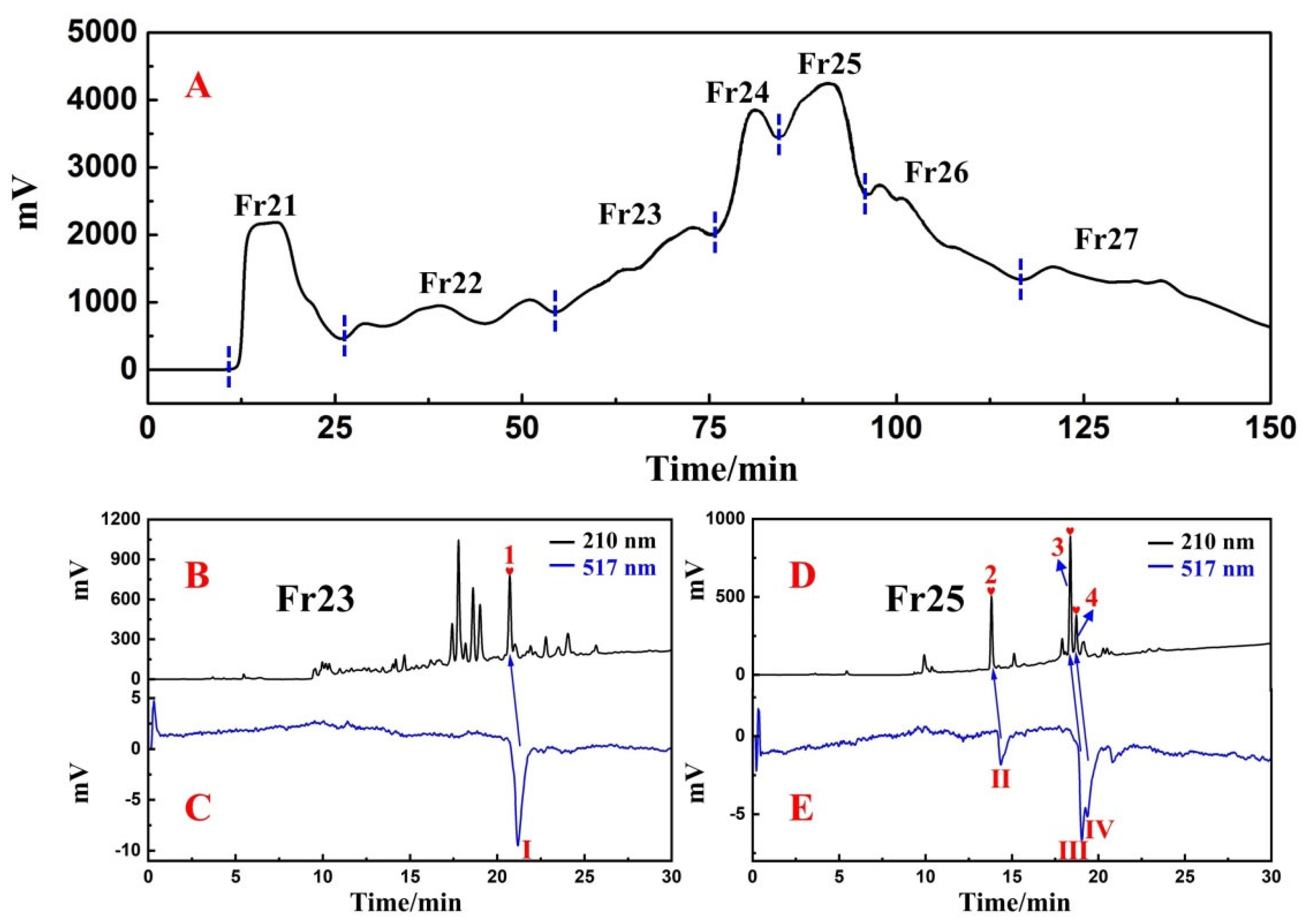
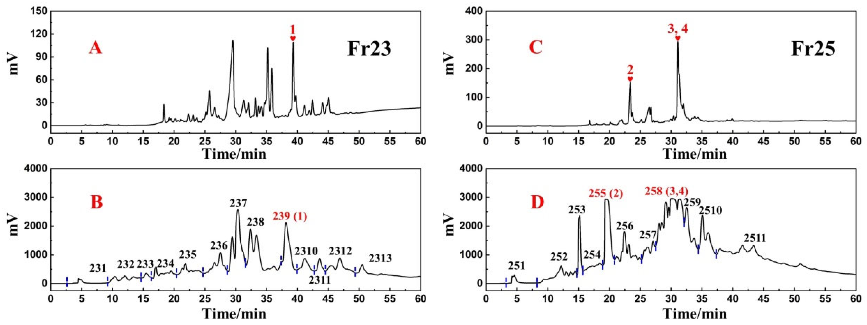
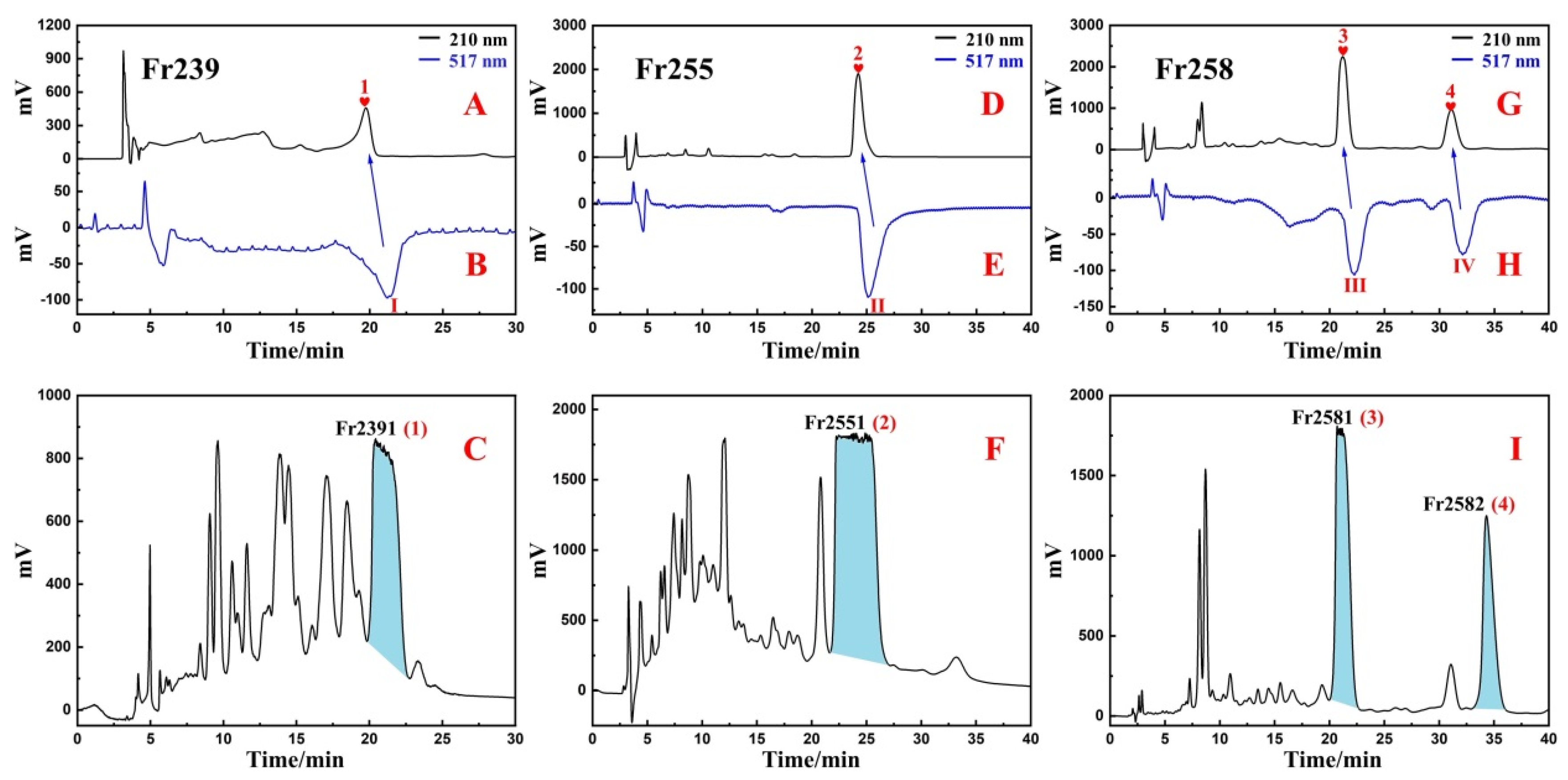
2.3. High-Pressure Liquid Chromatography Separation and Purification of Antioxidants from Fr23 and Fr25

Both Fr23 and Fr25 were further separated using a Click XIon (20 × 250 mm, 5 μm) preparative column, with trifluoroacetic acid in water (0.1% v/v) and ACN used as mobile phases A and B, respectively. In this case, 19.0 mL/min was selected as the flow rate, chromatograms were acquired at 210 nm, and the gradient elution step was performed with 100–60% B for 0–60 min. This separation process eventually yielded 50.63 mg of Fr239, 39.77 mg of Fr255, and 159.60 mg of Fr258. Dissolving these fractions in methanol and subsequent filtration through a 0.45 μm membrane resulted in the following sample solutions: Fr239 with an approximate concentration of 50.6 mg/mL, Fr255 with an approximate concentration of 39.8 mg/mL, and about 79.8 mg/mL of Fr258.
Additional purification of these three fractions was performed on a Kromasil 100-5 Phenyl (20 × 250 mm, 5 μm) preparative column under similar conditions as before (i.e., mobile phases A and B, flow rate and wavelength for acquiring chromatograms). For Fr239, the procedure for isocratic elution was performed for 30 min with 5% B, while for Fr255 and Fr258, it was carried out with 20% B for 40 min and 17% B for 40 min, respectively. Finally, after purification, 8.08 mg of Fr2391, 9.76 mg of Fr2551, 16.09 mg of Fr2581, and 8.75 mg of Fr2582 were obtained.

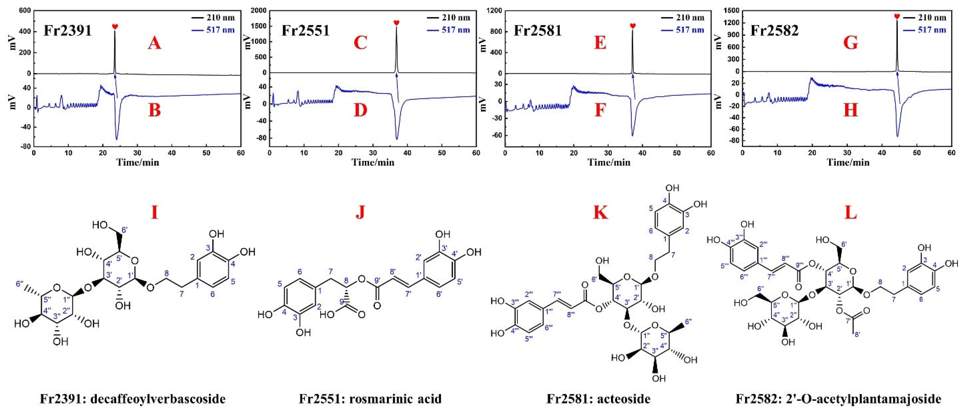
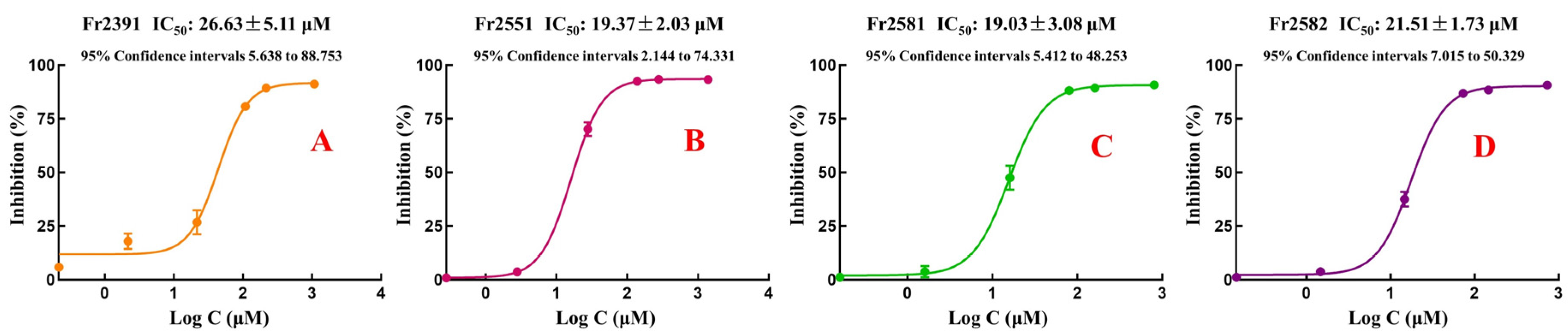
2.4. Purity and Antioxidant Activities of Fr2391, Fr2551, Fr2581, and Fr2582

Evaluation of the purity and activity of Fr2391, Fr2551, Fr2581, and Fr2582 was conducted using the on-line HPLC–DPPH system. A ReproSil-Pur C18 AQ (4.6 × 250 mm, 5 μm) analytical column was utilized to analyze the Fr2391, Fr2551, Fr2581, and Fr2582; 0.1% v/v trifluoroacetic acid in water and acetonitrile were used as mobile phase A and B, respectively. Linear gradient elution was based on 0–100% B for 60 min at a flow rate of 1.0 mL/min. The absorbance was tracked at 210 nm. The concentration of DPPH was 25 μg/mL, and the flow rate of the eluent was 0.8 mL/min. At 517 nm, the chromatograms of the ethanolic solution of DPPH were obtained.
DPPH was formulated in ethanol as a 25 μg/mL solution and stored at 0–4 °C in the dark. Each isolated antioxidative compound (1.0 mg) was dissolved in ethanol (1.0 mL) and used for preparing solutions of different concentrations (0.1, 1, 10, 50, 100, and 500 μg/mL). This was followed by the antioxidant assay, whereby 30 μL of each test solution and 70 μL of DPPH were added to a 96-well plate. The mixed solution was then incubated for 30 min in a dark environment before reading absorbance values at 517 nm. All experiments were performed in triplicate and repeated three times. Based on the recorded values, the scavenging rate of DPPH free radicals was subsequently determined based on the following equation:
DPPH   inhibition   ( % ) = 1 A A 0 A 1 × 100 %
where A, A0, and A1 represent the absorbance values of the sample, the blank group (ethanol), and the control group, respectively.

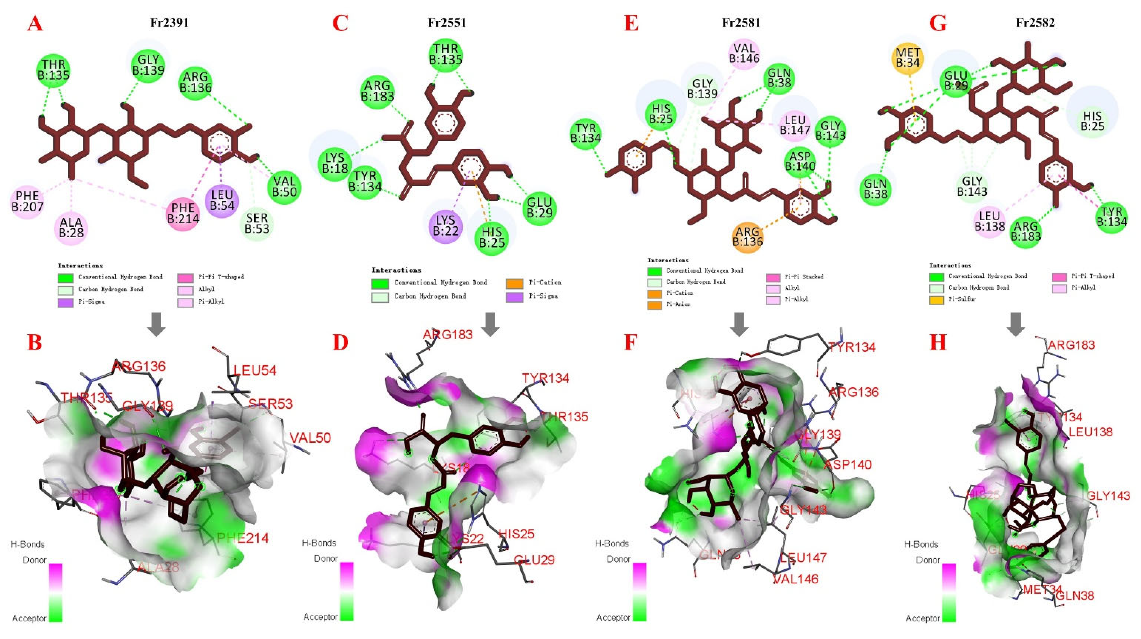
2.5. Molecular Docking

Potential interactions between the four antioxidants (Fr2391, Fr2551, Fr2581, and Fr2582), Nrf2, and HO-1 were determined by molecular docking using AutoDock. Nrf2 (PDB ID: 4IQK) and HO-1 crystal structures (PDB ID: 1N3U) were obtained from the RCSB Protein Data Bank (https://www.rcsb.org/ (accessed on 25 February 2022)) and, after removing ions and water molecules from the receptor, Kollman charges as well as polar hydrogen atoms were added. Grid boxes were then set with the AutoGrid program, while the Lamarckian genetic algorithm and the pseudo-Solis and Wets methods were used for minimization, with parameters set to default. Overall, based on dock score values, a total of 100 peptide conformations, with the minimum binding energy predefined for model development, were obtained. These results were visualized and analyzed using Discovery Studio 2020.

2.6. Statistical Analysis

Statistical analyses were carried out using SPSS version 20.0 software (SPSS, Chicago, IL, USA). Data were presented as means ± standard deviations of triplicate experiments, with the concentrations (0.1–500 μg/mL) converted into μM. The concentration of the isolated phenylethanoids at which 50% of antioxidant activity occurred was determined by nonlinear regression. Prism 8.0 software was also used for plotting the curves of DPPH scavenging activities.

3. Results and Discussion

3.1. Application of Medium-Pressure Liquid Chromatography for Pretreatment of D. heterophyllum Extracts

Being environmentally friendly and nontoxic, ethanol was selected as the solvent for extraction. In this study, from 10.0 kg of air-dried D. heterophyllum, about 1.3 kg of crude sample (after subtracting 1.5 kg of silica from 2.8 kg of silica mixture) was obtained, with the yield being around 13.4%. This crude sample was then pretreated with silica gel medium-pressure liquid chromatography to achieve visible separation while eliminating polymers and sugars. The separation process by medium-pressure liquid chromatography was performed by placing the dry mixture in an empty medium-pressure chromatography tower (49 × 100 mm), which was then connected to the silica gel medium-pressure column (49 × 460 mm) and the preparative HPLC system. Moreover, in order to identify as many chromatographic peaks in the sample as possible, a wavelength of 210 nm was selected and, after repeatedly performing separations 43 times, two fractions were eventually recovered, with 944.0 g of the target fraction Fr2 (recovery of 70.4%) collected. Therefore, overall, the preparation of TTM can be carried out using a medium-pressure chromatography column with silica gel fillers. The preparation diagram shown in Figure 1A indicates that baseline separation was achievable between the two fractions. Furthermore, the on-line HPLC–DPPH system (Figure S15) was also successful for rapidly recognizing antioxidative peaks from complex mixtures. Figure 1B,C further show that, between 20 and 45 min, the four main antioxidative peaks from Fr2 (negative peaks I–IV in Figure 1C correspond to heart-labeled peaks 1–4 in Figure 1B, respectively) could be observed. In this case, the addition of trifluoroacetic acid to the mobile phase could reduce the adsorption of the sample in the dead space of the chromatographic column, thereby prolonging the latter’s lifetime. Following the above experiment, 10.8 g of Fr2 was used for further separation.
The Fr2 contained a lot of chlorophyll, which had to be removed when enriching the active ingredients in the mixture due to its ability to be adsorbed onto the stationary phase of the preparative column. In the current study, MCI GEL®CHP20P medium-pressure liquid chromatography was used for this purpose, with the resulting chromatogram shown in Figure 2A. Fractions were subsequently collected and analyzed with an on-line HPLC–DPPH system on a Click XIon analytical column, and it was found that the four key antioxidative peaks from Fr2 were efficiently enriched in fractions Fr23 (peak 1 of Figure 2B corresponds to the negative DPPH peak I of Figure 2C) and Fr25 (peaks 2–4 in Figure 2D correspond to the negative DPPH peaks II–IV in Figure 2E, respectively). These two fractions were therefore selected and, after evaporating their solvent, 331.0 mg of Fr23 and 536.0 mg of Fr25 were obtained, with these values reflecting an overall 8.0% recovery yield.
Altogether, these results show that highly efficient enrichment can be achieved via pretreatment of crude extracts with silica and MCI GEL®CHP20P medium-pressure liquid chromatography. When combined with preparative HPLC, these pretreatment methods can not only allow sample preparation processes to be visualized, but also help in enriching the target fractions, thereby simplifying the subsequent preparation steps.

3.2. Targeted Purification of Antioxidants Fr23 and Fr25 with High-Pressure Liquid Chromatography

After obtaining the results from the on-line HPLC-DPPH recognition, the analytical conditions for Fr23 and Fr25 were time-optimized prior to additional analysis on the Click XIon analytical column. The resulting chromatograms, shown in Figure 3A,C, indicated that peak 1 from Figure 3A was inseparable from the other chromatographic peaks. Similarly, as observed from Figure 3C, although peaks 2–4 displayed retention times of 20–35 min, peak 2 was quite distinct, while peaks 3 and 4 could not achieve baseline separation, and merged into one peak. These results indicated that the four peaks could be initially separated by Click XIon preparative columns. Therefore, using the system shown in Figure S16, their preparative separation was performed on the Click XIon column, with Figure 3B,D showing the preparative chromatograms of fractions Fr23 and Fr25, respectively. The retention times for Fr239 (peak 1), Fr255 (peak 2), and Fr258 (peaks 3 and 4) were consistent in the analytical (Figure 3A,C) and preparative (Figure 3B,D) chromatograms. In the case of Fr258, given that peaks 3 and 4 were combined, further separation was required. The collected fractions were subsequently concentrated under low pressure to yield 50.63 mg of Fr239 (15.3% recovery), 39.77 mg of Fr255, and 159.60 mg of Fr258 (37.2% recovery). For subsequent analysis and purification, these fractions were again dissolved in methanol.
In order to pinpoint the active peaks 1–4 present in Fr239, Fr255, and Fr258, re-analysis was performed on a Click XIon analytical column under similar conditions as described in Figure 2B, with the results shown in Figure 4A–E. When comparing the analytical chromatograms of Fr23 and Fr239 (Figure 4A,B), it was observed that the latter contained the active peak 1 (Figure 4B). Similarly, Figure 4C–E compare the analytical chromatograms of Fr25, Fr255, and Fr258, respectively. In this case, active peak 2 was separated in Fr255 (Figure 4D), while peaks 3 and 4 were separated in Fr258 (Figure 4E). However, as far as fractions Fr239 and Fr255 were concerned, they appeared to have some impurities (red dashed lines 5, 6, 7, and 8; Figure 4B,D), thereby indicating the need for further purification. It should also be noted that, as shown in Figure 4E, if only the elution conditions of the Click XIon column were optimized, it would take at least 50 min for peaks 3 and 4 to achieve baseline separation, thus making the process very inefficient.
To further improve the purity of peaks 1 and 2, while achieving efficient separation and better resolution of peaks 3 and 4 within a short time, Fr239, Fr255, and Fr258 were re-analyzed on a Kromasil 100-5 Phenyl analytical column using isocratic elution. As shown in Figure 5A–H, peak 1 (Figure 5A corresponds to the DPPH negative peak I in Figure 5B), peak 2 (Figure 5D corresponds to the DPPH negative peak II in Figure 5E), and the well-resolved peaks 3 and 4 (Figure 5G corresponds to DPPH negative peaks III and IV in Figure 5H) could be observed after applying the on-line HPLC–DPPH system under optimized conditions. Following linear amplification, Fr239, Fr255, and Fr258 were separated on the Kromasil 100-5 Phenyl preparative column at a flow rate of 19.0 mL/min, with Figure 5C,F,I showing the preparative chromatograms of the three fractions, respectively. A comparison of Figure 5A,C indicated that peak 1 had similar retention times on the preparative column and the analytical column, and similar observations could be made for peak 2 in Fr255 (Figure 5D,F) as well as peaks 3 and 4 in Fr258 (Figure 5G,I). After preparative separations, 8.08 mg of Fr2391 (peak 1, 15.9% recovery), 9.76 mg of Fr2551 (peak 2, 24.5% recovery), 16.09 mg of Fr2581 (peak 3, 10.1% recovery), and 8.75 mg of Fr2582 (peak 4, 5.5% recovery) were collected. The current findings highlighted that the Click XIon and Kromasil 100-5 Phenyl columns had complementary selectivity in helping to separate peaks 1–4, and this could have been due to differences in the compounds’ polarities as well as in the way in which they interact with the stationary phase. In addition, the four compounds were prepared from 10.8 g of the Fr2 fraction. We actually had a total of 944.0 g of Fr2. Based on the yield, if all of the Fr2 was prepared to isolate these four antioxidants, we would get about 0.71 g of Fr2391, 0.85 g of Fr2551, 1.41 g of Fr2581, and 0.76 g of Fr2582.

3.3. Purity, Activity, and Structure of Fr2391, Fr2551, Fr2581, and Fr2582

The purity and activity of Fr2391, Fr2551, Fr2581, and Fr2582 were again determined using the on-line HPLC–DPPH system along with a ReproSil-Pur C18 AQ analytical column and, as shown in Figure 6A–H, all four antioxidative compounds were over 95% pure. The ESI-MS and NMR spectra of fractions Fr2391, Fr2551, Fr2581, and Fr2582 were also subsequently compared with available literature to elucidate their structures. Figures S2–S13 showed the full spectra generated of Fr2391, Fr2551, Fr2581 and Fr2582 from Supplementary Materials in the present study. The findings indicated that Fr2391, Fr2551, Fr2581, and Fr2582 had NMR and MS data that matched those of decaffeoylverbascoside, rosmarinic acid, acteoside, and 2′-O-acetylplantamajoside, respectively, with their corresponding chemical structures shown in Figure 6I–L, respectively.
Fr2391: (peak 1, decaffeoylverbascoside, 8.08 mg, yellow powder, ESI-MS m/z: 461.38 [M-H]). 1H NMR (600 MHz, MeOH-d4) δ: 6.67 (1H, d, J = 2.0 Hz, 2-H), 6.66 (1H, d, J = 8.0 Hz, 5-H), 6.54 (1H, dd, J = 8.0, 2.0 Hz, 6-H), 5.14 (1H, d, J = 1.8 Hz, 1″-H), 4.28 (1H, d, J = 7.9 Hz, 1′-H), 2.77 (2H, m, 7-H, 8-H), 1.24 (3H, d, J = 6.3 Hz, 6″-H). 13C NMR (151 MHz, MeOH-d4) δ: 146.1 (C-3), 144.7 (C-4), 131.5 (C-1), 121.2 (C-6), 117.1 (C-2), 116.3 (C-5), 104.2 (C-1′), 102.8 (C-1″), 84.5 (C-3′), 77.9 (C-5′), 75.6 (C-2′), 74.0 (C-4′), 72.4 (C-8), 72.2 (C-3″), 72.1 (C-2″), 70.2 (C-5″), 70.1 (C-4″), 62.7 (C-6′), 36.6 (C-7), 17.9 (C-6″). The findings support the compound’s identity as decaffeoylverbascoside [32].
Fr2551: (peak 2, rosmarinic acid, 9.76 mg, white powder, ESI-MS m/z: 359.25 [M-H]). 1H NMR (600 MHz, MeOH-d4) δ: 7.54 (1H, d, J = 15.9 Hz, 7′-H), 7.03 (1H, d, J = 2.1 Hz, 2′-H), 6.94 (1H, dd, J = 8.1, 2.1 Hz, 6′-H), 6.77 (1H, d, J = 8.2 Hz, 5′-H), 6.74 (1H, d, J = 2.1 Hz, 2-H), 6.69 (1H, d, J = 8.0 Hz, 5-H), 6.60 (1H, dd, J = 8.0, 2.1 Hz, 6-H), 6.26 (1H, d, J = 15.9 Hz, 8′-H), 5.18 (1H, m, 8-H), 3.09 (1H, dd, J = 14.4, 4.3 Hz, 7b-H), 3.00 (1H, dd, J = 14.4, 8.5 Hz, 7a-H). 13C NMR (151 MHz, MeOH-d4) δ: 173.4 (C-9), 168.4 (C-9′), 149.8 (C-4′), 147.7 (C-7′), 146.8 (C-3′), 146.2 (C-3), 145.3 (C-4), 129.2 (C-1), 127.7 (C-1′), 123.2 (C-6′), 121.8 (C-6), 117.6 (C-2), 116.5 (C-5′), 116.3 (C-5), 115.2 (C-8′), 114.4 (C-2′), 74.6 (C-8), 37.9 (C-7). The data correspond to rosmarinic acid [33].
Fr2581: (peak 3, acteoside, 16.09 mg, white powder, ESI-MS m/z: 623.51 [M-H]). 1H NMR (600 MHz, MeOH-d4) δ: 7.58 (1H, d, J = 16.1 Hz, 7‴-H), 7.05 (1H, d, J = 2.1 Hz, 2‴-H), 6.94 (1H, dd, J = 8.0, 2.1 Hz, 6‴-H), 6.77 (1H, d, J = 8.0 Hz, 5‴-H), 6.69 (1H, d, J = 2.1 Hz, 2-H), 6.66 (1H, d, J = 8.0 Hz, 5-H), 6.55 (1H, dd, J = 8.0, 2.1 Hz, 6-H), 6.26 (1H, d, J = 16.1 Hz, 8‴-H), 5.18 (1H, d, J = 1.4 Hz, 1″-H), 4.36 (1H, d, J = 7.9 Hz, 1′-H), 4.04 (1H, m, 5″-H), 3.91 (1H, dd, J = 3.3, 1.8 Hz, 2″-H), 3.81 (1H, m, 8b-H), 3.71 (1H, m, 8a-H), 3.29 (2H, m, 6′-H), 2.78 (2H, m, 7-H), 1.08 (3H, d, J = 6.2 Hz, 6″-H). 13C NMR (151 MHz, MeOH-d4) δ: 168.3 (C-CO), 149.8 (C-4‴), 148.0 (C-3‴), 146.8 (C-7‴), 146.1 (C-3), 144.7 (C-4), 131.5 (C-1), 127.7 (C-1‴), 123.2 (C-6‴), 121.3 (C-6), 117.1 (C-2), 116.5 (C-5‴), 116.3 (C-5), 115.2 (C-2‴), 114.7 (C-8‴), 104.2 (C-1′), 103.0 (C-1″), 81.6 (C-3′), 76.2 (C-5′), 76.0 (C-2′), 73.8 (C-4″), 72.3 (C-2″), 72.2 (C-3″), 72.0 (C-8), 70.6 (C-4′), 70.4 (C-5″), 62.4 (C-6′), 36.6 (C-7), 18.4 (C-6″). The data support the compound’s identity as acteoside [34].
Fr2582: (peak 4, 2′-O-acetylplantamajoside, 8.75 mg, white powder, ESI-MS m/z: 627.46 [M-H]). 1H NMR (600 MHz, MeOH-d4) δ: 7.56 (1H, d, J = 15.9 Hz, 7‴-H), 7.06 (1H, d, J = 2.1 Hz, 2‴-H), 6.98 (1H, dd, J = 8.2, 2.1 Hz, 6‴-H), 6.77 (1H, d, J = 8.2 Hz, 5‴-H), 6.66 (1H, d, J = 8.0 Hz, 5-H), 6.63 (1H, d, J = 2.0 Hz, 6-H), 6.51 (1H, dd, J = 8.0, 2.0 Hz, 2-H), 6.34 (1H, d, J = 15.9 Hz, 8‴-H), 4.95 (2H, m, 2′-H, 4′-H), 4.49 (1H, d, J = 8.1 Hz, 1′-H), 4.26 (1H, d, J = 7.7 Hz, 1″-H), 4.14 (1H, m, 3′-H), 4.09 (1H, m, 8-H), 3.76 (1H, dd, J = 11.6, 2.8 Hz, 6′a-H), 3.63(2H, m, 5′-H, 5″-H), 3.56 (1H, dd, J = 12.0, 5.6 Hz, 6″a-H), 3.42 (1H, dd, J = 11.6, 6.8 Hz, 6′b-H), 3.22 (1H, m, 3″-H), 3.19 (1H, ddd, J = 9.4, 6.8, 2.4 Hz, 6″b-H), 3.02 (2H, m, 2″-H, 4″-H), 2.69 (1H, m, 7-H), 1.98 (3H, s, 8′-H). 13C NMR (151 MHz, MeOH-d4) δ: 171.9 (C-7′), 168.4 (C-9‴), 149.7 (C-4‴), 147.3 (C-8‴), 146.8 (C-3‴), 146.0 (C-4), 144.6 (C-3), 131.8 (C-1), 127.8 (C-1‴), 123.0 (C-6‴), 121.3 (C-6), 117.2 (C-5), 116.5 (C-5‴), 116.2 (C-2‴), 115.3 (C-2), 115.3 (C-7‴), 105.1 (C-1″), 102.1 (C-1′), 80.4 (C-3′), 78.0 (C-3″), 77.8 (C-5′), 76.0 (C-5″), 74.8 (C-2′), 74.5 (C-2″), 71.9 (C-8), 71.8 (C-4″), 70.4 (C-4′), 63.2 (C-6′), 62.2 (C-6″), 36.3 (C-8‴), 21.1 (C-8′). These data are consistent with the available literature on 2′-O-acetylplantamajoside [35].

3.4. DPPH Free Radical Scavenging Activity and Molecular Docking Studies

The antioxidant activities of Fr2391, Fr2551, Fr2581, and Fr2582 were determined via the DPPH free radical scavenging assay, using a slightly modified approach [36]. As shown in Figure 7A–D, strong antioxidant activities were observed for all four isolated compounds, with their IC50 values being 26.63 ± 5.11 µM, 19.37 ± 2.03 µM, 19.03 ± 3.08 µM, and 21.51 ± 1.73 µM for Fr2391, Fr2551, Fr2581, and Fr2582, respectively. Although Fr2581 and Fr2582 had similar parent nuclei when comparing their structures, the hydroxyl-substituted C2′ and rhamnose-substituted C3′ for Fr2581 resulted in a lower IC50 value (19.03 µM) compared with the acetoxy-substituted C2′ and glucose-substituted C3′ in the case of Fr2582 (IC50 value: 21.51 µM). Furthermore, when comparing Fr2581 and Fr2391, the caffeoyl-substituted C4′ in Fr2581 led to a lower IC50 value (19.03 µM) compared with the non-substituted C4′ in Fr2391 (IC50 value of 26.63 µM).
With Nrf2/HO-1 being modulators of oxidative stress, Nrf2 (PDB ID: 4IQK) [37] and HO-1 (PDB ID: 1N3U) [38] were selected as targets for molecular docking studies, using a slightly modified approach [39]. Docking was performed as reported in Section 2.5, with the results collected in Table 1 and Table 2. The 2D representations of each binding and docking pose are also shown in Figure 8 and Figure 9. The binding energies were in good accordance with the experimentally determined IC50 values in the DPPH assay. When the target protein was Nrf2, the binding energy values for Fr2391, Fr2551, Fr2581, and Fr2582 were −7.18, −8.29, −8.68, and −7.85 Kcal/mol, respectively (Table 1). Thus, Fr2581 had the lowest value for binding energy, and even showed optimal contact with the binding pocket residues consisting of Val463, Val465, Val420, Val608, Gly367, Val418, Gly417, and Ala366 amino acids (Figure 8E,F). Similarly, Fr2391 was bound to a pocket with Val606, Val608, Val369, Val420, Gly367, Gly419, Val467, Ala466, Ala366, and Ile559 as amino acid residues (Figure 8A,B), while Fr2551 was found in the pocket surrounded by Val463, Gly367, Val606, Ile559, Ala366, Ala607, and Val608 (Figure 8C,D). Finally, the pocket in which Fr2582 was located was surrounded by Val561, Val420, Val467, Val608, Cys513, Leu365, Gly367, Val606, and Val604 (Figure 8G,H). Most of the interactions between the amino acids and the four compounds occurred through hydrogen bonds, with most of the residues also being valine. These results indicate that each compound displayed good interactions with Nrf2. In addition, docking experiments with the target protein HO-1 yielded nearly the same results. Indeed, the binding energy value of Fr2581 was still found to be the lowest, at −7.17 Kcal/mol, with the values for Fr2391, Fr2551, and Fr2582 being −4.52, −6.88, and −4.79 Kcal/mol, respectively (Table 2). However, the predicted binding interaction of Fr2391, Fr2551, and Fr2582 was not as good as that of Fr2581. In all docking structures, small-molecule ligands were always located in pockets surrounded by amino acids. For instance, in the case of Fr2391, the surrounding amino acids were Thr135, Gly139, Arg136, Val50, Ser53, Leu54, Phe214, Ala28, and Phe207 (Figure 9A,B), while for Fr2551, the amino acids were Thr135, Arg183, Lys18, Try134, His25, Glu29, and Lys22 (Figure 9C,D). Similarly, for Fr2581, the amino acids included Try134, His25, Gly139, Val146, Gln38, Leu147, Gly143, Asp140, and Arg136 (Figure 9E,F). Finally, as shown in Figure 9G,H, Met36, Gln38, Glu29, Gly143, Leu138, Arg183, Try134, and His25 amino acid residues surrounded the pocket of Fr2582. These findings suggest that the predicted binding energy preliminarily reflected the antioxidant effects of Fr2391, Fr2551, Fr2581, and Fr2582. Therefore, overall, both the antioxidant assays and the molecular docking of the isolated phenylethanoids confirmed that medium- and high-pressure liquid chromatography, coupled with on-line HPLC–DPPH systems, can efficiently separate antioxidants from natural products.

4. Conclusions

The present study applied medium- and high-pressure liquid chromatography, combined with an on-line HPLC–DPPH system, as a fast and effective approach to recognize, separate, and purify antioxidative phenylethanoids from D. heterophyllum. Four successfully isolated compounds were eventually identified as decaffeoylverbascoside, rosmarinic acid, acteoside, and 2′-O-acetylplantamajoside. Subsequent DPPH assays were carried out to test the antioxidant capacity of the isolated compounds, and molecular docking experiments allowed interactions between the compounds and related proteins to be theoretically predicted. The results showed that the four compounds had good antioxidant capacity. Additional experiments would undoubtedly be required for characterizing the underlying mechanisms of the observed antioxidant activities, and while this will likely be the focus of future studies, the present results nevertheless provide a basis for successfully isolating antioxidant compounds from TTM and natural products. In fact, this approach is not only valuable for identifying and separating antioxidant phenylethanoids from D. heterophyllum, but may also be applied to other plant sources.

Supplementary Materials

The following supporting information can be downloaded at: https://www.mdpi.com/article/10.3390/separations9050111/s1, Figure S1: The graph of the isolation steps. Figure S2: ESI mass spectrum of decaffeoylverbascoside (Fr2391). Figure S3: 1H NMR spectrum (600 MHz) of decaffeoylverbascoside (Fr2391) (in MeOH-d4). Figure S4: 13C NMR spectrum (151 MHz) of decaffeoylverbascoside (Fr2391) (in MeOH-d4). Figure S5: ESI mass spectrum of rosmarinic acid (Fr2551). Figure S6: 1H NMR spectrum (600 MHz) of rosmarinic acid (Fr2551) (in MeOH-d4). Figure S7: 13C NMR spectrum (151 MHz) of rosmarinic acid (Fr2551) (in MeOH-d4). Figure S8: ESI mass spectrum of acteoside (Fr2581). Figure S9: 1H NMR spectrum (600 MHz) of acteoside (Fr2581) (in MeOH-d4). Figure S10: 13C NMR spectrum (151 MHz) of acteoside (Fr2581) (in MeOH-d4). Figure S11: ESI mass spectrum of 2′-O-acetylplantamajoside (Fr2582). Figure S12: 1H NMR spectrum (600 MHz) of 2′-O-acetylplantamajoside (Fr2582) (in MeOH-d4). Figure S13: 13C NMR spectrum (151 MHz) of 2′-O-acetylplantamajoside (Fr2582) (in MeOH-d4). Figure S14: The actual medium-pressure liquid chromatography system. Figure S15: The actual HPLC–DPPH system. Figure S16. The actual high-pressure liquid chromatography system.

Author Contributions

Conceptualization, methodology, J.D. and Z.W.; writing—original draft, Y.L.; formal analysis, G.L. and Q.W. (Qilan Wang); investigation, Y.F.; data curation, Q.W. (Qian Wu); supervision, project administration, and funding acquisition, J.D. and G.L. All authors have read and agreed to the published version of the manuscript.

Funding

This work was supported by the Applied Basic Research Project of Qinghai Province (2020-ZJ-706), the Young Scholars in Western China, Chinese Academy of Sciences (2022), the Joint Research Project of Three-River Headwaters National Park, Chinese Academy of Sciences, the People’s Government of Qinghai Province (LHZX-2021-02), and the Yantai Science and Technology Innovation Development Project (2021XDHZ078).

Institutional Review Board Statement

Not applicable.

Informed Consent Statement

Not applicable.

Data Availability Statement

Data are available upon request from the authors.

Conflicts of Interest

The authors declare no conflict of interest.

References

  1. Venditti, P.; Stefano, L.D.; Meo, S.D. Mitochondrial metabolism of reactive oxygen species. Mitochondrion 2013, 13, 71–82. [Google Scholar] [CrossRef] [PubMed]
  2. Valko, M.; Jomova, K.; Rhodes, C.J.; Kuca, K.; Musilek, K. Redox- and non-redox-metal-induced formation of free radicals and their role in human disease. Arch. Toxicol. 2016, 90, 1–37. [Google Scholar] [CrossRef] [PubMed]
  3. Kim, G.H.; Kim, J.E.; Rhie, S.J.; Yoon, S. The role of oxidative stress in neurodegenerative diseases. Exp. Neurol. 2015, 24, 325–340. [Google Scholar] [CrossRef]
  4. Reuter, S.; Gupta, S.C.; Chaturvedi, M.M.; Aggarwal, B.B. Oxidative stress, inflammation, and cancer how are they linked? Free Radic. Biol. Med. 2010, 9, 1603–1616. [Google Scholar] [CrossRef] [PubMed] [Green Version]
  5. Tu, W.J.; Wang, H.; Li, S.; Liu, Q.; Sha, H. The anti-Inflammatory and anti-oxidant mechanisms of the Keap1/Nrf2/ARE signaling pathway in chronic diseases. Aging Dis. 2019, 10, 637–651. [Google Scholar] [CrossRef] [Green Version]
  6. Pisoschi, A.M.; Pop, A. The role of antioxidants in the chemistry of oxidative stress: A review. Eur. J. Med. Chem. 2015, 97, 55–74. [Google Scholar] [CrossRef] [PubMed]
  7. Li, C.; Tan, D.X.; Liang, D.; Chang, C.; Jia, D.F.; Ma, F.W. Melatonin mediates the regulation of ABA metabolism, free-radical scavenging, and stomatal behaviour in two Malus species under drought stress. J. Exp. Bot. 2015, 66, 669–680. [Google Scholar] [CrossRef] [PubMed] [Green Version]
  8. Manessis, G.; Kalogianni, A.I.; Lazou, T.; Moschovas, M.; Bossis, I.; Gelasakis, A.I. Plant-Derived Natural Antioxidants in Meat and Meat Products. Antioxidants 2020, 9, 1215. [Google Scholar] [CrossRef]
  9. Zeng, Q.; Jin, H.Z.; Qin, J.J.; Fu, J.J.; Hu, X.J.; Liu, J.H.; Yan, L.; Chen, M.; Zhang, W.D. Chemical constituents of plants from the genus Dracocephalum. Chem. Biodivers. 2010, 7, 1911–1929. [Google Scholar] [CrossRef]
  10. Liu, J.Y.; He, J.X.; Shi, G.X.; An, L.Z.; Öpik, M.; Feng, H.Y. Diverse communities of arbuscular mycorrhizal fungi inhabit sites with very high altitude in Tibet Plateau. FEMS Microbiol. Ecol. 2011, 78, 355–365. [Google Scholar] [CrossRef] [Green Version]
  11. Numonov, S.R.; Usmanova, S.K.; Aisa, H.A. Chemical composition of Dracocephalum heterophyllum. Chem. Nat. Compd. 2013, 49, 511–513. [Google Scholar] [CrossRef]
  12. Numonov, S.R.; Qureshi, M.N.; Aisa, H.A. Development of HPLC protocol and simultaneous quantification of four free flavonoids from Dracocephalum heterophyllum. Benth. Int. J. Anal. Chem. 2015, 2015, 5. [Google Scholar] [CrossRef] [Green Version]
  13. Zhou, S.; Wei, C.; Zhang, C.; Han, C.; Kuchkarova, N.; Shao, H. Chemical composition, phytotoxic, antimicrobial and insecticidal activity of the essential oils of Dracocephalum integrifolium. Toxins 2019, 11, 598. [Google Scholar] [CrossRef] [PubMed] [Green Version]
  14. Shi, Q.Q.; Zhao, J.Q.; Dang, J.; Yuan, X.; Wang, Q.L. Triterpenes, flavonoids, and lignans from Dracocephalum heterophyllum. Chem. Nat. Compd. 2018, 54, 970–972. [Google Scholar] [CrossRef]
  15. Wang, L.M.; Wang, S.Q.; Yang, S.; Guo, X.J.; Lou, H.X.; Ren, D.M. Phenolic alkaloids from the aerial parts of Dracocephalum heterophyllum. Phytochemistry 2012, 82, 166–171. [Google Scholar] [CrossRef] [PubMed]
  16. Zhang, C.J.; Li, W.; Li, H.Y.; Wang, Y.L.; Yun, T.; Song, Z.P.; Zhao, X.W. In vivo and in vitro antiviral activity of five Tibetan medicinal plant extracts against herpes simplex virus type 2 infection. Pharm Biol. 2009, 47, 598–607. [Google Scholar] [CrossRef]
  17. Zheng, W.; Wang, Q.L.; Lu, X.H.; Shi, Q.Q.; Zou, J.H.; Tao, Y.D.; Wang, P. Protective effects of Dracocephalum heterophyllum in ConA-Induced acute hepatitis. Mediators Inflamm. 2016, 9, 2684321. [Google Scholar] [CrossRef] [Green Version]
  18. Shi, Q.Q.; Dang, J.; Wen, H.X.; Yuan, X.; Tao, Y.D.; Wang, Q.L. Anti-hepatitis, antioxidant activities and bioactive compounds of Dracocephalum heterophyllum extracts. Bot. Stud. 2016, 57, 16. [Google Scholar] [CrossRef] [Green Version]
  19. Fu, L.; Wei, J.C.; Gao, Y.M.; Chen, R. Antioxidant and antitumoral activities of isolated macamide and macaene fractions from Lepidium meyenii (Maca). Talanta 2021, 221, 121635. [Google Scholar] [CrossRef]
  20. Liu, C.; Lei, Y.Q.; Dang, J.; Wang, W.D.; Zhang, J.; Mei, L.J.; Liu, Z.G.; Tao, Y.D.; Shao, Y. Preparative isolation of 1,1-diphenyl-2-picrylhydrazyl inhibitors from Ribes himalense using medium-pressure and two-dimensional reversed-phase/reversed-phase liquid chromatography guided by an online HPLC-1, 1-diphenyl-2-picrylhydrazyl assay. J. Sep. Sci. 2021, 44, 1345–1352. [Google Scholar] [CrossRef]
  21. Dang, J.; Wang, Q.; Wang, Q.L.; Yuan, C.; Li, G.; Ji, T.F. Preparative isolation of antioxidative gallic acid derivatives from Saxifraga tangutica using a class separation method based on medium-pressure liquid chromatography and reversed-phase liquid chromatography. J. Sep. Sci. 2021, 44, 3734–3746. [Google Scholar] [CrossRef] [PubMed]
  22. Li, A.F.; Xuan, H.Z.; Sun, A.L.; Liu, R.M.; Cui, J.C. Preparative separation of polyphenols from water-soluble fraction of Chinese propolis using macroporous absorptive resin coupled with preparative high performance liquid chromatography. J. Chromatogr. B 2016, 1012, 42–49. [Google Scholar] [CrossRef] [PubMed]
  23. Wang, W.D.; Jiao, L.J.; Tao, Y.D.; Shao, Y.; Wang, Q.L.; Yu, R.T.; Mei, L.J.; Dang, J. On-line HPLC-DPPH bioactivity-guided system for isolated of antioxidative phenylpropanoids from Qinghai-Tibet Plateau medicinal plant Lancea tibetica. J. Chromatogr. B 2019, 1106–1107, 1–10. [Google Scholar] [CrossRef] [PubMed]
  24. Dawa, Y.Z.; Du, Y.R.; Wang, Q.; Chen, C.B.; Zou, D.L.; Qi, D.S.; Ma, J.B.; Dang, J. Targeted isolation of 1,1-diphenyl-2-picrylhydrazyl inhibitors from Saxifraga atrata using medium- and high- pressure liquid chromatography combined with online high performance liquid chromatography-1,1-diphenyl-2-picrylhydrazyl detection. J. Chromatogr. A 2021, 1635, 461690. [Google Scholar] [CrossRef]
  25. Pan, G.Q.; Shen, J.W.; Ma, Y.H.; He, Y.F.; Bao, Y.; Li, R.R.; Wang, S.S.; Wang, Q.; Lin, P.C.; Dang, J. Preparative separation of isoquinoline alkaloids from Corydalis impatiens using a middle-pressure chromatogram isolated gel column coupled with two-dimensional liquid chromatography. J. Sep. Sci. 2019, 42, 3182–3190. [Google Scholar] [CrossRef]
  26. Wang, X.; Zhang, Y.P.; Wu, N.; Cao, J.Y.; Tao, Y.D.; Yu, R.T. A Method to Separate Two Main Antioxidants from Lepidium latifolium L. Extracts Using Online Medium Pressure Chromatography Tower and Two-Dimensional Inversion/Hydrophobic Interaction Chromatography Based on Online HPLC-DPPH Assay. Separations 2021, 8, 238. [Google Scholar] [CrossRef]
  27. İlhami, G. Antioxidants and antioxidant methods: An updated overview. Arch. Toxicol. 2020, 94, 651–715. [Google Scholar] [CrossRef] [Green Version]
  28. Warren, J.J.; Tronic, T.A.; Mayer, J.M. Thermochemistry of Proton-Coupled Electron Transfer Reagents and its Implications. Chem. Rev. 2010, 110, 6961–7001. [Google Scholar] [CrossRef] [Green Version]
  29. Athar, M.; Lone, M.Y.; Khedkar, V.M.; Jha, P.C. Pharmacophore model prediction, 3D-QSAR and molecular docking studies on vinyl sulfones targeting Nrf2-mediated gene transcription intended for anti-Parkinson drug design. J. Biomol. Struct. Dyn. 2016, 34, 1282–1297. [Google Scholar] [CrossRef]
  30. Loboda, A.; Damulewicz, M.; Pyza, E.; Jozkowicz, A.; Dulak, J. Role of Nrf2/HO-1 system in development, oxidative stress response and diseases: An evolutionarily conserved mechanism. Cell. Mol. Life Sci. 2016, 73, 3221–3247. [Google Scholar] [CrossRef] [Green Version]
  31. Dang, J.; Zhang, L.; Shao, Y.; Mei, L.J.; Liu, Z.G.; Yue, H.L.; Wang, Q.L.; Tao, Y.D. Preparative isolation of antioxidative compounds from Dracocephalum heterophyllum using off-line two-dimensional reversed-phase liquid chromatography/hydrophilic interaction chromatography guided by on-line HPLC-DPPH system. J. Chromatogr. B 2018, 1095, 267–274. [Google Scholar] [CrossRef] [PubMed]
  32. Kanchanapoom, T.; Kasai, R.; Yamasaki, K. Phenolic glycosides from Markhamia stipulate. Phytochemistry 2002, 59, 557–563. [Google Scholar] [CrossRef]
  33. Dapkevicius, A.; van Beek, T.A.; Lelyveld, G.P.; van Veldhuizen, A.; de Groot, A.; Linssen, J.P.; Venskutonis, R. Isolation and structure elucidation of radical scavengers from Thymus vulgaris leaves. J. Nat. Prod. 2002, 65, 892–896. [Google Scholar] [CrossRef] [PubMed]
  34. Juliao, L.D.; Piccinelli, A.L.; Marzocco, S.; Leitao, S.G.; Lotti, C.; Autore, G.; Rastrelli, L. Phenylethanoid glycosides from Lantana fucata with in vitro anti-inflammatory activity. J. Nat. Prod. 2009, 72, 1424–1428. [Google Scholar] [CrossRef]
  35. Kyriakopoulou, I.; Magiatis, P.; Skaltsounis, A.L.; Aligiannis, N.; Harvala, C. Samioside, a New Phenylethanoid Glycoside with Free-Radical Scavenging and Antimicrobial Activities from Phlomis samia. J. Nat. Prod. 2001, 64, 1095–1097. [Google Scholar] [CrossRef]
  36. Wootton-Beard, P.C.; Moran, A.; Ryan, L. Stability of the total antioxidant capacity and total polyphenol content of 23 commercially available vegetable juices before and after in vitro digestion measured by FRAP, DPPH, ABTS and Folin-Ciocalteu methods. Food Res. Int. 2011, 44, 217–224. [Google Scholar] [CrossRef]
  37. Thomasa, N.S.; Georgeb, K.; Selvamc, A.A.A. Anticancer mechanism of troxerutin via targeting Nrf2 and NF-κB signaling pathways in hepatocarcinoma cell line. Toxicol. Vitro 2019, 54, 317–329. [Google Scholar] [CrossRef]
  38. França, A.L.d.Q.; Chaves, H.V.; Freire, J.M.d.O.; Sousa, L.H.T.d.; Pimenta, A.T.A.; Lima, M.A.S.; Oliveira, B.R.d.; Mattos, M.C.d.; Pinto, V.d.P.T.; Portela, A.M.L.R.; et al. Molecular docking study and antireabsorptive activity of a semi-synthetic coumarin derivative from Platymiscium floribundum in the ligature-induced periodontitis in rats: The involvement of heme oxygenase-1. Clin. Oral Investig. 2022, 26, 1701–1711. [Google Scholar] [CrossRef]
  39. Yuan, C.; Dang, J.; Han, Y.; Liu, C.; Yu, S.; Lv, Y.; Cui, Y.B.; Wang, Z.H.; Li, G. Preparative isolation of maltol glycoside from Dianthus superbus and its anti-inflammatory activity in vitro. RSC Adv. 2022, 12, 5031–5041. [Google Scholar] [CrossRef]
Figure 1. Chromatogram of D. heterophyllum extract separation (A) with a silica gel medium-pressure liquid chromatography system. The analytical chromatogram of the Fr2 fraction, acquired at a wavelength of 210 nm, from D. heterophyllum (B), after separation on a ReproSil-Pur C18 AQ analytical column. The HPLC was performed at a flow rate of 1.0 mL/min, using a column temperature of 30 °C, with 0.01% v/v trifluoroacetic acid in water and acetonitrile (ACN) as mobile phases A and B, respectively, and with a gradient of 0–60 min using 0–100% B. Scavenging profile of 1,1-diphenyl-2-picrylhydrazyl (DPPH) radicals (C), acquired at 517 nm while using a flow rate of 0.8 mL/min for the DPPH solution. The negative peaks I–IV correspond to active peaks 1–4, respectively.
Separations 09 00111 g001
Figure 2. The pretreatment chromatogram (A) of Fr2 from D. heterophyllum with MCI GEL®CHP20P middle-pressure liquid chromatography. The analytical chromatograms of fractions Fr23 and Fr25 on a Click XIon analytical column (B,D, respectively), acquired at a wavelength of 210 nm. The HPLC was performed at a flow rate of 1.0 mL/min, using a column temperature of 30 °C, with 0.1% v/v trifluoroacetic acid in water and acetonitrile (ACN) as mobile phases A and B, respectively, and with a gradient of 0–30 min using 100–60% B. Scavenging profile of 1,1-diphenyl-2-picrylhydrazyl (DPPH) radical for fractions Fr23 and Fr25 (C,E, respectively), acquired at 517 nm while using a flow rate of 0.8 mL/min for the DPPH solution. The negative peaks I–IV correspond to active peaks 1–4, respectively.
Separations 09 00111 g002
Figure 3. Chromatograms of fractions Fr23 and Fr25, acquired at 210 nm, after analytical (A,C, respectively) and preparative (B,D, respectively) chromatography on the Click XIon columns. Analysis was carried out at a flow rate of 1.0 mL/min, using a column temperature of 30 °C, with 0.1% v/v trifluoroacetic acid in water and acetonitrile (ACN) as mobile phases A and B, respectively, and with a gradient of 0–60 min using 100–60% B. For preparation, similar conditions were applied, except that the column was at room temperature and a flow rate of 19.0 mL/min was selected. Peaks 1–4 represent active fraction peaks, respectively.
Separations 09 00111 g003
Figure 4. HPLC analysis of Fr23 and Fr239 (A,B, respectively) on the Click XIon analytical column, with fractions Fr25, Fr255, and Fr258 also analyzed on the same type of column (CE, respectively). Analysis was performed at a flow rate of 1.0 mL/min, using a column temperature of 30 °C, with 0.1% v/v trifluoroacetic acid in water and acetonitrile (ACN) as mobile phases A and B, respectively, and with a gradient of 0–30 min using 100–60% B. The chromatograms were obtained at a wavelength of 210 nm. Peaks 1–4 represent active fraction peaks, respectively. Peaks 5–8 represent impurity peaks.
Separations 09 00111 g004
Figure 5. Optimized analytical chromatograms of fractions Fr239, Fr255, and Fr258, acquired at a wavelength of 210 nm, after separation on a Kromasil 100-5 Phenyl analytical column (A,D,G, respectively). The HPLC was performed at a flow rate of 1.0 mL/min, using a column temperature of 30 °C, with 0.1% v/v trifluoroacetic acid in water and acetonitrile (ACN) as mobile phases A and B, respectively, and with the following gradients: 0–30 min with 5% B, 0–40 min with 20% B, and 0–40 min using 17% B. The scavenging profile of 1,1-diphenyl-2- picrylhydrazyl (DPPH) radicals (B,E,H), acquired at 517 nm using a flow rate of 0.8 mL/min for the DPPH solution. Preparation chromatograms of fractions Fr239, Fr255, and Fr258 after separation on a Kromasil 100-5 Phenyl preparative column (C,F,I, respectively).
Separations 09 00111 g005
Figure 6. Chromatograms of Fr2391 (A,B), Fr2551 (C,D), Fr2581 (E,F) and Fr2582 (G,H) after separation on a ReproSil-Pur C18 AQ analytical column, and showing their purity levels as well as DPPH inhibitory activities. The chemical structures of Fr2391 (I), Fr2551 (J), Fr2581 (K), and Fr2582 (L) are also shown.
Separations 09 00111 g006
Figure 7. Antioxidant activities of Fr2391 (A), Fr2551 (B), Fr2581 (C), and Fr2582 (D) at different concentrations (µM). The IC50 values were obtained after fitting the data with a logistic function.
Separations 09 00111 g007
Figure 8. Molecular docking analysis of Nrf2 (PDB ID: 4IQK) during binding to Fr2391, Fr2551, Fr2581, and Fr2582. (A,C,E,G, respectively) indicates the 2D interaction diagrams, with (B,D,F,H) showing binding models of Nrf2’s interactions with Fr2391, Fr2551, Fr2581, and Fr2582, respectively.
Separations 09 00111 g008
Figure 9. Molecular docking analysis of HO-1 (PDB ID: 1N3U) during binding to Fr2391, Fr2551, Fr2581, and Fr2582. (A,C,E,G, respectively) indicates the 2D interaction diagrams, with (B,D,F,H) showing binding models of HO-1’s interactions with Fr2391, Fr2551, Fr2581, and Fr2582, respectively.
Separations 09 00111 g009
Table 1. Binding energy of Fr2391, Fr2551, Fr2581, and Fr2582 in Nrf2 (PDB ID: 4IQK).
CompoundsRMSDBinding Energy (Kcal/mol)
Fr2391 (decaffeoylverbascoside)36.75−7.18
Fr2551 (rosmarinic acid)37.83−8.29
Fr2581 (acteoside)36.69−8.68
Fr2582 (2′-O-acetylplantamajoside)35.51−7.85
Table 2. Binding energy of Fr2391, Fr2551, Fr2581, and Fr2582 in HO-1 (PDB ID: 1N3U).
CompoundsRMSDBinding Energy (Kcal/mol)
Fr2391 (decaffeoylverbascoside)13.49−4.52
Fr2551 (rosmarinic acid)11.57−6.88
Fr2581 (acteoside)12.95−7.17
Fr2582 (2′-O-acetylplantamajoside)13.26−4.79
Publisher’s Note: MDPI stays neutral with regard to jurisdictional claims in published maps and institutional affiliations.

