Article

A Method to Separate Two Main Antioxidants from Lepidium latifolium L. Extracts Using Online Medium Pressure Chromatography Tower and Two-Dimensional Inversion/Hydrophobic Interaction Chromatography Based on Online HPLC-DPPH Assay

1
Qinghai Provincial Key Laboratory of Tibetan Medicine Research, Key Laboratory of Tibetan Medicine Research, Northwest Institute of Plateau Biology, Chinese Academy of Sciences, Xining 810008, China
2
University of Chinese Academy of Sciences, Beijing 100049, China
*
Author to whom correspondence should be addressed.
Separations 2021, 8(12), 238; https://doi.org/10.3390/separations8120238
Submission received: 17 September 2021 / Revised: 16 November 2021 / Accepted: 30 November 2021 / Published: 6 December 2021

Abstract

:
Free radicals, including 1,1-diphenyl-2-picrylhydrazyl, mediate oxidative stress to cause many chronic diseases (including cardiovascular diseases, diabetes and cancer). The extract of traditional Tibetan medicine Lepidium latifolium L. (L. latifolium) was reported to have free radical inhibition ability. Therefore, a system method was established to separate the ethanol extract of L. latifolium to prepare two main antioxidant compounds. First of all, silica gel and a medium-pressure liquid chromatography tower were used for pre-treatment of the ethanol extract of L. latifolium to obtain the main antioxidant active component fraction 4 through online high-performance liquid chromatography-1,1-diphenyl-2-picrylhydrazyl (HPLC-DPPH) assay. Then, fraction 4-1 was obtained by one-dimensional preparation using Megres C18 chromatographic column, and two active compounds with IC50 values 59.9 and 71.3 μg/mL were obtained by two-dimensional preparation using Click XIon chromatographic column. Through the study of the chemical components and separation methods of L. latifolium, the combination of HPLC-DPPH assay and two-dimensional preparative liquid chromatography was realized, providing a reference for the separation of active compounds from L. latifolium.

Graphical Abstract

1. Introduction

Oxidative stress is vital for organisms, however, in some given conditions, oxidative stress is inextricably linked to all kinds of diseases, such as Alzheimer, Parkinson, inflammation, aging, cancer, neuronal disorders, Diabetic Complications and cardiovascular diseases [1,2,3,4,5,6,7]. Thus, some antioxidants may play a role in prevention of above deceases. Antioxidants are divided into synthetic antioxidants and natural antioxidants. The most commonly used synthetic antioxidants are bhabutylated hydroxyanisole (BHA), butylated hydroxytolune (BHT), and 6-hydroxy-2,5,7,8-tetramethylchroman-2-carboxylic acid (Trolox) etc [8]. They all have good antioxidant activity, but, due to environmental and economic concerns, research is more and more inclined towards natural antioxidants. Natural antioxidants include flavonoids [9]. The plant Lepidium latifolium L. (L. latifolium) was reported for its better antioxidant activity of ethyl acetate site [10].
L. Latifolium, a perennial herb, belonging to the family Brassicaceae, which contains more than 175 species worldwide [11]. Although it was regarded as an aggressive foreign weed because of its competitiveness and adaptability [12], the drought resistance, salt tolerance, plant physiology, minerals, proteins and amino acids of L. latifolium were reported [13] and, it is now often used as an insect repellent, insecticidal, as well as to aid in bacteriostasis, the treatment of enteritis, bacillary dysentery and to detoxify [14]. L. latifolium also has diuretic, anti-cancer characteristics and a certain therapeutic effect on kidney stones [15,16,17,18,19]. In addition, L. latifolium is one of the main foods in cold regions, especially in the Himalayas [19]. At the same time, it was reported that the flavonoids isolated from L. latifolium have better 1,1-diphenyl-2-picrylhydrazyl (DPPH) inhibitory activity [10]. In order to use resources of L. latifolium well, which are cheap and easy to obtain, the experiment was designed to establish a two-dimensional (2D) preparation system for the first time to obtain the main antioxidant compounds in L. latifolium.
Classical antioxidant activity is based on electron donation mechanism [20]. DPPH is a stable free radical with unpaired single electrons on the nitrogen atomic bridge with a maximum absorption at 517 nm [21]. DPPH donors a pair of electrons paired to the antioxidants (free radical scavengers), transitioning from purple to yellow, the absorbance at 517 nm wavelength becomes smaller, and the degree of change is linear with the degree of free radical scavenging activity of antioxidants. The stronger scavenging ability of the free radical scavenger will show the lower absorbance. Microplate reader detection or offline 1,1-diphenyl-2-picrylhydrazyl-High Performance Liquid Chromatograph (DPPH-HPLC) method is commonly used to identify antioxidant compounds, but they are time consuming. The HPLC-DPPH-DAD online screening antioxidant system is a fast method which already finds a lot of antioxidants from natural resources [22,23,24,25]. With this method, the compounds in the sample are separated by the chromatographic column, and the DPPH solution is mixed with the sample through a tee and mixer, and the detection is carried out at 517 nm, and the degree of absorption reflects the antioxidant ability of the substance. The phytochemical composition is complex and the purified compounds cannot be prepared by one-dimensional (1D) preparation only. 2D preparation, especially the two-dimensional reversed-phase/hydrophobic interaction chromatograph (2D RPLC/HILIC) system, has the advantages of high orthogonality and rapidity [22,26]. Therefore, the online DPPH-HPLC combined with 2D preparation was employed to prepare the crude extract of L. latifolium.
Based on the above considerations, in this study, a 2D RPLC/HILIC method combined with online DPPH-HPLC assay and medium- and high-pressure chromatography were used to isolate two main DPPH inhibitors from the ethanol extract of whole L. latifolium. In combination, this research demonstrates that the 2D preparative method constructed in this research is a useful method to prepare antioxidative flavonoids with a similar structure to natural products. This is the first time an effective and systematic method to obtain DPPH inhibitors from L. latifolium has been established.

2. Materials and Methods

2.1. Apparatus and Reagents

The experimental instrument of online HPLC-DPPH assay consists of two instruments, LC-16 and LC-10AD (Shimadzu Enterprise Management Co., Ltd., Kyoto, Japan), which were linked by a three-way links and a reaction coil. The reaction coil was a polyether ether ketone (PEEK) tube (18.0 m × 0.25 mm inner diameter). Each instrument consists of a binary gradient pump, fraction collector, UV detector and software LC Workstation. The analysis was performed on High Performance Liquid Chromatograph LC 16-SPD (Shimadzu Enterprise Management Co., Ltd., Kyoto, Japan). Preparation was performed on NP 7005C (Jiangsu Hanbon Science and Technology Co., Ltd., Huaian, China). Microplate reader (Biotek, Winooski, VT, USA) containing full-wavelength spectrophotometer was used to assess the activity of isolated compounds. Liquid Chromatography Triple Quadrupole Mass Spectrometer (Agilent, Palo Alto, CA, USA) was used to get ESI-MS spectra. Bruker Avance 600 MHz (Billerica, MA, USA) was used to get 1HNMR, 13CNMR and HMBC spectra.
Silica gel (200–300 mesh) was from Qingdao Ocean Chemical Factory (Qingdao, China). The separation material, MCI GEL® CHP20P (75–100 μm) was from Mitsubishi Chemical Corporation (Tokyo, Japan). The Dubhe C18 analysis column (4.6 × 250 mm, 5 μm), Megres C18 analysis column (4.6 × 250 mm, 5 μm) and preparation column (20 × 250 mm, 5 μm) were from Jiangsu Hanbon Science and Technology Co., Ltd. (Huian, China) Analytical (4.6 × 250 mm, 5 μm), preparation (20 × 250 mm, 5 μm) Click XIon columns were manufactured by Acchrom Technologies Co., Ltd. (Beijing, China).
Methanol, ethanol, dichloromethane (analytical purity) were manufactured by Tianjin Komiou Chemical Reagent Co., Ltd (Tianjin, China). Formic acid (analytical purity) was applied by Chengdu Kelong Chemical Co., Ltd (Chengdu, China). Acetonitrile was from Yunnan Xinlanjing Chemical Industry Co., Ltd (Yunnan, China). DPPH was purchased from Sigma-Aldrich (St. Louis, MO, USA). Vitamin C was from NICPBP. The main scheme of the procedure is shown in Figure 1.

2.2. Sample Preparation, On-Line HPLC-DPPH Antioxidant Active Fractions Screening and Preparation

L. latifolium, collected from Dulan, Qinghai, China, was identified by Professor Mei L.J from Northwest Plateau Institute of Biology, Chinese Academy of Sciences. The voucher specimen (No. 0333409) was preserved in the Qinghai-Tibet Plateau Biological Museum. The air dried and powdered whole plant (1 kg) was extracted three times with 85% v/v ethanol (10 L and 2 h per extraction) at 55 °C. The 85% v/v ethanol extract was concentrated at 55 °C (filtered and then evaporated) to obtain 107 g of extract (extraction rate: 10.73%). In order to remove sugars and complexes from the extract, the extract was successively subjected to silica gel column (100–200 mesh, 100 g) and eluted with CH2Cl2-MeOH-H2O (7:3:0.5, v/v/v). Then 68 g sample was obtained (extraction rate: 63.45%). The online HPLC-DPPH analysis of the ethanol extract was analyzed by Dubhe C18 column (4.6 × 250 mm, 5 μm) and eluted with acetonitrile (mobile phase B) and 0.2% v/v formic acid aqueous solution (mobile phase A). The percentage of acetonitrile increased from 5 to 30% v/v B over 40 min. The wavelength was 254 nm. The flow rate was 1.0 mL/min. The injection volume was 20 μL. And the flow rate of the ethanol solution of DPPH (25 mg/L) was 0.8 mL/min, and the spectra were monitored at 517 nm. Column temperature for analysis was 40 °C.
122 g dried sample, a mixture of 60 g sample and 62 g silica gel, was injected into a 49 × 460 mm column, with 1.2 L of MCI GEL® CHP20P as stationary phase, and eluted with 85% v/v ethanol (mobile B) and water (mobile A). The percentage of 85% ethanol linearly increased from 0 to 100% v/v within 120 min and 100%–100% v/v over 30 min. The flow rate was 30.0 mL/min, and the spectra were monitored at 254 nm, sample loading was 30 g, column temperature was room temperature. After separation, Fr4 (target fraction) was concentrated to yield 4473.3 mg (recovery rate: 7.46%) target fraction sample.

2.3. 2D Preparation of Antioxidant Active Fractions Using High Performance Liquid Chromatography

The Fr4 was dissolved in 10 mL methanol and filtered with 0.45 μm organic membrane to obtain 447.3 g/mL sample. Then Megres C18 preparation column (20 × 250 mm, 5 μm) was used for 1D preparation. The mobile phase A was 0.2% v/v formic acid in aqueous solution, the mobile phase B was methanol. The linear gradient elution procedure for Fr4 was as follows: 0–40 min, 20%–45% v/v B. The flow rate was 19.0 mL/min. The loaded volume was 1 mL (12 times). Chromatograms were collected at 254 nm. Fr4-1 (620.7 mg, recovery rate: 13.88%) was obtained. Fr4-1 was analyzed on Megres C18 analytical column (4.6 × 250 mm, 5 μm) and Click XIon analytical column (4.6 × 250 mm, 10 μm, 100 Å). The mobile phase A was 0.2% v/v formic acid/aqueous solution and the mobile phase B was methanol or acetonitrile. The flow rate was 1.0 mL/min, and the injection volume was 10 μL. The following was the linear gradient elution for Fr4-1 on Megres C18 column: 0–60 min, 20–45% v/v B (methanol). The linear gradient elution for Fr4-1 on Click XIon analytical column was as follows: 0–60 min, 90–75% B (acetonitrile). Collection wavelength was 254 nm. Column temperature for analysis and preparation was 40 °C and room temperature, respectively.
Preparation Click XIon (20 × 250 mm, 5 μm, 100 Å) column was used to 2D purify with the mobile phase A being 0.2% v/v formic acid/aqueous solution and mobile phase B being acetonitrile. The linear gradient elution for Fr4-1 was as following: 0–60 min, 90–75% v/v B. The injection volume was 4 mL, and a total of eight needles were injected. The flow rate was 19.0 mL/min. Chromatographic wavelength was 254 nm. Column temperature was room temperature. Then, Fr4-1-1 (199.4 mg, recovery rate: 64.25%) and Fr4-1-2 (9.1 mg, recovery rate: 1.47%) were concentrated using a rotary evaporator (EYELA, Tokyo, Japan) and dried by lyophilization.

2.4. Purity, Activity Assessment, and Structural Identification of Two DPPH Inhibitors

A Dubhe C18 analytical column (4.6 × 250 mm, 5 μm) was used to evaluate the purity and activity of DPPH inhibitory active compounds with the same conditions for analysis of crude sample. To characterize the structure of isolated DPPH inhibitors, MS, 1HNMR, 13CNMR spectra and HMBC were collected and compared with the reported data.

2.5. In Vitro Bioassay against DPPH

In vitro, DPPH radical clearance assays of isolated compounds, with UV-detected, were performed using a slightly modified method [10]. 180 µL of DPPH ethanol solution (0.04 mg/mL) was added to 20 µL of diluted compound. The absorbance of the mixture was measured at 517 nm with a microplate reader containing full-wavelength spectrophotometer after 30 min of reaction in the dark. Clear the activity calculation formula: Scavenging activity (%) =   ( 1     A 1 A 0 A ) × 100 % .
A0 is the absorbance of the sample; A1 is the absorbance of the mixer of DPPH and the sample; A is the absorbance of DPPH without sample. All absorbance was collected at 517 nm.
The concentration of the sample to scavenge 50% DPPH free radicals (IC50 value) was analyzed by Graphpad Prism 7.0 (San Diego, CA. USA) for non-linear regression analysis, expressed as the average of three measurement results. Vitamin C was a reference compound for evaluating free radical scavenging activity.

3. Results

3.1. The Sample Screening and Preparation of the Main Antioxidant Active Fractions of Online HPLC-DPPH

In order to detect the antioxidant active substances in the extract of L. latifolium, the sample (68 g) after the complex and sugar was obtained, 5 mg of sample was weighed and dissolved in 1 mL methanol and analyzed with an analytical column Dubhe C18 with online DPPH inhibitory activity detection. As shown in Figure 2A, the retention time of the main antioxidant actives of L. latifolium was about 30 min. In order to further remove the complexes and sugars, and due to the poor solubility of the sample, the sample was mixed with silica gel (100–200 mesh, 60 g) and dried to obtain a mixture containing 62 g of the extract. In order to prevent the pigments and fat-soluble fractions in the extract from contaminating the chromatographic column, the extract was pre-treated again with medium pressure liquid chromatography which was filled with renewable MCI GEL® CHP20P sorbent. Using water and 85% v/v ethanol as the mobile phases, as shown in Figure 2B, the injection was repeated five times with 30 g per injection and five fractions (Fr1-Fr5) were collected. It was showed that the main target fraction was Fr4 (4473.3 mg) in Figure 2C. Fr4 was dissolved in 10 mL and filtrated with 0.45 μm organic microporous membrane filter for subsequent 2D preparation.

3.2. 2D Preparation of Target Fraction

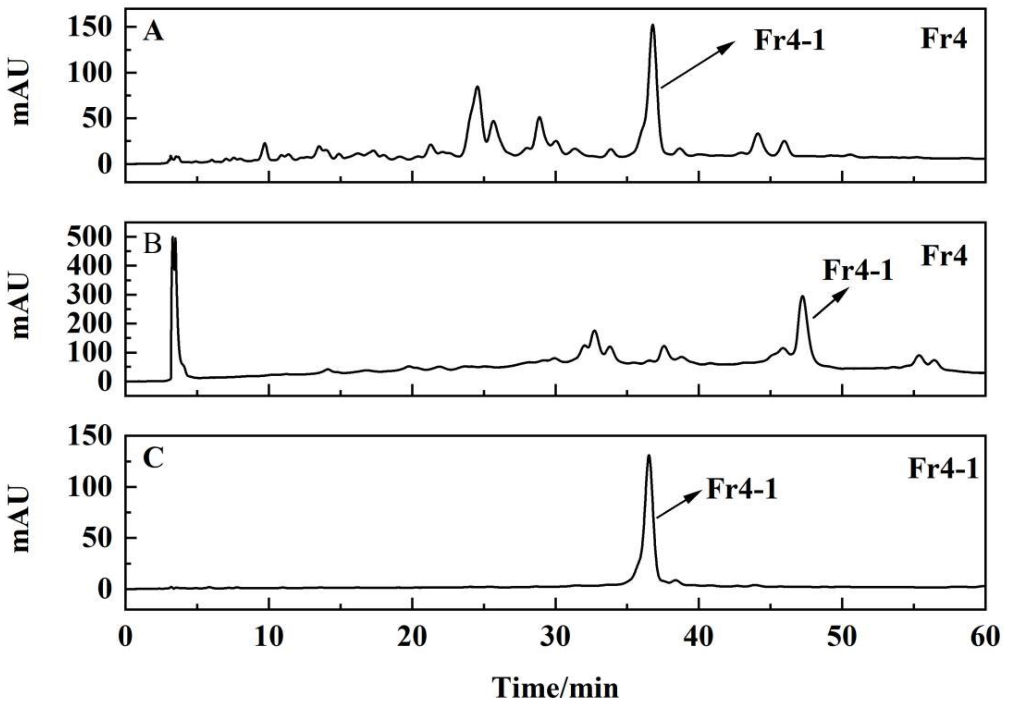
The 2D preparation system was constructed to obtain major DPPH inhibitors from Fr4. In order to purify and improve the peak resolution, the 1D preparation was carried on Megres C18 preparation column (20 × 250 mm, 5 μm), and the analysis and preparation chromatograms of the target fraction-Fr4 are shown in Figure 3. After fourteen repeated separations, 620.7 mg Fr4-1 was collected. As was showed in Figure 3C, the chromatograms of Fr4-1 exhibited one peak, but Fr4-1 was obviously impure and needed 2D purification.
In order to make it purification easier with the help of orthogonality [22], a hydrophilic column-Click XIon analytical column was used for analyzing Fr4-1. The result was shown in Figure 4. When injection volume was increased from 5, 10, 20, 40, to 80 μL with same conditions: mobile phase A: 0.2% v/v formic acid in water, and B: acetonitrile; gradient: 0–60 min, 90%–75% v/v B; monitoring wavelength: 254 nm; flow rate: 1.0 mL/min for analysis; injection volume: 10 μL; column temperature: 40 °C. The retention time for the two peaks marked Fr4-1-1 and Fr4-1-2 remained relatively unchanged while sample volume increased. In addition, better loadability was observed. When sample volume being increased, the chromatograms exhibited good resolution. Although flat head of peak 1 was observed as the volume increased to 80 μL, peak 2 maintained its shape, and the selectivity was sufficient for purification. The experimental result indicated that the Click XIon column has a higher loadability. As shown in Figure 4F, the preparation chromatograms of Fr4-1 on the Click XIon preparation column had the same conditions for analysis however the flow rate was 19.0 mL/min; the injection volume was 1 mL and the column temperature was room temperature. The 2D preparation had good reproducibility on the Click XIon preparation column, although retention time was delayed relative to analysis. Thus, it was possible to separate Fr4-1-1 and Fr4-1-2 using the Click XIon. Consequently, two compounds (Fr4-1-1: 199.4 mg and Fr4-1-2: 9.1 mg) were obtained.

3.3. Purity, Activity Assessment, and Structural Identification of Two DPPH Inhibitors

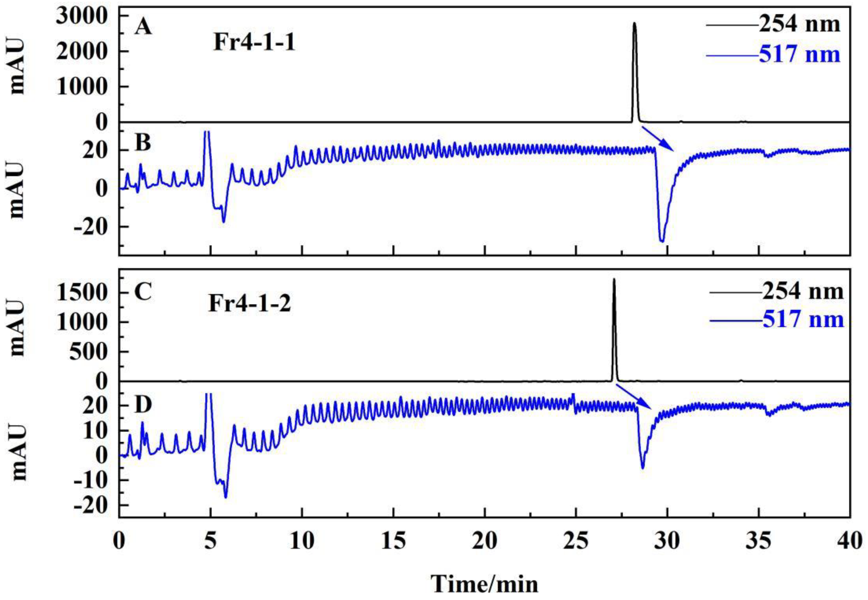
Using the online HPLC-DPPH system, the Dubhe C18 analytical column was used to reassess the purity and activity of Fr4-1-1 and Fr4-1-2 (isolated DPPH inhibitors). As shown in Figure 5A,C, the two compounds had purity above 98.0%. Figure 5B,D showed that the two compounds obtained have certain DPPH inhibitory activity based on better resolution and S/N ratio of negative peaks [27]. Obtained MS, 1HNMR and 13CNMR spectra were compared with the literature to clarify the structure of the two compounds (Figure 6). Figures S1–S8 showed the structure identification of Fr4-1-1 and Fr4-1-2 from Supplementary Materials.
Fr4-1-1 was identified as Quercetin-3-O-β-D-glucopyranoside-7-O-α-L-rhamnopyranoside according to spectrum data the literature reported [28], ESI-MS m/z 609.2 [M + Na] +, yellow powder. The purity reached 99.4%. Fr4-1-2 was identified as Quercetin-3-O-β-D-glucopyranoside-7-O-β-D-glucopyranosyl-(1→3)-α-L-rhamnopyranoside, according to the spectrum data reported by the literature [29]. ESI-MS m/z 771.3 [M + Na]+, yellow powder. The purity reached 98.5%.

3.4. Antioxidant Capacity Tests of the Isolated Antioxidants

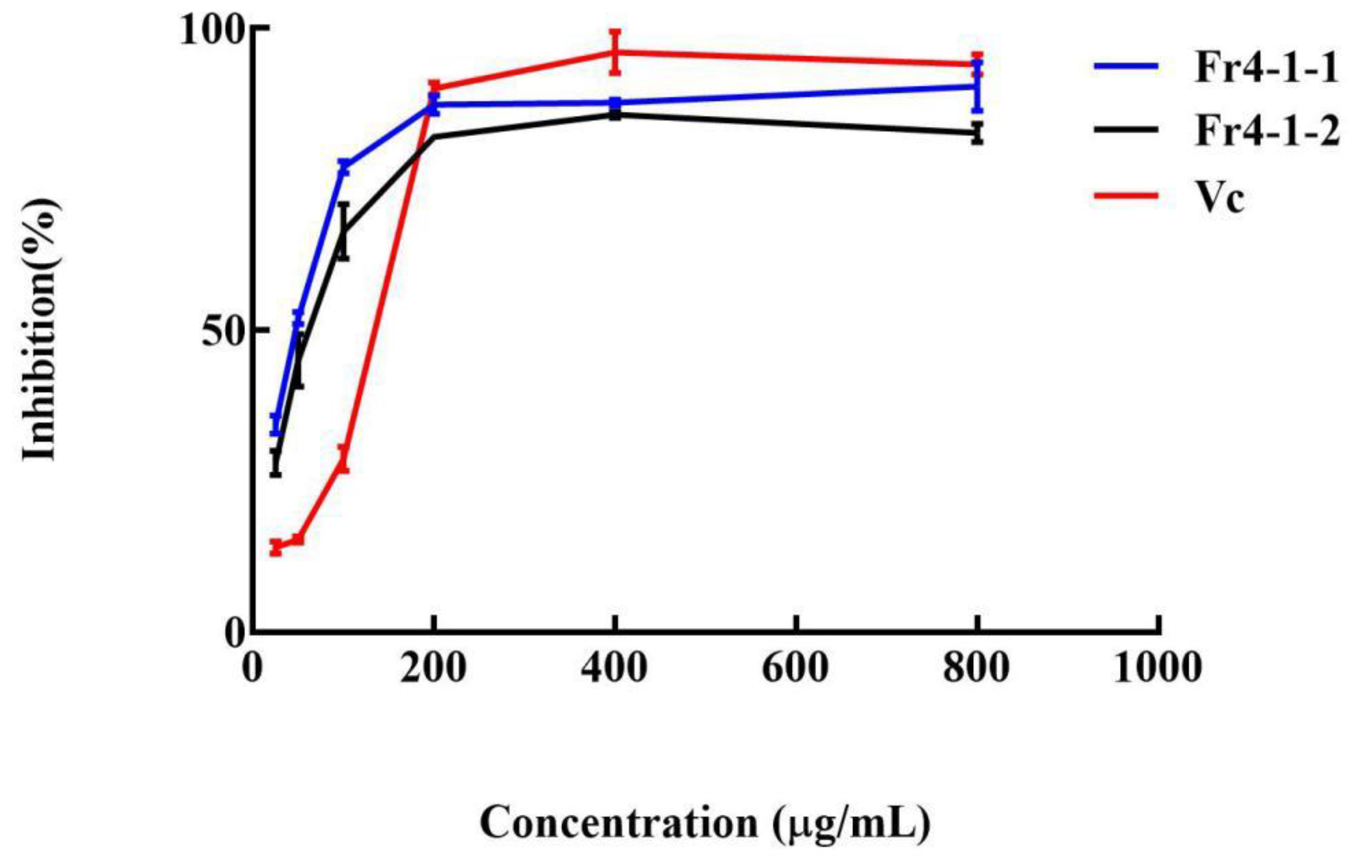
The scavenging ability assays against DPPH radical, was commonly used to determine the in vitro antioxidant activities. In vitro, DPPH radical clearance assays, of isolated compounds, with UV-detected, were completed. It is known that the lower IC50, the higher activity. The IC50 values of the Fr4-1-1, and Fr4-1-2 were 59.9 and 71.3 μg/mL, respectively (Figure 7), compared to 128.1 μg/mL for vitamin C. The activity of the two compounds showed a similar trend to VC, from concentrations of 20 μg/mL to 200 μg/mL: the activity increased sharply with increasing concentration, but the activity flattened between 200 μg/mL and 800 μg/mL. It can also be seen that at concentrations less than 200 μg/mL, the DPPH radical scavenging activities of compounds were evidently higher than VC, at concentrations exceeding 200 μg/mL, the DPPH radical scavenging activities of compounds were a little lower than VC, and the reason for this crossover was unclear, maybe due to structural differences in the compounds. The specific mechanism of this phenomenon remains to be clarified in later experiments. In sum, the experiment of antioxidant capacity tests of the isolated compounds verified that DPPH inhibitors Fr4-1-1, Fr4-1-2 obtained by the online 2D preparation HPLC-DPPH system have high antioxidant capacity.

4. Conclusions

In this study, a new method, medium- and high-pressure chromatography, combined with the online HPLC-DPPH assay, was implemented for screening, isolating, purifying of DPPH main inhibitors from L. latifolium. Initially, an MCI GEL® CHP20P sorbent was used to prepare the crude sample and collect Fr4-target fraction. Subsequently, a 2D RPLC/HILIC system was employed to separate DPPH inhibitors from Fr4. Finally, two DPPH inhibitors (Quercetin-3-O-β-D-glucopyranoside-7-O-α-L-rhamnopyranoside and Quercetin 3-O-β-D-glucopyranoside-7-O-β-D-glucopyranosyl-(1→3)-α-L-rhamnopyranoside) were obtained from L. latifolium, and exhibited high antioxidant activity with IC50 values 59.9 and 71.3 μg/mL, respectively. The two antioxidant flavonoids were not found by Yuan [10] who obtained nine antioxidant flavonoids from L. latifolium, so that the results of this study complemented the research on antioxidant compounds in L. latifolium. Consequently, these data suggest that the proposed method can prepare structurally similar antioxidant flavonoids from natural products. At the same time, because L. latifolium is so widely distributed and easy to obtain, and the isolated natural antioxidant Fr4-1-1 is the main compound of the plant, it is possible to extend longevity [30]. Furthermore, because it is greener and more economical than synthetic antioxidants, it can be prepared and used on a large scale with the constructed method.

Supplementary Materials

The following are available online at https://www.mdpi.com/article/10.3390/separations8120238/s1, Figure S1: ESI mass spectrum of Fr4-1-1. Figure S2: 1HNMR Spectrum (600 MHz) of Fr4-1-1 (in MeOD-d4). Figure S3: 13CNMR Spectrum (600 MHz) of Fr4-1-1 (in MeOD-d4). Figure S4: HMBC Spectrum (600 MHz) of Fr4-1-1 (in MeOD-d4). Figure S5: ESI mass spectrum of Fr4-1-2. Figure S6: 1HNMR Spectrum (600 MHz) of Fr4-1-2 (in MeOD-d4). Figure S7: 13CNMR Spectrum (600 MHz) of Fr4-1-2 (in MeOD-d4). Figure S8: HMBC Spectrum (600 MHz) of Fr4-1-2 (in MeOD-d4).

Author Contributions

Conceptualization, methodology, writing—original draft, X.W.; Formal analysis, Y.Z.; Investigation, N.W.; Data curation, J.C.; Supervision, Y.T. and Supervision, project administration, and funding acquisition R.Y. All authors have read and agreed to the published version of the manuscript.

Funding

This research was funded by Science and Technology Project of Qinghai Province (No. 2021-HZ-806), Natural Science Foundation of Qinghai Province (No. 2022-0302-ZJC-0055) and Science and Technology Project of Xining (No. 2018-Y-23) and Innovation platform for the development and construction of special project of Key Laboratory of Tibetan Medicine Research of Qinghai Province (No. 2021-ZJ-Y03).

Institutional Review Board Statement

Not applicable.

Informed Consent Statement

Not applicable.

Data Availability Statement

All data are contained within the article.

Conflicts of Interest

The authors declare no conflict of interest.

References

  1. Querfurth, H.W.; LaFerla, F.M. Mechanisms of Disease Alzheimer’s disease. N. Engl. J. Med. 2010, 362, 329–344. [Google Scholar] [CrossRef] [PubMed] [Green Version]
  2. Dauer, W.; Przedborski, S. Parkinson’s Disease: Mechanisms and Models. Neuron 2003, 39, 889–909. [Google Scholar] [CrossRef] [Green Version]
  3. Reuter, S.; Gupta, S.C.; Chaturvedi, M.M.; Aggarwal, B.B. Oxidative stress, inflammation, and cancer: How are they linked? Free Radic. Biol. Med. 2010, 49, 1603–1616. [Google Scholar] [CrossRef] [Green Version]
  4. Finkel, T.; Holbrook, N. Oxidants, oxidative stress and the biology of ageing. Nature 2000, 408, 239–247. [Google Scholar] [CrossRef]
  5. Sayre, L.M.; Perry, G.; Smith, M.A. Oxidative stress and neurotoxicity. Chem. Res. Toxicol. 2008, 21, 172–188. [Google Scholar] [CrossRef] [PubMed] [Green Version]
  6. Giacco, F.; Brownlee, M. Oxidative stress and diabetic complications. Circ. Res. 2010, 107, 1058–1070. [Google Scholar] [CrossRef] [Green Version]
  7. Heitzer, T.; Schlinzig, T.; Krohn, K.; Meinertz, T.; Münzel, T. Endothelial dysfunction, Oxidative stress, and risk of cardiovascular events in patients with coronary artery disease. Circulation 2001, 104, 2673–2678. [Google Scholar] [CrossRef] [Green Version]
  8. Boulebd, H. Comparative study of the radical scavenging behavior of ascorbic acid, BHT, BHA and Trolox: Experimental and theoretical study. J. Mol. Struct. 2020, 1201, 127210. [Google Scholar] [CrossRef]
  9. Ignacio, G.D.; Sara, L.I.; Patricia, M.C.; Álvaro, P.V.; Mateo, T.G.; Elisa, M.M.; Claudio, J.V.; Felipe, L. Terpenoids and Polyphenols as Natural Antioxidant Agents in Food Preservation. Antioxidants 2018, 10, 1264. [Google Scholar]
  10. Yuan, X.; Wang, H.X.; Mei, L.J.; Tao, Y.D. Isolation, purification and identification of antioxidants from Lepidium latifolium extracts. Med. Chem. Res. 2018, 27, 37–45. [Google Scholar]
  11. Bowman, J.L.; Bruggemann, H.; Lee, J.Y.; Mummenhoff, K. Evolutionary chances in floral structure within Lepidium L. (Brassicaceae). Int. J. Plant Sci. 1999, 160, 917–929. [Google Scholar] [CrossRef] [PubMed]
  12. Tarandeep, K.; Khadim, H.; Ushma, K.; Ram, V.; Dhiraj, V. Evaluation of nutritional and antioxidant status of Lepidium latifolium Linn.: A novel phytofood from Ladakh. PLoS ONE 2013, 8, e69112. [Google Scholar] [CrossRef] [Green Version]
  13. Ivica, B.; Sabine, M.; Franko, B.; Patrick, R. Glucosinolates: Novel Sources and Biological Potential. Glucosinolates; Springer: Berlin/Heidelberg, Germany, 2017; pp. 3–60. [Google Scholar]
  14. Northwest Plateau Institute of Biology, C.A.S. Economic Flora of Qinghai; Qinghai People’s Publishing House: Xining, China, 1987; Volume 227. [Google Scholar]
  15. Wright, C.I.; Van-Buren, L.; Kroner, C.I.; Koning, M.M.G. Herbal medicines as diuretics: A review of the scientific evidence. J. Ethnopharmacol. 2007, 114, 1–31. [Google Scholar] [CrossRef]
  16. Kadir, N.H.A.; David, R.; Rossiter, J.T.; Gooderham, N.J. The selective cytotoxicity of the alkenyl glucosinolate hydrolysis products and their presence in Brassica vegetables. Toxicology 2015, 334, 59–71. [Google Scholar] [CrossRef] [Green Version]
  17. Gianni, E.D.; Fimognari, C. Anticancer mechanism of sulfur-containing compounds. Enzymes 2015, 37, 167–192. [Google Scholar] [PubMed]
  18. Priya, D.K.D.; Gayathri, R.; Gunassekaran, G.R.; Murugan, S.; Sakthisekaran, D. Apoptotic role of natural isothiocyanate from broccoli (Brassica oleracea italica) in experimental chemical lung carcinogenesis. Pharm. Biol. 2013, 51, 621–628. [Google Scholar] [CrossRef] [PubMed] [Green Version]
  19. Navarro, E.; Alonso, J.; Rodriguez, R.; Trujillo, J.; Boada, J. Diuretic action of an aqueous extract of Lepidium latifolium L. J. Ethnopharmacol. 1994, 41, 65–69. [Google Scholar] [CrossRef]
  20. Shahidi, F. 1-Antioxidants: Principles and applications. In Handbook of Antioxidants for Food Preservation; Woodhead Publishing: Sawston, UK, 2015; pp. 1–14. [Google Scholar]
  21. Eklund, P.C.; Langvik, O.K.; Warna, J.P.; Salmi, T.O.; Willfor, S.M.; Sjoholm, R.E. Chemical studies on antioxidant mechanisms and free radical scavenging properties of lignans. Org. Biomol. Chem. 2005, 3, 3336–3347. [Google Scholar] [CrossRef] [PubMed]
  22. Dang, J.; Zhang, L.; Shao, Y.; Mei, L.J.; Liu, Z.G.; Yue, H.L.; Wang, Q.L.; Tao, Y.D. Preparative isolation of antioxidative compounds from Dracocephalum heterophyllum using off-line two-dimensional reversed-phase liquid chromatography/hydrophilic interaction chromatography guided by on-line HPLC-DPPH assay. J. Chromatogr. B 2018, 1095, 267–274. [Google Scholar] [CrossRef]
  23. Dang, J.; Chen, C.B.; Ma, J.B.; Dawa, Y.Z.; Wang, Q.; Tao, Y.D.; Wang, Q.L.; Ji, T.F. Preparative isolation of highly polar free radical inhibitor from Floccularia luteovirens using hydrophilic interaction chromatography directed by on-line HPLC-DPPH assay. J. Chromatogr. B 2020, 1142, 122043. [Google Scholar] [CrossRef]
  24. Cui, Y.L.; Shen, N.; Yuan, X.; Dang, J.; Shao, Y.; Mei, L.J.; Tao, Y.D.; Wang, Q.L.; Liu, Z.G. Two-dimensional chromatography based on on-line HPLC-DPPH bioactivity-guided assay for the preparative isolation of analogue antioxidant compound from Arenaria kansuensis. J. Chromatogr. B 2017, 1046, 81–86. [Google Scholar] [CrossRef] [PubMed]
  25. Liu, C.; Lei, Y.Q.; Dang, J.; Wang, W.D.; Zhang, J.; Mei, L.J.; Liu, Z.G.; Tao, Y.D.; Shao, Y. Preparative isolation of 1, 1-diphenyl-2-picrylhydrazyl inhibitors from Ribes himalense using mediu-pressure and two-dimensional reversed-phase/reversed-phase liquid chromatography guided by an online HPLC-1, 1-diphenyl-2-picrylhydrazyl assay. J. Sep. Sci. 2021, 44, 1345–1352. [Google Scholar] [CrossRef] [PubMed]
  26. Dang, J.; Zhang, L.; Wang, Q.L.; Mei, L.J.; Yue, H.L.; Liu, Z.G.; Shao, Y.; Gao, Q.B.; Tao, Y.D. Target separation of flavonoids from Saxifraga tangutica using two-dimensional hydrophilic interaction chromatography/reversed-phase liquid chromatography. J. Sep. Sci. 2018, 41, 4419–4429. [Google Scholar] [CrossRef] [PubMed]
  27. Chen, J.H.; Zhao, H.Q.; Shi, Q.; Zhang, D.L.; Cheng, H.Y.; Wang, X.R.; Lee, F.S.C. Rapid screening and identification of the antioxidants in Hippocampus japonicas Kaup by HPLC-ESI-TOF/MS and on-line ABTS free radical scavenging assay. J. Sep. Sci. 2010, 33, 672–677. [Google Scholar] [CrossRef] [PubMed]
  28. Aboushoer, M.I.; Fathy, H.M.; Abdel-Kader, M.S.; Goetz, G.; Omar, A.A. Terpenes and flavonoids from an Egyptian collection of Cleome droserifolia. Nat. Prod. Res. 2010, 24, 687–696. [Google Scholar] [CrossRef]
  29. Vitalini, S.; Braca, A.; Passarella, D.; Fico, G. New flavonol glycosides from Aconitum burnatii Gayer and Aconitum variegatum L. Fitoterapia. 2010, 81, 940–947. [Google Scholar] [CrossRef]
  30. Ahn, D.; Lee, E.B.; Kim, B.J.; Lee, S.Y.; Ahn, M.-S.; Eun, J.S.; Shin, T.-Y.; Kim, D.K. Lifespan extension property of quercetin-3-O-β-D-glucopyranoside-7-O-α-L-rhamnopyranoside from Curcuma longa L. in Caenorhabditis elegans. Saengyak Hakhoe Chi. 2014, 45, 275–281. [Google Scholar]
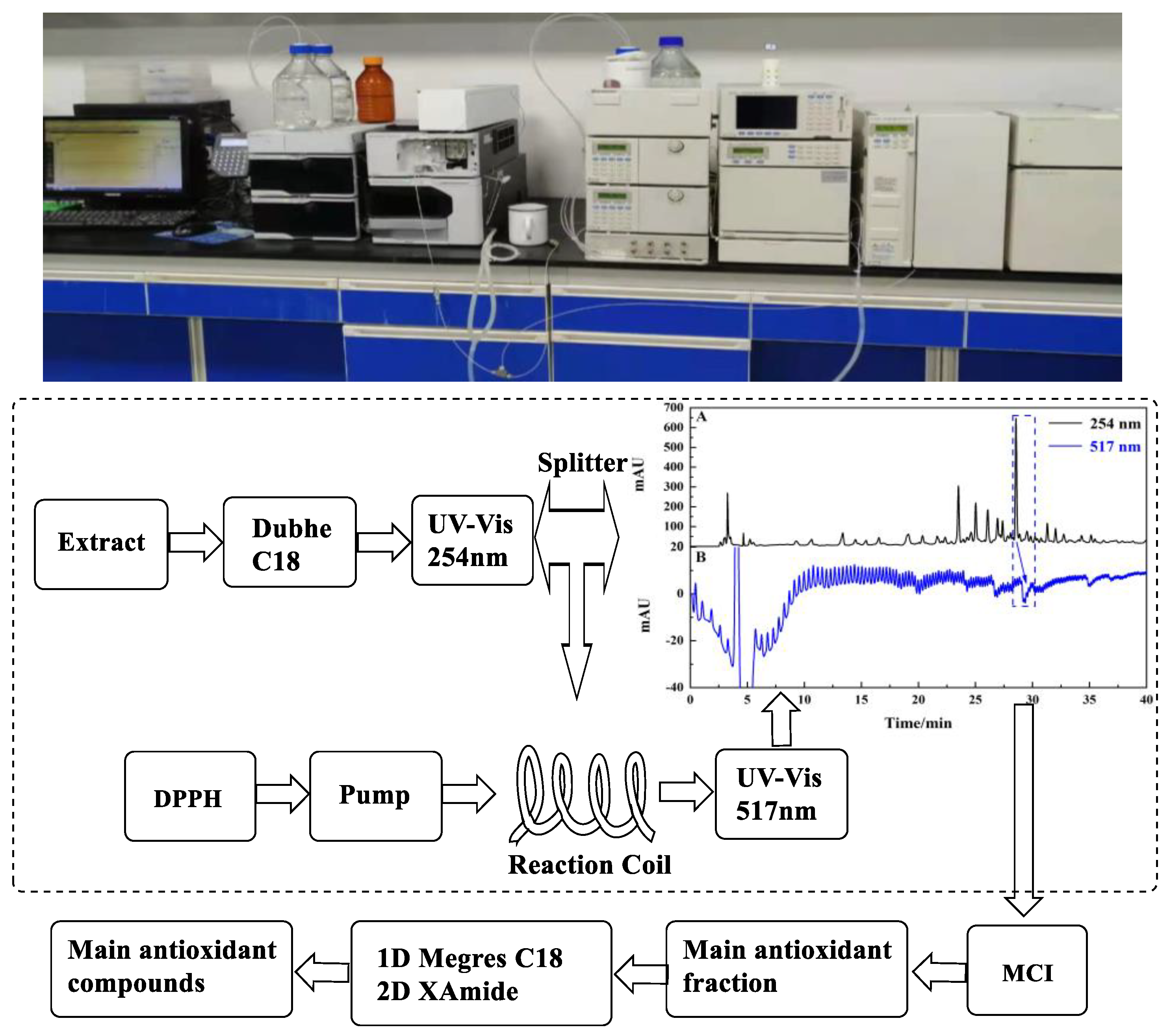
Figure 1. Scheme of performed experiment and used scientific equipment.
Separations 08 00238 g001
Figure 2. (A): The analytical spectra and 1,1-diphenyl-2-picrylhydrazyl (DPPH) inhibitory spectra on a Dubhe C18 analytical column of the L. latifolium crude sample. (B): the microgel® chp20p medium pressure liquid separation chromatography of L. latifolium. (C): HPLC analysis of the L. latifolium crude sample and Fr1-Fr5 on the analytical column (Dubhe C18) with the same conditions as 2A.
Separations 08 00238 g002
Figure 3. The first-dimensional analysis (A), preparation (B) of Fr4 and HPLC analysis of Fr4-1 (C) using the Megres C18 analytical and preparation columns.
Separations 08 00238 g003
Figure 4. (AE): HPLC analysis of Fr4-1 on Click XIon analytical column; (F): preparation chromatograms of Fr4-1 on the Click XIon preparation column.
Separations 08 00238 g004
Figure 5. Purity and DPPH inhibitory activity verification chromatograms of the isolated Fr4-1-1 (A,B), Fr4-1-2 (C,D), with the Dubhe C18 analytical column.
Separations 08 00238 g005
Figure 6. Chemical structures of the isolated compounds.
Separations 08 00238 g006
Figure 7. Antioxidant capacity of compounds Fr4-1-1, Fr4-1-2 and vitamin C.
Separations 08 00238 g007
Publisher’s Note: MDPI stays neutral with regard to jurisdictional claims in published maps and institutional affiliations.