Abstract
Extracts of Rheum palmatum L., Rhamnus purshiana DC., Rhamnus frangula L., and Cassia senna L. are used in traditional medicine thanks to their beneficial properties. These species contain hydroxyanthracene derivatives, considered genotoxic and possibly related to colorectal cancer development. This research aimed to study, using a micronucleus assay in vitro, the genotoxic potential of Rheum palmatum L., Rhamnus purshiana DC., Rhamnus frangula L. (bark), and Cassia senna L. (leaves and fruits) extracts. The extracts were evaluated at different concentrations: from 0 to 2000 µg/mL for Rhamnus purshiana DC, from 0 to 2500 µg/mL for Rheum palmatum L. and Rhamnus frangula L., and from 0 to 5000 µg/mL for Cassia senna L. The cytokinesis-block proliferation index was calculated to analyse if the used concentrations showed cytotoxicity. The hydroxyanthracene content varied between 0.06% and 0.23% for aloe-emodin, and between 0.07% and 0.16% for emodin and rhein. No cytotoxic effect was detected at any of these concentrations. Micronucleus analyses showed a lack of genotoxicity for all the extracts tested. These results show that Rheum palmatum L., Rhamnus purshiana DC, Rhamnus frangula L., and Cassia senna L. extracts do not induce genotoxicity since no increase in micronuclei formation in human lymphocytes in vitro was detected.
1. Introduction
In recent years, numerous studies have focused on the safety and toxicity of many botanical products that have been used as medicines and food supplements for many years. Botanical species and extracts are used as active pharmaceutical ingredients in traditional Chinese medicine, and their use is spreading all over the world [1].
Plant extracts are complex mixtures with biological properties. Among the components of several extracts, there are hydroxyanthracenes and their derivatives. The safety of hydroxyanthracenes was evaluated by the Panel of the EFSA, Food Additives, and Nutrient Sources added to Food (EFSA-ANS Panel) in 2018, which concluded that “hydroxyanthracene derivatives should be regarded as genotoxic and carcinogenic unless there are specific data to the contrary, […] and that there is a safety concern for extracts containing hydroxyanthracene derivatives although uncertainty persists” [2]. Many botanical preparations containing hydroxyanthracenes are under community scrutiny for new evaluations; such extracts include the ones obtained from the root or rhizome of Rheum palmatum L. and Rheum officinale Baillon, from the leaves and fruits of Cassia senna L., and from the bark of Rhamnus frangula L. and Rhamnus purshiana DC. [3].
According to the World Health Organization (WHO), the use of these types of products containing anthraquinones and hydroxyanthracenes should be limited to an average dose of 0.5–1.5 g of dried plant material or in decoction, a safe individual dose being 10–30 mg of hydroxyanthraquinones per day [4]. Moreover, they should not be used for more than 1–2 weeks due to their laxative properties that could potentially induce electrolyte imbalances. The European Medicines Agency (EMA) has stated that 30 mg/day should be the maximum dosage of hydroxyanthracene contents in medicinal products to be used as a laxative for adults, elderly, and adolescents over 12 years [5].
In traditional medicine, R. palmatum L. (Chinese rhubarb) is used to treat constipation, gastrointestinal haemorrhage, and ulcers [6,7]. Small doses of rhubarb extract show good digestive properties, while at higher doses it acts as a laxative. It is also used for its antibacterial, antioxidant, and anti-inflammatory effects [8,9]. C. senna L. is added in several herbal teas and used for purging and in weight loss [10]. R. frangula L. and R. purshiana L. are also used as purgatives. Hydroxyanthracene derivatives are present in these extracts as emodin, aloe-emodin, chrysophanol, physcion, and rhein [11].
We have recently demonstrated the lack of genotoxic effects in colon and kidney cells after in vivo treatment with Aloe ferox extract, containing hydroxyanthracene derivatives, and pure aloe-emodin in a comet assay [12,13]. Moreover, we have proved the absence of a genotoxic effect of a Rheum palmatum L. rhizome extract in Salmonella typhimurium, TA1535, TA1537, TA98, TA100, and Escherichia coli (WP2 uvrA) strains with the Ames test; the same extract was tested for micronuclei formation in isolated human lymphocytes showing negative results [14].
In this research, we investigated the genotoxic potential of five different extracts (R. palmatum L., R. purshiana DC, R. frangula L., and C. senna L. from fruits and leaves).
2. Materials and Methods
2.1. Extract Preparation
Extracts from R. palmatum L. (PAL), R. purshiana DC (PUR), and R. frangula L. (FRA), C. senna L. (leaves and fruits, CSL and CSF) were prepared and characterized analytically according to good agricultural and collection practices (GACP) and good manufacturing practices (GMP). Extract preparation is described in detail in the Supplementary Materials.
The PAL, PUR, and FRA extracts were kept at room temperature and prepared for the experiment immediately before use, dissolved in dimethylsulphoxide (DMSO, AppliChem, Darmstadt, Germany). One of the extracts of C. senna L. (from fruits; CSF) was kept at room temperature and prepared for the experiment immediately before use, dissolved in water, while the other (from leaves; CSL) was prepared in a complete culture medium.
2.2. Extract Characterization
Samples were characterized according to the specific analytical methods described in the pharmacopeia. In addition, aloin (A and B), aloe-emodin, rhein, and emodin were identified and quantified using an HPLC-UV method. More information on the extracts is available in the Supplementary Materials. The analytical methods, including the HPLC-UV analyses, were performed according to the relevant monographs for the four extracts reported in the current European Pharmacopoeia.
2.3. Peripheral Blood Lymphocyte Cultures
Blood samples were collected from healthy, young (under 35 years old), male, non-smoker donors, with no known illnesses or recent exposure to genotoxic agents, upon informed consent. Establishment of the blood cultures was done within 30 h from blood collection, preparing a 13% mixture of whole blood in a medium. The culture medium was Dulbecco’s Modified Eagles medium/Ham’s F12 (mixture 1:1) with 200 mM GlutaMAX™. The medium was completed with penicillin/streptomycin (100 U/mL/100 µg/mL), mitogen phytohaemagglutinin (PHA, 3 µg/mL as solvent lyophilizate), 10% foetal bovine serum (FBS), 10 mM HEPES, and the anticoagulant heparin (125 U.S.P.-U/mL). Cells were incubated at 37 °C with 5.5% CO2 in humidified air. For the analysis, only human lymphocytes were considered.
2.4. Mammalian Microsomal Fraction S9 Mix
The limited metabolic activation in in vitro systems is usually a major issue for the evaluation of genotoxic potential. For this reason, an exogenous metabolic activation was performed with the S9 mix obtained from rat liver. The S9 mix was prepared by ICCR-Roßdorf GmbH and stored following the currently valid version of the ICCR-Roßdorf standard operating procedure for rat liver S9 preparation.
The S9 supernatant was thawed and mixed with a cofactor solution and used at the final protein concentration of 0.75 mg/mL in the cultures. The S9 mix contains MgCl2 (8 mM), KCl (33 mM), glucose-6-phosphate (5 mM), and NADP (4 mM) in a sodium ortho-phosphate buffer (100 mM, pH 7.4).
2.5. Concentration Selection, Treatments, and Micronucleus Assay Preparation
First, the concentrations for the in vitro micronucleus test were selected according to the Organisation for Economic Co-operation and Development (OECD) guidelines. Different concentrations were chosen for each extract, taking into consideration the solubility properties of each of them. The concentrations analysed for the cytotoxicity evaluation ranged from 16.2 to 2500 µg/mL for PAL and FRA, from 13.0 to 2000 µg/mL for PUR, from 32.5 to 5000 µg/mL for CSF, and from 18.9 to 5000 µg/mL for CSL. No precipitation at the end of the treatment was observed either in the absence or the presence of the S9 mix for CSF, while it was observed starting at 466 µg/mL for PAL, at 816 µg/mL for FRA, at 1143 µg/mL for PUR, and at 544 µg/mL and above in the absence of the S9 mix and at 952 µg/mL and above in the presence of the S9 mix for CSL. The selected concentrations (expressed in w/v) for the treatment are reported in Table 1.

Table 1.
Doses selected for the treatments with the extracts of Rheum palmatum L. (PAL), Rhamnus purshiana DC (PUR), Rhamnus frangula L. (FRA), Cassia senna L. from fruits (CSF) and leaves (CSL). The concentrations selected for the treatments are reported in bold and are expressed in w/v.
Human lymphocytes proliferation was stimulated by adding the mitogen PHA to the culture medium for 48 h. To check the possible mutagenic effect of the extracts, two different treatments were performed. Forty-eight hours after seeding, 10 mL of the blood cultures was set up in parallel in 25 cm2 flasks for each extract concentration. For the treatment, the medium used was serum-free. For a set of samples, metabolic activation was promoted by the addition of the S9 mix (50 µL/mL culture medium). After 3 h, the cells were centrifuged for 5 min. The supernatant was removed, and the cells were resuspended and washed two times with saline G (pH 7.2, containing 8000 mg/L NaCl, 400 mg/L KCl, 1100 mg/L glucose × H2O, 192 mg/L Na2HPO4 × 2 H2O and 150 mg/L KH2PO4). The cells were resuspended in a culture medium with 10% FBS (v/v) in the presence of cytochalasin B (4–6 µg/mL) and cultured for 25 h [15].
Another set of cells were treated with the extracts and in the presence of cytochalasin B (4–6 µg/mL), and these cells were treated for 28 h [16].
For both treatments, the cultures were harvested by centrifugation 28 h after the beginning of each treatment. The cells were washed in saline G, resuspended in 5 mL KCl solution (0.0375 M), and incubated at 37 °C for 20 min. The cells were fixed with a 1 mL ice-cold mixture of methanol and glacial acetic acid (19 parts plus 1 part, respectively) and kept cold. The slides were prepared by dropping the cell suspension into fresh fixative. The cells were stained with Giemsa, mounted after drying, and covered with a coverslip.
2.6. Slide Evaluation and Cytotoxicity Analysis
The slides were evaluated with a 40× magnification. The micronuclei (MN) were counted in cells showing a clearly visible area of cytoplasm. MN were evaluated according to previously published criteria [17]. One thousand binucleated cells per culture were scored for cytogenetic damage on coded slides. The results are reported as % micronucleated cells. The cytotoxic effect was reported using the cytokinesis-block proliferation index (CBPI) (Formula (1) below) and cytotoxicity was expressed as % cytostasis (Formula (2)). A CBPI equal to 1 (all cells are mononucleated) means 100% cytostasis.
The cytotoxicity of a product is characterized by the percentages of reduction in the CBPI in comparison to the controls (% cytostasis) by counting 500 cells per culture. The data related to cytostasis and CBPI for the concentrations not evaluated for MN formation are reported in the Supplementary Material.
2.7. Statistical Analysis
Statistical significance was confirmed by a chi-squared test (p < 0.05), using a validated test script in R. The statistical analysis was conducted to detect significant differences in MN presence in treated samples compared to the concurrent solvent control. A linear regression was performed using R. A trend is considered significant whenever the p-value is below 0.05.
3. Results
3.1. Characterization
The samples were characterized according to the specific analytical methods described in the pharmacopeia. Table 2a,b report the percentage of hydroxyanthracene derivatives in the different botanical extracts tested in this study, identified by means of a high-performance liquid chromatograph-ultraviolet (HPLC-UV) detector.

Table 2.
(a) Composition of the extracts. All the data are expressed in percentages of the total weight of the extract. In addition, aloin (A and B), aloe-emodin, rhein, and emodin were identified and quantified using an HPLC-UV method. (b) Levels of aloin A and B, aloe emodin, rhein, and emodin in the tested extracts. All the data are expressed in percentages of the total weight of the extract.
3.2. Cytokinesis-Block Proliferation Index and Cytostasis
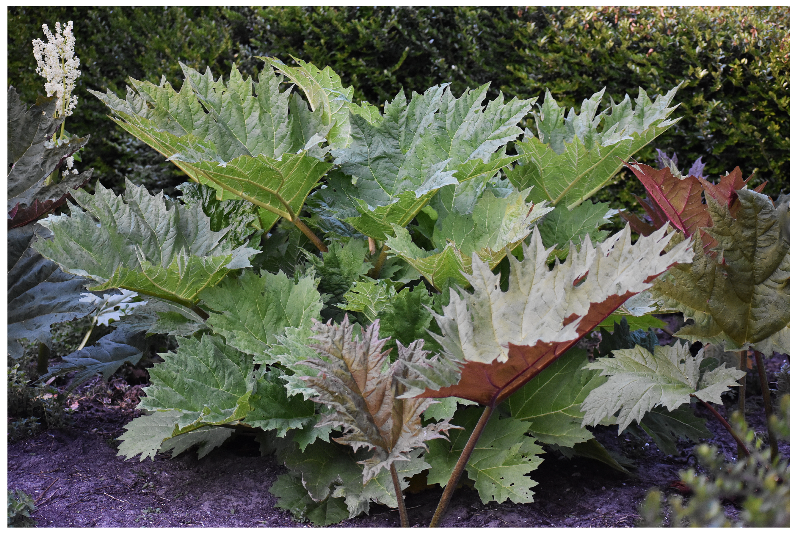
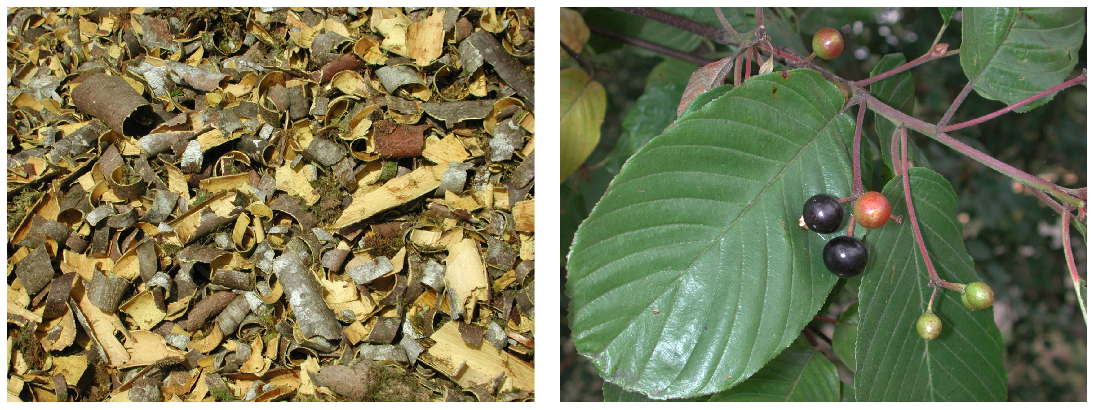
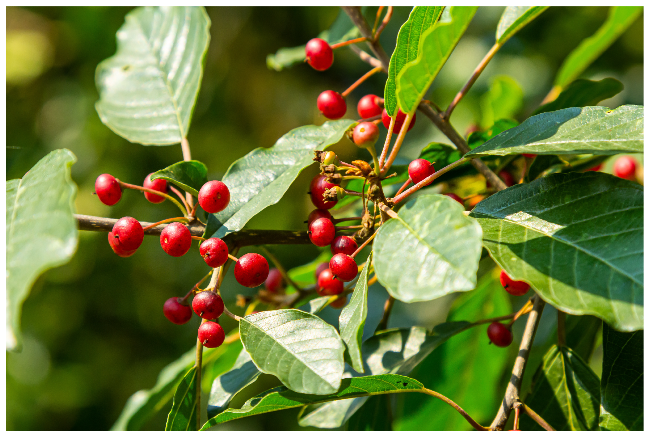
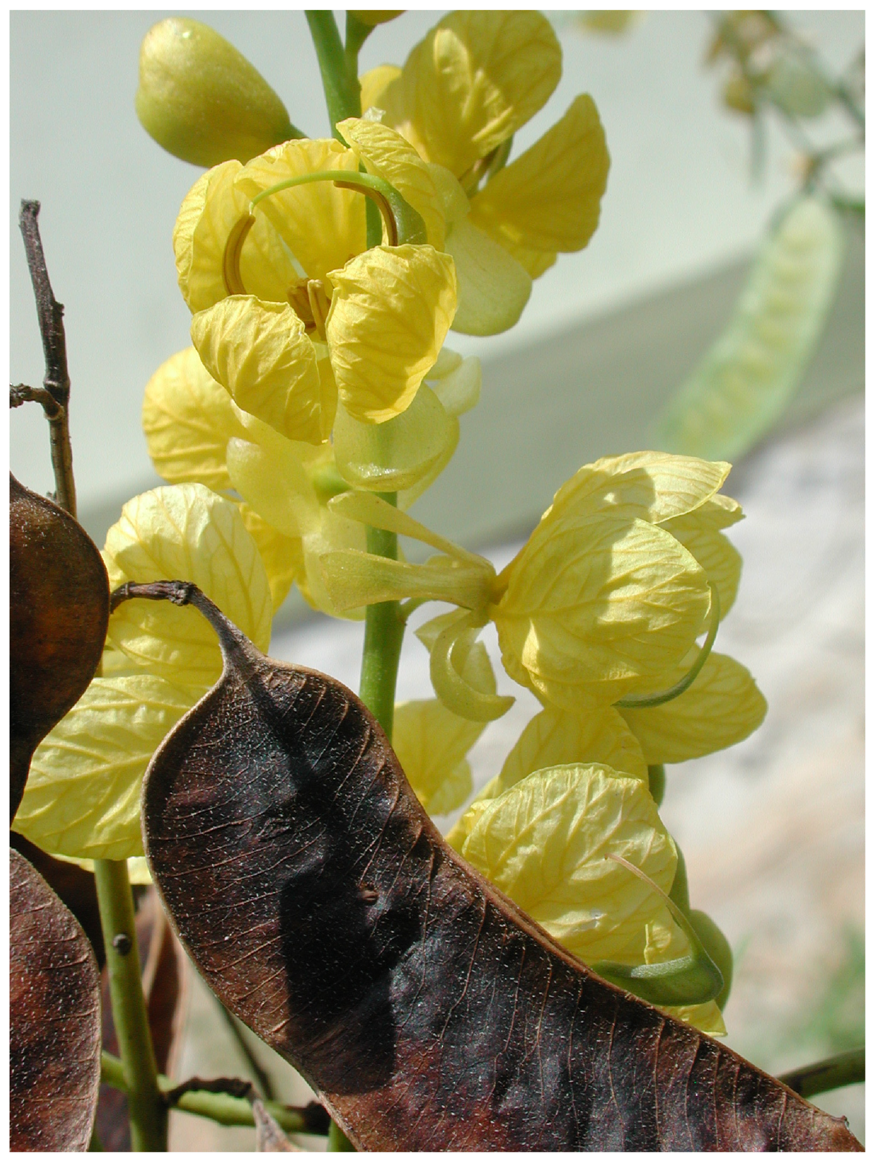
The CBPI and cytostasis were calculated for each of the treatments to obtain data on the cytotoxicity of the extracts. The results are presented in Table 3, Table 4, Table 5, Table 6 and Table 7. The treatment with PAL (Table 3) in the absence of the S9 mix at the highest concentrations, starting from 466 µg/mL, induced moderate cytotoxicity, while in the presence of the S9 mix, no cytotoxicity was detected. Following the long treatment, cytotoxicity was highlighted at the highest concentrations (from 833 µg/mL). For the treatment with PUR (Table 4), no cytotoxicity was observed at any concentration, while following the long treatment, the highest concentrations that showed precipitation at the end of the treatment (1333 and 2000 µg/mL) resulted in moderate cytotoxicity. For the treatment with FRA (Table 5), no cytotoxicity was observed at any concentration in the absence of the S9 mix, while in the presence of S9, the highest concentrations (starting from 816 µg/mL) induced moderate cytotoxicity. The treatment with CSF (Table 6) and CSL (Table 7) did not show cytotoxicity at any of the evaluated concentrations. All experimental plant specimens’ pictures are shown in Figure 1, Figure 2, Figure 3, Figure 4 and Figure 5.

Table 3.
Evaluation of the cytokinesis-block proliferation index (CBPI), cytostasis, and micronuclei in human lymphocytes after short treatment in the absence or presence of the S9 fraction (3 h) or long treatment in the absence of S9 fraction (28 h) with the extract of Rheum palmatum L. (PAL).

Table 4.
Evaluation of the cytokinesis-block proliferation index (CBPI), cytostasis, and micronuclei in human lymphocytes after short treatment in the absence or presence of the S9 fraction (3 h) or long treatment in the absence of the S9 fraction (28 h) with the extract of Rhamnus purshiana DC (PUR).

Table 5.
Evaluation of the cytokinesis-block proliferation index (CBPI), cytostasis, and micronuclei in human lymphocytes after short treatment in the absence or presence of the S9 fraction (3 h) or long treatment in the absence of the S9 fraction (28 h) with the extract of Rhamnus frangula L. (FRA).

Table 6.
Evaluation of the cytokinesis-block proliferation index (CBPI), cytostasis, and micronuclei in human lymphocytes after short treatment in the absence or presence of the S9 fraction (3 h) or long treatment in the absence of the S9 fraction (28 h) with the extract of Cassia senna L. fruits (CSF).

Table 7.
Evaluation of the cytokinesis-block proliferation index (CBPI), cytostasis, and micronuclei in human lymphocytes after short treatment in the absence or presence of the S9 fraction (3 h) or long treatment in the absence of the S9 fraction (28 h) with the extract of Cassia senna L. leaves (CSL).
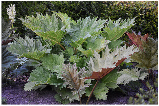
Figure 1.
Plant of Rheum palmatum L. (PAL). The extract tested in the study was obtained from bark.
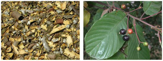
Figure 2.
Bark, fruits, and leaves of Rhamnus purshiana DC (PUR). The extract tested in the study was obtained from bark.
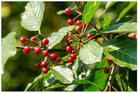
Figure 3.
Plant and fruits of Rhamnus frangula L. (FRA). The extract tested in the study was obtained from bark.
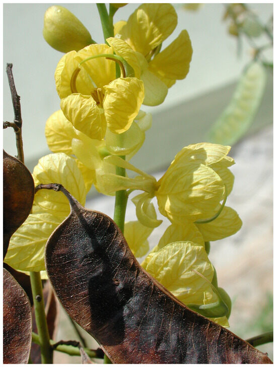
Figure 4.
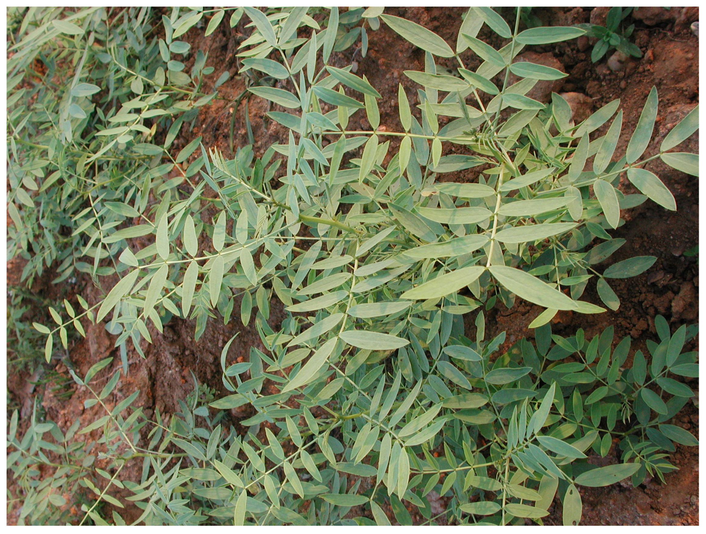
Leaves and flowers of Cassia senna L. fruits (CSF). The extract tested in the study was obtained from fruits.
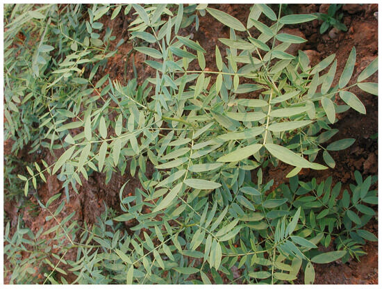
Figure 5.
Leaves of Cassia senna L. leaves (CSL).
3.3. Micronuclei
The results of the MN assay are reported in Table 3, Table 4, Table 5, Table 6 and Table 7. Statistically significant increases in the percentage of micronucleated cells were observed following treatments with the positive controls cyclophosphamide, mitomycin C, and vinblastine, indicating correct functioning of the assay. The treatment with the different extracts did not induce a statistically significant increase in the percentage of micronucleated cells compared to the control value at the tested concentrations, with the only exception being the highest concentration evaluated for PAL (466 µg/mL in the presence of the S9 mix, which is 1.10%). Nevertheless, the induction of MN at this concentration was found to be within the range of historical controls for the laboratory (0.15–1.15% micronucleated cells). Moreover, no concentration-related increase was detected. Thus, no statistically significant increase or dose relationship was seen for these treatments. These extracts were not able to induce MN formation in human lymphocytes after in vitro treatment.
4. Discussion
Hydroxyanthracene derivatives are a class of naturally occurring chemicals, generally used in food or dietary supplements to improve intestinal function through their laxative effect. A high number of plants contains hydroxyanthracene derivatives, which also belonging to various botanical families and genera.
Many extracts from plants, such as Rheum palmatum L., Rhamnus purshiana DC, Rhamnus frangula L., and Cassia senna L. leaves or fruits, contain hydroxyanthracene derivatives (aloins, emodin, aloe-emodin, rhein, etc.) in quite comparable concentrations. Some of them, such as emodin and aloe-emodin, are also naturally present in other plant species of very common use, such as lettuce, beans, and peas, as well as in coffee and tea, even if contained in traces [18].
The safety of products containing these natural botanical components has been addressed by different health authorities (WHO, EMA), which reached consistent conclusions [4,5] regarding the negative genotoxic potential when aloe or selected hydroxyanthracene derivatives (emodin and aloe-emodin) were administered to mice or rats [19,20,21]. However, these authorities also report that a causal relationship between anthranoid laxative abuse and colorectal cancer was not found [22,23,24,25]. The EMA monograph states that 30 mg/day of hydroxyanthracene derivatives should be the maximum dosage in medicinal products used with laxative properties in adults, the elderly, and adolescents over 12 years, and the WHO recommends the use of these products for no longer than 1–2 weeks due to a possible risk of electrolyte imbalances.
The European Commission, in March 2021, decided to ban aloe-emodin, emodin, and all extracts in which these substances are present, as well as extracts from the leaf of Aloe species containing hydroxyanthracene derivatives, leaving the safety assessment of other botanical preparations, such as Rheum palmatum L., Rhamnus purshiana DC, Rhamnus frangula L., Cassia senna L. leaves or fruits, pending for up to two years, even though they contain concentrations of hydroxyanthracene derivatives comparable to those of banned aloe preparations [3].
According to expert judgement, the alleged genotoxic and carcinogenicity potential of hydroxyanthracene derivatives seems more related to an epigenetic mechanism than to direct genotoxic activity. This seems to be confirmed by the experiment in which Aloe arborescens Miller, used as a food additive, exerted equivocal carcinogenic potential at a 4% high-dose level in the 2-year carcinogenicity study but not in the 0.8% and control groups. In females, the same results were demonstrated. The authors concluded that aloe is not carcinogenic at nontoxic-dose levels, and that the appearance of tumours in the colon at the 4% high-dose level was likely due to irritation of the intestinal tract by chronic diarrhoea [26].
An interesting scientific/regulatory debate regarding the toxicity of mixtures is currently underway. The opinion of the authors of this work is that while it is best to chemically characterize the components of a mixture, the safety assessment should be performed on the whole mixture, which will then be diluted and used in commercial products. It seems reasonable to assert that when the main components of the extracts are toxicologically evaluated individually, at very high doses, very far from the concentrations actually present in the mixture used by consumers, the results are not reliable for evaluating the hazard of the mixture. In designing toxicity tests and considering the nature of the material to be tested, it should be recognized that there may be significant differences in the pharmacological/toxic potency between a whole botanical extract and the equivalent amount of an isolated active principle [27,28,29,30,31].
Considering individual hydroxyanthracenes, it is worthy of note that in humans, aloe-emodin is rapidly converted and totally oxidized to rhein by gut microbiota [20,32,33,34]. Rhein is recognized to be devoid of any genotoxic activity [35] and considered to have certain anti-tumour effects [36].
Aloe-emodin and emodin, ubiquitous components of botanical extracts, were found to be genotoxic when tested in vitro [37,38,39,40], interacting with DNA topoisomerase II, but gave negative [13,36,38] or allegedly positive [40] results when tested in vivo. The information available on extracts seems to confirm a lack of genotoxicity when tested both in vitro and in vivo [12,14,41,42].
Recently, two original papers [12,13] have reported that, following oral gavage, neither aloe-emodin (purity 97.12%), dosed at 0, 250, 500, 1000, and 2000 mg/kg/day, nor dried Aloe ferox juice, dosed at 0, 500, 1000, and 2000 mg/kg/day, containing a well characterized concentration of hydroxyanthracene derivatives, induced DNA damage in preparations of single cells from the colon in in vivo-modified comet genotoxicity studies (OECD 489).
In the present study, extracts from Rheum palmatum L., Rhamnus purshiana DC, Rhamnus frangula L., and Cassia senna L. were evaluated in an in vitro micronucleus assay (OECD 487) and were found to be not genotoxic. Equally negative results were also obtained using the same botanical preparations in the Ames test [41].
Considering these results, single components as hydroxyanthracene derivatives in botanical extracts have to be considered as not having genotoxic potential. The results of the study we proposed showed that these extracts are not genotoxic when tested in vivo, and that the same botanical preparations, quite well characterized qualitatively and quantitatively regarding the hydroxyanthracene derivatives, yielded negative results in both in vitro and in vivo mutagenicity tests. It can be concluded that they can be considered safe as far as their genotoxicity potential goes. The weight of evidence of all the overall scientific lines of evidence reported so far leads to the conclusion that the hydroxyanthracenes examined (emodin, aloe-emodin, and rhein) are not hazardous. The same conclusion can be applied to the multiple extracts tested both in vivo and in vitro using internationally recognized standard protocols (OECD 471, 487, 489).
5. Conclusions
On the basis of these results, a risk characterization can be undertaken. The content of hydroxyanthracene derivatives in the botanical preparations tested in this study varied between 0.06% and 0.23% for aloe-emodin, and between 0.07% and 0.16% for emodin and rhein (Table 2).
Since the risk characterization is based on hazard and exposure, it is interesting to quantify the possible exposure to the botanical extracts based on the content of hydroxyanthracene derivatives in some food supplements. The quantities of hydroxyanthracene derivatives declared by the manufacturers of the botanical preparations provided by the Società Italiana di Scienze Applicate alle Piante Officinali e ai Prodotti per la Salute (SISTE) are in the range of 100–780 mg/day for Cassia senna L. fruits, 100–800 mg/day for Cassia senna L. leaves, 50–780 mg/day for Rheum palmatum L., 50–200 mg/day for Rhamnus purshiana DC., and 50–200 mg/day for Rhamnus frangula L. (dry extract).
As an indication, an attempted safety evaluation was made by estimating the dose of aloe-emodin to which a consumer of food supplements would be exposed by consuming products containing the tested Cassia senna and Rhamnus frangula. The dose of ingested aloe-emodin would be 0.00228 and 0.00657 mg/kg bw, in the worst case, for Cassia senna (800 mg) and Rhamnus frangula (200 mg) in a 70 kg individual. Such doses are tens of thousands of times lower than the doses that did not produce any genotoxic effects in in vivo tests.
The risk characterization exercise carried out shows that the toxicological data on the hazard identification of different botanical extracts, which are very well characterized for their anthracene derivatives content, combined with worst-case data for a realistic exposure, indicate that the risk deriving from the use of the test extracts can be considered negligible.
Supplementary Materials
The following supporting information can be downloaded at: https://www.mdpi.com/article/10.3390/separations11020047/s1, Methods of extracts preparation.
Author Contributions
Conceptualization, C.L.G. and M.M.; methodology, G.M.; formal analysis, G.M.; investigation, G.M.; resources, C.L.G. and M.M.; data curation, G.M., C.L.G. and M.M.; writing—original draft preparation, G.M., C.L.G., and M.M.; writing—review and editing, G.M., C.L.G. and M.M.; supervision, C.L.G. and M.M.; project administration, M.M.; funding acquisition, C.L.G. All authors have read and agreed to the published version of the manuscript.
Funding
This project was funded by the Italian Society of Toxicology (SITOX). We are also grateful for the support of the TWINALT-EU project 952404.
Data Availability Statement
Data are available upon request.
Acknowledgments
We thank Rachel Stenner and Robert Burns of Language Consulting Congressi, Milan, for English proofreading. We also thank Steffen Naumann, Sigrid Kuehnel, Sibylle Battenfeld, Andrea Sokolowski of ICCR-Roßdorf GmbH, where the chemical analyses were performed. The authors acknowledge the support of the APC central fund of the university of Milan.
Conflicts of Interest
The authors declare no conflicts of interest.
References
- Pace, R.; Martinelli, E.M. The Phytoequivalence of Herbal Extracts: A Critical Evaluation. Fitoterapia 2022, 162, 105262. [Google Scholar] [CrossRef]
- EFSA ANS Panel; Younes, M.; Aggett, P.; Aguilar, F.; Crebelli, R.; Filipi, M.; Frutos, M.J.; Galtier, P.; Gott, D.; Gundert-Remy, U.; et al. Safety of Hydroxyanthracene Derivatives for Use in Food. EFSA J. 2018, 16, e05090. [Google Scholar] [CrossRef] [PubMed]
- European Commission Commission Regulation (EU) 2021/468 of 18 March 2021 Amending Annex III to Regulation (EC) No 1925/2006 of the European Parliament and of the Council as Regards Botanical Species Containing Hydroxyanthracene Derivatives 2021. Available online: https://eur-lex.europa.eu/legal-content/EN/TXT/HTML/?uri=CELEX:32021R0468 (accessed on 7 January 2024).
- WHO. WHO Monographs on Selected Medicinal Plants; World Health Organization: Geneva, Switzerland, 1999; Volume 1, pp. 39–40. [Google Scholar]
- Committee on Herbal Medicinal Products (HMPC) Assessment Report on Rheum palmatum L. and Rheum Officinale Baillon, Radix. 2019; Volume 31. Available online: https://www.fitoterapia.net/archivos/202008/final-assessment-report-rheum-palmatum-l-rheum-officinale-baillon-radix-revision-1_en.pdf?1 (accessed on 7 January 2024).
- European Pharmacopoeia. European Directorate for the Quality of Medicines, 4th ed.; European Pharmacopoeia, Directorate for the Quality of Medicines, Council of Europe: Strasbourg, France, 2001. [Google Scholar]
- State Pharmacopoeia of the People’s Republic of China. State Pharmacopoeia Committee; Press, C.I., Ed.; First Div.: Beijing, China, 2010. [Google Scholar]
- Öztürk, M.; Aydoǧmuş-Öztürk, F.; Duru, M.E.; Topçu, G. Antioxidant Activity of Stem and Root Extracts of Rhubarb (Rheum Ribes): An Edible Medicinal Plant. Food Chem. 2007, 103, 623–630. [Google Scholar] [CrossRef]
- Wang, J.; Zhao, H.; Kong, W.; Jin, C.; Zhao, Y.; Qu, Y.; Xiao, X. Microcalorimetric Assay on the Antimicrobial Property of Five Hydroxyanthraquinone Derivatives in Rhubarb (Rheum palmatum L.) to Bifidobacterium adolescentis. Phytomedicine 2010, 17, 684–689. [Google Scholar] [CrossRef]
- National Institute of Diabetes and Digestive and Kidney Diseases. LiverTox: Clinical and Research Information on Drug-Induced Liver Injury; National Institute of Diabetes and Digestive and Kidney Diseases: Bethesda, MD, USA, 2012. [Google Scholar]
- Van den Berg, A.J.J.; Labadie, R.P. Anthraquinones, Anthrones and Dianthrones in Callus Cultures of Rhamnus Frangula and Rhamnus Purshiana. Planta Med. 1984, 50, 449–451. [Google Scholar] [CrossRef] [PubMed]
- Galli, C.L.; Cinelli, S.; Ciliutti, P.; Melzi, G.; Marinovich, M. Lack of in Vivo Genotoxic Effect of Dried Whole Aloe Ferox Juice. Toxicol. Rep. 2021, 8, 1471–1474. [Google Scholar] [CrossRef] [PubMed]
- Galli, C.L.; Cinelli, S.; Ciliutti, P.; Melzi, G.; Marinovich, M. Aloe-Emodin, a Hydroxyanthracene Derivative, Is Not Genotoxic in an in vivo Comet Test. Regul. Toxicol. Pharmacol. 2021, 124, 104967. [Google Scholar] [CrossRef]
- Melzi, G.; Galli, C.L.; Ciliutti, P.; Marabottini, C.; Marinovich, M. Lack of Genotoxicity of Rhubarb (Rhizome) in the Ames and Micronucleus in Vitro Tests. Toxicol. Rep. 2022, 9, 1574–1579. [Google Scholar] [CrossRef]
- Lorge, E.; Thybaud, V.V.V.; Aardema, M.J.; Oliver, J.; Wakata, A.; Lorenzon, G.; Marzin, D.; Clare, M.G.; Lorenzon, G.; Akhurst, L.C.; et al. SFTG International Collaborative Study on in Vitro Micronucleus Test. Mutat. Res. Toxicol. Environ. Mutagen. 2006, 607, 13–36. [Google Scholar] [CrossRef]
- Whitwell, J.; Smith, R.; Chirom, T.; Watters, G.; Hargreaves, V.; Lloyd, M.; Phillips, S.; Clements, J. Inclusion of an Extended Treatment with Recovery Improves the Results for the Human Peripheral Blood Lymphocyte Micronucleus Assay. Mutagenesis 2019, 34, 217–237. [Google Scholar] [CrossRef]
- Countryman, P.I.; Heddle, J.A. The Production of Micronuclei from Chromosome Aberrations in Irradiated Cultures of Human Lymphocytes. Mutat. Res. Mol. Mech. Mutagen. 1976, 41, 321–331. [Google Scholar] [CrossRef]
- Xiang, H.; Zuo, J.; Guo, F.; Dong, D. What We Already Know about Rhubarb: A Comprehensive Review. Chin. Med. 2020, 15, 88. [Google Scholar] [CrossRef]
- Brown, J.P. A Review of the Genetic Effects of Naturally Occurring Flavonoids, Anthraquinones and Related Compounds. Mutat. Res. Genet. Toxicol. 1980, 75, 243–277. [Google Scholar] [CrossRef]
- Lang, W. Pharmacokinetic-Metabolic Studies with 14C-Aloe Emodin after Oral Administration to Male and Female Rats. Pharmacology 1993, 47 (Suppl. S1), 110–119. [Google Scholar] [CrossRef]
- Westendorf, J.; Marquardt, H.; Poginsky, B.; Dominiak, M.; Schmidt, J.; Marquardt, H. Genotoxicity of Naturally Occurring Hydroxyanthraquinones. Mutat. Res. Toxicol. 1990, 240, 1–12. [Google Scholar] [CrossRef] [PubMed]
- Loew, D. Pseudomelanosis Coli Durch Anthranoide. Z. Für Prakt. 1994, 16, 312–318. [Google Scholar]
- Patel, P.M.; Selby, P.J.; Deacon, J.; Chilvers, C.; McElwain, T.J. Anthraquinone Laxatives and Human Cancer: An Association in One Case. Postgrad. Med. J. 1989, 65, 216–217. [Google Scholar] [CrossRef] [PubMed]
- Siegers, C.-P. Anthranoid Laxatives Colorectal Cancer. Trends Pharmacol. Sci. 1992, 13, 229–231. [Google Scholar] [CrossRef] [PubMed]
- Siegers, C.P.; von Hertzberg-Lottin, E.; Otte, M.; Schneider, B. Anthranoid Laxative Abuse—A Risk for Colorectal Cancer? Gut 1993, 34, 1099–1101. [Google Scholar] [CrossRef] [PubMed]
- Yokohira, M.; Matsuda, Y.; Suzuki, S.; Hosokawa, K.; Yamakawa, K.; Hashimoto, N.; Saoo, K.; Nabae, K.; Doi, Y.; Kuno, T.; et al. Equivocal Colonic Carcinogenicity of Aloe Arborescens Miller Var. Natalensis Berger at High-Dose Level in a Wistar Hannover Rat 2-y Study. J. Food Sci. 2009, 74, T24–T30. [Google Scholar] [CrossRef] [PubMed]
- Al-Malahmeh, A.J.; Al-ajlouni, A.M.; Wesseling, S.; Vervoort, J.; Rietjens, I.M.C.M. Determination and Risk Assessment of Naturally Occurring Genotoxic and Carcinogenic Alkenylbenzenes in Basil-Containing Sauce of Pesto. Toxicol. Rep. 2017, 4, 1–8. [Google Scholar] [CrossRef]
- Alhusainy, W.; Paini, A.; van den Berg, J.H.J.; Punt, A.; Scholz, G.; Schilter, B.; van Bladeren, P.J.; Taylor, S.; Adams, T.B.; Rietjens, I.M.C.M. In Vivo Validation and Physiologically Based Biokinetic Modeling of the Inhibition of SULT-Mediated Estragole DNA Adduct Formation in the Liver of Male Sprague-Dawley Rats by the Basil Flavonoid Nevadensin. Mol. Nutr. Food Res. 2013, 57, 1969–1978. [Google Scholar] [CrossRef] [PubMed]
- Jeurissen, S.M.F.; Punt, A.; Delatour, T.; Rietjens, I.M.C.M. Basil Extract Inhibits the Sulfotransferase Mediated Formation of DNA Adducts of the Procarcinogen 1′-Hydroxyestragole by Rat and Human Liver S9 Homogenates and in HepG2 Human Hepatoma Cells. Food Chem. Toxicol. 2008, 46, 2296–2302. [Google Scholar] [CrossRef] [PubMed]
- Marabini, L.; Galli, C.L.; La Fauci, P.; Marinovich, M. Effect of Plant Extracts on the Genotoxicity of 1′-Hydroxy Alkenylbenzenes. Regul. Toxicol. Pharmacol. 2019, 105, 36–41. [Google Scholar] [CrossRef] [PubMed]
- van den Berg, S.J.P.L.; Klaus, V.; Alhusainy, W.; Rietjens, I.M.C.M. Matrix-Derived Combination Effect and Risk Assessment for Estragole from Basil-Containing Plant Food Supplements (PFS). Food Chem. Toxicol. 2013, 62, 32–40. [Google Scholar] [CrossRef] [PubMed]
- Cho, J.H.; Chae, J.-I.; Shim, J.-H. Rhein Exhibits Antitumorigenic Effects by Interfering with the Interaction between Prolyl Isomerase Pin1 and C-Jun. Oncol. Rep. 2017, 37, 1865–1872. [Google Scholar] [CrossRef] [PubMed]
- EMA. Assessment Report for Rhubarb (Rhei Radix); European Medicines Agency: London, UK, 2008. [Google Scholar]
- Lee, J.-H.; Kim, J.M.; Kim, C. Pharmacokinetic Analysis of Rhein in Rheum undulatum L. J. Ethnopharmacol. 2003, 84, 5–9. [Google Scholar] [CrossRef]
- Heidemann, A.; Miltenburger, H.G.; Mengs, U. The Genotoxicity Status of Senna. Pharmacology 1993, 47 (Suppl. S1), 178–186. [Google Scholar] [CrossRef]
- Mengs, U.; Krumbiegel, G.; Völkner, W. Lack of Emodin Genotoxicity in the Mouse Micronucleus Assay. Mutat. Res. Toxicol. Environ. Mutagen. 1997, 393, 289–293. [Google Scholar] [CrossRef]
- Chen, Y.-Y.; Chiang, S.-Y.; Lin, J.-G.; Yang, J.-S.; Ma, Y.-S.; Liao, C.-L.; Lai, T.-Y.; Tang, N.-Y.; Chung, J.-G. Emodin, Aloe-Emodin and Rhein Induced DNA Damage and Inhibited DNA Repair Gene Expression in SCC-4 Human Tongue Cancer Cells. Anticancer Res. 2010, 30, 945–951. [Google Scholar]
- Heidemann, A.; Völkner, W.; Mengs, U. Genotoxicity of Aloeemodin in Vitro and in Vivo. Mutat. Res. Toxicol. 1996, 367, 123–133. [Google Scholar] [CrossRef]
- Müller, S.O.; Eckert, I.; Lutz, W.K.; Stopper, H. Genotoxicity of the Laxative Drug Components Emodin, Aloe-Emodin and Danthron in Mammalian Cells: Topoisomerase II Mediated? Mutat. Res. 1996, 371, 165–173. [Google Scholar] [CrossRef] [PubMed]
- Nesslany, F.; Simar-meintières, S.; Ficheux, H.; Marzin, D. Aloe-Emodin-Induced DNA Fragmentation in the Mouse in vivo Comet Assay. Mutat. Res. Toxicol. Environ. Mutagen. 2009, 678, 13–19. [Google Scholar] [CrossRef] [PubMed]
- Haßler, S.; Jung, K.; Kelber, O.; Feistel, B.; Steinhoff, B.; Nieber, K. P08-22 Assessment of the Genotoxic Safety of Herbal Drug Preparations Containing Anthraquinone Derivatives. Toxicol. Lett. 2022, 368, S149–S150. [Google Scholar] [CrossRef]
- Williams, L.D.; Burdock, G.A.; Shin, E.; Kim, S.; Jo, T.H.; Jones, K.N.; Matulka, R.A. Safety Studies Conducted on a Proprietary High-Purity Aloe Vera Inner Leaf Fillet Preparation, Qmatrix®. Regul. Toxicol. Pharmacol. 2010, 57, 90–98. [Google Scholar] [CrossRef]
Disclaimer/Publisher’s Note: The statements, opinions and data contained in all publications are solely those of the individual author(s) and contributor(s) and not of MDPI and/or the editor(s). MDPI and/or the editor(s) disclaim responsibility for any injury to people or property resulting from any ideas, methods, instructions or products referred to in the content. |
© 2024 by the authors. Licensee MDPI, Basel, Switzerland. This article is an open access article distributed under the terms and conditions of the Creative Commons Attribution (CC BY) license (https://creativecommons.org/licenses/by/4.0/).

