Introduction
Functional neurological disorder (FND), previously known as conversion disorder (CD), represents a puzzling and debilitating condition that has attracted the attention of physicians, scientists and philosophers for centuries. FND/CD was part of the foundation of modern neurology and psychiatry, but has gone through many names and classifications during the last century, reflecting the different ways in which this clinical entity has been viewed and explained. These descriptions have ranged from “hysteria” (deriving from the antique theory of a “wandering womb”) to conversion (after Freud’s psychodynamic theory) or dissociative disorder (based on Janet’s conception of conscious and unconscious divisions in the mind). In the current brief review, I discuss definitions and epidemiology of FND/CD, and highlight brain imaging results that helped shed new light on the neural correlates of this perplexing disorder.
Definitions and epidemiology
FND/CD constitutes up to 16% of referrals to neurology clinics [1], accounting for 30 to 60% of neurological outpatients in some centres [2,3] and making up an estimated 20% of neurologists’ workload [4]. The prevalence of FND in general hospital inpatients ranges between 5 and 16% [5]. This disorder can cause an important disability rate [6], comparable to neurological disease associated with organic brain lesions [7]. Although serious research efforts have been dedicated to this chronic and disabling disorder, the underlying pathogenic mechanisms remain poorly understood. Motor FND/CD is among the most common clinical presentations, and will be the main focus of the current review, given that it is also the symptom most researched with neuroimaging. The patients exhibit one or more symptoms of altered voluntary motor function, such as weakness or paralysis, abnormal movement (e.g., tremor, dystonia, myoclonus, gait disorder). According to the Diagnostic and Statistical Manual of Mental Disorders (DSM-5) [8], the associated clinical findings are incompatible with lesions in the central or peripheral nervous system (
Table 1).
The patients do not appear to feign their symptoms in order to obtain material or psychological gain, but their deficit seems to arise from a genuine disorder of the subjective experience of the patients, who maintain they have lost a function despite intact anatomical pathways [9].
Early neurobiological accounts of functional/conversion disorder
Physicians have tried to explain functional symptoms since the dawn of ages. Hippocrates (c. 460 – c. 370 BC), the famous doctor of ancient Greece, linked the emergence of hysterical symptoms with a displaced uterus wandering in the brain and causing symptoms in women. But theories formed in the late part of the 19th century had the greatest impact on our current conception of FND/CD. Jean-Martin Charcot (1825–1893), who provided handmade illustrations of these patients, linked functional motor symptoms labelled as hysteria with a strong “fixed idea” of impotence that permeates consciousness in the terrain of a particularly charged affective state, causing disturbances in internal representation of movement [10].
Table 1.
Definition and classification of functional neurological disorder / conversion disorder in DSM-5.
Table 1.
Definition and classification of functional neurological disorder / conversion disorder in DSM-5.
For Joseph Babinski (1857–1932), hysteria was a disorder of “persuasion”, in that the rise of symptoms was due to self-suggestion and symptoms could be abolished by persuasion [11]. Like Charcot, Babinski believed that functional symptoms arise as a result of disturbances in the subjective experience of the movement or sensation rather than organic damage. Josef Breuer (1842–1925) and, afterwards, Sigmund Freud (1856–1939) postulated that such symptoms arise in grounds of psychodynamic conflict between the conscious and unconscious mind [12]. Such conflicts were unconsciously repressed (often due to their sexual origin) and transformed into symbolic complaints in the body to alleviate such a conflict, hence the term “conversion” that was later used to refer to this condition in other versions of the DSM. Freud famously quipped that these “patients suffer from reminiscences” – that is “invading representations of memories of phantasies” that somehow took control of their consciousness and behaviour. Thus, according to him, patients might overvalue certain past events (e.g. ,related to childhood trauma), which could subsequently engender a somatic or neurological reaction through non-conscious processes of symbolisation. Although all these early theories were based only on clinical observation and speculation, they partly echo recent findings from brain imaging studies, including ours (see below).
Neuroimaging evidence in FND/CD
The advent of functional neuroimaging in the last 20 years allowed more detailed systemic investigations of FND/CD. Research focused mainly on motor symptoms and more rarely explored non-motor domains. Different motor paradigms were used to investigate active versus attempted movements [13,14,15,16,17], passive movements or vibration [18,19,20], motor imagery [21,22], and motor observation [23]. Other studies have used non-motor tasks such as emotional faces, autobiographical memory and so on [24,25]. Overall, the view has emerged that symptoms of motor conversion may arise as a result of some form of top-down regulation exerted by higher-order brain regions, which could ultimately interfere with motor function [26,27,28,29]. However, the source(s) of such influences remain unresolved and debated. Growing neuroimaging evidence points to a key role for areas such as the ventral medial prefrontal cortex (VMPFC), anterior cingulate cortex (ACC), or supplementary motor area (SMA) found to be frequently hyperactive in FND patients, whereas other areas such as the temporoparietal junction (TPJ) or caudate display hypoactivation. Of note, activity in the primary motor cortex is often found to be comparable to healthy controls, suggesting that motor deficits cannot be accounted for simply from abnormalities in primary motor pathways. Here, a question of particular clinical importance is whether FND patients intentionally produce or fake their motor symptoms. In order to explore this question, several studies have included a group of healthy controls who feigned the conversion symptoms of patients. Functional brain imaging was performed in feigning group simulating upper-limb motor loss [30], leg weakness [14], left-hand weakness [16], right or left side conversion paresis [22], or tremor [15]. These studies have clearly shown distinctive changes in neural systems recruited by conversion patients as compared with those simulating symptoms [9,31]. Taken together, this evidence shows that patients do not just fake or fabricate their symptoms, but they present with a genuine disorder of subjective experience.
Studies have clearly shown distinctive changes in neural systems recruited by conversion patients as compared with those simulating symptoms.
It has been suggested that top-down influences from the prefrontal cortices might result in exaggerated self monitoring [16,32] and disrupt conscious recall of unwanted memories [25]. Other studies imputed deficits to abnormal voluntary action selection [28], impaired motor intentions [30,33], or impaired execution with intact intention and preparation [16,29]. Abnormal recruitment of midline prefrontal regions critical for self-referential representations, emotional regulation, and autobiographical memory [16,29] may imbue motor action with affective valence ultimately interfering with motor function [9]. Dysfunction in the VMPFC has been repeatedly linked to pathophysiology in FND patients [13,16,34], in line with a key role for the VMPFC in promoting patterns of behaviour through processes that integrate affective value with autobiographical memory information and providing modulatory input to sensorimotor circuits [9,16,18,34].
Anomalies in the limbic system associated with affect and motivation were also often invoked [18,29,35,36,37]. Several studies reported abnormal emotion processing resulting in an abnormal response to the startle reflex [38], a higher state of arousal [39,40] and stress [41,42], as well as increased engagement of defensive behaviour [43,44,45,46]. Finally, abnormal emotion-movement interactions have been suspected based on findings of increased functional connectivity between brain regions implicated in motor control and emotion processing such as SMA and amygdala, respectively [24,25,47,48].
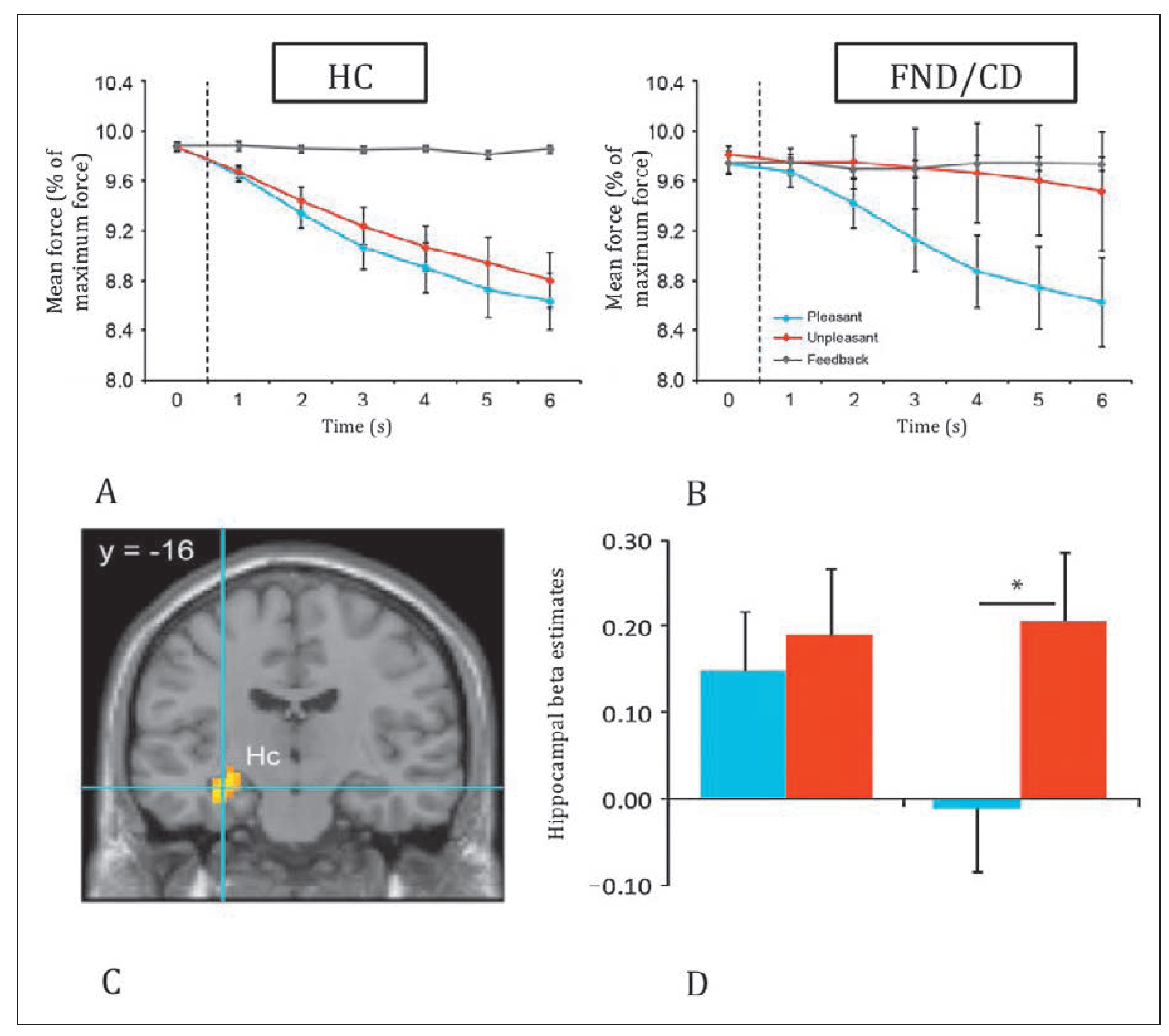
In our own research [46], we directly tested for the neural pathways linking emotion appraisal in limbic regions and impaired motor function in patients with motor FND/CD. We presented patients and controls with pleasant and unpleasant emotional images while they produced an isometric motor action (precision-grip contraction at 10% of maximum force). Both groups could either visually assess their ongoing motor output (control condition) or continue to view emotional images (pleasant and aversive conditions) while trying to maintain the target motor force. For controls, as expected, force output decayed over time while viewing images, either pleasant or aversive (fig. 1A). Patients, however, produced a constant, maintained motor output, with no force decay, while they viewed aversive images, but exhibited a normal reduction pattern during pleasant images (fig. 1B). These results reveal a significant effect of negative affect on motor output in FND/CD patients. At the brain level, such modulation of force control by emotional valence was associated with increased activity in the right inferior frontal cortex and the presupplementary motor area with the aversive compared with the pleasant conditions in controls. By contrast, patients recruited more the posterior cingulate cortex and hippocampus (shown in fig. 1C,D), two areas critically implicated in emotional memory, in addition to the cerebellum (vermis) which is involved in motor and emotional learning. These findings highlight a cerebellar-limbic circuit whose recruitment might distinctively arise through the activation of memory traces associated with past traumatic and negative affective memories, leading to impaired motor function in FND/CD patients.
Taken together, these findings demonstrate for the first time an intriguing link between motor control and associated activation of brain areas implicated in memory and emotional learning, rather than strictly motor pathways. These data point to a surprising convergence with the Freudian proposal of memory reminiscence in the emergence of FND, and more generally support the notion of top-down interference from affective and/or cognitive system on motor function, which may alter subjective experience and self-monitoring processes during motor action. Further research is needed to extend these findings to other neurological symptoms observed in FND, such a tremor, blindness and others.
A role for metacognitive deficits in motor FND/CD
A central feature of FND/CD is the dissociation between patients’ intact motor function (investigated with clinical and routine imaging) and their subjective conscious experience of these abilities (they’re unable to move). Such awareness of one’s own action and success of performance is tightly linked to the capacity of self monitoring. There are numerous situations in both health and disease where people judge the accuracy of their actions, perceptions or decisions in a manner that does not reflect their actual objective performance, thus either overestimating or underestimating their capacity.
Notably, dissociations between objective and subjective performance are common for classic neurological disorders such as blindsight [49] and anosognosia [51]. They are also observed in normal individuals under special experimental conditions [50]. Recent theoretical accounts in neuroscience have linked subjective conscious experience associated with self monitoring to metacognitive processes [49,51,52,53,54] – cognitive processes operating on other cognitive/internal processes. For instance, blindsight is a condition that results from damage in the primary visual (striate) cortex (V1), in which individuals can discriminate visual stimuli above chance level in simple visual tasks, even though they claim to not subjectively perceive the stimuli [55]. According to Rosenthal [51], blindsight may result thus from a specific deficit producing “a meta-representation” of the visual information, which could form the basis of conscious experience. According to a signal detection theory (SDT) account put forward by Ko and Lau [49], blindsight entails a metacognition failure, reflected by abnormally conservative criterion for monitoring visual performances, leading to overall low confidence ratings during discrimination tasks and poor correlation between such ratings and actual accuracy.
Figure 1.
Summary of findings in the study by Blakemore et al. [46]. Top panel, behavioural results. Mean force (expressed as % maximum force) produced by participants over each 1-second epoch, for each experimental condition (pleasant, unpleasant, feedback). Bottom panel, functional magnetic resonance imaging (fMRI) results. Statistical maps (C) and mean activity estimates (D) of brain areas activated in the aversive versus pleasant (U>P) condition contrast in FND/CD relative healthy controls (HC) in the hippocampus. Error bars represent standard error.
Figure 1.
Summary of findings in the study by Blakemore et al. [46]. Top panel, behavioural results. Mean force (expressed as % maximum force) produced by participants over each 1-second epoch, for each experimental condition (pleasant, unpleasant, feedback). Bottom panel, functional magnetic resonance imaging (fMRI) results. Statistical maps (C) and mean activity estimates (D) of brain areas activated in the aversive versus pleasant (U>P) condition contrast in FND/CD relative healthy controls (HC) in the hippocampus. Error bars represent standard error.
Conversely, anosognosia is another common neurological disorder, often resulting from parietal and frontal lesions in the right hemisphere, and characterised by erroneous beliefs of intact motor function despite stark evidence of paralysis. In a recent neuropsychological study, Vocat et al. [56] asked anosognosic individuals, brain-damaged patients without anosognosia and healthy subjects, to perform a guessing task where target words had to be identified while participants received successive informative clues. After each clue, patients and controls had not only to suggest their guess to solve this riddle by proposing a word solution, but also to rate their confidence on the accuracy of this solution. Results showed that patients with anosognosia were overconfident of their solutions despite insufficient information. Furthermore, even after presentation of clues debunking their previously suggested words, anosognosic patients (unlike nosognosics or healthy controls) had difficulty in modifying their original solutions. This behaviour was interpreted as an inability to update beliefs about current state, and thus constitutes a failure to produce adjusted metacognitive representations concerning a novel state of sensorimotor function (here due to brain pathology), which may ultimately lead to unawareness of the deficit.
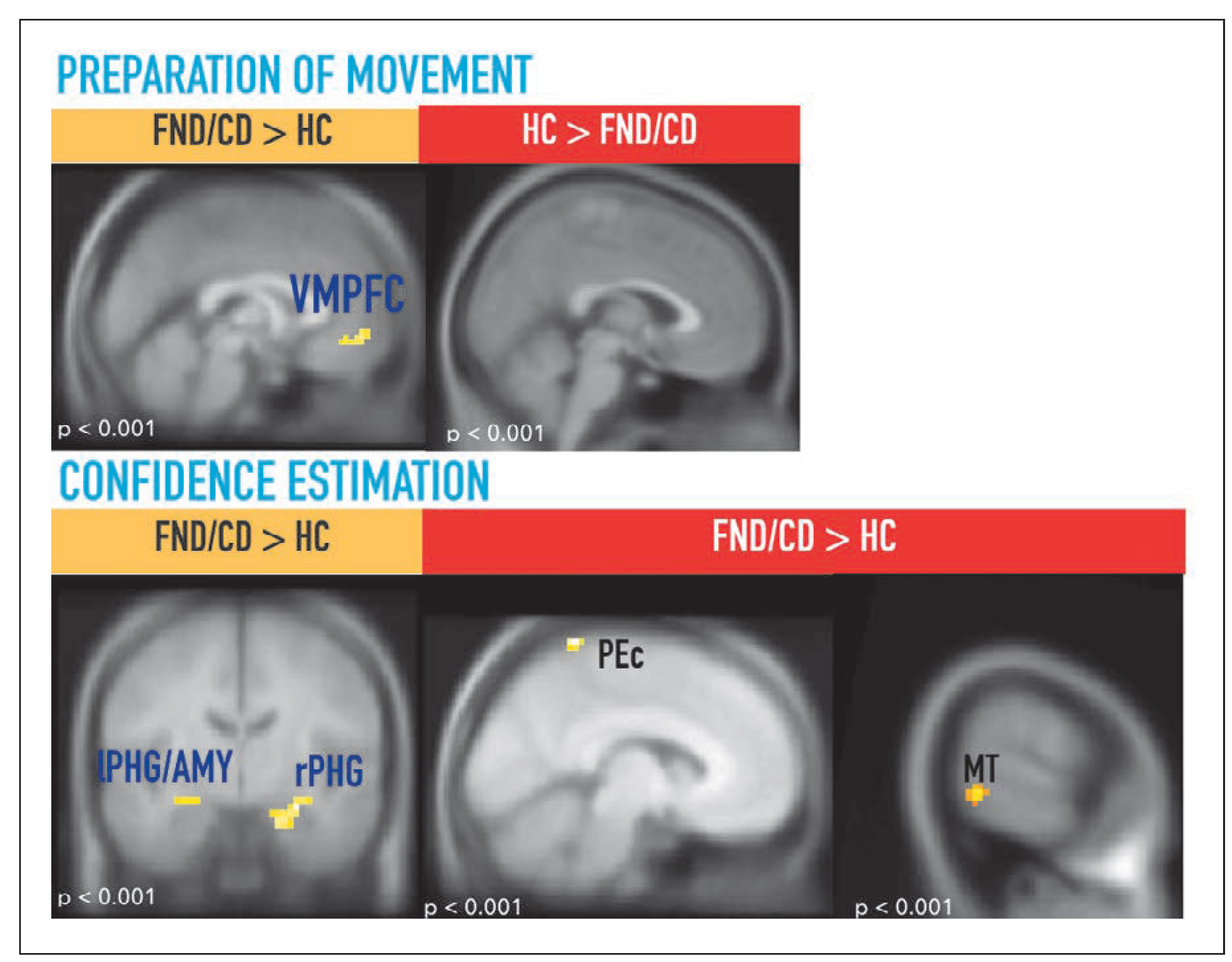
By analogy, in conversion disorder, metacognitive function might be impaired in a distinctive manner, with these patients experiencing a form of “motor blindsight”, namely preserved motor function with impaired subjective experience. Alternatively, metacognitive function may be intact per se, yet internal information used for judging motor performance may be fundamentally different in patients compared with healthy controls, ultimately resulting in impaired subjective experience. Specifically, based on our work in patients and healthy individuals [46,57], we hypothesise that self-monitoring processes governing metacognition in FND/CD may stem from information retrieved from past memories and affectively tagged associations, through abnormal recruitment of limbic and memory circuits. This account echoes classic interpretations by Freud and others pointing to the role of past traumatic memories and personally relevant affective appraisals. Indeed, seemingly neutral judgements may not be entirely neutral when people monitor their own doings, as they may be engaged and tagged by the value of associated memories, even during simple perceptual tasks [58,59,60]. At the brain level, recent studies indicate that such metacognitive processes applied to neutral stimuli may recruit the hippocampus [58,59] and VMPFC [59,60], both regions associated with self-relevant memory and frequently over-activated in FND/CD [9,16,46]. How does motor metacognition operate in conversion disorder patients, who frequently complain of being paralysed without organic damage to motor pathways? How do they evaluate their own motor performance in basic visuo-motor control tasks? To address this question, we employed an experimental paradigm previously used in healthy people [57] to test for metacognitive monitoring of motor action in FND/CD patients. Participants had to make movements, with their hand hidden from direct view, in order to draw straight trajectories on a screen, while deviations were occasionally introduced in the visible reaching trajectory. In these conditions, it is known that participants can make corrective movements with or without awareness of the deviation [61]. In our study, they had to first report whether they detected such deviations and subsequently evaluate their confidence in the accuracy of their response. Our hypothesis was that if patients display “motor blindsight”, then we should find significant differences in metacognition indices in the presence of an intact motor response. Alternatively, if patients have intact metacognition ability but use distinctive internal information to self monitor, then we should observe accurate metacognition scores, but differential recruitment of brain systems during their own performance judgements, in particular in memory and related limbic regions, in line with other recent findings [24,46] and theoretical accounts [9]. In agreement with the second hypothesis, our results showed different brain activation patterns for motor self monitoring in healthy controls and conversion patients. Notably, controls engaged the left superior parietal cortex (responsible for proprioceptivemotor integration) and extrastriate visual cortex (encompassing middle temporal region responsible for motion processing) when evaluating their visuo-motor reaching movement and confidence [34]. By contrast, motor FND/CD patients recruited bilateral parahippocampal regions when making confidence judgement about their visuo-motor action, suggesting that these patients relied on memory and contextual associative processing, rather than sensorimotor and visual information (fig. 2).
Seemingly neutral judgements may not be entirely neutral when people monitor their own doings, as they may be engaged and tagged by the value of associated memories.
Interestingly, heightened VMPFC recruitment was found during the preparation of upcoming movement in patients but not controls (fig 2, top panel). Taken together, this pattern of activity may possibly account for how affect and personal memories (e.g., linked to past trauma or personal contextual associations) may imbue self monitoring and current motor experience in these patients.
These recent results therefore complement previous models in FND/CD that have suggested that an influence of internal representations arising from imagery and/or memory might interfere with the control of motor behaviour [9,12]. According to a model developed recently [9], imagery and memory regions may provide modulatory inputs to sensorimotor regions through the integration of sensorimotor activity with internal priors derived from memory or imagery. Such effects may be partly similar to the influence of hypnosis on motor function [16], as proposed by Charcot [10], but mediated through different neural pathways (engaged by suggestion in hypnosis rather than emotional memory and self-monitoring in FND/ CD) [9,62]. Future studies should further elucidate the more exact role of memory related systems, including VMPFC, PCC, and PHG, in CD/FND, and verify their involvement in other non-motor symptoms.
Figure 2.
Summary of MRI results showing different effects in FND/CD patients and HC taken from Begue et al. [34]. Top panel: Statistical map showing different effects during movement preparation, with greater activation in VMPFC in FND/CD compared with HC (left) but no activation for HC > FND/CD (right). Bottom panel: Statistical map showing different effects during confidence, with greater activation in hippocampal-amygdala regions in FND/CD compared with HC during self monitoring (confidence estimation about trajectory correction accuracy). Statistical map showing greater activation in superior medial parietal cortex (PEc) and visual extrastriate cortex (MT) for HC compared with FND/CD during the same condition of confidence judgment. VMPFC: ventromedial prefrontal cortex; rPHG: right parahippocampal gyrus; lPHG/lAmy: left parahippocampal gyrus/left amygdala; PEc: superior anterior portion of the precuneus; MT: middle temporal gyrus.
Figure 2.
Summary of MRI results showing different effects in FND/CD patients and HC taken from Begue et al. [34]. Top panel: Statistical map showing different effects during movement preparation, with greater activation in VMPFC in FND/CD compared with HC (left) but no activation for HC > FND/CD (right). Bottom panel: Statistical map showing different effects during confidence, with greater activation in hippocampal-amygdala regions in FND/CD compared with HC during self monitoring (confidence estimation about trajectory correction accuracy). Statistical map showing greater activation in superior medial parietal cortex (PEc) and visual extrastriate cortex (MT) for HC compared with FND/CD during the same condition of confidence judgment. VMPFC: ventromedial prefrontal cortex; rPHG: right parahippocampal gyrus; lPHG/lAmy: left parahippocampal gyrus/left amygdala; PEc: superior anterior portion of the precuneus; MT: middle temporal gyrus.
Our current ongoing projects
In this context, our current ongoing projects aim to investigate FND/CD patients with different symptoms and follow changes in brain activity patterns as a function of clinical follow-up. In particular, our research will focus on patients presenting with pseudo-seizure as well as positive motor symptoms (tremor, dystonia) or non-motor symptoms (blindness). Similar brain imaging protocols will be conducted in different patient populations with the goal to identify both commonalities and specificities of different FND/CD symptoms. In addition, in the longer term, longitudinal studies are envisaged in order to assess patients after specific treatment protocols, which are now being put in place through a multidisciplinary approach [63,64]. In this context, we are currently recruiting patients suffering from a variety of functional neurological symptoms (motor, sensory, visual, speech, etc.).
Brain imaging protocols will be conducted in different patient populations with the goal to identify both commonalities and specificities of different FND/CD symptoms.
We plan to test these patients in a comprehensive array of tasks using functional neuroimaging measures and hope to compare distinct phenotypes in order to determine differences in underlying brain mechanisms potentially related to different symptom generation, as well as any similarities reflecting potentially shared causes for their emergence. Better understanding the neural substrates of particular symptoms and their origin will in turn help to define better therapeutic management for these patients, who often remain difficult to treat.
Recruitment criteria for ongoing studies
Inclusion criteria:
Ages eligible for study: 18–65 years old
Sexes eligible for study: M or F
Functional neurological disorders diagnosed by a neurologist:
− Functional motor symptoms: functional tremor, functional
− gait, astasia-abasia, functional dystonia, functional tic, functional jerk, functional myoclonus, functional weakness, functional paralysis, functional (psychogenic non epileptic) seizures
− Functional sensory symptoms: functional visual loss affecting acuity or field of view, functional hemianopia, tinnitus
− Functional speech and swallowing symptoms: dysphonia, dysarthria, functional dysphagia
− Compatibility with MRI (no metal in the body, absence of pregnancy, no major surgery in the last 6 months, no claustrophobia)
Study design:
− Patients willing to participate will undergo a full clinical examination and interview, followed by functional MRI of the brain.
− Free clinical examination session of approximately 1 hour and MRI scanning session of approximately 1 hour on different days. If additional sessions are needed, it will be discussed directly with the patients.
− MRI will be acquired during both passive (rest) and active (task) conditions. MRI has no radiation and is safe if patients are compatible (e.g. no metal in the body, or other exclusion criteria mentioned above).
− Patients will perform simple motor and perceptual tasks in the scanner, with their response given through a dedicated joystick interface connected with a computer outside the MRI room.
− Longitudinal follow up (6 to 12 months) will be proposed and discussed with the patients and their primary physicians on an individual basis.
− No invasive procedures (e.g., injections) will be performed.
− Patients will receive coverage for their transportation fees (e.g. train, bus, etc.)
− Patients can interrupt the study at any time, no justification needed.
Physicians or patients can contact for more information: neurofon-geneve[at]unige.ch, Centre Medical Universitaire, Université de Genève, 1 rue Michel-Servet 1211, Genève 4
Conclusion
Modern neuroscience is only beginning to unravel the neural mechanisms underlying FND/CD disorder and will increasingly help to bridge the gap between the brain and the mind. Patients suffering from FND/ CD are not faking or simulating their symptoms. Instead, top-down interference with motor function resulting from abnormal recruitment of internal selfmonitoring systems, associated with memory and emotional processes, appear to be implicated in the emergence of their symptoms. These recent results add novel insights enriching classic psychodynamic hypotheses formed by Charcot, Freud and others more than a century ago. However, although modern neuroscience approaches now better highlight the neural pathways linking memory and self awareness to FND/CD (how symptoms emerge), more efforts still must be made to better elucidate the exact causes of such changes in brain function (why symptoms emerge) [65].