Next Article in Journal
Neurological manifestations of Behçet’s disease
Previous Article in Journal
Neuroborreliose: klinisches Spektrum, Diagnose und Therapie
 
 
Swiss Archives of Neurology, Psychiatry and Psychotherapy is published by MDPI from Volume 176 Issue 1 (2026). Previous articles were published by another publisher in Open Access under a CC-BY (or CC-BY-NC-ND) licence, and they are hosted by MDPI on mdpi.com as a courtesy and upon agreement with EMH Editores Medicorum Helveticorum.
Opinion

The neurological manifestations of HIV infection

by
Renaud A. Pasquier
* and
I. J. Koralnik
Neurology Department and Division of Viral Pathogenesis, Beth Israel Deaconess Medical Center, Harvard Medical School, Research East, room 217 330 Brookline Avenue, Boston MA 02215, USA
*
Author to whom correspondence should be addressed.
Swiss Arch. Neurol. Psychiatry Psychother. 2003, 154(4), 178-185; https://doi.org/10.4414/sanp.2003.01373
Published: 1 January 2003

Summary

At the end of 2002, an estimated 42 million people worldwide were living with human immunodeficiency virus (HIV). In Switzerland, the number of newly diagnosed HIV infections had been declining between 1992 and 1999. However, since the year 2000, this trend has not continued and more than 50% of these new infections are now due to heterosexual contact. This increased risk-taking behaviour comes in large part from the fact that highly active anti-retroviral therapy (HAART) is perceived by many in the general population as a cure for HIV, which, unfortunately, is not true. Nevertheless, HAART has brought a major improvement in the prognosis of HIV+ people who can afford these costly drugs. As a result of this longer survival, the number of people living with HIV has increased. In this context, the neurological manifestations of HIV infection continue to represent a diagnostic and therapeutic challenge. Indeed, most antiretroviral medications have a poor ability to cross the blood-brain barrier and some subtypes of HIV seem to have a higher affinity for the central nervous system (CNS). Thus, the CNS may serve as a sanctuary for the virus. In addition, novel neurological syndromes have recently been recognised: such as a severe demyelinating leukoencephalopathy in AIDS patients receiving antiretroviral therapy as well as paradoxical reaction against known opportunistic infections in the context of the immune reconstitution inflammatory syndrome. Finally, antiretroviral therapies themselves can cause neurological dysfunction. Knowledge of the patients’ level of immunosuppression, as reflected by their CD4+ T lymphocytes count is of paramount importance. At the time of seroconversion, a patient can present with aseptic meningitis, mononeuritis such as peripheral facial nerve palsy, or inflammatory demyelinating polyradiculopathy, similar to Guillain-Barré syndrome. It is important for the clinician to recognise these conditions since they can be the lead to the diagnosis of HIV infection. Mononeuritis multiplex, myopathy or distal sensory polyneuropathy can occur in patients with CD4+ T cells <500/µl. Myopathy can be caused by HIV itself or by zidovudine. When the CD4+ T cells count goes below 200/µl, opportunistic infections and tumours of the brain have to be expected. In this category of patients, neurological complications directly attributable to HIV include HIV-associated dementia and its lesser form, HIV-associated minor cognitive/motor disorder, and vacuolar myelopathy. The diagnosis of an intracranial mass lesion in an HIV+ patient with a CD4+ T cells count less than 200/µl can be particularly challenging. Indeed, several types of opportunistic infections or tumours can present as a space-occupying lesion. In addition, two or more pathogenic processes can coexist in the brain of an HIV+ individual, and these can also be superimposed to HIV encephalopathy (HIVE). We here review the diagnostic criteria and therapeutic modalities concerning the major neurological opportunistic infections, which include toxoplasma encephalitis and progressive multifocal leukoencephalopathy (PML), as well as primary CNS lymphoma.We propose an algorithm for the clinical management of such patients and discuss new trends in HIV-associated neurological disorders.

Introduction

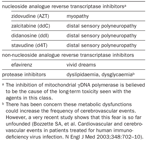
The availability of highly active anti-retroviral therapy (HAART) has dramatically improved the overall prognosis of patients infected with the human immunodeficiency virus (HIV). However, neurological disorders are still a frequent occurrence during the course of HIV infection and can represent serious diagnostic and therapeutic challenges. First, most antiretroviral medications have a poor ability to cross the blood-brain barrier. Second, it is known that some subtypes of HIV have a high affinity for the central nervous system (CNS). For these reasons, the CNS may serve as a sanctuary for the virus. Third, the spectrum of the classical opportunistic infections of the brain is now different from what it was at the beginning of this epidemic. For instance, while the frequency of primary CNS lymphoma has strikingly decreased, new conditions such as severe demyelinating leukoencephalopathy in AIDS patients on antiretroviral therapy [1] or the immune reconstitution inflammatory syndrome [2] have emerged. Finally, antiretroviral therapies can have metabolic and neurological toxicity (Table 1).
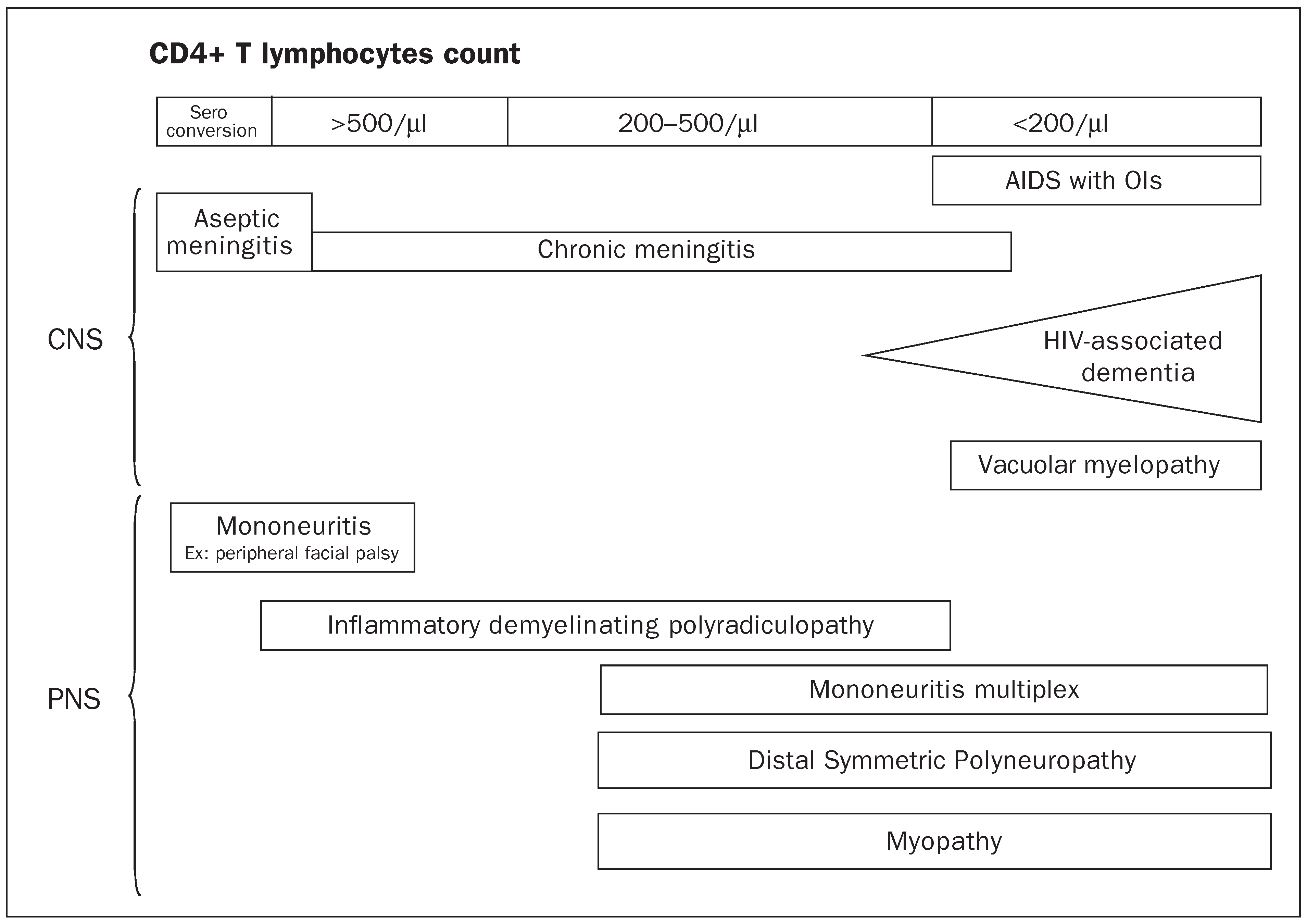
Because of this constant evolution of neurological manifestations of HIV infection, caring for such patients can be demanding for the neurologists. The goal of this article is not to do an exhaustive review of all the possible neurological manifestations of HIV, but rather to focus on those which are frequent and/or which represent a diagnostic challenge. Neurological manifestations of HIV can be divided between those caused by HIV itself and those caused by other organisms, which become pathogens in the setting of the immunodeficiency caused by HIV. These are referred to as opportunistic infections.The first question to ask when taking care of HIV+ patients is: what is the level of their immunosuppression? This will condition our entire approach. The level of immunodeficiency is reflected by the CD4+ T lymphocytes count. The spectrum of the HIV-related neurological manifestations is presented in Figure 1 and detailed below.

CD4+ T cells count >500/µl

In this situation, HIV+ patients are only mildly immunosuppressed, thus, they are not susceptible to opportunistic infections. Nevertheless, HIV itself can be responsible for various neurological conditions early in the course of the infection. At the time of the seroconversion, a subset of patients present with an aseptic meningitis. The CSF shows a lymphocytic pleocytosis, elevated protein concentration and normal glucose. Others can have a peripheral facial palsy. This affection can occur at any stage of the HIV infection but predominantly at the time of the seroconversion. Contrary to idiopathic peripheral facial palsy, the CSF is always abnormal [3]. Inflammatory demyelinating polyradiculopathy is a condition that clinically resembles the Guillain-Barré syndrome. It occurs at the time of the seroconversion or during the long asymptomatic phase of HIV infection. The major distinction with Guillain-Barré syndrome is that there is a pleocytosis in the CSF. This condition responds to plasmapheresis. Often, the patients in this CD4+ T cells count range are not aware of their HIV seropositivity, therefore an HIV serology testing should be systematically performed in patients with these neurological conditions.

CD4+ T cells count between 200 and 500/µl

In this category, neurological opportunistic infections are rare. Among the HIV-associated conditions, there is mononeuritis multiplex, a vasculitic process which causes nerve infarction and can happen before as well as after the onset of AIDS [4]. Myopathies caused either by HIV itself or by zidovudine can occur at any stage of the disease. They can be difficult to distinguish from each other clinically as they both cause proximal muscle weakness. Myalgias seem to be more common in zidovudine myopathy. In HIV-related myopathy, CD8+ T lymphocytes infiltrate the muscle, while zidovudine has a toxic effect on mitochondria, leading to a focal degeneration called raggedred fibres.
Distal sensory polyneuropathy is the most frequent peripheral nerve complication of HIV infection, affecting approximately 30% of the patients. This is a distal axonal degeneration involving both myelinated and unmyelinated fibres. Patients develop sensory loss in the feet, then in the hands, often with painful dysesthesias. It generally occurs in patients with advanced immunosuppression, though it can start at a CD4+ T cells count as high as 500/µl. This condition can be complicated by the fact that some members of the NRTIs family, ddI, ddC and d4T can aggravate it. The component of the polyneuropathy due to these medications is dose dependent and can be reversible after cessation of these drugs. Gabapentine is a good therapeutic option, since it often brings a significant relief to the patient and does not interact with any of the antiretroviral medications [4].

CD4+ T cells count <200/µl

These patients present with opportunistic infections as well as several neurological complications directly caused by HIV. It is important to remember that, in immunosupressed individuals, multiple pathologies frequently coexist.Thus, a positive culture for a given pathogen or even the result of a brain biopsy does not ensure that the sole diagnosis has been found. Several opportunistic infections and tumours can coexist in the brain of an HIV+ individual, and these can also be superimposed on HIV encephalopathy (HIVE).

HIV encephalopathy (HIVE) and HIV-associated dementia (HAD)

Although these two denominations are often used as synonymous, they encompass different entities. HIVE is a histopathological condition, consisting in multiple microglial nodules, multinucleated giant cells, and reactive astrocytes. Diffuse myelin pallor frequently accompanies HIVE [4]. Brain MRI is a good indicator of such an affection. It may reveal a subcortical atrophy associated with multiple hyperintense signals in T2-weighted images on MRI. These lesions are generally non enhancing and localised bilaterally in the periventricular white matter. Checking for the presence of HIV RNA (HIV viral load) in the CSF is recommended since the level of HIV viral load in this compartment is significantly higher in patients with HIVE than in HIV+ individuals without this condition. This is in contrast with the level of plasma HIV viral load, for which no correlation has been found [5].
HAD is a clinical diagnosis and consists in a subcortical-type dementia [6]. It is always the repercussion of some degree of HIVE, but this is a loose correlation. HAD usually occurs when the CD4+ T cells count is lower than 200/µl. Onset is insidious and the clinical syndrome is characterised by cognitive abnormalities including mental slowness, forgetfulness and poor concentration. In addition, affected individuals can present with behavioural abnormalities such as lethargy, and a decrease in spontaneity and emotional responses. Finally, some may present with motor deficits. When there is a cognitive dysfunction with mild functional impairment, the term “HIV-associated minor cognitive/motor disorder” is used, whereas for marked cognitive and functional impairment, one speaks of HAD.
Since the availability of HAART, the occurrence of HAD has decreased from 30 to 5% of AIDS patients. HIV-associated minor cognitive/ motor disorder is more frequent and can be missed by the examiner if a detailed neuropsychological examination is not performed. When HAD was first recognised, there was great concern that cognitive deterioration might begin at the time of the initial infection and progress to late stages dementia. However, most longitudinal studies have failed to demonstrate a slow progressive deterioration in the cognitive functions in HIV+ persons prior to the onset of severe immunodeficiency [7]. In HIV+ patients with minor cognitive/motor disorder or HAD, we advocate performing a spinal tap to measure the HIV viral load in the CSF. If HIV is detectable in the CSF, anti-HIV medications with a good penetration through the blood-brain barrier should be added. Among the NRTIs, these include: zidovudine, stavudine, and abacavir, and among the NNRTIs, efavirenz and nevirapine. Except for indinavir, the medications belonging to the protease inhibitors group have a poor penetration through the blood-brain barrier.

Vacuolar myelopathy

In autopsy series, 20 to 55% of patients with AIDS have pathological signs consistent with vacuolar myelopathy, but the actual number of HIV+ individuals who develop a medullary syndrome is considerably smaller. Vacuolar myelopathy is characterised pathologically by the presence of intramyelinic and periaxonal vacuoles, containing foamy macrophages, in the lateral and posterior columns of the thoracic portion of the spinal cord. A deficiency of the B12-dependent transmethylation pathway rather than direct HIV infection seems to be the pathological explanation for vacuolar myelopathy [8]. Clinically, vacuolar myelopathy manifests itself late in the course of HIV infection. The onset is usually insidious with sphincterian and erectile dysfunction, then weakness appears and some patients may become wheelchair bound. Except for hyperreflexia, the upper extremities are usually spared. Consistent with the hypothetical pathogenesis of vacuolar myelopathy, treatment with high doses of L-methionine has been proposed [9].

Approach of an HIV+ patient with CNS mass lesion(s)

The diagnosis of a CNS mass lesion in HIV+ individuals represents a particular challenge for the clinician. Indeed, several opportunistic infections or HIV-associated tumours can be the cause of such findings.The MRI is much more sensitive than the CT and should be preferred whenever it is available.

Toxoplasma encephalitis

Although the frequency of this affection has decreased since the introduction of HAART – from 5.4 per 1000 person-years in 1990–1992 to 2.2 per 1000 person-years in 1996–1998 – this is still the most common cerebral mass lesion in patients with AIDS who have a CD4+ T cells count <200/µl [10]. Patients with toxoplasma encephalitis typically present with headache, confusion and fever, which are present in 55, 52 and 47% of patients respectively, but any kind of focal neurologic deficit is possible. The MRI shows multiple lesions in 66% of the patients and ringenhancing lesion(s) in >90% of patients. The lesions are located in the white matter, the corticomedullar junction or the basal ganglia and often associated with a mass effect. PCR for T. gondii in the CSF is available, but although this test has a specificity of 100%, its sensitivity is relatively poor, between 44 and 65% [11]. Definitive diagnosis of cerebral toxoplasmosis is made by pathologic examination of brain tissue by stereotactic biopsy. However, this procedure has its limitations (see below). Therefore empirical treatment trial is warranted. Toxoplasma encephalitis responds promptly to treatment and usually the clinical improvement precedes the neuro-radiological improvement. The first-line therapy associates pyrimethamine with either sulfadiazine or clindamycin. Alternatively, pyrimethamine can be associated with azithromycin or atovaquone. The duration of the treatment is 6 weeks and is followed by a lifetime prophylaxis including the same medications, but at lower doses. Primary prophylaxis is indicated for HIV+ patients with a CD4 count <100 cells/µl and who are seropositive for T. gondii. The first choice is trimethoprim-sulfamethoxazole (TMP-SMX). This prophylaxis is also efficient against Pneumocystis carinii pneumonia. Finally, if the CD4 count rises above 200/µl for more than 4–6 weeks, primary prophylaxis can be safely discontinued [12].

Primary CNS lymphoma

This disorder is encountered in AIDS patients who are profoundly immunosupressed, usually with a CD4+ count <50/µl, thus primary CNS lymphoma is rarely an initial AIDS-defining illness. It accounts for up to 15% of non-Hodgkin lymphomas (NHLs) in HIV+ patients compared to only 1% of NHLs in HIV individuals. The most common histology is diffuse large B-cell lymphoma. Primary CNS lymphoma can present with a variety of focal and nonfocal signs and symptoms, such as lethargy, confusion, memory loss, aphasia, hemiparesis and/ or seizures. Solitary lesions and multiple lesions occur with equal frequency. The majority of the lesions show some degree of contrast enhancement, most commonly nodular or patchy, however, ring enhancement, identical to that commonly seen in toxoplasma encephalitis, can occur. Lesions that involve the corpus callosum, the periventricular, or the periependymal areas are more likely to be due to a lymphoma. Less than 10% of lymphomas invade the posterior fossa [13]. On SPECT, an accumulation of Thallium-201, resulting from increased metabolic activity, has been seen in patients with lymphoma, compared to a decreased activity in those with necrotic abscesses due to toxoplasma encephalitis. This test has 100% sensitivity for the detection of primary CNS lymphoma, but its specificity is lower, approximately at 60%. Perfusion MRI also shows an increase cerebral blood flow in lymphomas compared to lesions of toxoplasmosis. PCR amplification of EBV DNA in the CSF has a sensitivity of 83 to 100% and a specificity of 93 to 100% for the diagnosis of primary CNS lymphoma [14]. Therefore, the combination of Thallium 201 SPECT and PCR for EBV DNA achieves a very high sensitivity and specificity for the detection of primary CNS lymphoma. The standard first-line therapy is radiation therapy and corticosteroids which lead to a complete response in 20–50% of patients. Chemotherapy given with radiation therapy may provide some benefit to the subgroup of patients with relatively high CD4 cell counts [13]. Before HAART era, the median survival was only 3.5 months. HAART has not only allowed a significant decrease in the incidence of primary CNS lymphoma, but also seems to confer a better prognosis to the patients once they developed primary CNS lymphoma [15]. Other opportunistic infections can be associated with mass effect. These include cryptococcomas, fungal infections, tuberculosis, and CMV encephalitis. However, all of these entities are now much less common and, when they occur, they are usually associated with a disseminated infection.

Progressive multifocal leukoencephalopathy (PML)

This opportunistic infection, caused by polyomavirus JC (JCV), is a demyelinating disease of the central nervous system which occurs in HIV+ individuals with CD4+ T cell count <200/µl. The clinical presentation is subacute; neurological signs and symptoms indicate multiple localisations within the brain. Focal motor deficits, visual defects, and cognitive dysfunction are the most frequent inaugural manifestations of this disease [4]. The radiological hallmarks of PML are patchy or confluent areas of low attenuation on CT or hyperintensities on T2-weighted images on MRI. In 8% of the patients, the lesions can have some faint peripheral and irregular enhancement [16]. A substantial mass effect is almost always absent. Lesions are usually bilateral, asymmetric, and localised preferentially in the periventricular areas and the subcortical white matter. Involvement of the deep gray structures, including basal ganglia and thalamus, can nevertheless be found in up to 17% of cases. A normal neuro-imagery does not rule out PML since microscopic lesions may be smaller than the power of resolution of these tests. It has to be emphasised that HIVE can masquerade as PML. However, HIVE lesions are usually symmetrical, less demarcated than lesions of PML, and may or may not be associated with symptoms of AIDS dementia but never with focal motor or sensory deficits. An additional difficulty is that HIVE can coexist with other neurological opportunistic infections, such as PML. 1H-MR spectroscopy is a useful additional tool to help differentiating HIVE from PML lesions. In PML, CSF studies show normal values of leukocytes, proteins and glucose. However, PCR detection for JCV DNA in the CSF has a sensitivity of 74–93% and a specificity of 92–100% [14,17]. This diagnostic test has thus replaced the brain biopsy. In HIV+ patients, cytosine arabinoside (ARA-C), cifofovir and interferon 2b, although associated with anecdotal successes, have all failed to demonstrate an additional benefit to HAART in controlled studies [18]. HAART is the only treatment which has been shown to improve survival of PML patients from 10 to 50% after one year. This effect is likely due to reconstitution of the immune system, and is mediated by JCV-specific cytotoxic T-lymphocytes [19].

Corticosteroids

CNS mass lesions in AIDS are often associated with significant swelling and oedema, inducing sometimes a mass effect on the surrounding structures. Thus, many patients are treated with steroids before a diagnosis is established. However, steroid administration frequently complicates the management of these patients. Indeed, the majority of patients who receive steroids will also be on treatment trial for toxoplasma encephalitis, therefore this may cloud the picture. The administration of steroids might cause false-negative results on the brain biopsy in patients with primary CNS lymphoma. In addition, steroids have a strong immunosuppressive action, which should be considered very carefully in patients with AIDS.Therefore, they should be reserved for the case of impending brain herniation or for patients in whom the diagnosis of lymphoma has been established.

Brain biopsy

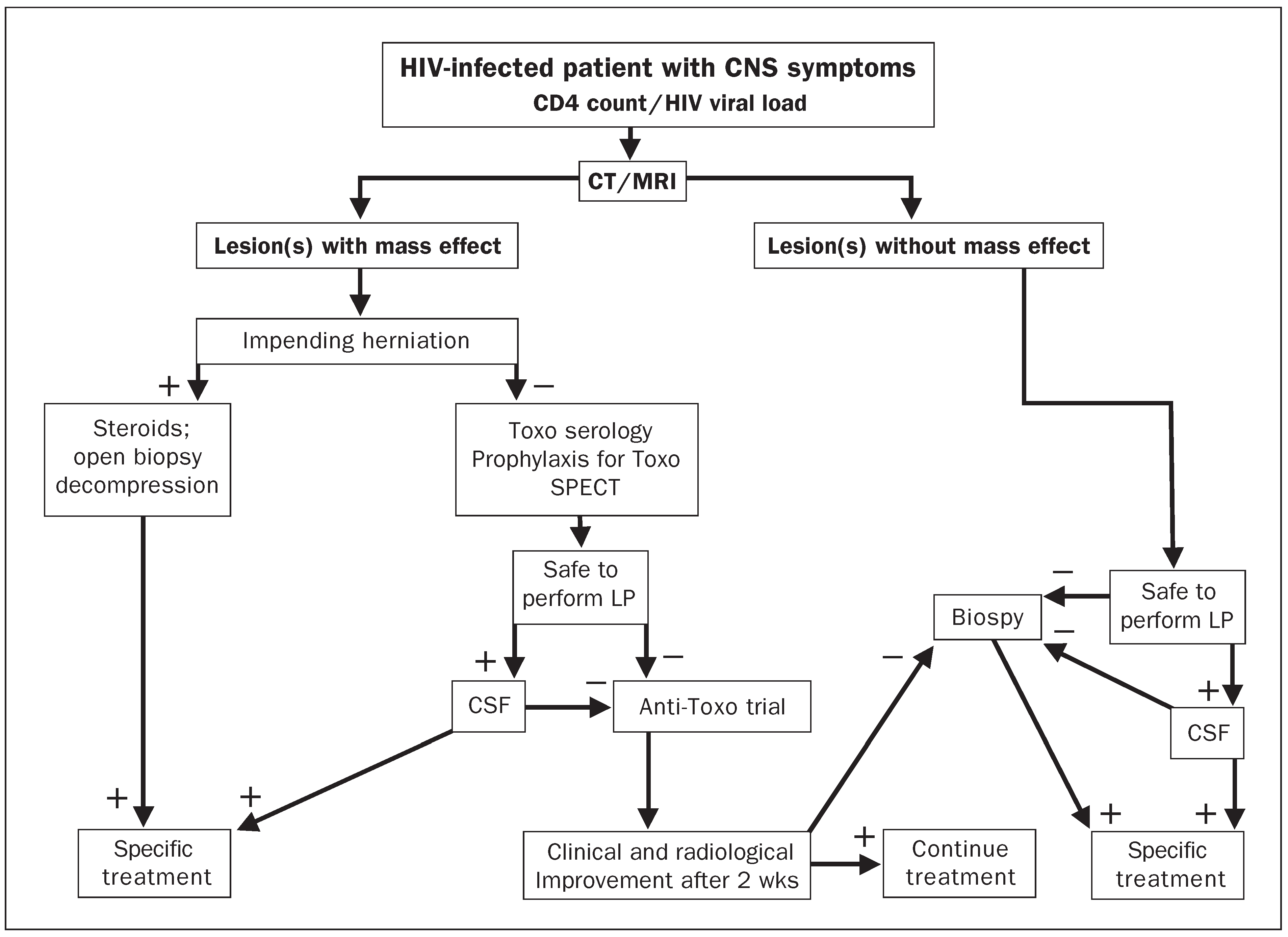
This examination remains the gold standard, but is not without significant risks. It has up to 3.1% mortality, and 2 to 12% morbidity [11,14]. In addition, it is often not feasible due to the localisation of the lesions. Finally, several disease processes can coincide in patients with multiple lesions, but multiple biopsies are rarely performed. Investigators performed a decision-making analysis on 136 consecutive HIV patients presenting with CNS mass lesions between 1991 and 1995 [14]. Following three weeks of empiric therapy for toxoplasma encephalitis, patients with progressive disease underwent a brain biopsy. The probability of toxoplasma encephalitis was 87% in Toxoplasma-seropositive patients with mass effect who were not on TMP-SMX, but only 59% for those receiving prophylaxis. For Toxoplasma-seropositive patients receiving TMP-SMX, the probability of primary CNS lymphoma was 36%. In Toxoplasma-seronegative patients with mass effect, the likelihood of primary CNS lymphoma was 74%, which increased to 96% if PCR for EBV DNA was positive in the CSF. Among focal brain lesions without mass effect, the probability of progressive multifocal leukoencephalopathy was 81%, which increased to 99% if JCV DNA was detected in the CSF. An algorithm for the management of CNS mass lesions in HIV+ patients is shown in Figure 2.

Temporal trends in CNS events and CNS mass lesions

A study compared the incidence of CNS mass lesions in HIV+ patients in the pre-HAART (1991 to 1996) and HAART periods (1997–1998) and found the following: (1) The diagnosis of toxoplasma encephalitis remained stable during the HAART period, with a frequency of 28% among CNS lesions in 1998. (2) On the other hand, there was a significant decline in the proportion of primary CNS lymphoma cases from 1996 to 1998 (34.9 vs 12%). (3) By contrast, progressive multifocal leukoencephalopathy, as a cause of CNS mass lesion, increased from 16.3 to 28% from 1996 to 1998. (4) The incidence of focal brain lesions other than toxoplasma encephalitis, primary CNS lymphoma, and progressive multifocal leukoencephalopathy increased steadily from 1991 to 1996 and 1998 (5.6 vs 30.2 and 32%, respectively) [20]. These cases were attributed to viral encephalitis or focal HIVE, which are diagnoses of exclusion. It is possible that these are, in fact, progressive multifocal leukoencephalopathy or primary CNS lymphoma with JCV or EBV viral load below the level of detection of the PCR assays. Alternatively, these may also represent new clinical entities. As a support of this theory, there was a recent description of a new form of HIV-associated leukoencephalopathy. This is a demyelinating condition which occurs in HAART-treated patients. No other pathogens than HIV are found in the brain and the severity of the leukoencephalopathy is more severe than that described prior to the use of HAART [1]. The precise cause of this new form of leukoencephalopathy arising in HAART experimented patients is unknown. It could result from a direct or indirect damage caused by antiretroviral medications themselves. For example, certain NRTIs such as stavudine (d4T) can cause mitochondrial injury and impaired cell metabolism. Proteases inhibitors can disrupt normal lipid metabolism (table 1). Either mechanism could damage the CNS. Finally, this severe leukoencephalopathy might be an expression of the so-called immune reconstitution inflammatory syndrome (IRIS). As a matter of fact, in some patients, tissue injury can occur when the newly reconstituted immune system responds overzealously to clinical or subclinical infections that are present when HAART is initiated [2]. IRIS has been described in cryptococcal meningitis, CMV retinitis and progressive multifocal leukoencephalopathy [16].This example illustrates the fact that the field of HIV-associated neurological conditions is evolving rapidly. Therefore, if a molecular diagnosis is not obtained in the CSF, a brain biopsy should be considered.

References

  1. Langford TD, Letendre SL, Marcotte TD, Ellis RJ, McCutchan JA, Grant I, et al. Severe, demyelinating leukoencephalopathy in AIDS patients on antiretroviral therapy. AIDS 2002;16:1019–29.
  2. DeSimone JA, Pomerantz RJ, Babinchak TJ. Inflammatory reactions in HIV-1-infected persons after initiation of highly active antiretroviral therapy. Ann Intern Med 2000;133:447–54.
  3. Kohler A, Chofflon M, Sztajzel R, Magistris MR. Cerebrospinal fluid in acute peripheral facial palsy. J Neurol 1999;246:165–9.
  4. Koralnik IJ. Human Immunodeficiency Virus-Positive Patients. In: Samuels MA, editor. Hospitalist Neurology. Boston: Butterworth Heinemann; 1999. p. 175–210.
  5. Cinque P, Vago L, Ceresa D, Mainini F, Terreni MR, Vagani A, et al. Cerebrospinal fluid HIV-1 RNA levels: correlation with HIV encephalitis. AIDS 1998;12:389–94.
  6. Power C, Selnes OA, Grim JA, McArthur JC. HIV Dementia Scale: a rapid screening test. J Acquir Immune Defic Syndr Hum Retrovirol 1995;8:273–8.
  7. Selnes OA, Miller E, McArthur J, Gordon B, Munoz A, Sheridan K, et al. HIV-1 infection: no evidence of cognitive decline during the asymptomatic stages. The Multicenter AIDS Cohort Study. Neurology 1990;40:204–8.
  8. Di Rocco A, Bottiglieri T, Werner P, Geraci A, Simpson D, Godbold J, et al. Abnormal cobalamin-dependent transmethylation in AIDS-associated myelopathy. Neurology 2002;58:730–5.
  9. Di Rocco A, Tagliati M, Danisi F, Dorfman D, Moise J, Simpson DM. A pilot study of L-methionine for the treatment of AIDS-associated myelopathy. Neurology 1998;51:266–8.
  10. Sacktor N, Lyles RH, Skolasky R, Kleeberger C, Selnes OA, Miller EN, et al. HIV-associated neurologic disease incidence changes: Multicenter AIDS Cohort Study, 1990–1998. Neurology 2001;56:257–60.
  11. Holloway RG, Mushlin AI. Intracranial mass lesions in acquired immunodeficiency syndrome: using decision analysis to determine the effectiveness of stereotactic brain biopsy. Neurology 1996;46:1010–5.
  12. Kovacs JA, Masur H. Prophylaxis against opportunistic infections in patients with human immunodeficiency virus infection. N Engl J Med 2000;342:1416–29.
  13. Forsyth PA, DeAngelis LM. Biology and management of AIDS-associated primary CNS lymphomas. Hematol Oncol Clin North Am 1996;10:1125–34.
  14. Antinori A, Ammassari A, De Luca A, Cingolani A, Murri R, Scoppettuolo G, et al. Diagnosis of AIDS-related focal brain lesions: a decision-making analysis based on clinical and neuroradiologic characteristics combined with polymerase chain reaction assays in CSF. Neurology 1997;48:687–94.
  15. Antinori A, Cingolani A, Alba L, Ammassari A, Serraino D, Ciancio BC, et al. Better response to chemotherapy and prolonged survival in AIDS-related lymphomas responding to highly active antiretroviral therapy. AIDS 2001;15:1483–91.
  16. Du Pasquier RA, Koralnik IJ. Inflammatory reaction in Progressive Multifocal Leukoencephalopathy: harmful or beneficial? J Neurovirol 2003;9:1–7.
  17. Koralnik IJ, Boden D, Mai VX, Lord CI, Letvin NL. JC virus DNA load in patients with and without progressive multifocal leukoencephalopathy. Neurology 1999;52:253–60.
  18. Marra CM, Rajicic N, Barker DE, Cohen BA, Clifford D, Donovan Post MJ, et al. A pilot study of cidofovir for progressive multifocal leukoencephalopathy in AIDS. AIDS 2002;16:1791–7.
  19. Du Pasquier RA, Clark KW, Smith PS, Joseph JT, Mazullo JM, De Girolami U, et al. Favorable clinical outcome in HIV-infected individuals with Progressive Multifocal Leukoencephalopathy correlates with JCV-specific cellular immune response. J Neurovirol 2001;7:318–22.
  20. Ammassari A, Cingolani A, Pezzotti P, De Luca DA, Murri R, Giancola ML, et al. AIDS-related focal brain lesions in the era of highly active antiretroviral therapy. Neurology 2000;55:1194–200.
Figure 1. Occurrence of HIV-related neurological conditions according to the degree of immunosupression. Ols = opportunistic infections; CNS = central nervous system; PNS = peripheral nervous system.
Sanpp 154 00178 g001
Figure 2. Management of CNS mass lesions in HIV+ patients. CT = computerised tomography; MRI = magnetic resonance imagery; LP = lumbar puncture; CSF = cerebrospinal fluid.
Sanpp 154 00178 g002
Table 1. Neurological toxicity of anti-HIV drugs.
Sanpp 154 00178 i001