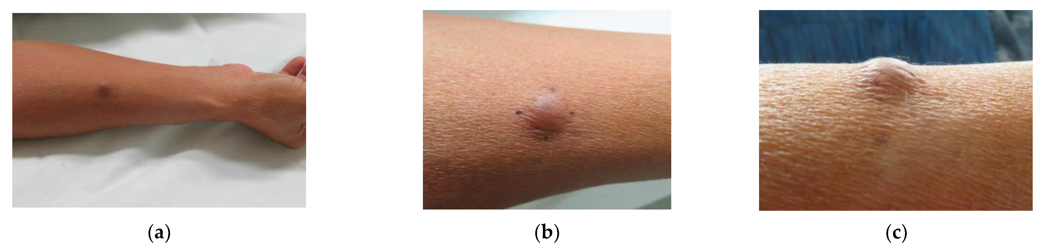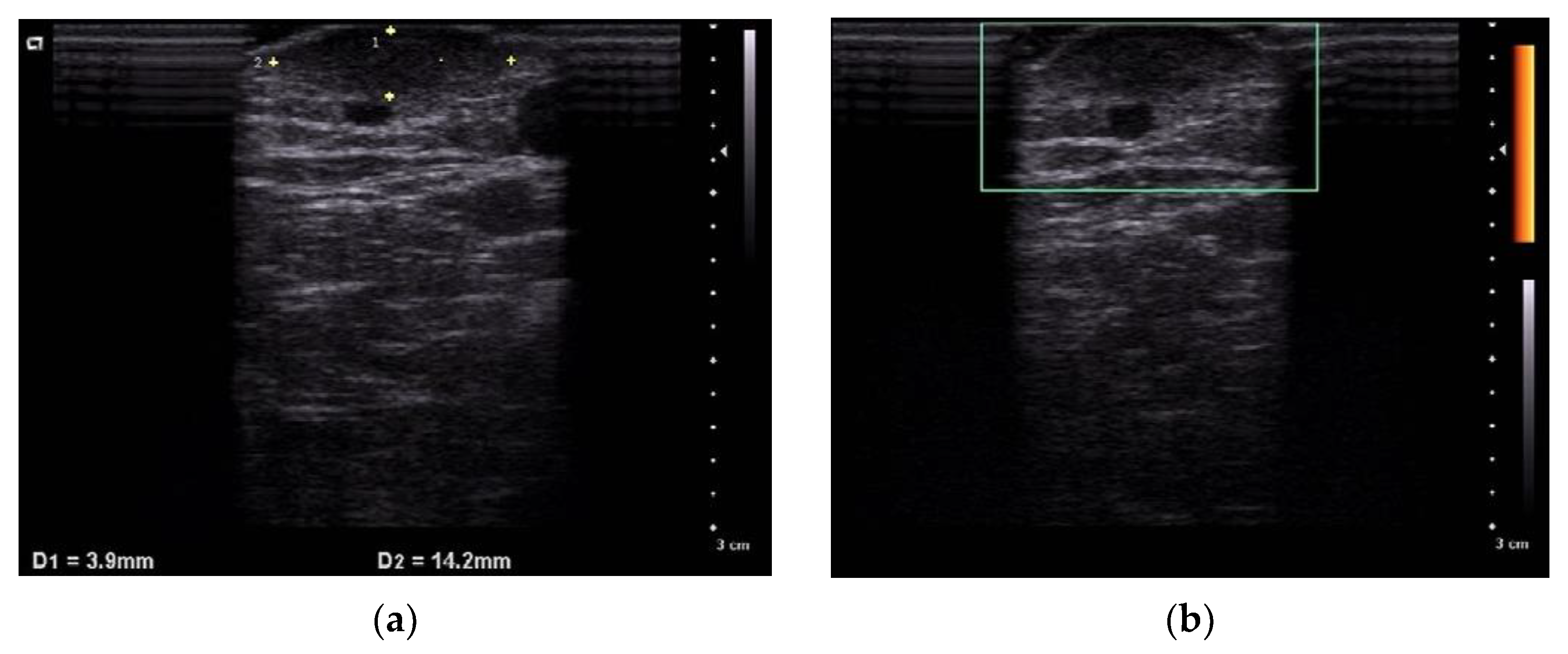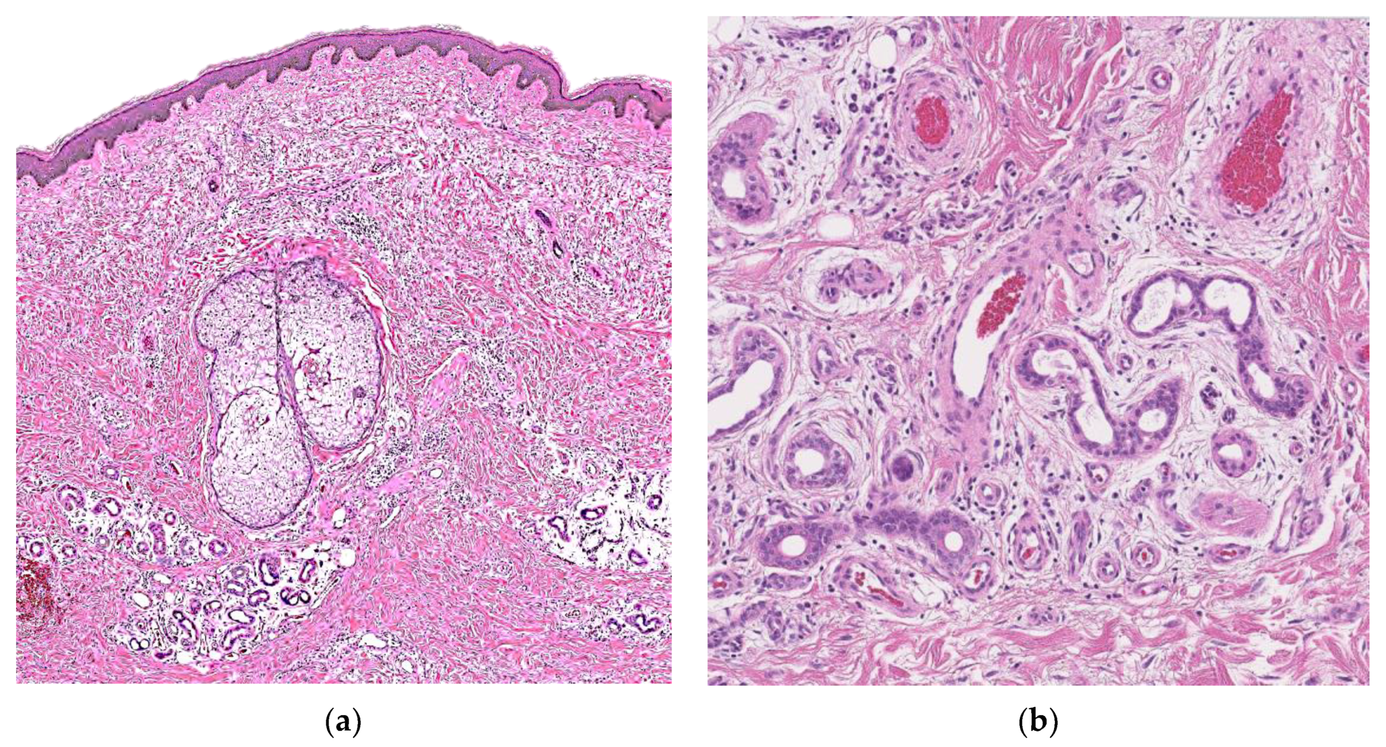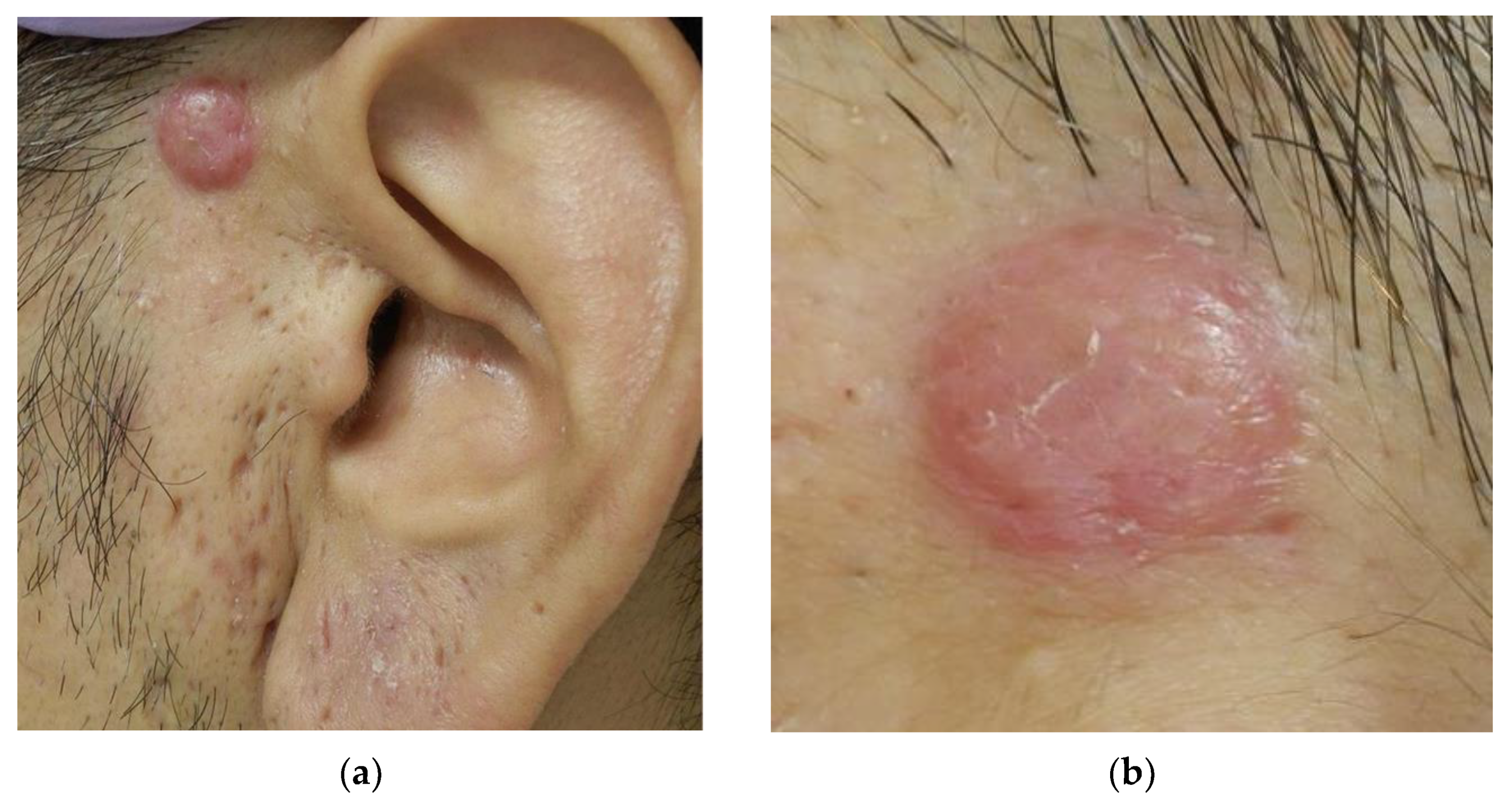

A Case of Adult-Onset Eccrine Angiomatous Hamartoma—The Comparison with Epithelioid Hemangioma
 ,
, Abstract
Share and Cite
Nishimura, M.; Matsushima, Y.; Nakai, Y.; Habe, K.; Hayashi, A.; Yamanaka, K. A Case of Adult-Onset Eccrine Angiomatous Hamartoma—The Comparison with Epithelioid Hemangioma. Dermatopathology 2022, 9, 94-99. https://doi.org/10.3390/dermatopathology9020012
Nishimura M, Matsushima Y, Nakai Y, Habe K, Hayashi A, Yamanaka K. A Case of Adult-Onset Eccrine Angiomatous Hamartoma—The Comparison with Epithelioid Hemangioma. Dermatopathology. 2022; 9(2):94-99. https://doi.org/10.3390/dermatopathology9020012
Chicago/Turabian StyleNishimura, Mai, Yoshiaki Matsushima, Yasuo Nakai, Koji Habe, Akinobu Hayashi, and Keiichi Yamanaka. 2022. "A Case of Adult-Onset Eccrine Angiomatous Hamartoma—The Comparison with Epithelioid Hemangioma" Dermatopathology 9, no. 2: 94-99. https://doi.org/10.3390/dermatopathology9020012
APA StyleNishimura, M., Matsushima, Y., Nakai, Y., Habe, K., Hayashi, A., & Yamanaka, K. (2022). A Case of Adult-Onset Eccrine Angiomatous Hamartoma—The Comparison with Epithelioid Hemangioma. Dermatopathology, 9(2), 94-99. https://doi.org/10.3390/dermatopathology9020012