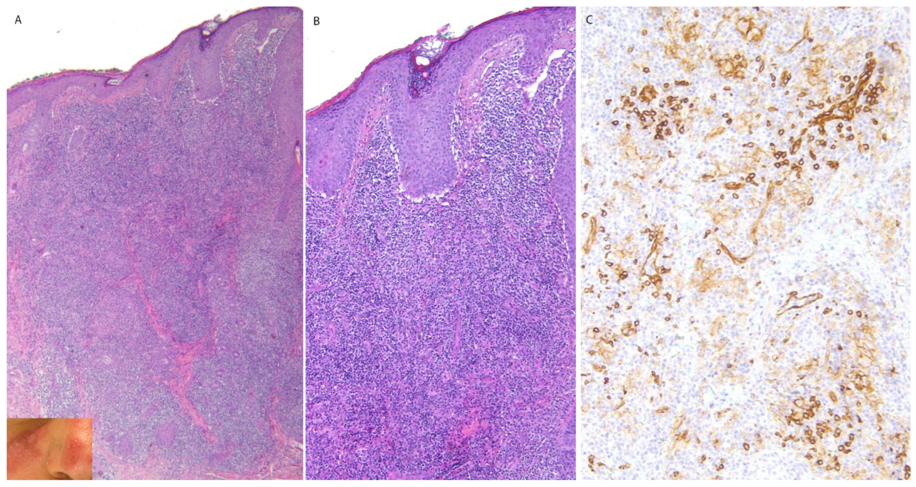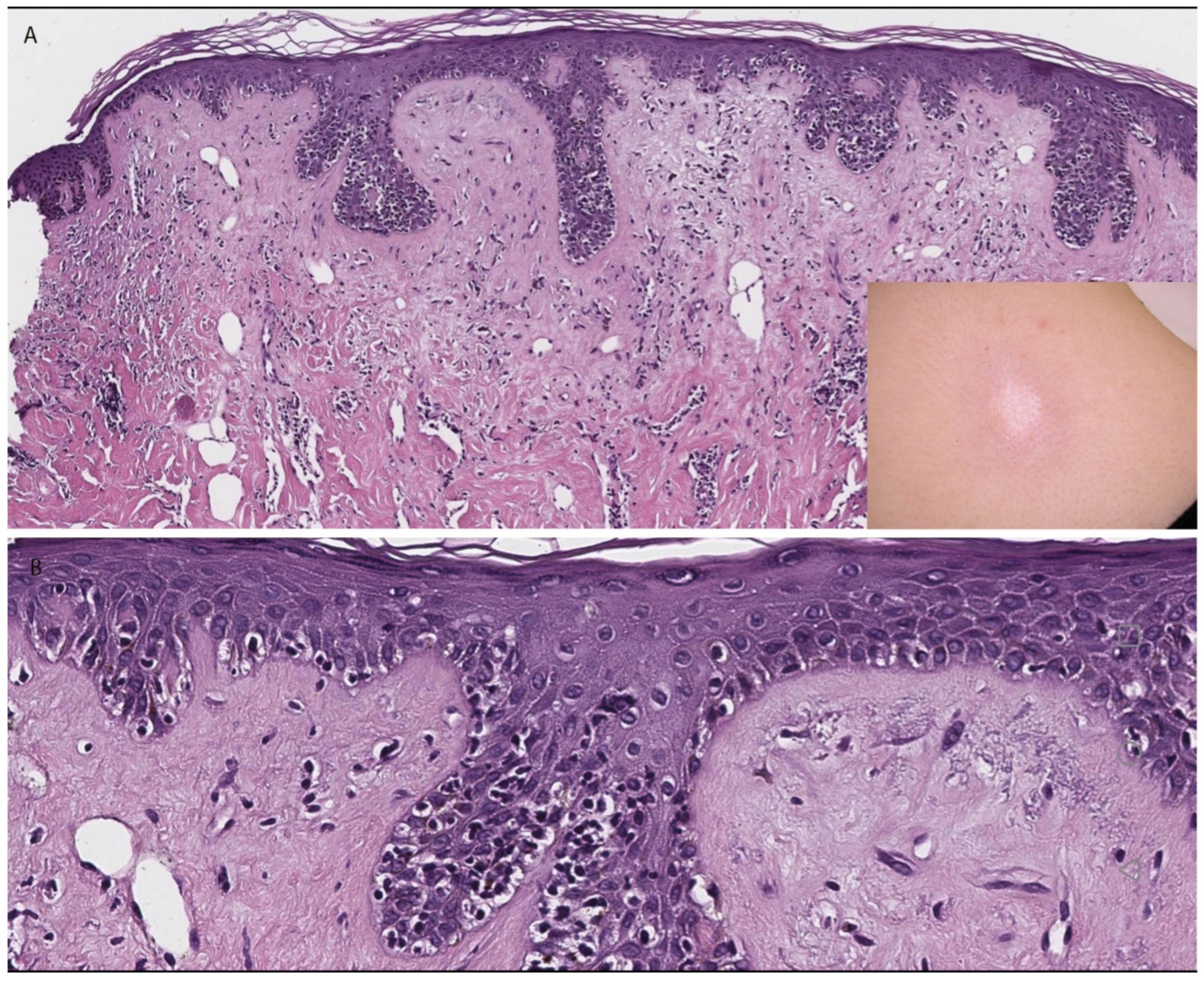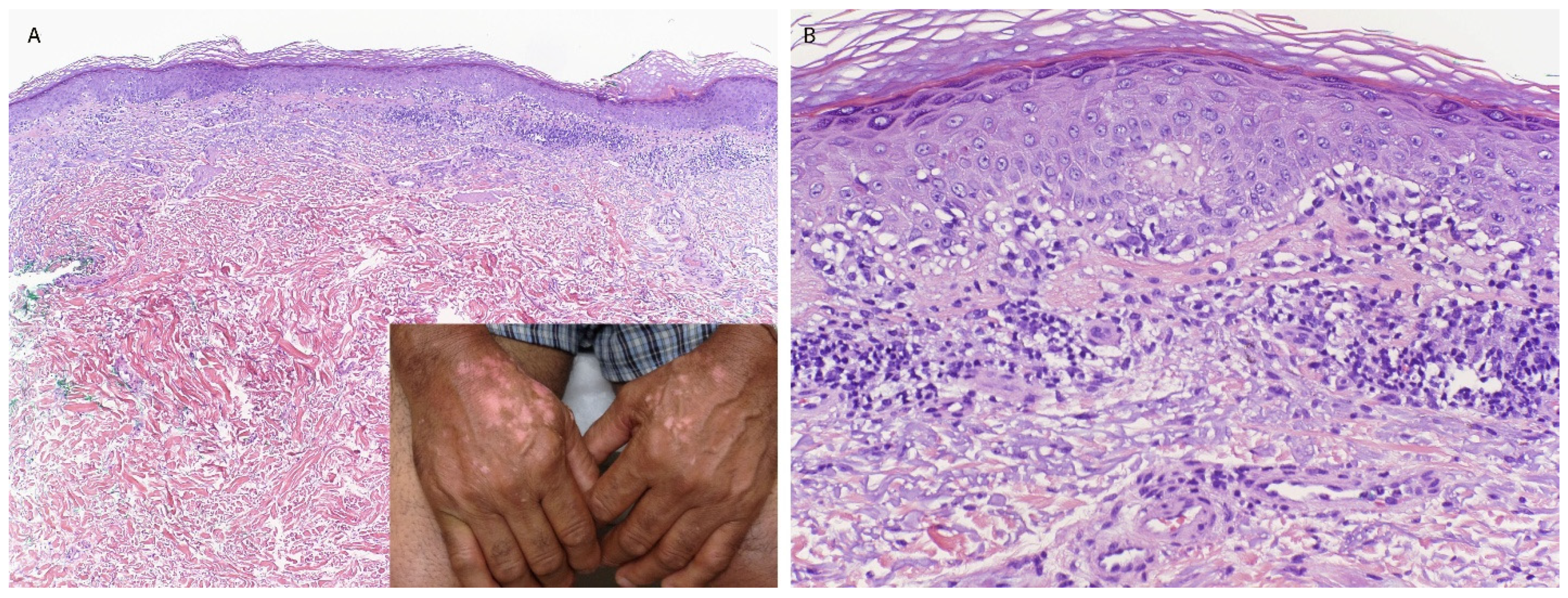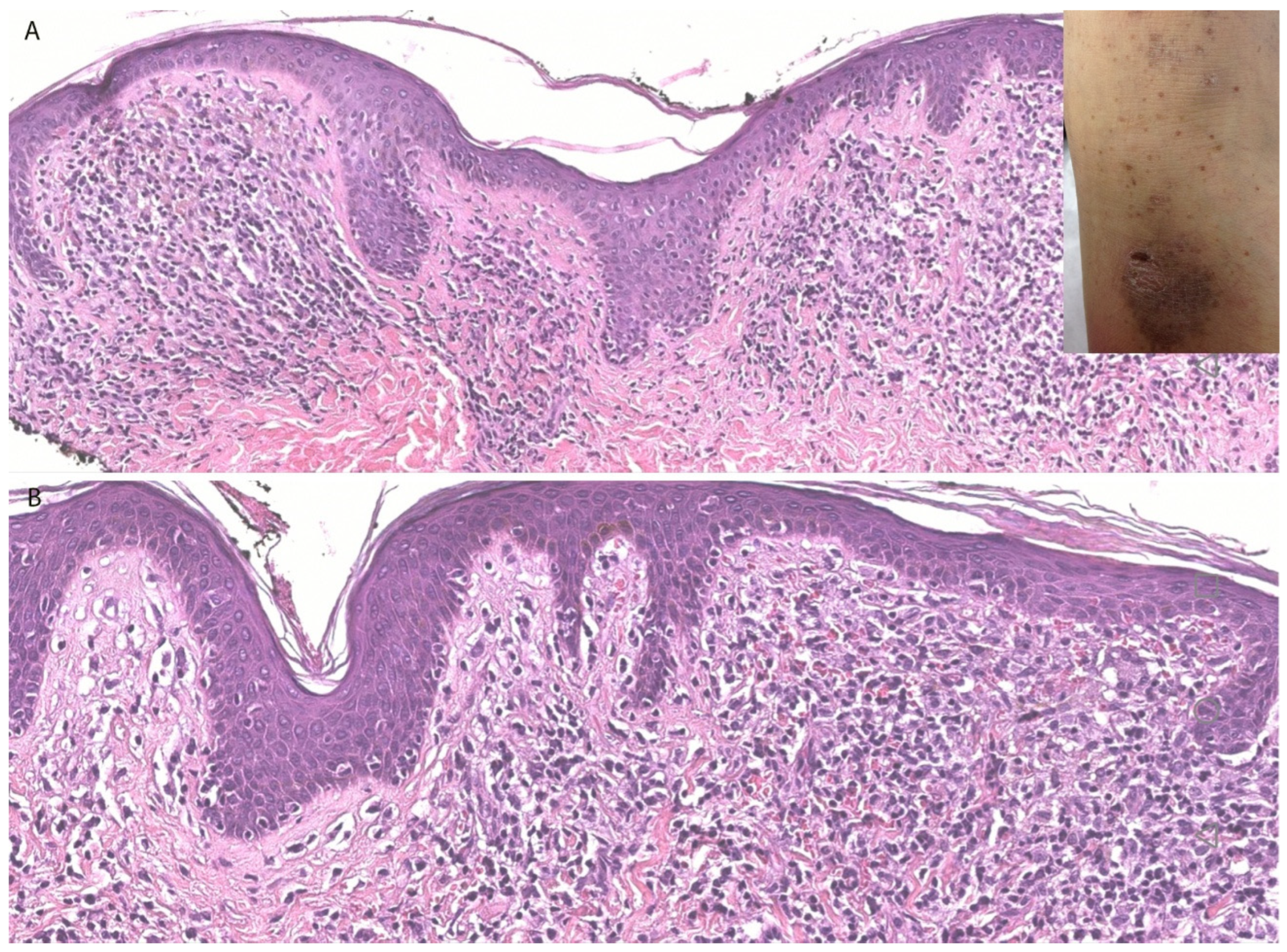

Inflammatory and Infectious Cutaneous Entities Resembling Cutaneous T-Cell Lymphoma (CTCL): An Integrated Clinicopathological Review



1
Dermatology Department, American University of Beirut-Medical Center, Beirut P.O. Box 11-0236, Lebanon
2
Dermatopathology Section, Department of Dermatology, Boston University School of Medicine, Boston, MA 02118, USA
*
Author to whom correspondence should be addressed.
†
These authors contributed equally to this work.
Dermatopathology 2026, 13(2), 23; https://doi.org/10.3390/dermatopathology13020023
Submission received: 22 February 2026
/
Revised: 26 March 2026
/
Accepted: 25 May 2026
/
Published: 27 May 2026
Simple Summary
Cutaneous pseudolymphomas are non-cancerous skin conditions that can closely resemble skin lymphomas, especially a type called mycosis fungoides, both in appearance and under the microscope. They can be triggered by many causes such as autoimmune diseases, infections, medications, insect bites, or even tattoos. Because they can look very similar to true lymphoma, they can sometimes be misdiagnosed, leading to unnecessary anxiety or treatment. This review explains the main conditions that mimic lymphoma and highlights key clinical and microscopic clues that help doctors tell them apart. These include features like the type of immune cells present, patterns in the skin, and how the condition behaves over time. Understanding these differences is essential to ensure patients receive the correct diagnosis and avoid overtreatment.
Abstract
Cutaneous pseudolymphomas are benign reactive lymphoid proliferations that often mimic cutaneous lymphomas both clinically and histologically. A diverse array of inflammatory, infectious, and drug-induced dermatoses can closely resemble cutaneous T-cell lymphomas (CTCLs), particularly mycosis fungoides (MFs), posing significant diagnostic challenges. These mimickers may show histopathological features such as epidermotropism, dense lymphocytic infiltrates, or even clonality, making accurate differentiation crucial to avoid overtreatment. This review endeavors to comprehensively discuss the clinicopathologic features of the various inflammatory and infectious dermatoses that may simulate CTCL, drawing on illustrative examples across disease categories. By highlighting important comparative features and emphasizing the importance of clinicopathologic correlation, this review outlines practical strategies for distinguishing true lymphoma from its inflammatory mimics.