Introduction
Histoplasma capsulatum and
Pneumocystis jirovecii are opportunistic fungal pathogens primarily implicated in respiratory infections. While both have been extensively studied as individual entities, their concurrent infection is infrequently reported, particularly in immunocompromised individuals without HIV.[
1]
H. capsulatum is the etiological agent of histoplasmosis, a systemic fungal infection of significant public health concern. It has the capacity to infect both immunocompetent and immunocompromised hosts, with the latter experiencing more severe clinical manifestations. Transmission occurs via the inhalation of aerosolized microconidia, which subsequently transforms into yeast-like forms within the respiratory tract, facilitating pulmonary and disseminated infection.[
2]
P. jirovecii, the causative agent of pneumocystosis, gained prominence during the AIDS epidemic as a leading cause of opportunistic pneumonia and mortality in patients with HIV. Despite the reduced prevalence of pneumocystosis due to the widespread implementation of antiretroviral therapy,
P. jirovecii continues to pose a significant risk to individuals with non-HIV-related immunosuppression.[
3]
While reports of coinfection with
H. capsulatum and
P. jirovecii are limited, most documented cases are found in patients with AIDS.[
4] This case underscores the diagnostic complexity associated with multiple opportunistic infections in non-HIV-related immunosuppression and emphasizes the importance of heightened clinical suspicion in these populations.
Case report
A 58-year-old female with a medical history of systemic lupus erythematosus (SLE), lupus nephritis, and rheumatoid arthritis was admitted to the emergency department with a 15-day history of fever and chills, without accompanying respiratory symptoms. She was undergoing treatment with prednisolone (15 mg/day) and monthly intravenous cyclophosphamide. There was no indication that the patient had traveled to rural areas. In recent months, she remained at home in Bogotá D.C., Colombia. On admission, she presented with tachycardia and fever; however, no other significant findings were noted on physical examination. Oxygen saturation was found to be below the lower normal limit for the altitude of Bogotá, leading to the initiation of low-flow oxygen supplementation.
A chest X-ray revealed bilateral alveolar infiltrates, prompting the initiation of empirical antibiotic therapy with ampicillin-sulbactam (3 g qid) and clarithromycin (500 mg bid). The initial blood count showed lymphopenia (320/mcL). Additional tests showed a negative HIV result, as well as negative initial cultures. Despite treatment, the patient exhibited no clinical improvement and continued to experience fever and desaturation. A subsequent chest computed tomography scan revealed multiple randomly distributed micronodules in both lungs, along with ground-glass opacities, lymphadenopathies and subpleural areas of honeycombing (
Figure 1). Given these findings, the antibiotic regimen was modified to piperacillin-tazobactam (4.5 g every six hours) on the second day of hospitalization, and a bronchoscopy with bronchoalveolar lavage (BAL) was performed. By day 7 of hospitalization, the report was received. The BAL analysis identified
Pneumocystis jirovecii, leading to the initiation of trimethoprim/sulfamethoxazole at a dose of 15 mg/kg/day. Galactomannan was negative.
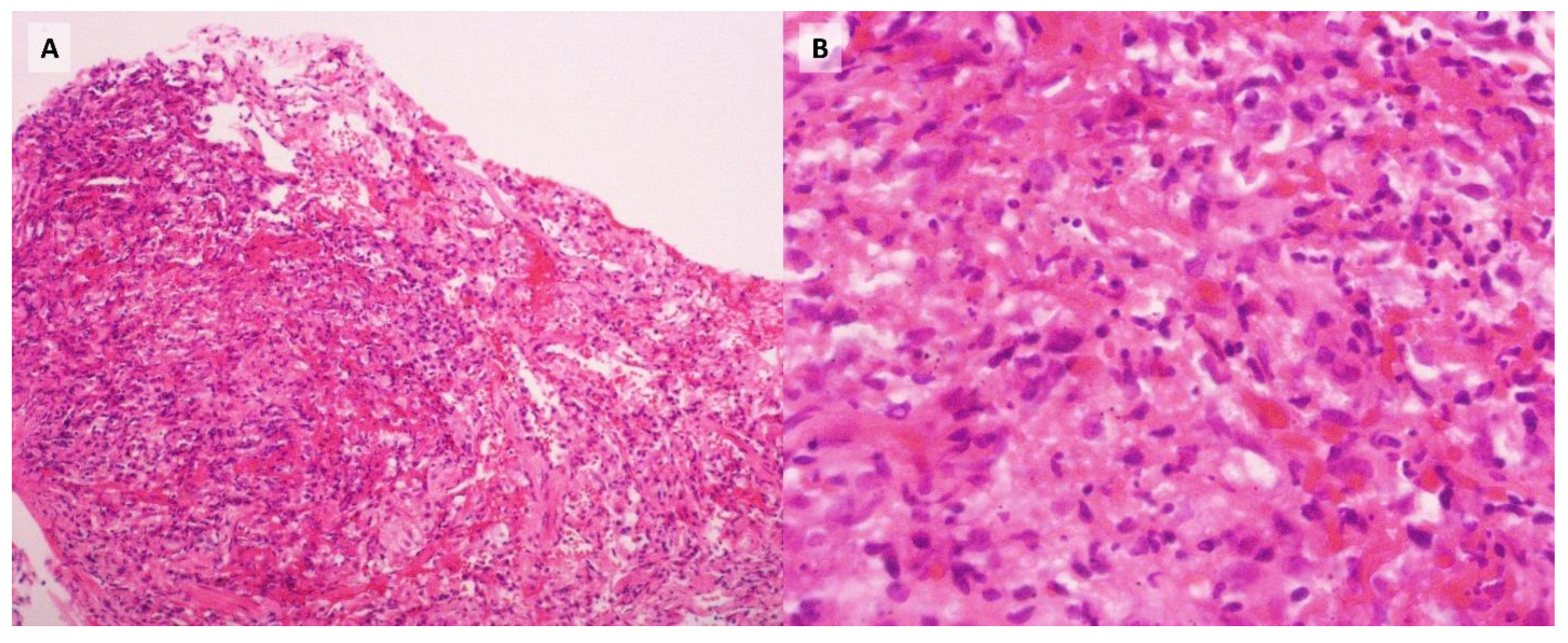
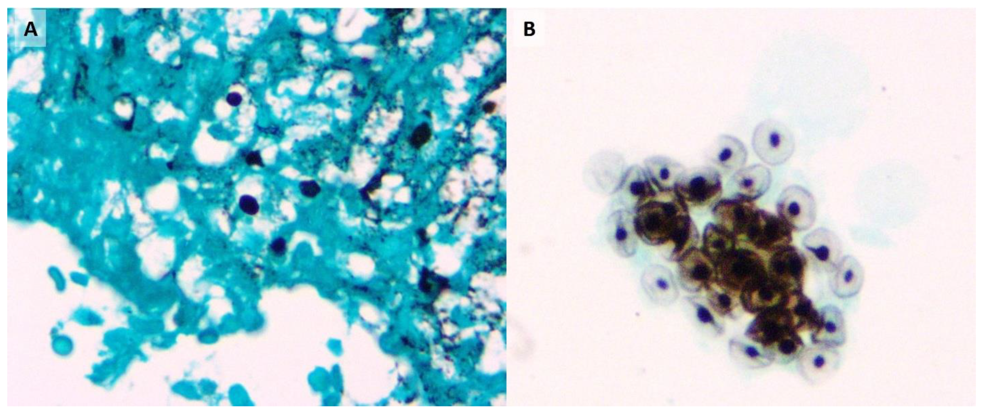
Despite targeted therapy, the patient experienced progressive clinical deterioration, ultimately developing respiratory failure that required orotracheal intubation and admission to the intensive care unit (ICU) on day 10 of hospitalization. By day 12, the report was received. Subsequent transbronchial biopsies were positive for
Pneumocystis jirovecii and fungi morphologically consistent with
Histoplasma (
Figure 2,
Figure 3), with no other infectious agents detected in the BAL samples or the blood and bone marrow cultures. Consequently, antifungal therapy was adjusted to include liposomal amphotericin B (5 mg/kg/day) and itraconazole (200 mg tid). However, despite intensive therapeutic interventions, the patient's condition continued to worsen, culminating in a fatal outcome on day 31 of her hospitalization.
Discussion
Co-infection with
Histoplasma capsulatum and
Pneumocystis jirovecii has been primarily reported in immunocompromised patients, particularly those with HIV/AIDS.[
4] Although this coinfection is rarely documented, an increasing number of cases have been reported in recent years, likely due to the use of more sensitive diagnostic methods, such as polymerase chain reaction (PCR) assays.[
5] However, in patients without HIV, reports of this coinfection remain uncommon. A study by Carreto et al.[
1] identified only three cases, but the authors were unable to determine whether the patients were immunosuppressed and, if so, the specific nature of their immunosuppression.
Infections are a common complication in patients with SLE, with approximately 30% experiencing at least one significant infection over a 10-year period. This increased susceptibility is multifactorial and stems from both disease-related and treatment-induced immunosuppression. Dysregulation of the innate and adaptive immune responses plays a central role, including aberrant expression of IFN-γ, IL-1, and TNF-α; impaired cytolytic activity of T lymphocytes; and suppression of natural killer (NK) cell function. Additionally, defective chemotaxis and phagocytosis of polymorphonuclear cells, abnormal microbial recognition and binding, and acquired complement deficiencies further contribute to increased infection risk. Importantly, immunosuppressive therapies exacerbate this vulnerability: glucocorticoids increase the risk of serious infections by 18%, and cyclophosphamide by 16.2%. In the context of the present case, the patient's immunosuppressed state likely heightened the risk of serious infections.[
6]
Corticosteroids contribute to the risk of invasive fungal infections by exerting multiple immunosuppressive effects. They impair macrophage function by stabilizing lysosomal membranes during phagocytosis, which interferes with phagolysosome formation. Additionally, corticosteroids reduce the number of circulating monocytes and macrophages by inhibiting myelopoiesis and limiting their release from the bone marrow. These effects are mediated through activation of the glucocorticoid receptor.[
6]
Cyclophosphamide modulates the immune response by reducing the secretion of interferongamma and interleukin-12, while increasing the production of Th2 cytokines such as IL-4 and IL-10. These anti-inflammatory cytokines may promote the proliferation of certain pathogens. Moreover, cyclophosphamide is known to induce leukopenia, which further increases the risk of infection.[
7]
The study by Carreto et al. demonstrated an increased mortality associated with co-infection,[
1] underscoring the necessity of considering multiple factors in the presented case. Although
Pneumocystis pneumonia is rare, particularly in patients receiving mycophenolate and cyclophosphamide (with an incidence of 1.8 cases per 1,000 patients), a significant risk factor is the presence of underlying structural pulmonary disease, as observed in the patient.[
8] Moreover, it is well-established that lymphopenia is a critical risk factor for
Pneumocystis pneumonia in patients receiving intermediate doses of corticosteroids (≥15 to <30 mg/day).[
9] While there is no consensus on the appropriate timing for
Pneumocystis prophylaxis in patients with autoimmune diseases and immunosuppressive therapy, these risk factors underscore the necessity for individualized case management.
Additionally, it is crucial to consider the diagnostic methods specific to each infectious agent. In histoplasmosis, the most common radiological findings are diffuse or interstitial opacities, although micronodules or nodules may also be present. Furthermore, in both acute and subacute histoplasmosis, lymphadenopathy can be observed, a finding that is less common in
Pneumocystis pneumonia, where the predominant manifestation is ground-glass opacity. The coexistence of combined tomographic patterns, along with the presence or absence of lymphadenopathy, as seen in this patient, underscores the necessity of considering a broad range of infectious agents in immunosuppressed individuals.[
3,
10]
Furthermore,
Pneumocystis is particularly challenging to culture, with detection primarily relying on PCR testing or direct visualization in induced sputum or bronchoalveolar lavage (BAL) samples.[
3] In contrast,
Histoplasma can be isolated, but the sensitivity of respiratory samples typically ranges from 0% to 60%. It is important to note that blood cultures and bone marrow cultures demonstrate higher sensitivity, ranging from 60% to 90%. However, fungal cultures require extended periods for growth (2-3 weeks), making PCR studies a valuable alternative, as they provide rapid results with a sensitivity ranging from 67% to 100%.[
10] Unfortunately, our institution lacks the capability to perform PCR testing for fungal infections. In our case fungal cultures were negative, and histoplasmosis was only detected in biopsy samples, not in the BAL. This delay in diagnosis hindered the timely initiation of targeted antifungal therapy. As diagnostic methods, such as PCR for fungal infections, become more widely available, it is likely that more cases of co-infection, such as the one presented, will be identified.
This case underscores the complexity of diagnosing opportunistic infections in immunocompromised patients without HIV, where classical clinical reasoning tools may fall short. In the 14th century, William of Ockham formulated the principle of parsimony, known as Ockham’s Razor, “entities must not be multiplied beyond necessity”, implying that the simplest explanation is usually correct. However, John B. Hickam later challenged this notion with his dictum: “A man can have as many diseases as he damn well pleases.” While Ockham’s principle remains useful in routine clinical practice, Hickam’s dictum is particularly relevant in the care of immunosuppressed patients, in whom multiple concurrent pathologies may coexist. This is especially true in complex autoimmune diseases such as SLE, where the risk of severe infection is high. The overall infectious risk must be assessed not only in relation to the underlying disease but also considering the cumulative effects of immunosuppressive therapy. In resource-limited settings, a detailed clinical history becomes essential for identifying diseasespecific risk factors and anticipating likely pathogens. Radiological evaluation must also be guided by an awareness of imaging patterns characteristic of specific infectious agents, thereby supporting more accurate diagnostic and therapeutic decision-making.
Conclusions
This case highlights the diagnostic and therapeutic challenges associated with opportunistic coinfections in immunocompromised patients without HIV, particularly in the context of autoimmune diseases such as systemic lupus erythematosus. The concomitant use of immunosuppressive agents, including corticosteroids and cyclophosphamide, significantly alters the host immune response, thereby increasing susceptibility to rare and potentially fatal infections such as Histoplasma capsulatum and Pneumocystis jirovecii. Limitations in diagnostic resources, especially in low-resource settings, further complicate timely and accurate identification of causative pathogens. A high index of suspicion, combined with a thorough clinical history and careful interpretation of radiologic findings, remains essential. Ultimately, this case underscores the importance of individualized risk assessment and the potential need to reconsider traditional diagnostic heuristics when evaluating immunocompromised hosts. In such patients, Hickam’s dictum may offer a more clinically appropriate lens than Ockham’s Razor, acknowledging the reality that multiple concurrent infections may coexist and require targeted, multifaceted management strategies.


