Article

A Combination Extract of Gardeniae Fructus and Perillae Folium Exerts Anti-Inflammatory Effects on LPS-Activated RAW 264.7 Mouse Macrophages via an ER Stress-Induced CHOP Pathway

Department of Pathology, College of Korean Medicine, Gachon University, Seongnam 13120, Korea
Processes 2021, 9(9), 1632; https://doi.org/10.3390/pr9091632
Submission received: 10 July 2021 / Revised: 3 September 2021 / Accepted: 7 September 2021 / Published: 10 September 2021
(This article belongs to the Section Pharmaceutical Processes)

Abstract

:
The aim of this study is to investigate the effects of a combination extract of Gardeniae Fructus and Perillae Folium (GP) on inflammatory reactions in lipopolysaccharide (LPS)-activated mouse macrophages RAW 264.7 cells. Multiplex cytokine assay, Fluo-4 calcium assay, Flow cytometry assay for phospho-P38 MAPK, and quantitative PCR were carried out. GP significantly reduced LPS-induced productions of macrophage inflammatory protein (MIP)-1α and monokine induced by gamma interferon (MIG) and release of intracellular calcium in LPS-activated RAW 264.7 cells. GP also significantly inhibited P38 MAPK phosphorylation and mRNA levels of Chop, Camk2a, Stat1, Stat3, Jak2, Fas, Nos2, and Ptgs2 in LPS-activated RAW 264.7 cells. Taken together, this study represents that GP exerts anti-inflammatory effects on LPS-activated RAW 264.7 cells via ER stress-induced CHOP pathway.

1. Introduction

Inflammatory reactions are one of the most important immune responses that protect our body from infection by pathogens [1]. Many pathogens, fungi, viruses, etc., cause inflammation through immune responses. Lipopolysaccharides (LPS), also known as endotoxins, are released by Gram-negative bacteria, which cause endotoxemia if not properly removed [2]. Macrophages are one of the main immune cells for immune function, and are responsible for innate immunity [3]. It is well known that LPS stimulation makes macrophages produce various proinflammatory mediators such as nitric oxide (NO), cytokines, chemokines, growth factors, and prostaglandins. Interleukins (ILs) are well known to be a type of cytokine with immunoregulatory functions and key signaling molecules which are mainly expressed by leukocytes, including macrophages. Interestingly, mRNA expressions of inflammatory genes such as Nos2 and Ptgs2 are increased in LPS-activated macrophages via P38 Mitogen-Activated Protein Kinase (MAPK) activation [4]. Janus kinase (JAK) signaling is known to be involved in inflammatory reactions in LPS-activated macrophages [5].
Despite the development of many antibiotics, there are still many infectious diseases that remain unsolved and many types of infectious diseases can also cause troublesome inflammatory conditions in the human body. In general, natural products are known to be less toxic and have fewer side effects than synthetic chemicals. In fact, there has been constant research into less toxic natural products that have anti-inflammatory effects because uncontrolled inflammation causes many problems in the human body, including pain and dysfunction. For example, it was reported in 2009 that Scutellaria Radix water extract decreases levels of cytokines in LPS-activated RAW 264.7 cells [6]. Gardeniae Fructus and Perillae Folium are both found to treat inflammatory diseases in ‘Donguibogam’, a representative classic of Korean Medicine and a Registered Heritage of the Memory in the World Register of UNESCO. Since Gardeniae Fructus extract has antioxidative properties [7,8] and Perillae Folium extract has anti-inflammatory effects [9,10], we set the hypothesis that a combination extract of Gardeniae Fructus and Perillae Folium (GP) has anti-inflammatory activity and conducted in vitro experiments with RAW 264.7 cells. In this study, GP significantly decreased levels of cytokines as well as Ca2+ release in LPS-activated RAW 264.7 cells. These mean that GP might be a candidate for modulating hyper-inflammatory phenomena related with LPS-stimulated macrophages.

2. Materials and Methods

2.1. Materials

Phosphate buffer saline, Dulbecco’s modified Eagle medium (DMEM), fetal bovine serum (FBS), penicillin, streptomycin, LPS, baicalein, indomethacin, and Griess reagent were obtained from Thermo Fisher Scientific (Waltham, MA, USA).

2.2. Preparation of GP

Gardeniae Fructus and Perillae Folium were obtained from Omniherb company (Daegu, Korea). The voucher specimens of Gardeniae Fructus (no. 2018-007) and Perillae Folium (no. 2018-008) were deposited at the Department of Pathology in Gachon University’s College of Korean Medicine. Gardeniae Fructus and Perillae Folium were extracted together with boiling water for 2 h, filtered, and then lyophilized (yield: 20.75%). The powdered extract was dissolved in saline and then filtered through a 0.22 µM syringe filter [11,12].

2.3. Total Flavonoid Content of GP

The total flavonoid content of GP was determined using the diethylene glycol colorimetric method [11,12]. Total flavonoid content was expressed as milligrams of rutin equivalents (mg RE/g extract).

2.4. Cell Culture

The RAW 264.7 cell lines (second passage) were purchased from Korea Cell Line Bank (Seoul, Korea). Cells were cultured in DMEM, 10% heat-inactivated FBS, 1% of penicillin-streptomycin [11,12]. The cells were incubated in atmosphere of 5% CO2 and 95% humidity at 37 °C [11,12]. Cell viability was accessed by MTT (3-(4,5-dimethylthiazol-2-yl)-2,5-diphenyltetrazolium bromide) assay [11,12]. Briefly, cell viabilities in RAW 264.7 cells which were incubated with GP at concentrations of 25, 50, 100, and 200 µg/mL for 24 h were 100.49 ± 7.32%, 100.76 ± 4.39%, 116.6 ± 12.66%, and 239.54 ± 11.09% of the control group treated with media only, respectively. Subsequently, the cells were treated with a range of concentrations (25~200 µg/mL) of GP in this study.

2.5. NO Production Measurement

After 24 h of treatment, NO production from RAW 264.7 (1 × 10⁴ cells/well) was accessed by the Griess reagent kit (Thermo Fisher Scientific). Briefly, concentrations of nitrite from RAW 264.7 cells treated with LPS and GP were quantified colorimetrically at 540 nm with a spectrophotometer (Bio-Rad, Hercules, CA, USA) by Griess reagent assay [11,12].

2.6. Assay for Intracellular Calcium Release

After 18 h of treatment of RAW 264.7 cells (1 × 10⁵ cells/well) with LPS and GP, Ca2+ levels in cells were identified using Fluo-4 NW Calcium Assay Kits (Thermo Fisher Scientific) following the previous studies [11,12]. Briefly, after treatment, the cell culture medium was removed and cells were incubated with 100 µL of the Fluo-4 dye loading solution (Thermo Fisher Scientific) for 30 min at 37 °C. After incubation, Ca2+ levels in each well were determined using a spectrofluorometer (Dynex, West Sussex, UK) with excitation and emission filters of 485 and 535 nm, respectively [11,12].

2.7. Multiplex Cytokine Assay

A fluorescent bead-based multiplex cytokine assay allows the simultaneous detection of the cytokines in a well containing cell culture supernatant. Briefly, after 24 h of treatment, productions of various cytokines from RAW 264.7 (1 × 10⁴ cells/well) were evaluated with MILLIPLEX MAP Mouse Cytokine/Chemokine Magnetic Bead Panel kits (Millipore) and a Bio-Plex 200 suspension array system (Bio-Rad) according to the previous studies [11,12]. The following cytokine productions were analyzed: MIP-1α (CCL3); MIG (CXCL9); MCP-1 (CCL2); GM-CSF (CSF2); vascular endothelial growth factor (VEGF); IL-1β; LIX (CXCL5); leukemia inhibitory factor (LIF; CD118); IL-6.

2.8. Assay for mRNA Expressions of Inflammatory Genes

After 18 h treatment, total RNA of RAW 264.7 (3 × 10⁵ cells/well) was isolated using NucleoSpin RNA kit (Macherey-Nagel, Duren, Germany). Then, cDNA of the RNA samples was produced using iScript cDNA Synthesis kit (Bio-Rad) and checked with an Experion Automatic Electrophoresis System (Bio-Rad). Quantitative PCR was performed with iQ SYBR Green Supermix (Bio-Rad). The mRNA expressions of Chop (NM_007837), Camk2a (NM_012920), Jak2 (NM_008413), Fas (NM_007987), Stat1 (NM_009283.4), Stat3 (NM_213659.2), Nos2 (NM_010927.3), and Ptgs2 (NM_011198) were evaluated with quantitative PCR assay using Bio-Rad CFX 96 Optical Reaction Module (Bio-Rad). The relative expression of each gene was calculated by the 2−ΔΔCT method normalized to β-Actin. Primers used for this study are listed in Table 1.

2.9. Assay for Phosphorylation of P38 MAPK

Phosphorylation of P38 MAPK was evaluated with Flow cytometry assay using an Attune NxT flow cytometer (Thermo Fisher Scientific) according to the previous studies [11,12]. Briefly, after 18 h of treatment, RAW 264.7 cells (3 × 10⁵ cells/well) were stained with 5 µg/mL of phospho-p38 MAPK Antibody (T180/Y182) (eBioscience 17-9078-42). Data were analyzed with Attune NxT software (Thermo Fisher Scientific).

2.10. Statistics

Data are presented as means ± SD. All data were analyzed by one-way analysis of variance (ANOVA) test followed by Tukey’s multiple comparison test using GraphPad Prism (version 4; GraphPad Software, San Diego, CA, USA).

3. Results

3.1. Determination of Total Flavonoid Content of GP

The total flavonoid content of GP was 10.81 mg RE/g extract.

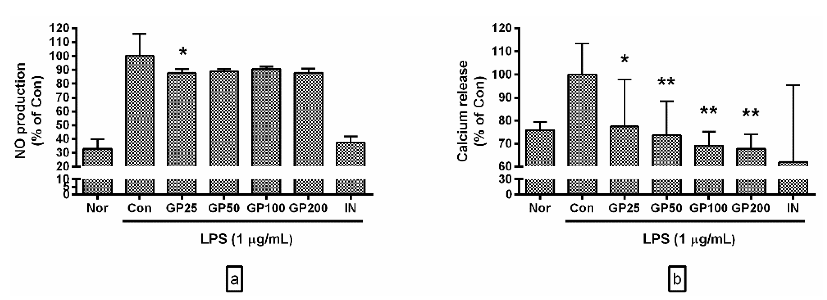
3.2. Effects of GP on Levels of NO and Ca2+

GP inhibited LPS-induced production of NO in RAW 264.7 cells (Figure 1a). Percentages of NO production in LPS-activated RAW 264.7 treated with GP at concentrations of 25, 50, 100, and 200 µg/mL were 87.85 ± 2.79%, 88.94 ± 1.64%, 90.51 ± 1.8%, and 87.93 ± 3% of the control group treated with LPS only, respectively.
GP also inhibited calcium release in LPS-activated RAW 264.7 (Figure 1b). Percentages of calcium release in LPS-activated RAW 264.7 treated with GP at concentrations of 25, 50, 100, and 200 µg/mL were 77.47 ± 20.29%, 73.73 ± 14.66%, 69.15 ± 6.06%, and 67.77 ± 6.24% of the control group treated with LPS only, respectively.

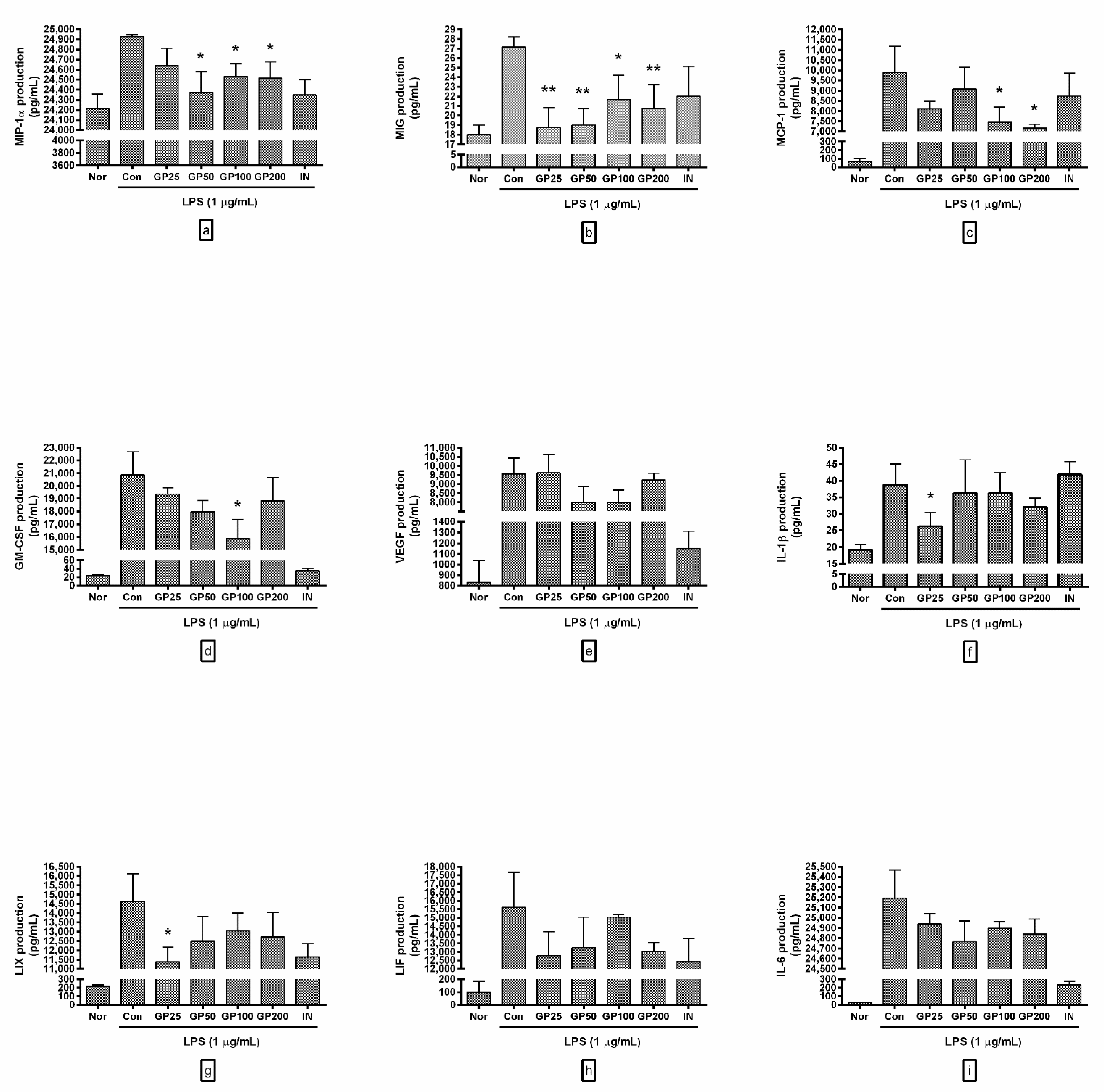
3.3. Effect of GP on Cytokine Production

GP significantly reduced MIP-1α and MIG production in LPS-activated RAW 264.7 (Figure 2). Concrete experimental results, including other cytokines, are as follows:
-
MIP-1α productions from RAW 264.7 with GP at concentrations of 25, 50, 100, and 200 µg/mL were 98.84 ± 0.7%, 97.78 ± 0.85%, 98.41 ± 0.52%, and 98.34 ± 0.66% of the control group treated with LPS only;
-
MIG were 69.02 ± 7.59%, 69.94 ± 6.38%, 79.75 ± 9.44%, and 76.38 ± 9.2%;
-
MCP-1 were 81.91 ± 3.86%, 91.69 ± 10.81%, 75.36 ± 7.39%, and 72.45 ± 1.89%;
-
GM-CSF were 92.83 ± 2.62%, 86.17 ± 4.82%, 76.12 ± 9.42%, and 90.23 ± 9.65%;
-
VEGF were 100.61 ± 10.56%, 83.55 ± 10.99%, 83.44 ± 8.75%, and 96.4 ± 4.14%;
-
IL-1β were 67.65 ± 10.6%, 93.43 ± 25.89%, 93.43 ± 15.8%, and 82.8 ± 6.69%;
-
LIX were 77.78 ± 5.39%, 85.26 ± 9.17%, 89.21 ± 6.45%, and 86.92 ± 9%;
-
LIF were 81.82 ± 9.14%, 84.81 ± 11.59%, 96.35 ± 1.02%, and 83.56 ± 3.29%;
-
IL-6 were 99 ± 0.4%, 98.31 ± 0.82%, 98.83 ± 0.27%, and 98.61 ± 0.59%.
These data suggest that GP could inhibit excessive productions of pro-inflammatory chemokines such as MIP-1α and MIG, leading it to alleviate a hyper-inflammatory syndrome caused by endotoxemia.

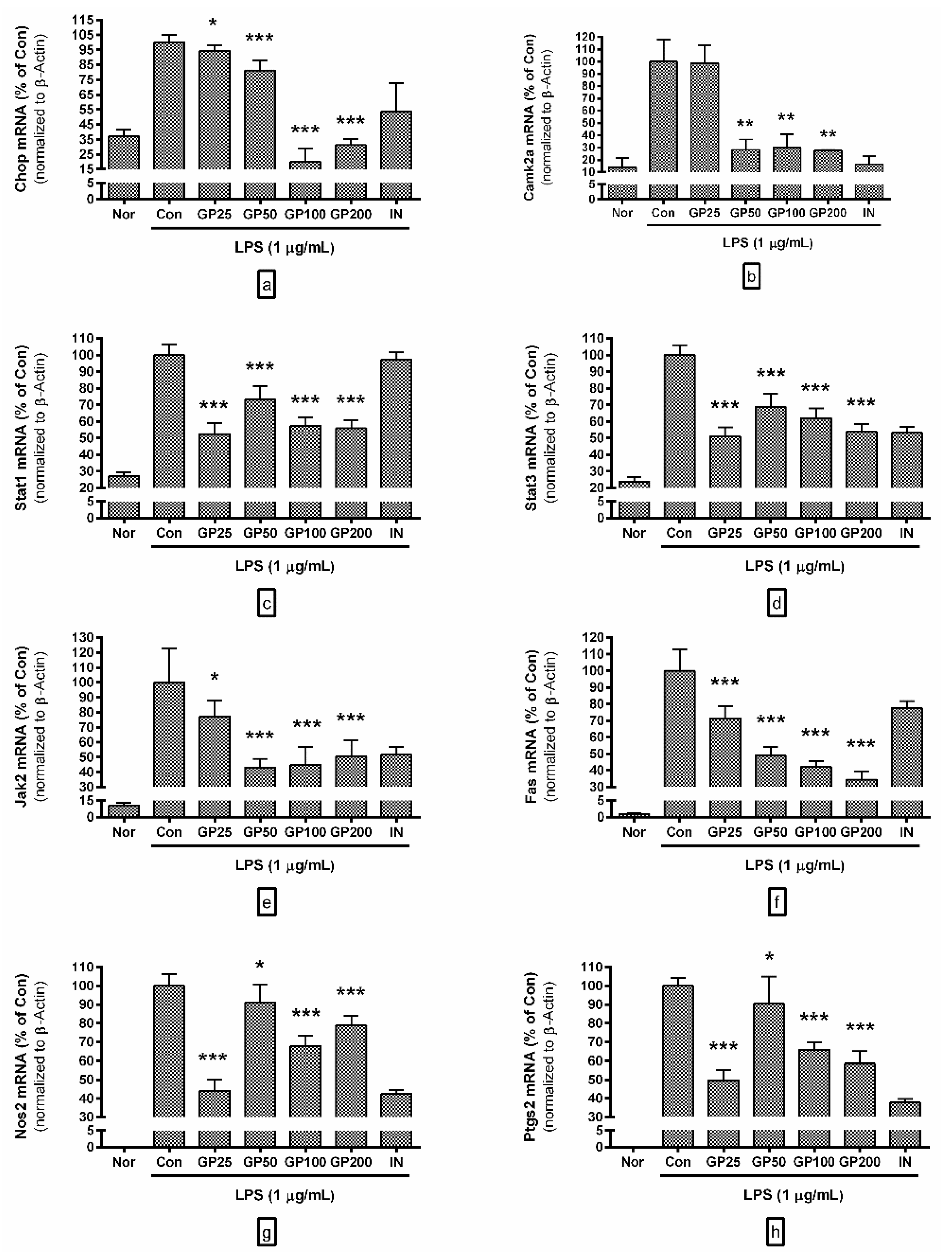
3.4. Effect of GP on mRNA Expressions of Inflammatory Genes

GP significantly inhibited mRNA expressions of Chop (NM_007837), Camk2a (NM_012920), Jak2 (NM_008413), Fas (NM_007987), Stat1 (NM_009283.4), Stat3 (NM_213659.2), Nos2 (NM_010927.3), and Ptgs2 (NM_011198) (Figure 3). Concrete experimental results are as follows:
-
Chop mRNA levels in LPS-activated RAW 264.7 with GP at concentrations of 25, 50, 100, and 200 µg/mL were 94.24 ± 4%, 81.17 ± 6.88%, 19.87 ± 9.02%, and 31.12 ± 4.13% of the control group treated with LPS only;
-
Camk2a were 98.74 ± 14.5, 28.17 ± 8.37%, 30.13 ± 10.88%, and 27.62 ± 0.56%;
-
Stat1 were 52.52 ± 6.34, 73.17 ± 8.11%, 57.45 ± 5.04%, and 55.89 ± 4.94%;
-
Stat3 were 51.04 ± 5.49, 68.85 ± 7.68%, 62.08 ± 5.93%, and 53.83 ± 4.5%;
-
Jak2 were 77.22 ± 10.46, 43.18 ± 5.75%, 44.82 ± 12.07%, and 50.42 ± 10.86%;
-
Fas were 71.59 ± 7.23, 49.15 ± 4.99%, 42.11 ± 3.66%, and 34.57 ± 4.67%;
-
Nos2 were 44.11 ± 5.77, 91.15 ± 9.71%, 67.8 ± 5.61%, and 78.73 ± 5.26%;
-
Ptgs2 were 49.65 ± 5.36, 90.5 ± 14.47%, 65.88 ± 3.94%, and 58.52 ± 6.52%.

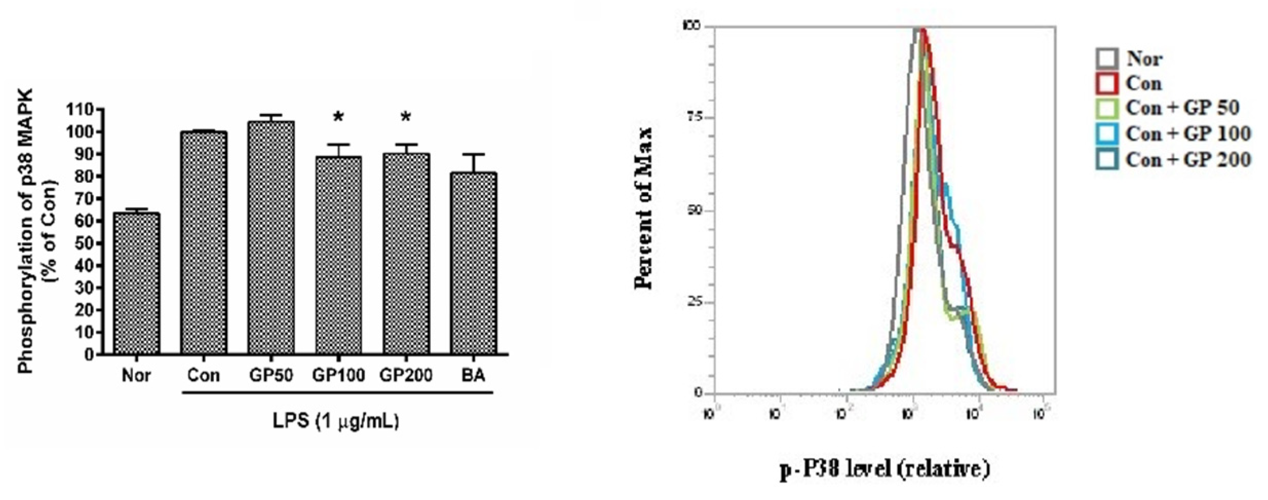
3.5. Effect of GP on Phosphorylation of P38 MAPK

According to the results of the experiment, GP generally inhibited the phosphorylation of P38 MAPK in LPS-activated RAW 264.7 (Figure 4). To be specific, phosphorylation of P38 MAPK in LPS-activated RAW 264.7 treated with GP at concentrations of 50, 100, and 200 µg/mL was 104.55 ± 3.05%, 88.4 ± 5.69%, and 90.16 ± 4.06% of the control group treated with LPS only, respectively. These data mean that GP alleviates inflammatory reactions in LPS-activated macrophages via P38 MAPK signaling.

4. Discussion

Despite the development of many antibiotics and anti-inflammatory drugs, interest in effective drugs to cope with infectious diseases, namely new substances with low toxicity and side effects, continues. As research on infectious diseases progresses, not only the prevention of intrusion into the human body or removal of infectious pathogens, but also the response and control of immune inflammatory reactions after infection are recognized as an important part of treating infectious diseases. This is because many pathogens such as bacteria, virus, fungi, rickettsia, and protozoa cause hyper-inflammatory reactions in our bodies, and life is often at risk if those hyper-inflammatory reactions are not properly controlled [13,14,15,16]. In other words, inflammation is the body’s natural response to infection, but it is also a double-edged sword that, if not properly controlled, causes an acute or chronic disease [1]. In this regard, there are considerable reports on natural products that are less toxic and can modulate inflammatory reactions, apart from drugs that fully suppress immune inflammatory reactions while showing toxicity to immune cells in the body [17,18].
Gardeniae Fructus and Perillae Folium are both found to treat inflammatory diseases in Donguibogam. Modern experimental studies have reported antioxidative properties of Gardeniae Fructus extract [7,8] and anti-inflammatory effects of Perillae Folium extract [9,10]. Minutely, Liu et al. reported in 2020 that Gardeniae Fructus has diuretic, anti-inflammatory, and hemostatic effects; it also protects the liver and gallbladder from infringement and treats cardiovascular and cerebrovascular diseases [19] and its chemical composition includes iridoid glycosides (geniposide, gardenoside, etc.) [20], crocins [21], phenylpropanoids [22], glycoprotein [23], and polysaccharides. Interestingly, Cai et al. reported in 2011 that the maximum extraction yield of flavonoid from Gardeniae Fructus crude extract was 5.05 (mg/g) [24]. Because the current data represented that the total flavonoid content of GP was 10.81 mg RE/g extract, it is possible to estimate that GP will show anti-inflammatory effects. Ruan et al. reported in 2019 that oil from Fructus Gardeniae exerts the antidepressant activity [25]. Kim et al. suggested in 2020 that Gardeniae Fructus 50% EtOH extract protects the esophagus mucosal membrane by attenuating oxidative stress and inflammatory response under reflux esophagitis condition through the antioxidant pathway [26]. However, there are also various reports of the toxicity of Gardeniae Fructus. Recently, Zhou et al. reported in 2019 that Fructus Gardenia can damage the gastrointestinal tract [27]. Li et al. reported in 2019 that the hepatotoxicity of Fructus Gardeniae is caused by iridoids compounds such as genipin [28]. Luo et al. reported in 2021 that Gardeniae Fructus causes plasma biochemical and liver histopathological alterations in rats [29].
In 2015, Zhao et al. reported that ingredients of Perillae Folium include rosmarinic acid, elemicin, perillaldehyde, and dillapiole [30]. It is well known that Perillae Folium contains a lot of polyphenols and flavonoids such as luteolin, 4-hydroxycinnamic acid, scutellarin, caffeic acid, apigenin, chrysoeriol, malvidin, cyanidin, and kaempferol [31,32,33] as well as triterpene acids such as antitumor-promoting tormentic acid [34]. Ueda et al. reported in 2002 that oral administration of Perillae Folium extract to mice shows anti-inflammatory and antiallergic activity with inhibiting TNF-α production; luteolin isolated from Perillae Folium extract inhibits serum TNF-α production [35]. Recently, Yang et al. reported in 2020 that Perillae Folium extract may alleviate allergic airway inflammation [36]. Bae et al. reported in 2017 that Perillae Folium extract decreases MMP-1/MMP-3 expression and ERKs/JNKs phosphorylation in ultraviolet radiation-activated human dermal fibroblasts, which is accompanied by a reduction in reactive oxygen species generation [37]. Kong et al. reported that luteolin from Perillae Folium was reported to inhibit on the expression of VCAM-1 (CD106) in microvascular endothelial cells [38]. Huang et al. reported in 2014 that Perillae Folium extract significantly decreases NO production, PGE2 secretion, and mRNA expressions of pro-inflammatory genes such as IL-6, TNF-α, iNOS, Nos2, and Ptgs2 via the inhibition of MAPK as well as NF-kB signaling in RAW 264.7 cells activated with LPS [39], which makes it possible to estimate that GP might inhibit productions of inflammatory mediators via MAPK signaling pathway in RAW 264.7 cells activated with LPS. In addition, Kangwan et al. reported in 2019 that rosmarinic acid has antioxidant, anti-inflammatory, and anticancer effects; rosmarinic acid-enriched fraction from Perillae Folium strongly protects the stomach against gastric ulcers induced by indomethacin. Considering Gardeniae Fructus-induced gastrointestinal injury, this gastrointestinal protection effect of rosmarinic acid-enriched Perillae Folium fraction makes the investigation into anti-inflammatory effects of the mixture of Gardeniae Fructus and Perillae Folium reasonable in the current study.
Research on bioactive natural products has been increasing in recent years. However, little is known about the effects of a mixture of Gardeniae Fructus and Perillae Folium on inflammatory reactions of macrophages. Naturally, this study examined how a combination of Gardeniae Fructus and Perillae Folium works in LPS-activated macrophages using multiple cytokine assay and signal pathway assay. Because LPS is known to cause endotoxemia [2] and macrophages are responsible for innate immunity [3], LPS-activated macrophages are a representative model of in vitro experiments that identify anti-inflammatory effects. It is well known that LPS stimulation makes RAW 264.7 produce various inflammatory mediators via P38 phosphorylation [4] and JAK-STAT signaling [5]. Interestingly, LPS-induced endoplasmic reticulum (ER) stress increases Chop (GADD153) gene expression, which turns out to be accompanied by P38 phosphorylation [40,41]. In macrophage-related inflammatory process, calcium release with ER stress induces Camk2a, which might enable macrophage apoptosis via Fas induction and/or activation of Stat1 [42,43,44]; cyclooxygenase-2 (COX-2; Ptgs2) is also an important inflammatory mediator related with ER stress [45].
In the current study, GP significantly inhibits productions of MIP-1α and MIG as well as intracellular calcium release and P38 phosphorylation in LPS-activated RAW 264.7, and GP decreased mRNA expressions of Chop (NM_007837), Camk2a (NM_012920), Jak2 (NM_008413), Fas (NM_007987), Stat1 (NM_009283.4), Stat3 (NM_213659.2), Nos2 (NM_010927.3), and Ptgs2 (NM_011198) in LPS-activated RAW 264.7. These results can lead to a hypothesis that GP-mediated reduction of proinflammatory mediators in LPS-activated RAW 264.7 might be achieved through the ER stress-induced CHOP pathway (Figure 5).
However, this study leaves some room for improvement. This study could not evaluate effects of GP on NF-κB activation in LPS-activated RAW 264.7. Data in this study have sometimes failed to represent experimental results in a dose-dependent manner. It seems that GP at concentrations of 100 and 200 µg/mL has affected the results of the experiment by increasing cell viabilities of RAW 264.7. The analytic investigation for phytochemical ingredients of GP has not been determined. Further studies are needed to elucidate the exact mechanisms for anti-inflammatory activities of GP with endotoxemia.

5. Conclusions

GP inhibits productions of MIP-1α and MIG in LPS-activated RAW 264.7 mediated via ER stress-induced CHOP pathway with an accompanying reduction in mRNA expressions of Chop (NM_007837), Camk2a (NM_012920), Jak2 (NM_008413), Fas (NM_007987), Stat1 (NM_009283.4), Stat3 (NM_213659.2), Nos2 (NM_010927.3), and Ptgs2 (NM_011198). More detailed research into the anti-inflammatory effects of GP will help develop treatments for bacterial infectious inflammatory diseases.

Funding

This research was supported by the Basic Science Research Program through the National Research Foundation of Korea, funded by the Ministry of Science, ICT, and Future Planning (2017R1A2B4004933).

Acknowledgments

The author thanks Ji-Young Lee, Young-Jin Kim, and Hyun-Joo Kim (College of Korean Medicine, Gachon University) for their technical assistance.

Conflicts of Interest

The author declares no conflict of interest. The funders had no role in the design of the study, in the collection, analyses, or interpretation of data, in the writing of the manuscript, or in the decision to publish the results.

References

  1. Si-Tahar, M.; Touqui, L.; Chignard, M. Innate immunity and inflammation—Two facets of the same anti-infectious reaction. Clin. Exp. Immunol. 2009, 156, 194–198. [Google Scholar] [CrossRef]
  2. Mills, C.D. M1 and M2 macrophages: Oracles of health and disease. Crit. Rev. Immunol. 2012, 32, 463–488. [Google Scholar] [CrossRef] [Green Version]
  3. Rietschel, E.T.; Kirikae, T.; Schade, F.U.; Mamat, U.; Schmidt, G.; Loppnow, H.; Ulmer, A.J.; Zahringer, U.; Seydel, U.; Di Padova, F. Bacterial endotoxin: Molecular relationships of structure to activity and function. FASEB J. 1994, 8, 217–225. [Google Scholar] [CrossRef]
  4. Kim, A.R.; Lee, M.S.; Shin, T.S.; Hua, H.; Jang, B.C.; Choi, J.S.; Byun, D.S.; Utsuki, T.; Ingram, D.; Kim, H.R. Phlorofucofuroeckol a inhibits the LPS-stimulated iNOS and COX-2 expressions in macrophages via inhibition of NF-kappaB, Akt and p38 MAPK. Toxicol. In Vitro 2011, 25, 1789–1795. [Google Scholar] [CrossRef]
  5. Liu, H.; Yao, Y.M.; Yu, Y.; Dong, N.; Yin, H.N.; Sheng, Z.Y. Role of Janus kinase/signal transducer and activator of transcription pathway in regulation of expression and inflammation-promoting activity of high mobility group Box protein 1 in rat peritoneal macrophages. Shock 2007, 27, 55–60. [Google Scholar] [CrossRef]
  6. Yoon, S.; Lee, Y.; Park, S.K.; Kim, H.; Bae, H.; Kim, H.M.; Ko, S.; Choi, H.Y.; Oh, M.S.; Park, W. Anti-Inflammatory effects of scutellaria baicalensis water extract on LPS-activated RAW 264.7 macrophages. J. Ethnopharmacol. 2009, 125, 286–290. [Google Scholar] [CrossRef] [PubMed]
  7. Liu, H.; Chen, Y.F.; Li, F.; Zhang, H.Y. Fructus Gardenia (Gardenia Jasminoides J. Ellis) phytochemistry, pharmacology of cardiovascular and safety with the perspective of new drugs development. J. Asian Nat. Prod. Res. 2013, 15, 94–110. [Google Scholar] [CrossRef]
  8. Cheng, S.; Zhou, F.; Xu, Y.; Liu, X.; Zhang, Y.; Gu, M.; Su, Z.; Zhao, D.; Zhang, L.; Jia, Y. Geniposide regulates the miR-101/MKP-1/p38 pathway and alleviates atherosclerosis inflammatory injury in ApoE(-/-) mice. Immunobiology 2019, 224, 296–306. [Google Scholar] [CrossRef] [PubMed]
  9. Jin, C.H.; Park, H.C.; So, Y.; Nam, B.; Han, S.N.; Kim, J.B. Comparison of the anti-inflammatory activities of supercritical carbon dioxide versus ethanol extracts from leaves of perilla frutescens britt. radiation mutant. Molecules 2017, 22, 311. [Google Scholar] [CrossRef] [PubMed] [Green Version]
  10. Yang, J.H.; Yoo, J.M.; Lee, E.; Lee, B.; Cho, W.K.; Park, K.I.; Yeul Ma, J. Anti-Inflammatory effects of perillae herba ethanolic extract against TNF-alpha/IFN-Gamma-Stimulated human keratinocyte HaCaT cells. J. Ethnopharmacol. 2018, 211, 217–223. [Google Scholar] [CrossRef] [PubMed]
  11. Kim, D.H.; Lee, J.Y.; Kim, Y.J.; Kim, H.J.; Park, W. Rubi fructus water extract alleviates LPS-Stimulated macrophage activation via an ER stress-induced calcium/CHOP signaling pathway. Nutrients 2020, 12, 3577. [Google Scholar] [CrossRef]
  12. Kim, Y.J.; Lee, J.Y.; Kim, H.J.; Kim, D.H.; Lee, T.H.; Kang, M.S.; Park, W. Anti-Inflammatory effects of angelica sinensis (Oliv.) diels water extract on RAW 264.7 induced with lipopolysaccharide. Nutrients 2018, 10, 647. [Google Scholar] [CrossRef] [Green Version]
  13. Hartley, M.A.; Ronet, C.; Zangger, H.; Beverley, S.M.; Fasel, N. Leishmania RNA virus: When the host pays the toll. Front. Cell. Infect. Microbiol. 2012, 2, 99. [Google Scholar] [CrossRef] [Green Version]
  14. Barsness, K.A.; Bensard, D.D.; Partrick, D.A.; Calkins, C.M.; Hendrickson, R.J.; McIntyre, R.C., Jr. Endotoxin induces an exaggerated interleukin-10 response in peritoneal macrophages of children compared with adults. J. Pediatr. Surg. 2004, 39, 912–915. [Google Scholar] [CrossRef] [PubMed]
  15. Bradford, W.D.; Croker, B.P.; Tisher, C.C. Kidney lesions in rocky mountain spotted fever: A light-, immunofluorescence-, and electron-microscopic study. Am. J. Pathol. 1979, 97, 381–392. [Google Scholar] [PubMed]
  16. Jalbert, E.; Crawford, T.Q.; D’Antoni, M.L.; Keating, S.M.; Norris, P.J.; Nakamoto, B.K.; Seto, T.; Parikh, N.I.; Shikuma, C.M.; Ndhlovu, L.C.; et al. IL-1Beta enriched monocytes mount massive il-6 responses to common inflammatory triggers among chronically HIV-1 infected adults on stable anti-retroviral therapy at risk for cardiovascular disease. PLoS ONE 2013, 8, e75500. [Google Scholar] [CrossRef]
  17. Yuk, S.; Lim, E.; Lee, J.Y.; Lee, Y.; Kim, Y.; Lee, T.H.; Park, S.K.; Bae, H.; Kim, H.M.; Ko, S. Antiinflammatory effects of epimedium brevicornum water extract on lipopolysaccharide-activated RAW264.7 macrophages. Phytother. Res. 2010, 24, 1781–1787. [Google Scholar] [CrossRef] [PubMed]
  18. Kim, H.K.; Lee, J.Y.; Han, H.S.; Kim, Y.J.; Kim, H.J.; Kim, Y.S.; Kim, H.M.; Ko, S.G.; An, H.J.; Lee, Y.J.; et al. Immunomodulatory effects of liriope platyphylla water extract on lipopolysaccharide-activated mouse macrophage. Nutrients 2012, 4, 1887–1897. [Google Scholar] [CrossRef] [Green Version]
  19. Liu, F.; Li, Y.; Li, M.; Wang, J.; Zhang, Y.; Du, Y.; Yang, Y. Study on mechanism of iridoid glycosides derivatives from fructus gardeniae in jiangxi province by network pharmacology. Evid. Based Complement. Altern. Med. 2020, 2020, 4062813. [Google Scholar] [CrossRef]
  20. Wang, Y.; Liu, H.; Shen, L.; Yao, L.; Ma, Y.; Yu, D.; Chen, J.; Li, P.; Chen, Y.; Zhang, C. Isolation and purification of six iridoid glycosides from gardenia jasminoides fruit by medium-pressure liquid chromatography combined with macroporous resin chromatography. J. Sep. Sci. 2015, 38, 4119–4126. [Google Scholar] [CrossRef]
  21. Zhang, C.; Xiao, Y.; Li, L.; Li, W.; Yin, X. Contents of diterpenoid pigments in didifferent parts of fruit from gardenia jasminoides. Zhongguo Zhong Yao Za Zhi 2009, 34, 1395–1397. [Google Scholar] [PubMed]
  22. Zuo, Y.M.; Xu, Y.L.; Zhang, Z.L.; Liu, D.H.; Cai, M.T. Study on phenylpropanoids of gardenia jasminoides. Zhong Yao Cai 2015, 38, 2311–2313. [Google Scholar] [PubMed]
  23. Lu, D.; Zhang, W.; Jiang, Y.; Zhang, Y.; Pan, D.; Zhang, D.; Yao, X.; Yu, Y. Two new triterpenoids from gardenia jasminoides fruits. Nat. Prod. Res. 2019, 33, 2789–2794. [Google Scholar] [CrossRef] [PubMed]
  24. Cai, D.J.; Shu, Q.; Xu, B.Q.; Peng, L.M.; He, Y. Orthogonal test design for optimization of the extraction of flavonid from the fructus gardeniae. Biomed. Environ. Sci. 2011, 24, 688–693. [Google Scholar]
  25. Ruan, J.; Liu, L.; Shan, X.; Xia, B.; Fu, Q. Anti-Depressant effects of oil from fructus gardeniae via PKA-CREB-BDNF signaling. Biosci. Rep. 2019, 39. [Google Scholar] [CrossRef] [Green Version]
  26. Kim, S.H.; Shin, M.R.; Lee, A.R.; Seo, B.I.; Park, H.J.; Roh, S.S. Improvement of inflammation through antioxidant pathway of gardeniae fructus 50% EtOH Extract (GE) from acute reflux esophagitis rats. Biomed. Res. Int. 2020, 2020, 4826176. [Google Scholar] [CrossRef] [Green Version]
  27. Zhou, J.; Yao, N.; Wang, S.; An, D.; Cao, K.; Wei, J.; Li, N.; Zhao, D.; Wang, L.; Chen, X.; et al. Fructus gardeniae-induced gastrointestinal injury was associated with the inflammatory response mediated by the disturbance of vitamin b6, phenylalanine, arachidonic acid, taurine and hypotaurine metabolism. J. Ethnopharmacol. 2019, 235, 47–55. [Google Scholar] [CrossRef]
  28. Li, C.; Lan, M.; Lv, J.; Zhang, Y.; Gao, X.; Gao, X.; Dong, L.; Luo, G.; Zhang, H.; Sun, J. Screening of the hepatotoxic components in fructus gardeniae and their effects on rat liver BRL-3A cells. Molecules 2019, 24, 3920. [Google Scholar] [CrossRef] [Green Version]
  29. Luo, Y.; Gao, F.; Chang, R.; Zhang, X.; Zhong, J.; Wen, J.; Wu, J.; Zhou, T. Metabolomics based comprehensive investigation of gardeniae fructus induced hepatotoxicity. Food Chem. Toxicol. 2021, 153, 112250. [Google Scholar] [CrossRef]
  30. Zhao, B.T.; Lee, K.R.; Lee, J.H.; Min, B.S.; Son, J.K.; Woo, M.H. Quality evaluation of perillae folium by HPLC/PDA. Arch. Pharm. Res. 2015, 38, 1521–1529. [Google Scholar] [CrossRef]
  31. Jiang, T.; Guo, K.; Liu, L.; Tian, W.; Xie, X.; Wen, S.; Wen, C. Integrated transcriptomic and metabolomic data reveal the flavonoid biosynthesis metabolic pathway in perilla frutescens (L.) leaves. Sci. Rep. 2020, 10, 16207. [Google Scholar] [CrossRef]
  32. Xu, L.; Ma, T.; Wang, X.; Liu, W.; Li, J.; Wang, X. Preparative separation of seven polyphenols from perillae folium via pH-zone-refining counter-current chromatography combined with high-speed counter-current chromatography. Anal. Methods 2021, 13, 1232–1237. [Google Scholar] [CrossRef] [PubMed]
  33. Liu, Y.; Hou, Y.; Si, Y.; Wang, W.; Zhang, S.; Sun, S.; Liu, X.; Wang, R.; Wang, W. Isolation, characterization and xanthine oxidase inhibitory activities of flavonoids from the leaves of perilla frutescens. Nat. Prod. Res. 2020, 34, 2566–2572. [Google Scholar] [CrossRef] [PubMed]
  34. Banno, N.; Akihisa, T.; Tokuda, H.; Yasukawa, K.; Higashihara, H.; Ukiya, M.; Watanabe, K.; Kimura, Y.; Hasegawa, J.; Nishino, H. Triterpene acids from the leaves of perilla frutescens and their anti-inflammatory and antitumor-promoting effects. Biosci. Biotechnol. Biochem. 2004, 68, 85–90. [Google Scholar] [CrossRef] [Green Version]
  35. Ueda, H.; Yamazaki, C.; Yamazaki, M. Luteolin as an anti-inflammatory and anti-allergic constituent of perilla frutescens. Biol. Pharm. Bull. 2002, 25, 1197–1202. [Google Scholar] [CrossRef] [PubMed] [Green Version]
  36. Yang, H.; Sun, W.; Ma, P.; Yao, C.; Fan, Y.; Li, S.; Yuan, J.; Zhang, Z.; Li, X.; Lin, M.; et al. Multiple components rapidly screened from perilla leaves attenuate asthma airway inflammation by synergistic targeting on Syk. J. Inflamm. Res. 2020, 13, 897–911. [Google Scholar] [CrossRef]
  37. Bae, J.S.; Han, M.; Shin, H.S.; Kim, M.K.; Shin, C.Y.; Lee, D.H.; Chung, J.H. Perilla frutescens leaves extract ameliorates ultraviolet radiation-induced extracellular matrix damage in human dermal fibroblasts and hairless mice skin. J. Ethnopharmacol. 2017, 195, 334–342. [Google Scholar] [CrossRef] [PubMed]
  38. Kong, X.; Huo, G.; Liu, S.; Li, F.; Chen, W.; Jiang, D. Luteolin suppresses inflammation through inhibiting cAMP-phosphodiesterases activity and expression of adhesion molecules in microvascular endothelial cells. Inflammopharmacology 2019, 27, 773–780. [Google Scholar] [CrossRef]
  39. Huang, B.P.; Lin, C.H.; Chen, Y.C.; Kao, S.H. Anti-Inflammatory effects of perilla frutescens leaf extract on lipopolysaccharide-stimulated RAW264.7 cells. Mol. Med. Rep. 2014, 10, 1077–1083. [Google Scholar] [CrossRef] [Green Version]
  40. Endo, M.; Mori, M.; Akira, S.; Gotoh, T. C/EBP Homologous Protein (CHOP) is crucial for the induction of caspase-11 and the pathogenesis of lipopolysaccharide-induced inflammation. J. Immunol. 2006, 176, 6245–6253. [Google Scholar] [CrossRef] [Green Version]
  41. Wang, X.Z.; Ron, D. Stress-Induced phosphorylation and activation of the transcription factor CHOP (GADD153) by p38 MAP kinase. Science 1996, 272, 1347–1349. [Google Scholar] [CrossRef] [Green Version]
  42. Stout, B.A.; Melendez, K.; Seagrave, J.; Holtzman, M.J.; Wilson, B.; Xiang, J.; Tesfaigzi, Y. STAT1 activation causes translocation of bax to the endoplasmic reticulum during the resolution of airway mucous cell hyperplasia by IFN-gamma. J. Immunol. 2007, 178, 8107–8116. [Google Scholar] [CrossRef] [Green Version]
  43. Timmins, J.M.; Ozcan, L.; Seimon, T.A.; Li, G.; Malagelada, C.; Backs, J.; Backs, T.; Bassel-Duby, R.; Olson, E.N.; Anderson, M.E.; et al. Calcium/calmodulin-Dependent protein kinase II links Er stress with Fas and mitochondrial apoptosis pathways. J. Clin. Investig. 2009, 119, 2925–2941. [Google Scholar] [CrossRef] [PubMed] [Green Version]
  44. Tabas, I.; Seimon, T.; Timmins, J.; Li, G.; Lim, W. Macrophage apoptosis in advanced atherosclerosis. Ann. N. Y. Acad. Sci. 2009, 1173 (Suppl. 1), E40–E45. [Google Scholar] [CrossRef] [PubMed] [Green Version]
  45. Cho, H.K.; Cheong, K.J.; Kim, H.Y.; Cheong, J. Endoplasmic reticulum stress induced by hepatitis B virus X protein enhances cyclo-oxygenase 2 expression via activating transcription factor 4. Biochem. J. 2011, 435, 431–439. [Google Scholar] [CrossRef] [PubMed]
Figure 1. Effects of a combination extract of Gardeniae Fructus and Perillae Folium (GP) on levels of NO (a) and Ca2+ (b). Values are mean ± SD of the three independent experiments. Nor, normal group (media only); Con, control group (LPS alone); IN, indomethacin (0.5 µM). GP25, GP50, GP100, and GP200 indicate 25, 50, 100, and 200 µg/mL of GP, respectively. * p < 0.05 vs. Con; ** p < 0.01 vs. Con.
Processes 09 01632 g001
Figure 2. Effects of a combination extract of Gardeniae Fructus and Perillae Folium (GP) on productions of MIP-1α (CCL-3) (a); MIG (CXCL9) (b); MCP-1 (CCL2) (c); GM-CSF (CSF2) (d); VEGF (e); IL-1β (f), LIX (CXCL5) (g); LIF (h), and IL-6 (i) in LPS-activated RAW 264.7. Each cytokine in the culture medium was measured by a Multiplex cytokine assay. Values are the mean ± SD of the three independent experiments. Nor, normal group (media only); Con, control group (LPS alone); IN, indomethacin (0.5 µM). GP25, GP50, GP100, and GP200 indicate 25, 50, 100, and 200 µg/mL of GP, respectively. * p < 0.05 vs. Con; ** p < 0.01 vs. Con.
Processes 09 01632 g002
Figure 3. Effects of a combination extract of Gardeniae Fructus and Perillae Folium (GP) on mRNA expressions of Chop (NM_007837) (a), Camk2a (NM_012920) (b); Stat1 (NM_009283.4) (c); Stat3 (NM_213659.2) (d); Jak2 (NM_008413) (e); Fas (NM_007987) (f); Nos2 (NM_010927.3) (g), and Ptgs2 (NM_011198) (h) in LPS-activated RAW 264.7. mRNA expressions were evaluated by real-time quantitative PCR. β-Actin was used as an internal control. Values are the mean ± SD of the three independent experiments. Nor, normal group (media only); Con, control group (LPS alone); IN, indomethacin (0.5 µM). GP25, GP50, GP100, and GP200 indicate 25, 50, 100, and 200 µg/mL of GP, respectively. * p < 0.05 vs. Con; ** p < 0.01 vs. Con; *** p < 0.001 vs. Con.
Processes 09 01632 g003
Figure 4. Effects of a combination extract of Gardeniae Fructus and Perillae Folium (GP) on the phosphorylation of P38 in LPS-activated RAW 264.7. Values are the mean ± SD of the three independent experiments. Nor, normal group (media only); Con, control group (LPS alone); BA, baicalein (25 µM). GP25, GP50, GP100, and GP200 indicate 25, 50, 100, and 200 µg/mL of GP, respectively. * p < 0.05 vs. Con.
Processes 09 01632 g004
Figure 5. A combination extract of Gardeniae Fructus and Perillae Folium (GP) inhibits hyper-inflammatory reactions in LPS-activated RAW 264.7 via ER stress-induced CHOP pathway.
Processes 09 01632 g005
Table 1. Primers used for quantitative PCR.
Name 1Forward Primer (5′–3′)Reverse Primer (5′–3′)
ChopCCACCACACCTGAAAGCAGTCCTCATACCAGGCTTCCA
Camk2aAGCCATCCTCACCACTATATTCCTTCACGCCATCATT
Stat1TGAGATGTCCCGGATAGTGGCGCCAGAGAGAAATTCGTGT
Stat3GTCTGCAGAGT TCAAGCACCTTCCTCAGTCACGATCAAGGAG
Jak2TTGGTTTTGAATTATGGTGTCTGTTCCAAATTTTACAAATTCTTGAACC
FasCGCTGTTTTCCCTTGCTGCCTTGAGTATGAACTCTTAACTGTGAG
Nos2TGGAGGTTCTGGATGAGAGCAATGTCCAGGAAGTAGGTGAGG
Ptgs2TCAAACAGTTTCTCTACAACAACTCCACATTTCTTCCCCCAGCAA
β-ActinCTAAGGCCAACCGTGAAAAGACCAGAGGCATACAGGGACA
1 Primers’ names: C/EBP homologous protein (Chop); calcium/calmodulin dependent protein kinase II alpha (Camk2a); signal transducers and activators of transcription 1 (Stat1), Stat3; Janus kinase 2 (Jak2); first apoptosis signal receptor (Fas); nitric oxide synthase 2 (Nos2); prostaglandin-endoperoxide synthase 2 (Ptgs2), and β-Actin.
Publisher’s Note: MDPI stays neutral with regard to jurisdictional claims in published maps and institutional affiliations.

