Review

Challenges towards Targeted Drug Delivery in Cancer Nanomedicines

by
Muhammad Nadeem Hafeez
1,
Christian Celia
1 and
Vilma Petrikaite
2,3,*
1
Department of Pharmacy, University of Chieti—Pescara “G. d’Annunzio”, I-66100 Chieti, Italy
2
Laboratory of Drug Targets Histopathology, Institute of Cardiology, Lithuanian University of Health Sciences, Sukilėlių pr. 13, LT-50162 Kaunas, Lithuania
3
Institute of Biotechnology, Life Sciences Center, Vilnius University, Saulėtekio al. 7, LT-10257 Vilnius, Lithuania
*
Author to whom correspondence should be addressed.
Processes 2021, 9(9), 1527; https://doi.org/10.3390/pr9091527
Submission received: 28 June 2021 / Revised: 20 August 2021 / Accepted: 25 August 2021 / Published: 27 August 2021
(This article belongs to the Special Issue Application of Nanoparticles in Cancer Therapy)

Abstract

:
Despite cancer nanomedicine celebrates already thirty years since its introduction, together with the achievements and progress in cancer treatment area, it still undergoes serious disadvantages that must be addressed. Since the first observation that macromolecules tend to accumulate in tumor tissue due to fenestrated endothelial of vasculature, considered as the “royal gate” in drug delivery field, more than dozens of nanoformulations have been approved and introduced into the practice for cancer treatment. Lipid, polymeric, and hybrid nanocarriers are biocompatible nano-drug delivery systems (NDDs) having suitable physicochemical properties and modulate payload release in response to specific chemical or physical stimuli. Biopharmaceutical properties of NDDs and their efficacy in animal models and humans can significantly affect their impact and perspective in nanomedicine. One of the future directions could be focusing on personalized cancer treatment, considering the heterogeneity and complexity of each patient tumor tissue and the designing of multifunctional targeted NDDs combining synthetic nanomaterials and biological components, like cellular membranes, circulating proteins, RNAi/DNAi, which enforce the efficacy of NDDs and boost their therapeutic effect.

1. Introduction

Combination-chemotherapy for cancer treatment has recently emerged to address some drawbacks of chemotherapy, which entails the simultaneous administration of many anticancer drugs. Despite its numerous challenges, anticancer drug co-administration is a widely used conventional goal in clinical practice [1]. Blood circulation, bio-distribution, and the targeted effect of combination drugs are all influenced by the solubility, biopharmaceutical, and pharmacokinetic properties of payloads [1,2]. Nano-sized drug delivery systems (NDDs) are gaining widespread interest among scientists as a promising and innovative strategy in combination therapy to increase the effect of co-delivered chemotherapeutic drugs due to their enhanced retention and permeability, as well as their extended blood circulation time, controlled drug release, and prevention of drug degradation [3]. NDDs are frequently made by loading nanocarriers with natural and synthetic drugs, having different physicochemical properties, and co-delivering in the targeting tissue. Nanocarriers are made from a variety of materials, including polymer micelles, organic and inorganic nanoparticles, supramolecular aggregates/devices, and liposomes. The physical properties of nanocarriers, such as shape and size [4], as well as the microenvironment surrounding the tumor, as alteration in temperature [5,6,7,8] and pH [9], could be used to control drug release. The pH difference in the tumor microenvironment causes a charge transition from negative to positive, which might be exploited for endocytosis absorption of specially tailored carriers [9]. NDDs are made of pH-sensitive organic and inorganic materials that allow the payload to be released at the tumor location by simple bond breakage [10,11].
Tumor cells overexpressed certain receptors on their surfaces. Active targeting involves the overexpression of specific ligand-binding receptors in tumor tissues. Specific ligands are attached to the surface of drug-loaded nanocarriers with a high affinity for targeting receptors overexpressed on tumor surface. Active targeting provides several advantages over passive targeting, including reduced systemic administration, increased effectiveness, and increased therapeutic drug delivery [12,13]. Anticancer nanoparticles with sizes ranging from 80 to 200 nm are made up of a combination of organic and inorganic elements [14]. Several liposomal-based NDDs have been approved for cancer therapy, with more under clinical testing. The shell-core structure of polymer lipid nanocarriers, which consists of a phospholipid shell and a polymeric core, combines the benefits of polymers and liposomes [15]. They are mechanically stable, have a wide surface area, and are evenly dispersed [16]. They are also highly biocompatible [17], have increased loading capacity [18], and make transporting both hydrophilic and hydrophobic medicines easier [19].
Nanoparticles made from metals (such as platinum, gold, silver, and iron) and non-metallic nanocarriers (Fe3O4, mesoporous silica) and their derivatives [20] are becoming more popular in cancer research because of their capacity to monitor the release process and drug delivery mechanism. Carbon nanotubes (CNTS) on the other hand, are unsuitable for receptor-mediated cancer treatment. Conjugated halloysite magnetic CNTS with folate and chitosan oligosaccharides improved intracellular medication delivery [21]. The cytotoxic effects of CNTS are a major impediment to their widespread adoption as a cancer therapy. However, PEGylated CNTS with a diameter of 300 nm were shown to be nontoxic and safe for doxorubicin administration [22]. Vogtle was the first to introduce dendrimers into the realm of cancer treatment [23]. Like carbon nanotubes, they are tiny and can easily penetrate cancer cells. When negatively charged poly-(amido amine)-2,3-dimethylmaleic monoamide was exposed to acidic pH, it transformed into a positively charged dendrimer at the tumor site, resulting in increased membrane permeability and minimal cytotoxicity [24].
In this review we provide a critical overview of drug delivery systems as nanomedicine and their potential treatment in anticancer therapy.

2. Drug Delivery Strategies in Cancer Therapy

Cancer is one of the leading causes of death around the world. Chemotherapy is an extensively used treatment approach for cancer. The chemotherapeutic drugs have several drawbacks including development of resistance, interference with metabolic activities of normal cells, and nonspecific targeting [25]. Combination therapy in which multiple anticancer drugs are used simultaneously has emerged recently to overcome shortcomings of chemotherapy. Despite having numerous challenges, co-delivery of anticancer drugs is widely used as the standard goal in clinical practice [1]. Blood circulation time, bio-distribution, and targeting effect of combined drugs are considerably affected by solubility and pharmacokinetic properties of drugs [1,2]. Nanosized drug delivery systems (NDDs) are widely gaining attraction among scientific community as promising and innovative approach in combination therapy to increase the effect of co-delivered chemotherapeutic drugs because of their enhanced retention, permeability, extended blood circulation time, controlled release of drug, prevention of drug from degradation, maximum accumulation rate at targeted site, and minimum side effects on normal cells [3]. Typically, NDDs are developed from nanocarriers loaded with anti- drugs for their simultaneous co-delivery. Materials used for the synthesis of nanocarriers are polymer micelles, they have inert properties to prevent events such as systemic toxicity and inflammation. Another key advantage of NDDs is real-time tracking of drug, achieved through co-loading of tracking agent which allows tracking of delivery process and biodistribution of drugs, thus predicting their therapeutic effects [26]. However, if loading capacity of NDDs is quite low (<10%), it is highly desirable to enhance their loading efficiency to improve the proficiency of the system [2].

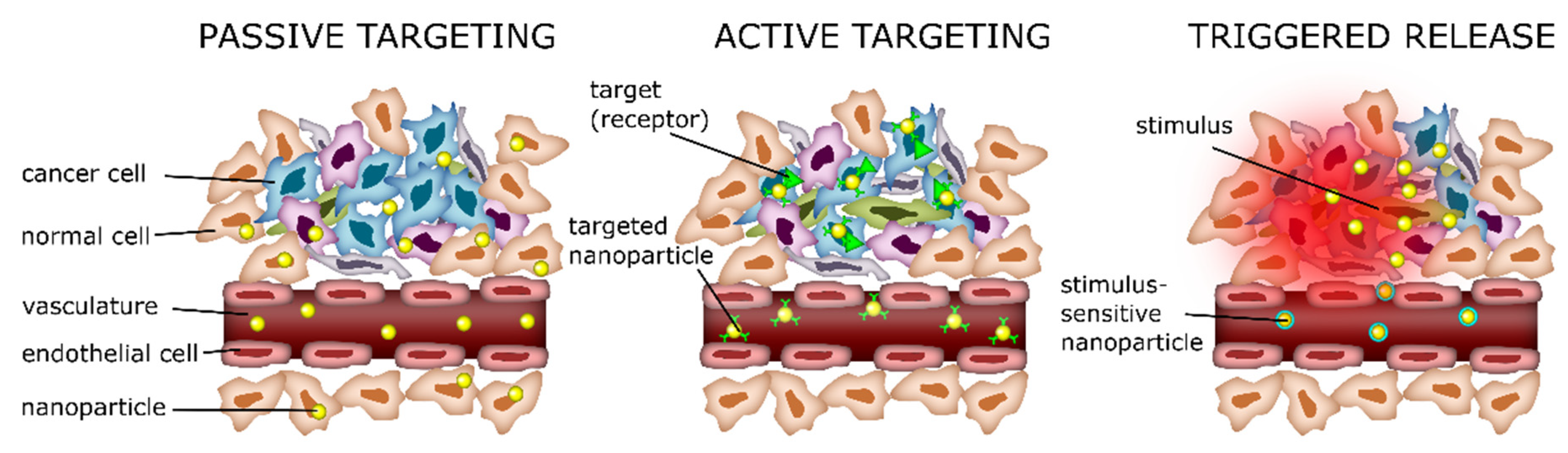
3. Drug Targeting Strategies in Cancer Therapy

The distribution of co-delivered anticancer drugs to normal cells is the main challenge in combination chemotherapy [5,6]. The co-delivered drug usually interferes with metabolic activities, causes side effects, and damages the healthy cells along with cancerous cells. The concentration and efficacy of delivered anticancer drugs at targeted site was reduced due to the distribution of drugs to normal tissues [7]. To overcome this limitation, targeted strategies must be improved. Nanocarriers, in combination chemotherapy, were found useful in targeted drug delivery to cancer cells with reduced cytotoxicity and enhanced blood circulation time [27]. Two types of targeting strategies are usually utilized: active and passive targeted drug delivery. The active targeting is based on ligands conjugated to nanocarriers which can bind with the overexpressed receptors on the surface of the targeted cells [28], the passive targeting exploits the presence of abnormal fenestrations on the surface of tumor cells [13].
Tumor cells multiply quickly, thus they produce vascular endothelial growth factor (VEGF) to stimulate formation of new blood vessels [29]. Newly formed vessels are structurally and anatomically abnormal, having saccular endothelium with large fenestrations [28,29]. Platelets and leukocytes can easily extravasate through fenestrations during inflammation. In passive targeting, nanocarriers, having size range of leukocytes are usually designed to exploit this underlying phenomenon (Figure 1). Drug-loaded nanocarriers come out from the endothelial fenestrations of tumor microvasculature and very low blood flow allows them to accumulate at tumor site. The physical properties of nanocarriers, e.g., shape and size [4] and microenvironment around tumor, increase in temperature [5,6,7,8] and decrease in pH [9], could allow to exploit for controlled drug release. The pH difference at tumor microenvironment results in switching negative to positive charge which could allow to take advantage of the uptake of specifically designed carriers by endocytosis [9]. In fact, NDDs are developed pH sensitive organic and inorganic materials which allow the release of payload at tumor site by easy rupture of bonds [10,11,30]. The primary disadvantage of pH sensitive NDDs is they are usually active at specific pH range and also toxic to normal cells in addition to cancer cells [11]. The temperature in the different body parts varies, depending upon different factors such as blood flow and energy generated by cellular metabolism. Tumor cells usually have higher metabolic activity and blood flow than that of surrounding healthy tissue, an increase in temperature is observable in the tumor tissue [1,2,3]. Increased temperature at tumor site could allow us to take advantage through thermo responsive NDDs. Temperature control stimuli-targeted system has greater advantages in terms of passive targeting ability, versatility in design, in situ phase transition, and tunability of phase transition [31]. As an effort to further exploit existing temperature responsive systems, current innovative applications have combined temperature with other stimuli such as pH and light. The result has demonstrated suitable control over drug release at targeted site [4].
Tumor cells overexpress specific receptors on their surface. Active targeting exploits the overexpression of specific ligand-binding receptors in tumor tissues (Figure 1). These ligands are attached on the surface of drug-loaded nanocarriers which has significant affinity with overexpressed receptors of tumor site [12]. Choice of ligands is usually critical in appropriate binding with receptors to deliver higher amount of drug to cancer cells rather than in healthy tissues. Several ligands have been exploited in active targeting of drug delivery including peptides (e.g., glycine, asparagine, arginine, octreotide), [32], proteins (e.g., antibodies, transferrin, growth factors), mono- and polysaccharides (e.g., galactose, hyaluronic acid), and aptamers (e.g., biotin, bisphosphonates) [33]. Active targeting has several advantages over passive targeting such as lower systemic administration, higher efficacy, and higher delivery of therapeutic drug [12,13]. Some tumor cells highly express integrin αvβ3 receptors [11] that actively bind with cyclic peptide iRGD (tumor-penetrating and tumor-homing peptide), exhibited enhanced tumoricidal effects of doxorubicin [32]. Overexpression of transferrin receptor 1 on tumor cells can potentially be exploited by transferrin-conjugated NDDs [34]. High dose of transferrin conjugated NDDs was also found toxic to normal cells, as well [35]. EGFR (epidermal growth factor receptor) conjugated NDDs via endocytic pathway, unlike transferrin receptor 1, promote the transportation of drug into the nucleus in rapidly dividing cells [36]. ErbB2 (erythroblastic leukemia viral oncogene homolog 2) is overexpressed in breast carcinomas. F5-scFv containing doxorubicin exhibited antitumor activity and significant reduction in tumor size of xenografted mice [37]. Treatment of malignancies with VEGF (vascular endothelial growth factor) inhibitors are under clinical trials. Bevacizumab, Avastin® (anti-VEGF monoclonal antibody), got approval from FDA for the treatment of colorectal cancer with conventional chemotherapy [38,39]. There are several disadvantages of these NDDS including: (i) Their therapeutic effect might be altered if bounded antibody (ligand) is not degraded properly; (ii) if the fractions involved in antibody-DDS binding are sterically hampered then affinity between antibody and NDDS might be; (iii) they are poorly immunogenic and readily excreted via liver and kidneys; (iv) their synthesis is quite expensive; (v) these are degraded by peptidase enzymes and make them biologically ineffective [5].
Triggered release depends on biological and physical stimuli and can modulate the release of payloads in tumor macroenvironment. pH, temperature, physical, and chemical properties of nanomaterials make nanoparticles to modulate the active/passive delivery of chemotherapeutic drugs and promote their accumulation in the tumor sites (Figure 1).

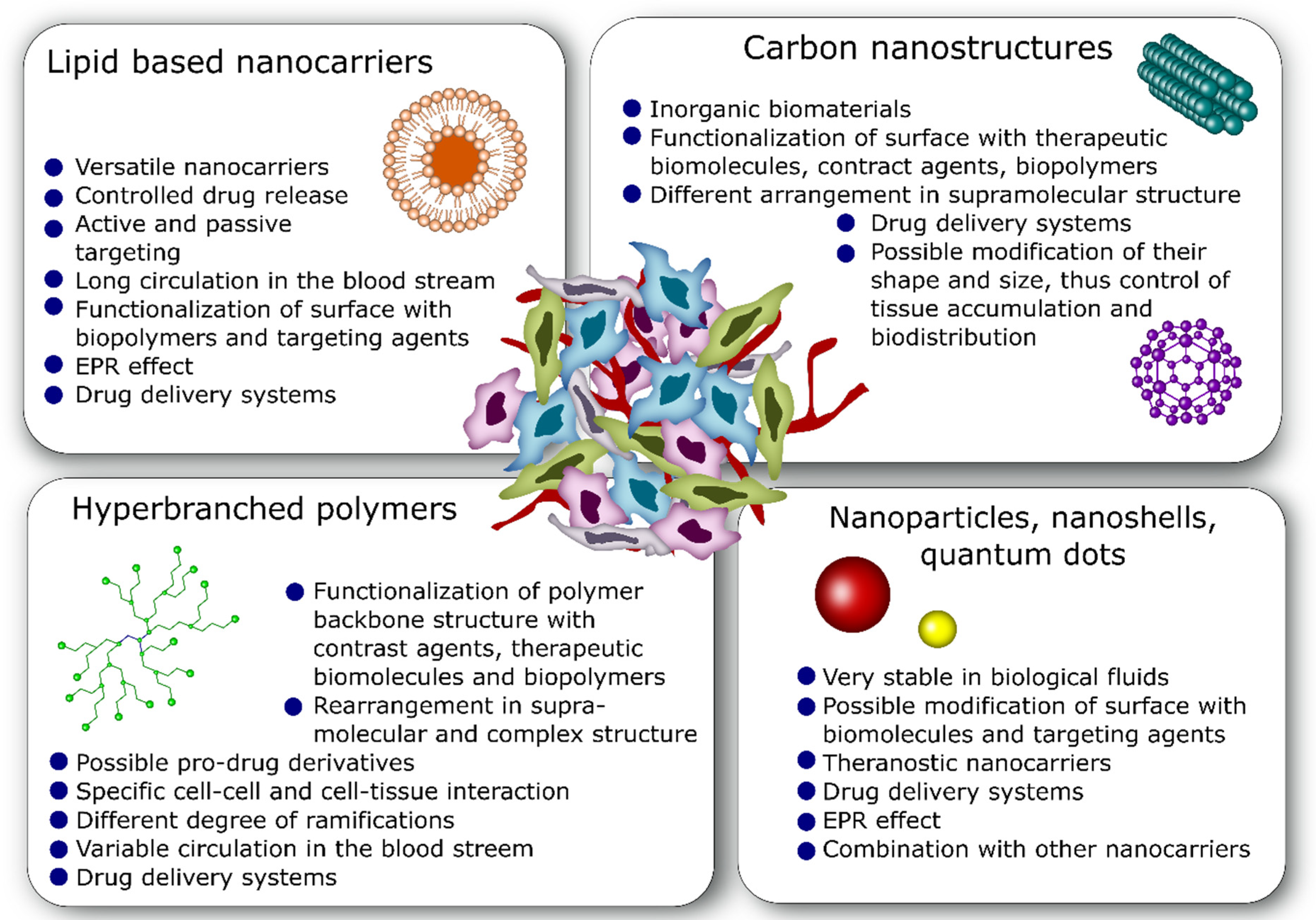
4. Commonly Used Nano-Drug Delivery Systems in Anticancer Therapy

Despite having several limitations, chemotherapy is a widely used treatment approach for cancer. Chemotherapeutic drugs are mostly administered intravenously, and they are highly cytotoxic to normal cells. Targeted delivery of anticancer drugs through nanocarriers is an emerging technology, which commits to overcome drawbacks of chemotherapy in cancer treatment. Nanosized anticancer particles (anticancer drug loaded) having size from 80–200 nm are made up of combination of different organic and inorganic materials [14]. Targeted drug delivery, minimum side effects, lower toxicity toward normal cells, and increased size to surface area ratio are the main advantages of NDDs over chemotherapy [40].
Several lipids-based NDDs have been approved to date for cancer treatment and some others are under clinical trials. Lipo-Dox (Doxil®) was the first lipid based nanodrug approved by FDA in 1995. It exhibited better toxicity profile as comparted with free drug and generally used for treatment of Kaposi’s sarcoma, multiple myeloma, ovarian neoplasms, and breast cancer [41]. Another nanodrug, DaunoXome®, for the treatment of Kaposi’s sarcoma approved in 1996 by FDA. In 2009, E.U approved liposomal mifamurtide, Mepact®, for the treatment of osteosarcoma. To treat myelogenous leukemia, Marqibo® (liposomal vincristine sulfate) was introduced in 2012. In 2015, lipo-irinotecan (Onivyde®) was introduced in the market as chemotherapeutic agent for treatment of pancreatic cancer. Adjuvant liposomal AS15, active ingredient monophosphoryl lipid-A, is under phase-II clinical trial. SP1-077 (cisplatin-loaded PEG-liposome) for treatment of neck, lung and head cancers, under phase II clinical trial. The ALN-VSP (lipid-based NDDS containing kinesin spindle protein siRNAs and VEGF-A) is under phase I clinical trial for the treatment of advanced liver metastases [14]. Nanocarrier-based NDDs PTX-loaded albumin (Abraxane®) got approval in 2005 for pancreatic neo-plasma lung carcinoma therapy. A monoclonal antibody (radioimmunotherapy, Zevalin®) was released in the market in 2004 for the treatment of transformed B cell non-Hodgkin’s lymphoma [42].
Here we overview a classification of different nanocarriers based on material chemistry and compositions, which delivered payloads as well as contrast agents, and are used for the treatment of liquid and solid tumors (Figure 2).

4.1. Lipid-Based Nanocarriers

Lipid-based nanocarriers are commonly used NDDs for cancer treatment. Polymer lipid nanocarriers with their unique shell-core structure are composed of phospholipid shell and polymeric core and exhibit collective advantages of polymers and liposomes [15]. They are mechanically stable with large surface area, uniformly distributed [16], highly biocompatible [17], enhanced loading capacity [18] and facilitates the transportation of both hydrophilic and hydrophobic drugs [19]. More than 90% encapsulation efficiency and release profile of docetaxel (anticancer drug) has been obtained with polymer-lipid carriers. Chemotherapeutic efficacy, hydrophobicity, payload capacity (>40) and controlled release kinetics, synergistic cytotoxicity, and higher accumulation at targeted site of metformin and topotecan has been achieved when delivered through lipid-based nanocarriers [43].
To increase water solubility and bioavailability, lipid carriers are usually conjugated with natural molecules. The first polymer-lipid hybrid carrier was developed by conjugating furanocoumarin psoralen [44], which showed lower toxicity and higher chemotherapeutic activity of doxorubicin. Combination of therapeutic and diagnostic agent in single formulation of lipid-based carrier is gaining momentum in nanodrug cancer therapy. Doxorubicin and docosahexaenoic acid demonstrated higher feasibility in breast cancer therapy when co-loaded into the radiolabeled lipid-based NC-Tc-99m [45]. Similarly, miR-181a and melphalan exhibited enhanced release profile when encapsulated in single formulation of lipid carriers for retinoblastoma treatment [46].

4.2. Nanoparticles

Nanorattles, nanoshells, metallic, non-metallic nanoparticles, and quantum dots are widely used in various types of cancer treatment. Nanoparticles are made up of metals (e.g., platinum, gold, silver, iron, zinc, titanium, thallium, and cerium) or their derivatives (e.g., sulfides, hydroxides, oxides, chlorides, fluorides and phosphates) [20]. Magneto-plasmonic metallic nanohybrid containing Ag/Au shell coated with poly (co-methacrylic acid-butyl methacrylate-co-acrylamide) and Fe3O4 co-loaded letrozole showed enhanced targeted and controlled delivery at tumor site [20]. Plasmonic bimetallic Au-Ag and Ag-Au (core) nanocarriers produce heat energy which increases the solubility of polymer and its diffusion and also kill cancer cells with photothermal radiations [47]. Encapsulated hallow plasmonic Ag-Au nanoshells demonstrated effective and efficient release of 5-fluorouracil in prostate cancer treatment [48].
Fluorescent labelled non-metallic nanocarriers (Fe3O4, inorganic-hydroxyapatite and mesoporous silica) are gaining interest in cancer research because of their ability to monitor the release process and drug delivery pathway. Nanocomposites comprising quantum dots graphenes (GQDs), lectin protein, concanavalin A, and Fe3O4 co-loaded with doxorubicin, exhibited significant increase in accumulation of drug and 13% higher cytotoxicity against HeLa cells [49]. Doxorubicin-targeted delivery is also achieved by using modified GQD-mesoporous silica-NPs with hyaluronic acid for fluorescent imaging [50]. WS2 QDs co-loaded with DOX and mesoporous organosilicas nanoparticles demonstrated high potential for synergistic chemo-PTT. Ultrasmall WS2 quantum dots open new horizons for cancer therapy [51]. WS2 quantum dots co-loaded mesoporous organosilicas and doxorubicin nanocarriers exhibited high potential for synergistic chemo-PTT [51].

4.3. Carbon Nanostructures

Carbon nanotubes (CNTS) are third allotrope of carbon and cylindrical fullerenes [52]. CNTS comprised rolled concentric graphene sheets having diameter of 1 nm [53] and has two subdivisions per sheet in concentric cylinder; single walled CNTS (SCNTS) having 0.4–3.0 nm diameter and multi walled CNTS (MCNTS) having 2–100 nm diameter [54]. SWCNTS are suitable vehicle for nucleic acid delivery, these can easily penetrate into the cell due to their narrow needle like structure [55]. However, SCNTS, are not suitable for receptor-mediated cancer therapy. Enhanced intracellular drug delivery was achieved through conjugated halloysite magnetic SCNTS with folate and chitosan oligosaccharides [21]. Carbon dots can be used as fluorescent therapeutic pH responsive NDDs containing doxorubicin for the treatment of gastric cancer instead of titanium dioxide and carbon nanotubes. The optical labeling of carbon dots enables the tracking of drug delivery process within 48 h. The carboxyl rich carbon dots, exhibited no cytotoxicity with 90% survival rate of human gastric epithelial cells (GES-1) and human gastric cancer cells (MGC-803) [44]. Recently, titanium dioxide nanotubes co-loaded liposomes have successfully been demonstrated for extended delivery of anticancer drug into HeLa cells [56]. Cytotoxic effects of MCNTS are major barrier in their safe and extensive use for cancer treatment. However, PEGylated MCNTS (300 nm), exhibited safe delivery of doxorubicin with no cytotoxic effects [22].

4.4. Hyperbranched Polymers

Dendrimers, hyperbranched polymers were first time introduced for cancer therapy by Vogtle [23]. These are nanoscopic three-dimensional macromolecules. They are widely used in cancer nanomedicines because they are highly interactive with other potential macromolecules [23]. Here we reviewed recent advancement about crucial role of hyperbranched polymers in cancer treatment and drug delivery. They are smaller in size and can easily penetrate into the cancer cells like carbon nanotubes. Negatively charged poly-(amido amine)-2,3-dimethylmaleic monoamide converted into positively charged dendrimers at tumor site when they encountered acidic pH and they exhibit enhanced membrane permeability and low cytotoxicity when co-administered with doxorubicin [24]. Cytotoxicity of poly-amidoamine G4 was reduced by conjugation with PEG in mice myoblast C2C12 cell lines [57]. Edges of graphene sheets of magnetic dendrimers are conjugated with hydrazone group which impart magnetic property and increase the solubility of carrier, allowing them to be used for co-delivery of hydrophilic (doxorubicin) and hydrophobic (curcumin) drugs [58].

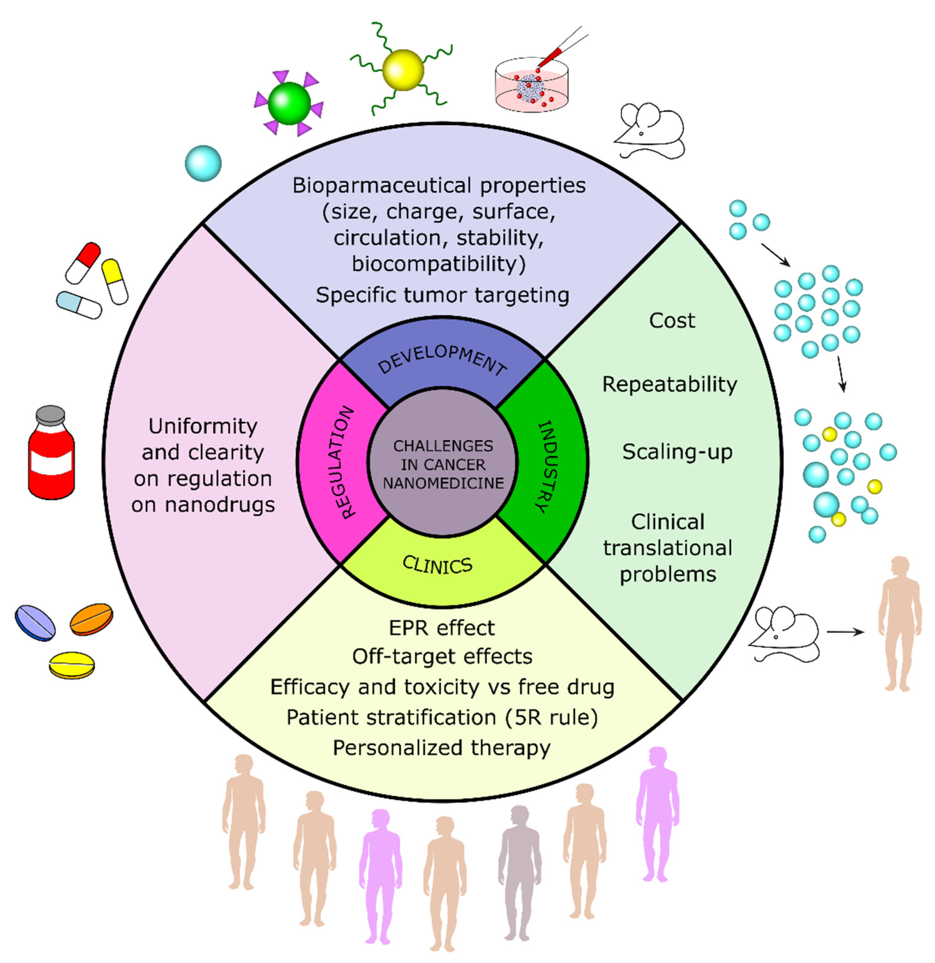
5. Challenges toward Drug Delivery Systems in Anticancer Therapy

Although cancer nanomedicine celebrates thirty years since its introduction, together with the achievements and progress in cancer treatment area, it still ha serious disadvantages that have to be addressed yet [59] (Figure 3). Since the first observation that macromolecules tend to accumulate in tumor tissue due to fenestrated endothelial of vasculature [60], considered as the “royal gate” in drug delivery field [61], more than dozens of cancer nanoformulations have been approved and introduced as potential treatment approach [62]. Moreover, about ~2000 of designed nanoformulations intended to be used for cancer treatment are under clinical trials (search terms: nanoparticle/liposome/micelle/quantum dots/nano) [63,64]. However, the extensive development of NDDs is facing more and more challenges in scientific and clinical community, including high developmental cost, technological issues, and clinical translational failures. Due to these reasons, only few out of hundreds developed NDDs successfully get their place in the market. Nanomedicines are complex formulations, thus even small variations in their technological process could influence the treatment efficacy and alter the profile of their side effects [65]. The importance of specific issues related to the development of nanomedicines, are officially recognized by European Medicines Agency (EMA), which prepared several scientific guidelines for medicine developers addressing surface coatings, data requirements for intravenous colloidal nanoproducts [66]. Due to these factors, nanomedicines remain less popular as compared to the conventional, usually non-specific and relatively toxic chemotherapeutics, regardless that nanodrugs are more specific and are more clinically efficacious [67].
There are many publications in which the challenges on anticancer nanomedicines with suggestions on how to resolve them are discussed [68,69,70]. As one of the strategies exploited by industries to resolve the translational problem, is the application of AstraZeneca’s 5R principle: right target, right patient, right tissue, right safety and right commercial potential [71]. Hare et al. [72] emphasize the need to use more clinically relevant models for testing nanomedicines, by focusing more on tumor biology and targeting the right patients. Regardless the excellent efficacy in preclinical models, many nanomedicines fail under clinical trials [73,74]. The majority of approved nanodrugs in clinical trials were compared with the standard chemotherapy or combinations, not with free drugs, due to ethical reasons [73].
Nevertheless, the development of nanoformulations aims to enhance the clinical efficacy and reduce toxicity of anticancer drugs. Nanoformulations may improve the therapeutic efficacy of anticancer agents by optimizing their pharmacokinetic properties, both by exploiting the enhanced permeability and retention (EPR) effect [75] and targeted therapy approach [76]. NDDs usually have a long systemic circulation and accumulate less in the normal organs compared to tumors tissue, which reduces the side effects. In addition to diminishing side effects, characteristic of the free anticancer drug, nanomedicines could cause the off-target effects [77,78]. Efficacy, side effects, biopharmaceutical properties, off-target effects of nanomedicines are discussed in further subsections.

5.1. Efficacy and Side Effects

NDDs are considered as the most promising systems to increase anticancer drug efficacy and reduce side effects [79]. This could be achieved by focusing drug effect on tumor site and avoiding their transport to the healthy tissues. Currently, approved anticancer nanoformulations take advantage of EPR effect of NDDs [80]. However, in past decade researchers are extensively discussing if NDDs design based on EPR effect could be the right strategy for the development of anticancer nanomedicines [73,81]. It has been established that EPR effect is highly heterogenous, it could change during the cancer treatment [82]. Moreover, in some patients this effect is not expressed enough to contribute to the clinical efficiency of nanomedicines, so, it is necessary to stratify patients according to EPR criteria in order to increase therapeutic outcome [83]. Interestingly, based on more than 100 of preclinical studies, researchers established that overall average accumulation of NDDs in tumors is about 0.7% of administered dose [26], but this small dose could still be sufficient for patient to achieve a beneficial clinical effect [84]. Hare. et al. [72] in their review raised the question if nanomedicines can enhance drug accumulation in tumors by EPR, as compared to free drugs. It has been demonstrated that macromolecular drugs could also favorably accumulate in the tumor due to EPR effect [85]. The lack of the clinical data regarding direct and consistent comparison of accumulation of the free drug and NDDs containing the same drug, make the evaluation of EPR effect contribution to nanomedicines efficacy even more complicated. Out of eight NDDs tested under Phase III clinical trials, only two were established to be more efficacious as compared to free drugs [72].
Regardless of the widespread opinion that NDDs reduce side effects, Luan, et al. [86] noted this is not always the case. Anticancer nanomedicines could induce some specific toxicities. For instance, PEGylated doxorubicin (Caelyx/Doxil®) reduced cardiotoxicity, but at the same time increased hand-foot syndrome, rash, and pigmentation as comparison to the free drug [87]. Another nanotherapeutic Abraxane® (albumin-bound paclitaxel) reduces the neutropenia, but increases the neuropathy [88]. However, the decreased side effects of Abraxane® might be associated with the lack of Cremophor El® in the formulation [89]. Moreover, it was determined that NDDs can accumulate not only in tumors due to EPR, but also in other tissues with fenestrated endothelium, such as spleen, liver, and pancreas [90]. Majority of the nanomedicines act like intracellular organelles and other molecules in the body and thus could be involved in various biological interactions and induce unique cytotoxic effects, depending on their size, physicochemical properties, etc. These effects are studied by so-called nanotoxicology research [91].

5.2. Biopharmaceutical Properties and Anticancer Activity

According to the criteria by FDA [92], nanotechnological products are those containing materials or that range from 1 to 100 nm, or larger but exhibit properties or phenomena that are attributable to this range of dimensions. It should be noted that the maximum size of nanomaterial cannot clearly define the psychochemical and biological properties of the materials because these do not differ greatly at 100 nm scale, thus other characteristics should be considered, too [93]. Size determines the transport of nanoparticles through the bloodstream and then the accumulation in the tumor tissue. Smaller nanomedicines can be delivered to the tumor better as compared to the larger ones, but they might be distributed to the healthy tissue as well. Conversely, it is much more difficult for larger particles to distribute in the tumor site [94]. Usually, NDDs in the range of 10–100 nm are desired due to their accumulation in the targeted tissues [95]. The charge can also affect biopharmaceutical properties of nanomedicines. Positively charged particles tend to distribute better in tumor tissue as compared to the negatively charged ones, but neutral NDDs diffused faster [96]. The charge of nanocarriers can be changed easily by attaching various ligands, and this could also impact blood circulation time and cellular uptake [97]. Moreover, the modification of surface properties of NDDs cause the adsorption of circulating proteins which modify nanoparticle targeting properties and their relative accumulation in the tumor tissues [98,99]. The protein corona effect tailors NDDs by changing their potentiality for biodistribution and pharmacokinetics and impacts their efficacy in anticancer therapy because, protein corona of NDDs modifies their interactions with targeting tissues and modulates their specific anticancer responses in patients [98].
According to the data provided in a review of Patra et al. [100], the advantages of FDA approved nanomedicines mainly include improved stability, enhanced site specific delivery, and increased drug loading and bioavailability, which contribute to the lower toxicity and higher efficacy. Nanoformulations may improve biopharmaceutical properties of drugs by several ways. As it was mentioned earlier, NDDs can increase distribution of anticancer agents at targeted site (tumor) [101]. It could be achieved by passive targeting, active targeting, or triggered drug release [102]. Usually, NDDs are characterized by a prolonged circulation half-life, controlled drug delivery, and higher bioavailability, compared to small molecule anticancer drugs. Arranja. et al. [103] noted, to achieve targeted drug delivery in tumor, nanomedicines have to overcome obstacles such as blood-brain barrier, opsonization, glomerular filtration, EPR heterogeneity, and others, etc.
NDDs help to overcome the problems of poorly water soluble and unstable drugs, which affect bioavailability [104]. For solubility enhancement, PEGylation strategy is used, which also help to overcome rapid renal clearance, might improve pharmacokinetics, and also reduce immunogenicity [105]. At the same time PEGylation increase the molecular mass and serum stability of the drugs, and thus it is considered as a method of choice for passive targeting of anticancer agents [106]. For instance, the first FDA-approved nanomedicine Doxil® (a PEGylated liposomal formulation of doxorubicin) has a much slower plasma clearance as compared to free doxorubicin, and its volume of distribution is very small [78]. These properties are considered to be the most contributing to enhance the therapeutic efficacy of anticancer drugs [75]. Some types of nanoparticles, such as inorganic, are coated with lipids in order to enhance their biocompatibility [107]. Such coatings also help to control the release of drugs and improve their biopharmaceutical properties [108]. However, the criteria of long circulation time are widely debated recently, because it could be beneficial only for those NDDs that have a better tumor accumulation due to EPR effect [72]. Conversely, it could even decrease the anticancer efficacy as compared to free drugs in patients, having lower EPR effect [109].
Not only biopharmaceutical properties of NDDs, but also the loaded anticancer drugs must be considered. Each small molecule has unique pharmacodynamics and pharmacokinetic properties, which affect their efficacy and toxicity, thus it is highly important to choose the proper nanoformulation with the right strategy [72,110,111]. However, the encapsulation of several anticancer agents may result in synergistic effect and thus improve cancer therapy [74,112].

5.3. Off-Targeting Effects and Other Side Effects

Regardless of the success rate in development of anticancer nanomedicines, off-target effects remain a major issue to be addressed [81]. Foulkes et al. [113] support the cautiousness of regulatory agencies, as recently the medicinal product of iron oxide nanoparticles (Sinerem®) has been withdrawn from the market due to their unfavorable risk/benefit ratio [114]. The importance of regulation and monitoring the side effects of nanomedicines could be illustrated by the joint initiative of FDA, Environmental Protection Agency (EPA) and Health and Consumer Protection Directorate of the European Commission to deal with potential risks of nanoparticles [115], as well as collaboration between different communities seeking to define criteria for the regulation of nanomedicines used in clinical practice [116].
Encapsulation of anticancer drugs into NDDs is considered to decrease their side effects by several mechanisms: (i) They improve the accumulation of drugs in tumor tissue; (ii) thus affecting less the healthy cells; (iii) and also due to the reduction of the dose required to achieve therapeutic efficacy [117]. For instance, doxorubicin incorporation into the liposomes (Doxil®) reduced its cardiotoxicity [118]. It is worthy to note that due to the long half-life, it induced skin toxicity—the effect that is not characteristic to doxorubicin alone [119].
Another important advantage of NDDs over free anticancer drugs, which are usually hydrophobic and not very soluble, is the possibility to exclude the organic solvents and solubilizers used to dissolve drugs in conventional pharmaceutical dosage forms [120,121]. However, nanodrugs may induce different side effects compared to free drugs, due to their specific pharmacokinetics and other characteristics [122]. This could be illustrated by mesothelioma induction by carbon nanotubes (the shape of tube) [123], or toxicity related to the small size of the metal nanoparticles [124]. Brand et al. [122] compared the side effects of five groups of nanomedicines and found, the cytotoxicity of cytostatic is associated mainly with active substances, not with nanocarrier/nanoformulation.
Wolfram et al. [117] in their review summarized the specific side effects associated with nanomedicines at molecular, cellular, and tissue level. The immunological reactions are considered as the most often induced side effects by majority of nanomedicines due to their accumulation in the spleen and liver, their nature being a foreign substances, generation of reactive oxygen species (ROS), and other factors [125].
One of the possible solutions to reduce the toxicity of nanomedicines, is surface modification of nanoparticles with targeting agents specifically bind to the overexpressed receptors by cancer cells [13]. By selecting the suitable anchors, which are specific to the overexpressed molecules by tumor of a particular patient, it is possible to construct personalized anticancer nanotherapeutics [126]. Another possible approach for the improvement of safety, is double vectorization, the nanocarrier must contain the combination of specifically cell and particularly tissue targeting motifs [13]. For instance, albumin base nanocarriers preferably accumulate in tumor tissue due to EPR effect and actively bind to their target receptors, glycoprotein gp60 [127].

6. Emerging Targeted Drug Delivery Systems

Nanotechnology has revolutionized the existing approach of managing cancer by resurrecting conventional anticancer drugs reducing their toxicity, improving stability, bioavailability, and hence enhancing clinical efficacy. Regardless of high expectations, the field of nanomedicines often receives criticism in various aspects, including safety and debatable benefits. Many reasons could be responsible for such debates, and these issues are widely discussed in several critical reviews [72,73,113]. The lack of high affinity of targeted tissue could be one of the important reasons, which do not allow sufficient selectivity to be achieved. The first actively targeted nanomedicines used in clinics are based on transferrin anchored to the particle surface to bind transferrin receptors upregulated in many types of cancer [128]. Such NDDs are considered to be less toxic, less immunogenic, and more efficient [129]. Currently, thousands of actively targeting or tumor-triggered therapeutics are being investigated in different stages in vitro and in vivo, providing a hope of more specific and personalized drug approval in the nearest future. More efficient strategies including NDDs surface covering with cell-membrane components or development of nanomedicines by integrating therapy and diagnosis (theragnostic systems) are considered of high importance [100].

7. Conclusions

NDDS-based nanocarriers recently gained attentions as emerging cancer therapy among the scientific community. Lipid, polymeric, and hybrid nanocarriers are biocompatible NDDS having suitable physicochemical properties and release payload modulating by response-specific chemical or physical stimuli. Biomaterials making nanocarriers have several suitable properties which provide selective targeting approaches of NDDs and affect anticancer therapy. Biopharmaceutical properties of NDDs and their efficacy in animal models and humans can significantly affect their impact and perspective in nanomedicine. In fact, modifications of nanomaterials, forming NDDs, and their supramolecular structure after interaction with biological fluids can modify their fate and affect NDDs efficacy, thus causing sometimes off-targeting side effects or unexpected therapeutic effects. One of the future directions could be to focus on personalized cancer treatment, considering the heterogeneity and complexity of each patient tumor tissue and the design of multifunctional-targeted NDDs combining synthetic nanomaterials and biological components, like cellular membranes, circulating proteins, RNAi/DNai, which enforce the efficacy of NDDs and boost their therapeutic effect.

Funding

The research of Nadeem Muhammad Hafeez is funded by the Italian Ministry of Instruction, University, and Research under the national project Programma Operativo Nazionale Ricerca e Innovazione (PON) 2014–2020 (CCI 2014IT16M2OP005) Fondo Sociale Europeo, Azione I.1, Dottorati Innovativi con Caratterizzazione Industriale.

Institutional Review Board Statement

Not applicable.

Informed Consent Statement

Not applicable.

Data Availability Statement

No new data were created or analyzed in this study. Data sharing is not applicable to this article.

Conflicts of Interest

The authors declare no conflict of interest.

References

  1. Zhang, W.; Wen, Y.; He, D.-X.; Wang, Y.-F.; Liu, X.-L.; Li, C.; Liang, X.-J. Near-Infrared AIEgens as Transformers to Enhance Tumor Treatment Efficacy with Controllable Self-Assembled Redox-Responsive Carrier-Free Nanodrug. Biomaterials 2019, 193, 12–21. [Google Scholar] [CrossRef]
  2. Zhao, R.; Zheng, G.; Fan, L.; Shen, Z.; Jiang, K.; Guo, Y.; Shao, J.-W. Carrier-Free Nanodrug by Co-Assembly of Chemotherapeutic Agent and Photosensitizer for Cancer Imaging and Chemo-Photo Combination Therapy. Acta Biomater. 2018, 70, 197–210. [Google Scholar] [CrossRef]
  3. Shi, J.; Kantoff, P.W.; Wooster, R.; Farokhzad, O.C. Cancer Nanomedicine: Progress, Challenges and Opportunities. Nat. Rev. Cancer 2017, 17, 20–37. [Google Scholar] [CrossRef]
  4. Palange, A.L.; Palomba, R.; Rizzuti, I.F.; Ferreira, M.; Decuzzi, P. Deformable Discoidal Polymeric Nanoconstructs for the Precise Delivery of Therapeutic and Imaging Agents. Mol. Ther. 2017, 25, 1514–1521. [Google Scholar] [CrossRef] [PubMed] [Green Version]
  5. Feng, W.; Han, X.; Wang, R.; Gao, X.; Hu, P.; Yue, W.; Chen, Y.; Shi, J. Nanocatalysts-Augmented and Photothermal-Enhanced Tumor-Specific Sequential Nanocatalytic Therapy in Both NIR-I and NIR-II Biowindows. Adv. Mater. 2019, 31, e1805919. [Google Scholar] [CrossRef]
  6. Kang, S.; Kim, E.H.; Hwang, J.-E.; Shin, J.-H.; Jeong, Y.S.; Yim, S.Y.; Joo, E.W.; Eun, Y.G.; Lee, D.J.; Sohn, B.H.; et al. Prognostic Significance of High Metabolic Activity in Breast Cancer: PET Signature in Breast Cancer. Biochem. Biophys. Res. Commun. 2019, 511, 185–191. [Google Scholar] [CrossRef] [PubMed]
  7. Rossmanna, C.; Haemmerich, D. Review of Temperature Dependence of Thermal Properties, Dielectric Properties, and Perfusion of Biological Tissues at Hyperthermic and Ablation Temperatures. Crit. Rev. Biomed. Eng. 2014, 42, 467–492. [Google Scholar] [CrossRef] [Green Version]
  8. Kateb, B.; Yamamoto, V.; Yu, C.; Grundfest, W.; Gruen, J.P. Infrared Thermal Imaging: A Review of the Literature and Case Report. NeuroImage 2009, 47, T154–T162. [Google Scholar] [CrossRef]
  9. Guaita-Esteruelas, S.; Gumà, J.; Masana, L.; Borràs, J. The Peritumoural Adipose Tissue Microenvironment and Cancer. The Roles of Fatty Acid Binding Protein 4 and Fatty Acid Binding Protein 5. Mol. Cell Endocrinol. 2018, 462, 107–118. [Google Scholar] [CrossRef]
  10. Vermeulen, L.M.P.; De Smedt, S.C.; Remaut, K.; Braeckmans, K. The Proton Sponge Hypothesis: Fable or Fact? Eur. J. Pharm. Biopharm. 2018, 129, 184–190. [Google Scholar] [CrossRef]
  11. Liu, J.; Huang, Y.; Kumar, A.; Tan, A.; Jin, S.; Mozhi, A.; Liang, X.-J. PH-Sensitive Nano-Systems for Drug Delivery in Cancer Therapy. Biotechnol. Adv. 2014, 32, 693–710. [Google Scholar] [CrossRef] [PubMed]
  12. Kutova, O.M.; Guryev, E.L.; Sokolova, E.A.; Alzeibak, R.; Balalaeva, I.V. Targeted Delivery to Tumors: Multidirectional Strategies to Improve Treatment Efficiency. Cancers 2019, 11, 68. [Google Scholar] [CrossRef] [PubMed] [Green Version]
  13. Villaverde, G.; Baeza, A. Targeting Strategies for Improving the Efficacy of Nanomedicine in Oncology. Beilstein J. Nanotechnol. 2019, 10, 168–181. [Google Scholar] [CrossRef] [Green Version]
  14. Aftab, S.; Shah, A.; Nadhman, A.; Kurbanoglu, S.; Ozkan, S.A.; Dionysiou, D.D.; Shukla, S.S.; Aminabhavi, T.M. Nanomedicine: An Effective Tool in Cancer Therapy. Int. J. Pharm. 2018, 540, 132–149. [Google Scholar] [CrossRef]
  15. Yang, X.-Z.; Dou, S.; Wang, Y.-C.; Long, H.-Y.; Xiong, M.-H.; Mao, C.-Q.; Yao, Y.-D.; Wang, J. Single-Step Assembly of Cationic Lipid–Polymer Hybrid Nanoparticles for Systemic Delivery of SiRNA. ACS Nano 2012, 6, 4955–4965. [Google Scholar] [CrossRef]
  16. Beija, M.; Salvayre, R.; Lauth-de Viguerie, N.; Marty, J.-D. Colloidal Systems for Drug Delivery: From Design to Therapy. Trends Biotechnol. 2012, 30, 485–496. [Google Scholar] [CrossRef]
  17. Peetla, C.; Stine, A.; Labhasetwar, V. Biophysical Interactions with Model Lipid Membranes: Applications in Drug Discovery and Drug Delivery. Mol. Pharm. 2009, 6, 1264–1276. [Google Scholar] [CrossRef]
  18. Zhang, R.X.; Cai, P.; Zhang, T.; Chen, K.; Li, J.; Cheng, J.; Pang, K.S.; Adissu, H.A.; Rauth, A.M.; Wu, X.Y. Polymer-Lipid Hybrid Nanoparticles Synchronize Pharmacokinetics of Co-Encapsulated Doxorubicin-Mitomycin C and Enable Their Spatiotemporal Co-Delivery and Local Bioavailability in Breast Tumor. Nanomedicine 2016, 12, 1279–1290. [Google Scholar] [CrossRef] [Green Version]
  19. Pokharkar, V.B.; Jolly, M.R.; Kumbhar, D.D. Engineering of a Hybrid Polymer-Lipid Nanocarrier for the Nasal Delivery of Tenofovir Disoproxil Fumarate: Physicochemical, Molecular, Microstructural, and Stability Evaluation. Eur. J. Pharm. Sci. 2015, 71, 99–111. [Google Scholar] [CrossRef]
  20. Hadilou, N.; Khoshgenab, A.N.; Amoli-Diva, M.; Sadighi-Bonabi, R. Remote Trice Light, Temperature, and PH-Actuation of Switchable Magneto-Plasmonic Nanocarriers for Combinational Photothermal and Controlled/Targeted Chemotherapies. J. Pharm. Sci. 2018, 107, 3123–3133. [Google Scholar] [CrossRef]
  21. Dramou, P.; Fizir, M.; Taleb, A.; Itatahine, A.; Dahiru, N.S.; Mehdi, Y.A.; Wei, L.; Zhang, J.; He, H. Folic Acid-Conjugated Chitosan Oligosaccharide-Magnetic Halloysite Nanotubes as a Delivery System for Camptothecin. Carbohydr. Polym. 2018, 197, 117–127. [Google Scholar] [CrossRef]
  22. Zhao, X.; Tian, K.; Zhou, T.; Jia, X.; Li, J.; Liu, P. PEGylated Multi-Walled Carbon Nanotubes as Versatile Vector for Tumor-Specific Intracellular Triggered Release with Enhanced Anti-Cancer Efficiency: Optimization of Length and PEGylation Degree. Colloids Surf. B Biointerfaces 2018, 168, 43–49. [Google Scholar] [CrossRef]
  23. Dufès, C.; Uchegbu, I.F.; Schätzlein, A.G. Dendrimers in Gene Delivery. Adv. Drug Deliv. Rev. 2005, 57, 2177–2202. [Google Scholar] [CrossRef] [PubMed] [Green Version]
  24. Cao, J.; Wang, C.; Guo, L.; Xiao, Z.; Liu, K.; Yan, H. Co-Administration of a Charge-Conversional Dendrimer Enhances Antitumor Efficacy of Conventional Chemotherapy. Eur. J. Pharm. Biopharm. 2018, 127, 371–377. [Google Scholar] [CrossRef]
  25. Ghorbani, M.; Mahmoodzadeh, F.; Nezhad-Mokhtari, P.; Hamishehkar, H. A Novel Polymeric Micelle-Decorated Fe3O4/Au Core–Shell Nanoparticle for PH and Reduction-Responsive Intracellular Co-Delivery of Doxorubicin and 6-Mercaptopurine. New J. Chem. 2018, 42, 18038–18049. [Google Scholar] [CrossRef]
  26. Wilhelm, S.; Tavares, A.J.; Dai, Q.; Ohta, S.; Audet, J.; Dvorak, H.F.; Chan, W.C.W. Analysis of Nanoparticle Delivery to Tumours. Nat. Rev. Mater. 2016, 1, 16014. [Google Scholar] [CrossRef]
  27. Chen, T.; Gong, T.; Zhao, T.; Fu, Y.; Zhang, Z.; Gong, T. A Comparison Study between Lycobetaine-Loaded Nanoemulsion and Liposome Using NRGD as Therapeutic Adjuvant for Lung Cancer Therapy. Eur. J. Pharm. Sci. 2018, 111, 293–302. [Google Scholar] [CrossRef]
  28. Maeda, H.; Khatami, M. Analyses of Repeated Failures in Cancer Therapy for Solid Tumors: Poor Tumor-Selective Drug Delivery, Low Therapeutic Efficacy and Unsustainable Costs. Clin. Transl. Med. 2018, 7, 11. [Google Scholar] [CrossRef] [PubMed]
  29. Bergers, G.; Benjamin, L.E. Tumorigenesis and the Angiogenic Switch. Nat. Rev. Cancer 2003, 3, 401–410. [Google Scholar] [CrossRef]
  30. Behr, J.-P. The Proton Sponge: A Trick to Enter Cells the Viruses Did Not Exploit. Chim. Int. J. Chem. 1997, 51, 34–36. [Google Scholar]
  31. Bikram, M.; West, J.L. Thermo-Responsive Systems for Controlled Drug Delivery. Expert Opin. Drug Deliv. 2008, 5, 1077–1091. [Google Scholar] [CrossRef]
  32. Song, X.; Wan, Z.; Chen, T.; Fu, Y.; Jiang, K.; Yi, X.; Ke, H.; Dong, J.; Yang, L.; Li, L.; et al. Development of a Multi-Target Peptide for Potentiating Chemotherapy by Modulating Tumor Microenvironment. Biomaterials 2016, 108, 44–56. [Google Scholar] [CrossRef]
  33. Dassie, J.P.; Hernandez, L.I.; Thomas, G.S.; Long, M.E.; Rockey, W.M.; Howell, C.A.; Chen, Y.; Hernandez, F.J.; Liu, X.Y.; Wilson, M.E.; et al. Targeted Inhibition of Prostate Cancer Metastases with an RNA Aptamer to Prostate-Specific Membrane Antigen. Mol. Ther. 2014, 22, 1910–1922. [Google Scholar] [CrossRef]
  34. Shen, Y.; Li, X.; Dong, D.; Zhang, B.; Xue, Y.; Shang, P. Transferrin Receptor 1 in Cancer: A New Sight for Cancer Therapy. Am. J. Cancer Res. 2018, 8, 916–931. [Google Scholar]
  35. Khajavinia, A.; Varshosaz, J.; Dehkordi, A.J. Targeting Etoposide to Acute Myelogenous Leukaemia Cells Using Nanostructured Lipid Carriers Coated with Transferrin. Nanotechnology 2012, 23, 405101. [Google Scholar] [CrossRef]
  36. Mao, J.; Ran, D.; Xie, C.; Shen, Q.; Wang, S.; Lu, W. EGFR/EGFRvIII Dual-Targeting Peptide-Mediated Drug Delivery for Enhanced Glioma Therapy. ACS Appl. Mater. Interfaces 2017, 9, 24462–24475. [Google Scholar] [CrossRef]
  37. Nielsen, U.B.; Kirpotin, D.B.; Pickering, E.M.; Hong, K.; Park, J.W.; Shalaby, M.R.; Shao, Y.; Benz, C.C.; Marks, J.D. Therapeutic Efficacy of Anti-ErbB2 Immunoliposomes Targeted by a Phage Antibody Selected for Cellular Endocytosis. Biochim. Biophys. Acta 2002, 1591, 109–118. [Google Scholar] [CrossRef] [Green Version]
  38. Riley, R.S.; June, C.H.; Langer, R.; Mitchell, M.J. Delivery Technologies for Cancer Immunotherapy. Nat. Rev. Drug. Discov. 2019, 18, 175–196. [Google Scholar] [CrossRef]
  39. Koshkaryev, A.; Sawant, R.; Deshpande, M.; Torchilin, V. Immunoconjugates and Long Circulating Systems: Origins, Current State of the Art and Future Directions. Adv. Drug Deliv. Rev. 2013, 65, 24–35. [Google Scholar] [CrossRef] [Green Version]
  40. Etheridge, M.L.; Campbell, S.A.; Erdman, A.G.; Haynes, C.L.; Wolf, S.M.; McCullough, J. The Big Picture on Nanomedicine: The State of Investigational and Approved Nanomedicine Products. Nanomedicine 2013, 9, 1–14. [Google Scholar] [CrossRef] [Green Version]
  41. Jahan, S.T.; Sadat, S.M.A.; Walliser, M.; Haddadi, A. Targeted Therapeutic Nanoparticles: An Immense Promise to Fight against Cancer. J. Drug Deliv. 2017, 2017, 9090325. [Google Scholar] [CrossRef]
  42. Farjadian, F.; Ghasemi, A.; Gohari, O.; Roointan, A.; Karimi, M.; Hamblin, M.R. Nanopharmaceuticals and Nanomedicines Currently on the Market: Challenges and Opportunities. Nanomedicine 2019, 14, 93–126. [Google Scholar] [CrossRef] [PubMed]
  43. Banala, V.T.; Sharma, S.; Barnwal, P.; Urandur, S.; Shukla, R.P.; Ahmad, N.; Mittapelly, N.; Pandey, G.; Dwivedi, M.; Kalleti, N.; et al. Synchronized Ratiometric Codelivery of Metformin and Topotecan through Engineered Nanocarrier Facilitates In Vivo Synergistic Precision Levels at Tumor Site. Adv. Healthc. Mater. 2018, 7, e1800300. [Google Scholar] [CrossRef] [PubMed]
  44. Duan, Q.; Ma, Y.; Che, M.; Zhang, B.; Zhang, Y.; Li, Y.; Zhang, W.; Sang, S. Fluorescent Carbon Dots as Carriers for Intracellular Doxorubicin Delivery and Track. J. Drug Deliv. Sci. Technol. 2019, 49, 527–533. [Google Scholar] [CrossRef]
  45. Fernandes, R.S.; Silva, J.O.; Mussi, S.V.; Lopes, S.C.A.; Leite, E.A.; Cassali, G.D.; Cardoso, V.N.; Townsend, D.M.; Colletti, P.M.; Ferreira, L.A.M.; et al. Nanostructured Lipid Carrier Co-Loaded with Doxorubicin and Docosahexaenoic Acid as a Theranostic Agent: Evaluation of Biodistribution and Antitumor Activity in Experimental Model. Mol. Imaging Biol. 2018, 20, 437–447. [Google Scholar] [CrossRef] [PubMed]
  46. Tabatabaei, S.N.; Derbali, R.M.; Yang, C.; Superstein, R.; Hamel, P.; Chain, J.L.; Hardy, P. Co-Delivery of MiR-181a and Melphalan by Lipid Nanoparticles for Treatment of Seeded Retinoblastoma. J. Control. Release 2019, 298, 177–185. [Google Scholar] [CrossRef]
  47. Amoli-Diva, M.; Sadighi-Bonabi, R.; Pourghazi, K.; Hadilou, N. Tunable Surface Plasmon Resonance–Based Remote Actuation of Bimetallic Core-Shell Nanoparticle-Coated Stimuli Responsive Polymer for Switchable Chemo-Photothermal Synergistic Cancer Therapy. J. Pharm. Sci. 2018, 107, 2618–2627. [Google Scholar] [CrossRef]
  48. Poudel, B.K.; Soe, Z.C.; Ruttala, H.B.; Gupta, B.; Ramasamy, T.; Thapa, R.K.; Gautam, M.; Ou, W.; Nguyen, H.T.; Jeong, J.-H.; et al. In Situ Fabrication of Mesoporous Silica-Coated Silver-Gold Hollow Nanoshell for Remotely Controllable Chemo-Photothermal Therapy via Phase-Change Molecule as Gatekeepers. Int. J. Pharm. 2018, 548, 92–103. [Google Scholar] [CrossRef]
  49. Chowdhury, A.D.; Ganganboina, A.B.; Tsai, Y.; Chiu, H.; Doong, R. Multifunctional GQDs-Concanavalin A@Fe3O4 Nanocomposites for Cancer Cells Detection and Targeted Drug Delivery. Anal. Chim. Acta 2018, 1027, 109–120. [Google Scholar] [CrossRef]
  50. Gui, W.; Zhang, J.; Chen, X.; Yu, D.; Ma, Q. N-Doped Graphene Quantum Dot@mesoporous Silica Nanoparticles Modified with Hyaluronic Acid for Fluorescent Imaging of Tumor Cells and Drug Delivery. Microchim. Acta 2017, 185, 66. [Google Scholar] [CrossRef]
  51. Liao, W.; Zhang, L.; Zhong, Y.; Shen, Y.; Li, C.; An, N. Fabrication of Ultrasmall WS2 Quantum Dots-Coated Periodic Mesoporous Organosilica Nanoparticles for Intracellular Drug Delivery and Synergistic Chemo-Photothermal Therapy. Onco Targets Ther. 2018, 11, 1949–1960. [Google Scholar] [CrossRef] [Green Version]
  52. Iijima, S. Helical Microtubules of Graphitic Carbon. Nature 1991, 354, 56–58. [Google Scholar] [CrossRef]
  53. Elhissi, A.M.A.; Ahmed, W.; Hassan, I.U.; Dhanak, V.R.; D’Emanuele, A. Carbon Nanotubes in Cancer Therapy and Drug Delivery. J. Drug Deliv. 2012, 2012, 837327. [Google Scholar] [CrossRef] [PubMed]
  54. Kostarelos, K.; Lacerda, L.; Partidos, C.D.; Prato, M.; Bianco, A. Carbon Nanotube-Mediated Delivery of Peptides and Genes to Cells: Translating Nanobiotechnology to Therapeutics. J. Drug Deliv. Sci. Technol. 2005, 15, 41–47. [Google Scholar] [CrossRef]
  55. Charbgoo, F.; Nikkhah, M.; Behmanesh, M. Size of Single-Wall Carbon Nanotube Affects the Folate Receptor-Mediated Cancer Cell Targeting. Biotechnol. Appl. Biochem. 2018, 65, 328–337. [Google Scholar] [CrossRef] [PubMed]
  56. Heidari Khoee, M.; Khoee, S.; Lotfi, M. Synthesis of Titanium Dioxide Nanotubes with Liposomal Covers for Carrying and Extended Release of 5-FU as Anticancer Drug in the Treatment of HeLa Cells. Anal. Biochem. 2019, 572, 16–24. [Google Scholar] [CrossRef] [PubMed]
  57. Narmani, A.; Mohammadnejad, J.; Yavari, K. Synthesis and Evaluation of Polyethylene Glycol- and Folic Acid-Conjugated Polyamidoamine G4 Dendrimer as Nanocarrier. J. Drug Deliv. Sci. Technol. 2019, 50, 278–286. [Google Scholar] [CrossRef]
  58. Pourjavadi, A.; Asgari, S.; Hosseini, S.H.; Akhlaghi, M. Codelivery of Hydrophobic and Hydrophilic Drugs by Graphene-Decorated Magnetic Dendrimers. Langmuir 2018, 34, 15304–15318. [Google Scholar] [CrossRef] [PubMed]
  59. Salvioni, L.; Rizzuto, M.A.; Bertolini, J.A.; Pandolfi, L.; Colombo, M.; Prosperi, D. Thirty Years of Cancer Nanomedicine: Success, Frustration, and Hope. Cancers 2019, 11, 1855. [Google Scholar] [CrossRef] [Green Version]
  60. Matsumura, Y.; Maeda, H. A New Concept for Macromolecular Therapeutics in Cancer Chemotherapy: Mechanism of Tumoritropic Accumulation of Proteins and the Antitumor Agent Smancs. Cancer Res. 1986, 46, 6387–6392. [Google Scholar]
  61. Danhier, F. To Exploit the Tumor Microenvironment: Since the EPR Effect Fails in the Clinic, What Is the Future of Nanomedicine? J. Control. Release 2016, 244, 108–121. [Google Scholar] [CrossRef]
  62. Nayak, P.P.; Nijil, S.; Narayanan, A.; Badekila, A.K.; Kini, S. Nanomedicine in Cancer Clinics: Are We There Yet? Curr. Pathobiol. Rep. 2021, 9, 43–55. [Google Scholar] [CrossRef]
  63. Home-ClinicalTrials.Gov. Available online: https://www.clinicaltrials.gov/ (accessed on 12 April 2021).
  64. EU Clinical Trials Register-Update. Available online: https://www.clinicaltrialsregister.eu/ (accessed on 12 April 2021).
  65. Desai, N. Challenges in Development of Nanoparticle-Based Therapeutics. AAPS J. 2012, 14, 282–295. [Google Scholar] [CrossRef] [PubMed] [Green Version]
  66. European Medicines Agency. Multidisciplinary: Nanomedicines. Available online: https://www.ema.europa.eu/en/human-regulatory/research-development/scientific-guidelines/multidisciplinary/multidisciplinary-nanomedicines (accessed on 11 April 2021).
  67. Narang, A.S.; Chang, R.-K.; Hussain, M.A. Pharmaceutical Development and Regulatory Considerations for Nanoparticles and Nanoparticulate Drug Delivery Systems. J. Pharm. Sci. 2013, 102, 3867–3882. [Google Scholar] [CrossRef] [PubMed]
  68. Prabhakar, U.; Maeda, H.; Jain, R.K.; Sevick-Muraca, E.M.; Zamboni, W.; Farokhzad, O.C.; Barry, S.T.; Gabizon, A.; Grodzinski, P.; Blakey, D.C. Challenges and Key Considerations of the Enhanced Permeability and Retention Effect for Nanomedicine Drug Delivery in Oncology. Cancer Res. 2013, 73, 2412–2417. [Google Scholar] [CrossRef] [Green Version]
  69. Lammers, T.; Kiessling, F.; Hennink, W.E.; Storm, G. Drug Targeting to Tumors: Principles, Pitfalls and (Pre-) Clinical Progress. J. Control. Release 2012, 161, 175–187. [Google Scholar] [CrossRef]
  70. Hua, S.; de Matos, M.B.C.; Metselaar, J.M.; Storm, G. Current Trends and Challenges in the Clinical Translation of Nanoparticulate Nanomedicines: Pathways for Translational Development and Commercialization. Front. Pharmacol. 2018, 9. [Google Scholar] [CrossRef]
  71. Morgan, P.; Brown, D.G.; Lennard, S.; Anderton, M.J.; Barrett, J.C.; Eriksson, U.; Fidock, M.; Hamrén, B.; Johnson, A.; March, R.E.; et al. Impact of a Five-Dimensional Framework on R&D Productivity at AstraZeneca. Nat. Rev. Drug Discov. 2018, 17, 167–181. [Google Scholar] [CrossRef] [PubMed]
  72. Hare, J.I.; Lammers, T.; Ashford, M.B.; Puri, S.; Storm, G.; Barry, S.T. Challenges and Strategies in Anti-Cancer Nanomedicine Development: An Industry Perspective. Adv. Drug Deliv. Rev. 2017, 108, 25–38. [Google Scholar] [CrossRef] [Green Version]
  73. Sun, D.; Zhou, S.; Gao, W. What Went Wrong with Anticancer Nanomedicine Design and How to Make It Right. ACS Nano 2020, 14, 12281–12290. [Google Scholar] [CrossRef]
  74. Tran, S.; DeGiovanni, P.-J.; Piel, B.; Rai, P. Cancer Nanomedicine: A Review of Recent Success in Drug Delivery. Clin. Transl. Med. 2017, 6, 44. [Google Scholar] [CrossRef] [PubMed] [Green Version]
  75. Gabizon, A.; Shmeeda, H.; Barenholz, Y. Pharmacokinetics of Pegylated Liposomal Doxorubicin: Review of Animal and Human Studies. Clin. Pharm. 2003, 42, 419–436. [Google Scholar] [CrossRef] [PubMed]
  76. Kreuter, J. Nanoparticulate Systems for Brain Delivery of Drugs. Adv. Drug Deliv. Rev. 2001, 47, 65–81. [Google Scholar] [CrossRef]
  77. Park, I.H.; Sohn, J.H.; Kim, S.B.; Lee, K.S.; Chung, J.S.; Lee, S.H.; Kim, T.Y.; Jung, K.H.; Cho, E.K.; Kim, Y.S.; et al. An Open-Label, Randomized, Parallel, Phase III Trial Evaluating the Efficacy and Safety of Polymeric Micelle-Formulated Paclitaxel Compared to Conventional Cremophor EL-Based Paclitaxel for Recurrent or Metastatic HER2-Negative Breast Cancer. Cancer Res. Treat. 2016, 49, 569–577. [Google Scholar] [CrossRef]
  78. Gabizon, A.; Catane, R.; Uziely, B.; Kaufman, B.; Safra, T.; Cohen, R.; Martin, F.; Huang, A.; Barenholz, Y. Prolonged Circulation Time and Enhanced Accumulation in Malignant Exudates of Doxorubicin Encapsulated in Polyethylene-Glycol Coated Liposomes. Cancer Res. 1994, 54, 987–992. [Google Scholar]
  79. Senapati, S.; Mahanta, A.K.; Kumar, S.; Maiti, P. Controlled Drug Delivery Vehicles for Cancer Treatment and Their Performance. Signal Transduct. Target. Ther. 2018, 3, 7. [Google Scholar] [CrossRef] [Green Version]
  80. Rosenblum, D.; Joshi, N.; Tao, W.; Karp, J.M.; Peer, D. Progress and Challenges towards Targeted Delivery of Cancer Therapeutics. Nat. Commun. 2018, 9, 1410. [Google Scholar] [CrossRef] [Green Version]
  81. Golombek, S.K.; May, J.-N.; Theek, B.; Appold, L.; Drude, N.; Kiessling, F.; Lammers, T. Tumor Targeting via EPR: Strategies to Enhance Patient Responses. Adv. Drug Deliv. Rev. 2018, 130, 17–38. [Google Scholar] [CrossRef]
  82. Tanaka, N.; Kanatani, S.; Tomer, R.; Sahlgren, C.; Kronqvist, P.; Kaczynska, D.; Louhivuori, L.; Kis, L.; Lindh, C.; Mitura, P.; et al. Whole-Tissue Biopsy Phenotyping of Three-Dimensional Tumours Reveals Patterns of Cancer Heterogeneity. Nat. Biomed. Eng. 2017, 1, 796–806. [Google Scholar] [CrossRef]
  83. Natfji, A.A.; Ravishankar, D.; Osborn, H.M.I.; Greco, F. Parameters Affecting the Enhanced Permeability and Retention Effect: The Need for Patient Selection. J. Pharm. Sci. 2017, 106, 3179–3187. [Google Scholar] [CrossRef]
  84. Lammers, T.; Kiessling, F.; Ashford, M.; Hennink, W.; Crommelin, D.; Storm, G. Cancer Nanomedicine: Is Targeting Our Target? Nat. Rev. Mater. 2016, 1, 16069. [Google Scholar] [CrossRef] [PubMed] [Green Version]
  85. Torchilin, V. Tumor Delivery of Macromolecular Drugs Based on the EPR Effect. Adv. Drug Deliv. Rev. 2011, 63, 131–135. [Google Scholar] [CrossRef] [PubMed]
  86. Luan, X.; Yuan, H.; Song, Y.; Hu, H.; Wen, B.; He, M.; Zhang, H.; Li, Y.; Li, F.; Shu, P.; et al. Reappraisal of Anticancer Nanomedicine Design Criteria in Three Types of Preclinical Cancer Models for Better Clinical Translation. Biomaterials 2021, 275, 120910. [Google Scholar] [CrossRef]
  87. O’Brien, M.E.R.; Wigler, N.; Inbar, M.; Rosso, R.; Grischke, E.; Santoro, A.; Catane, R.; Kieback, D.G.; Tomczak, P.; Ackland, S.P.; et al. Reduced Cardiotoxicity and Comparable Efficacy in a Phase III Trial of Pegylated Liposomal Doxorubicin HCl (CAELYX/Doxil) versus Conventional Doxorubicin for First-Line Treatment of Metastatic Breast Cancer. Ann. Oncol. 2004, 15, 440–449. [Google Scholar] [CrossRef]
  88. Sparreboom, A.; Scripture, C.D.; Trieu, V.; Williams, P.J.; De, T.; Yang, A.; Beals, B.; Figg, W.D.; Hawkins, M.; Desai, N. Comparative Preclinical and Clinical Pharmacokinetics of a Cremophor-Free, Nanoparticle Albumin-Bound Paclitaxel (ABI-007) and Paclitaxel Formulated in Cremophor (Taxol). Clin. Cancer Res. 2005, 11, 4136–4143. [Google Scholar] [CrossRef] [PubMed] [Green Version]
  89. Henderson, I.C.; Bhatia, V. Nab-Paclitaxel for Breast Cancer: A New Formulation with an Improved Safety Profile and Greater Efficacy. Expert Rev. Anticancer Ther. 2007, 7, 919–943. [Google Scholar] [CrossRef] [PubMed]
  90. Zein, R.; Sharrouf, W.; Selting, K. Physical Properties of Nanoparticles That Result in Improved Cancer Targeting. J. Oncol. 2020, 2020, 5194780. [Google Scholar] [CrossRef]
  91. Lewinski, N.; Colvin, V.; Drezek, R. Cytotoxicity of Nanoparticles. Small 2008, 4, 26–49. [Google Scholar] [CrossRef]
  92. Office of the Commissioner. Considering Whether an FDA-Regulated Product Involves the Application of Nanotechnology. Available online: https://www.fda.gov/regulatory-information/search-fda-guidance-documents/considering-whether-fda-regulated-product-involves-application-nanotechnology (accessed on 13 June 2021).
  93. Soares, S.; Sousa, J.; Pais, A.; Vitorino, C. Nanomedicine: Principles, Properties, and Regulatory Issues. Front. Chem. 2018, 6, 360. [Google Scholar] [CrossRef]
  94. Bregoli, L.; Movia, D.; Gavigan-Imedio, J.D.; Lysaght, J.; Reynolds, J.; Prina-Mello, A. Nanomedicine Applied to Translational Oncology: A Future Perspective on Cancer Treatment. Nanomedicine 2016, 12, 81–103. [Google Scholar] [CrossRef] [PubMed] [Green Version]
  95. Gonzalez-Valdivieso, J.; Girotti, A.; Schneider, J.; Arias, F.J. Advanced Nanomedicine and Cancer: Challenges and Opportunities in Clinical Translation. Int. J. Pharm. 2021, 599, 120438. [Google Scholar] [CrossRef]
  96. Stylianopoulos, T.; Poh, M.-Z.; Insin, N.; Bawendi, M.G.; Fukumura, D.; Munn, L.L.; Jain, R.K. Diffusion of Particles in the Extracellular Matrix: The Effect of Repulsive Electrostatic Interactions. Biophys. J. 2010, 99, 1342–1349. [Google Scholar] [CrossRef] [Green Version]
  97. Yue, Z.-G.; Wei, W.; Lv, P.-P.; Yue, H.; Wang, L.-Y.; Su, Z.-G.; Ma, G.-H. Surface Charge Affects Cellular Uptake and Intracellular Trafficking of Chitosan-Based Nanoparticles. Biomacromolecules 2011, 12, 2440–2446. [Google Scholar] [CrossRef] [PubMed]
  98. Hadjidemetriou, M.; Al-Ahmady, Z.; Mazza, M.; Collins, R.F.; Dawson, K.; Kostarelos, K. In Vivo Biomolecule Corona around Blood-Circulating, Clinically Used and Antibody-Targeted Lipid Bilayer Nanoscale Vesicles. ACS Nano 2015, 9, 8142–8156. [Google Scholar] [CrossRef]
  99. Hadjidemetriou, M.; Al-Ahmady, Z.; Kostarelos, K. Time-Evolution of In Vivo Protein Corona onto Blood-Circulating PEGylated Liposomal Doxorubicin (DOXIL) Nanoparticles. Nanoscale 2016, 8, 6948–6957. [Google Scholar] [CrossRef] [PubMed] [Green Version]
  100. Patra, J.K.; Das, G.; Fraceto, L.F.; Campos, E.V.R.; del Pilar Rodriguez-Torres, M.; Acosta-Torres, L.S.; Diaz-Torres, L.A.; Grillo, R.; Swamy, M.K.; Sharma, S.; et al. Nano Based Drug Delivery Systems: Recent Developments and Future Prospects. J. Nanobiotechnol. 2018, 16, 71. [Google Scholar] [CrossRef] [PubMed] [Green Version]
  101. Minchinton, A.I.; Tannock, I.F. Drug Penetration in Solid Tumours. Nat. Rev. Cancer 2006, 6, 583–592. [Google Scholar] [CrossRef] [PubMed]
  102. Bertrand, N.; Wu, J.; Xu, X.; Kamaly, N.; Farokhzad, O.C. Cancer Nanotechnology: The Impact of Passive and Active Targeting in the Era of Modern Cancer Biology. Adv. Drug Deliv. Rev. 2014, 66, 2–25. [Google Scholar] [CrossRef] [Green Version]
  103. Arranja, A.G.; Pathak, V.; Lammers, T.; Shi, Y. Tumor-Targeted Nanomedicines for Cancer Theranostics. Pharmacol. Res. 2017, 115, 87–95. [Google Scholar] [CrossRef] [Green Version]
  104. Narvekar, M.; Xue, H.Y.; Eoh, J.Y.; Wong, H.L. Nanocarrier for Poorly Water-Soluble Anticancer Drugs—Barriers of Translation and Solutions. AAPS PharmSciTech 2014, 15, 822–833. [Google Scholar] [CrossRef]
  105. Harris, J.M.; Chess, R.B. Effect of Pegylation on Pharmaceuticals. Nat. Rev. Drug Discov. 2003, 2, 214–221. [Google Scholar] [CrossRef] [PubMed]
  106. Mishra, P.; Nayak, B.; Dey, R.K. PEGylation in Anti-Cancer Therapy: An Overview. Asian J. Pharm. Sci. 2016, 11, 337–348. [Google Scholar] [CrossRef] [Green Version]
  107. Luchini, A.; Vitiello, G. Understanding the Nano-Bio Interfaces: Lipid-Coatings for Inorganic Nanoparticles as Promising Strategy for Biomedical Applications. Front. Chem. 2019, 7, 343. [Google Scholar] [CrossRef] [PubMed]
  108. Pavel, I.-A.; Girardon, M.; Hajj, S.E.; Parant, S.; Amadei, F.; Kaufmann, S.; Tanaka, M.; Fierro, V.; Celzard, A.; Canilho, N.; et al. Lipid-Coated Mesoporous Silica Microparticles for the Controlled Delivery of β-Galactosidase into Intestines. J. Mater. Chem. B 2018, 6, 5633–5639. [Google Scholar] [CrossRef] [PubMed]
  109. Gabizon, A.; Goren, D.; Horowitz, A.T.; Tzemach, D.; Lossos, A.; Siegal, T. Long-Circulating Liposomes for Drug Delivery in Cancer Therapy: A Review of Biodistribution Studies in Tumor-Bearing Animals. Adv. Drug Deliv. Rev. 1997, 24, 337–344. [Google Scholar] [CrossRef]
  110. Rizk, M.; Zou, L.; Savic, R.; Dooley, K. Importance of Drug Pharmacokinetics at the Site of Action. Clin. Transl. Sci. 2017, 10, 133–142. [Google Scholar] [CrossRef]
  111. Sandritter, T.L.; McLaughlin, M.; Artman, M.; Lowry, J. The Interplay between Pharmacokinetics and Pharmacodynamics. Pediatrics Rev. 2017, 38, 195–206. [Google Scholar] [CrossRef]
  112. Wang, L.; Shi, C.; Wright, F.A.; Guo, D.; Wang, X.; Wang, D.; Wojcikiewicz, R.J.H.; Luo, J. Multifunctional Telodendrimer Nanocarriers Restore Synergy of Bortezomib and Doxorubicin in Ovarian Cancer Treatment. Cancer Res. 2017, 77, 3293–3305. [Google Scholar] [CrossRef] [Green Version]
  113. Foulkes, R.; Man, E.; Thind, J.; Yeung, S.; Joy, A.; Hoskins, C. The Regulation of Nanomaterials and Nanomedicines for Clinical Application: Current and Future Perspectives. Biomater. Sci. 2020, 8, 4653–4664. [Google Scholar] [CrossRef]
  114. European Medicines Agency Sinerem: Withdrawn Application. Available online: https://www.ema.europa.eu/en/medicines/human/withdrawn-applications/sinerem (accessed on 13 June 2021).
  115. Tarhan, Ö. 16-Safety and regulatory issues of nanomaterials in foods. In Handbook of Food Nanotechnology; Jafari, S.M., Ed.; Academic Press: Cambridge, MA, USA, 2020; pp. 655–703. ISBN 978-0-12-815866-1. [Google Scholar]
  116. Refine Nanomed—Regulatory Science Framework. Available online: http://refine-nanomed.eu/ (accessed on 11 March 2021).
  117. Wolfram, J.; Zhu, M.; Yang, Y.; Shen, J.; Gentile, E.; Paolino, D.; Fresta, M.; Nie, G.; Chen, C.; Shen, H.; et al. Safety of Nanoparticles in Medicine. Curr. Drug Targets 2015, 16, 1671–1681. [Google Scholar] [CrossRef] [Green Version]
  118. Xing, M.; Yan, F.; Yu, S.; Shen, P. Efficacy and Cardiotoxicity of Liposomal Doxorubicin-Based Chemotherapy in Advanced Breast Cancer: A Meta-Analysis of Ten Randomized Controlled Trials. PLoS ONE 2015, 10, e0133569. [Google Scholar] [CrossRef] [Green Version]
  119. Lyass, O.; Uziely, B.; Ben-Yosef, R.; Tzemach, D.; Heshing, N.I.; Lotem, M.; Brufman, G.; Gabizon, A. Correlation of Toxicity with Pharmacokinetics of Pegylated Liposomal Doxorubicin (Doxil) in Metastatic Breast Carcinoma. Cancer 2000, 89, 1037–1047. [Google Scholar] [CrossRef]
  120. Ibrahim, N.K.; Desai, N.; Legha, S.; Soon-Shiong, P.; Theriault, R.L.; Rivera, E.; Esmaeli, B.; Ring, S.E.; Bedikian, A.; Hortobagyi, G.N.; et al. Phase I and Pharmacokinetic Study of ABI-007, a Cremophor-Free, Protein-Stabilized, Nanoparticle Formulation of Paclitaxel. Clin. Cancer Res. 2002, 8, 1038–1044. [Google Scholar]
  121. Hamid, R.; Manzoor, I. Nanomedicines: Nano Based Drug Delivery Systems Challenges and Opportunities; IntechOpen: London, UK, 2020; ISBN 978-1-83962-333-2. [Google Scholar]
  122. Brand, W.; Noorlander, C.W.; Giannakou, C.; De Jong, W.H.; Kooi, M.W.; Park, M.V.; Vandebriel, R.J.; Bosselaers, I.E.; Scholl, J.H.; Geertsma, R.E. Nanomedicinal Products: A Survey on Specific Toxicity and Side Effects. Int. J. Nanomed. 2017, 12, 6107–6129. [Google Scholar] [CrossRef]
  123. Johnston, H.J.; Hutchison, G.R.; Christensen, F.M.; Peters, S.; Hankin, S.; Aschberger, K.; Stone, V. A Critical Review of the Biological Mechanisms Underlying the in Vivo and in Vitro Toxicity of Carbon Nanotubes: The Contribution of Physico-Chemical Characteristics. Nanotoxicology 2010, 4, 207–246. [Google Scholar] [CrossRef] [PubMed]
  124. Carnovale, C.; Bryant, G.; Shukla, R.; Bansal, V. Identifying Trends in Gold Nanoparticle Toxicity and Uptake: Size, Shape, Capping Ligand, and Biological Corona. ACS Omega 2019, 4, 242–256. [Google Scholar] [CrossRef] [Green Version]
  125. Halamoda-Kenzaoui, B.; Bremer-Hoffmann, S. Main Trends of Immune Effects Triggered by Nanomedicines in Preclinical Studies. Int. J. Nanomed. 2018, 13, 5419–5431. [Google Scholar] [CrossRef] [PubMed] [Green Version]
  126. Fornaguera, C.; García-Celma, M.J. Personalized Nanomedicine: A Revolution at the Nanoscale. J. Pers. Med. 2017, 7, 12. [Google Scholar] [CrossRef] [Green Version]
  127. Yin, Q.; Tang, L.; Cai, K.; Yang, X.; Yin, L.; Zhang, Y.; Dobrucki, L.W.; Helferich, W.G.; Fan, T.M.; Cheng, J. Albumin as a “Trojan Horse” for Polymeric Nanoconjugates Transendothelial Transport across Tumor Vasculatures for Improved Cancer Targeting. Biomater. Sci. 2018, 6, 1189–1200. [Google Scholar] [CrossRef]
  128. Bobo, D.; Robinson, K.J.; Islam, J.; Thurecht, K.J.; Corrie, S.R. Nanoparticle-Based Medicines: A Review of FDA-Approved Materials and Clinical Trials to Date. Pharm. Res. 2016, 33, 2373–2387. [Google Scholar] [CrossRef]
  129. Pirollo, K.F.; Nemunaitis, J.; Leung, P.K.; Nunan, R.; Adams, J.; Chang, E.H. Safety and Efficacy in Advanced Solid Tumors of a Targeted Nanocomplex Carrying the P53 Gene Used in Combination with Docetaxel: A Phase 1b Study. Mol. Ther. 2016, 24, 1697–1706. [Google Scholar] [CrossRef] [PubMed] [Green Version]
Figure 1. Comparison of passive targeting, active targeting, and triggered release of nano-drugs.
Processes 09 01527 g001
Figure 2. Properties of the types of nanocarriers discussed in this review.
Processes 09 01527 g002
Figure 3. The main challenges toward drug delivery systems in anticancer therapy.
Processes 09 01527 g003
Publisher’s Note: MDPI stays neutral with regard to jurisdictional claims in published maps and institutional affiliations.

